
Improving the Diagnostic Performance by Adding Methylation Marker to Conventional Visual Examination in Identifying Oral Cancer

 , , , , ,
Abstract
:1. Introduction
2. Materials and Methods
2.1. Study Design and Study Population
2.2. Clinical Evaluations
2.3. DNA Preparation of the Oral Specimen
2.4. ZNF582 Methylation (ZNF582m) Assay
2.5. Statistical Analysis
3. Results
4. Discussion
5. Conclusions
Author Contributions
Funding
Institutional Review Board Statement
Informed Consent Statement
Data Availability Statement
Acknowledgments
Conflicts of Interest
References
- Reibel, J.; Gale, N.; Hille, J.; Hunt, J.L.; Lingen, M.; Muller, S.; Sloan, P.; Tilakaratne, W.M.; Westra, W.H.; Willams, M.D. Oral potentially malignant disorders and oral epithelial dysplasia. In WHO Classification of Head and Neck Tumours; El-Naggar, A.K., Chan, J.K.C., Grandis, J.R., Takata, T., Slootweg, P.J., Eds.; IARC: Lyon, France, 2017; Volume 4, pp. 112–115. [Google Scholar]
- Mehrotra, R.; Gupta, D.K. Exciting new advances in oral cancer diagnosis: Avenues to early detection. Head Neck Oncol. 2011, 3, 33. [Google Scholar] [CrossRef] [PubMed] [Green Version]
- Speight, P.M.; Epstein, J.; Kujan, O.; Lingen, M.W.; Nagao, T.; Ranganathan, K.; Vargas, P. Screening for oral cancer-a perspective from the Global Oral Cancer Forum. Oral. Surg. Oral Med. Oral Pathol. Oral Radiol. 2017, 123, 680–687. [Google Scholar] [CrossRef] [PubMed]
- Epstein, J.B.; Silverman, S., Jr.; Epstein, J.D.; Lonky, S.A.; Bride, M.A. Analysis of oral lesion biopsies identified and evaluated by visual examination, chemiluminescence and toluidine blue. Oral Oncol. 2008, 44, 538–544. [Google Scholar] [CrossRef] [PubMed]
- Laronde, D.M.; Williams, P.M.; Hislop, T.G.; Poh, C.; Ng, S.; Zhang, L.; Rosin, M.P. Decision making on detection and triage of oral mucosa lesions in community dental practices: Screening decisions and referral. Community Dent. Oral Epidemiol. 2014, 42, 375–384. [Google Scholar] [CrossRef] [PubMed] [Green Version]
- Macey, R.; Walsh, T.; Brocklehurst, P.; Kerr, A.R.; Liu, J.L.; Lingen, M.W.; Ogden, G.R.; Warnakulasuriya, S.; Scully, C. Diagnostic tests for oral cancer and potentially malignant disorders in patients presenting with clinically evident lesions. Cochrane Database Syst. Rev. 2015, 2015, CD010276. [Google Scholar] [CrossRef] [Green Version]
- Tatehara, S.; Satomura, K. Non-Invasive Diagnostic System Based on Light for Detecting Early-Stage Oral Cancer and High-Risk Precancerous Lesions-Potential for Dentistry. Cancers 2020, 12, 3185. [Google Scholar] [CrossRef]
- Su, Y.F.; Chen, Y.J.; Tsai, F.T.; Li, W.C.; Hsu, M.L.; Wang, D.H.; Yang, C.C. Current Insights into Oral Cancer Diagnostics. Diagnostics 2021, 11, 1287. [Google Scholar] [CrossRef]
- Oral Cancer Screening Quality Report 2020. Available online: https://www.hpa.gov.tw/Pages/Detail.aspx?nodeid=612&pid=11389 (accessed on 30 April 2022).
- Chierici, G.; Silverman, S., Jr.; Forsythe, B. A tumor registry study of oral squamous carcinoma. J. Oral Med. 1968, 23, 91–98. [Google Scholar]
- Wright, A.; Shear, M. Epithelial dysplasia immediately adjacent to oral squamous cell carcinomas. J. Oral Pathol. Med. 1985, 14, 559–564. [Google Scholar] [CrossRef]
- Mehanna, H.M.; Rattay, T.; Smith, J.; McConkey, C.C. Treatment and follow-up of oral dysplasia—A systematic review and meta-analysis. Head Neck 2009, 31, 1600–1609. [Google Scholar] [CrossRef]
- Davies, A.D.; Davies, C.; Delpo, M.C. Depression and anxiety in patients undergoing diagnostic investigations for head and neck cancers. Br. J. Psychiatry 1986, 149, 491–493. [Google Scholar] [CrossRef] [PubMed]
- Godoy, G.P.; Amorim, R.F.B.; Silva, R.T.; Muniz, P.A.; Carneiro, F.P. Anxiety in Patients Submitted to Oral Biopsies: An Overview. Appl. Cancer Res. 2010, 30, 322–324. [Google Scholar]
- Thomson, P.J. Field change and oral cancer: New evidence for widespread carcinogenesis? Int. J. Oral Maxillofac. Surg. 2002, 31, 262–266. [Google Scholar] [CrossRef] [PubMed]
- Sankaranarayanan, R.; Mathew, B.; Jacob, B.J.; Thomas, G.; Somanathan, T.; Pisani, P.; Pandey, M.; Ramadas, K.; Najeeb, K.; Abraham, E. Early findings from a community-based, cluster-randomized, controlled oral cancer screening trial in Kerala, India. The Trivandrum Oral Cancer Screening Study Group. Cancer 2000, 88, 664–673. [Google Scholar] [CrossRef]
- Ramadas, K.; Sankaranarayanan, R.; Jacob, B.J.; Thomas, G.; Somanathan, T.; Mahe, C.; Pandey, M.; Abraham, E.; Najeeb, S.; Mathew, B.; et al. Interim results from a cluster randomized controlled oral cancer screening trial in Kerala, India. Oral Oncol. 2003, 39, 580–588. [Google Scholar] [CrossRef]
- Sankaranarayanan, R.; Ramadas, K.; Thomas, G.; Muwonge, R.; Thara, S.; Mathew, B.; Rajan, B.; Trivandrum Oral Cancer Screening Study Group. Effect of screening on oral cancer mortality in Kerala, India: A cluster-randomised controlled trial. Lancet 2005, 365, 1927–1933. [Google Scholar] [CrossRef]
- Sankaranarayanan, R.; Ramadas, K.; Thara, S.; Muwonge, R.; Thomas, G.; Anju, G.; Mathew, B. Long term effect of visual screening on oral cancer incidence and mortality in a randomized trial in Kerala, India. Oral Oncol. 2013, 49, 314–321. [Google Scholar] [CrossRef]
- Subramanian, S.; Sankaranarayanan, R.; Bapat, B.; Somanathan, T.; Thomas, G.; Mathew, B.; Vinoda, J.; Ramadas, K. Cost-effectiveness of oral cancer screening: Results from a cluster randomized controlled trial in India. Bull. World Health Organ. 2009, 87, 200–206. [Google Scholar] [CrossRef]
- Ellington, T.D.; Henley, S.J.; Senkomago, V.; O’Neil, M.E.; Wilson, R.J.; Singh, S.; Thomas, C.C.; Wu, M.; Richardson, L.C. Trends in Incidence of Cancers of the Oral Cavity and Pharynx—United States 2007–2016. MMWR Morb. Mortal. Wkly. Rep. 2020, 69, 433–438. [Google Scholar] [CrossRef]
- Deaton, A.M.; Bird, A. CpG islands and the regulation of transcription. Genes Dev. 2011, 25, 1010–1022. [Google Scholar] [CrossRef] [Green Version]
- Ushijima, T. Detection and interpretation of altered methylation patterns in cancer cells. Nat. Rev. Cancer 2005, 5, 223–231. [Google Scholar] [CrossRef] [PubMed]
- Asokan, G.S.; Jeelani, S.; Gnanasundaram, N. Promoter hypermethylation profile of tumour suppressor genes in oral leukoplakia and oral squamous cell carcinoma. J. Clin. Diagn. Res. 2014, 8, ZC09-12. [Google Scholar] [CrossRef]
- Locke, W.J.; Guanzon, D.; Ma, C.; Liew, Y.J.; Duesing, K.R.; Fung, K.Y.C.; Ross, J.P. DNA Methylation Cancer Biomarkers: Translation to the Clinic. Front. Genet. 2019, 10, 1150. [Google Scholar] [CrossRef] [PubMed]
- Witjes, J.A.; Morote, J.; Cornel, E.B.; Gakis, G.; van Valenberg, F.J.P.; Lozano, F.; Sternberg, I.A.; Willemsen, E.; Hegemann, M.L.; Paitan, Y.; et al. Performance of the Bladder EpiCheck Methylation Test for Patients Under Surveillance for Non-muscle-invasive Bladder Cancer: Results of a Multicenter, Prospective, Blinded Clinical Trial. Eur. Urol. Oncol. 2018, 1, 307–313. [Google Scholar] [CrossRef] [PubMed]
- Song, L.; Jia, J.; Peng, X.; Xiao, W.; Li, Y. The performance of the SEPT9 gene methylation assay and a comparison with other CRC screening tests: A meta-analysis. Sci. Rep. 2017, 7, 3032. [Google Scholar] [CrossRef] [Green Version]
- Kan, Y.Y.; Liou, Y.L.; Wang, H.J.; Chen, C.Y.; Sung, L.C.; Chang, C.F.; Liao, C.I. PAX1 methylation as a potential biomarker for cervical cancer screening. Int. J. Gynecol. Cancer 2014, 24, 928–934. [Google Scholar] [CrossRef]
- Cheng, S.J.; Chang, C.F.; Lee, J.J.; Chen, H.M.; Wang, H.J.; Liou, Y.L.; Yen, C.; Chiang, C.P. Hypermethylated ZNF582 and PAX1 are effective biomarkers for detection of oral dysplasia and oral cancer. Oral Oncol. 2016, 62, 34–43. [Google Scholar] [CrossRef]
- Cheng, S.J.; Chang, C.F.; Ko, H.H.; Liu, Y.C.; Peng, H.H.; Wang, H.J.; Lin, H.S.; Chiang, C.P. Hypermethylated ZNF582 and PAX1 genes in oral scrapings collected from cancer-adjacent normal oral mucosal sites are associated with aggressive progression and poor prognosis of oral cancer. Oral Oncol. 2017, 75, 169–177. [Google Scholar] [CrossRef]
- Huang, Y.K.; Peng, B.Y.; Wu, C.Y.; Su, C.T.; Wang, H.C.; Lai, H.C. DNA methylation of PAX1 as a biomarker for oral squamous cell carcinoma. Clin. Oral Investig. 2014, 18, 801–808. [Google Scholar] [CrossRef]
- Cheng, S.J.; Chang, C.F.; Ko, H.H.; Lee, J.J.; Chen, H.M.; Wang, H.J.; Lin, H.S.; Chiang, C.P. Hypermethylated ZNF582 and PAX1 genes in mouth rinse samples as biomarkers for oral dysplasia and oral cancer detection. Head Neck 2018, 40, 355–368. [Google Scholar] [CrossRef]
- Sun, R.; Juan, Y.C.; Su, Y.F.; Zhang, W.B.; Yu, Y.; Yang, H.Y.; Yu, G.Y.; Peng, X. Hypermethylated PAX1 and ZNF582 genes in the tissue sample are associated with aggressive progression of oral squamous cell carcinoma. J. Oral Pathol. Med. 2020, 49, 751–760. [Google Scholar] [CrossRef] [PubMed]
- Juan, Y.C.; Su, Y.F.; Bai, C.H.; Fan, Y.C.; Kuo, T.T.; Ko, H.H.; Peng, H.H.; Chiang, C.P.; Fwu, C.W.; Cheng, S.J. ZNF582 hypermethylation as a prognostic biomarker for malignant transformation of oral lesions. Oral Dis. 2021. [Google Scholar] [CrossRef] [PubMed]
- Chiu, Y.W.; Su, Y.F.; Yang, C.C.; Liu, C.J.; Chen, Y.J.; Cheng, H.C.; Wu, C.H.; Chen, P.Y.; Lee, Y.H.; Chen, Y.L.; et al. Is OLP potentially malignant? A clue from ZNF582 methylation. Oral Dis. 2021. [Google Scholar] [CrossRef] [PubMed]
- Pencina, M.J.; D’Agostino, R.B., Sr. Evaluating Discrimination of Risk Prediction Models: The C Statistic. JAMA 2015, 314, 1063–1064. [Google Scholar] [CrossRef] [PubMed]
- Pencina, M.J.; D’Agostino, R.B., Sr.; D’Agostino, R.B., Jr.; Vasan, R.S. Evaluating the added predictive ability of a new marker: From area under the ROC curve to reclassification and beyond. Stat. Med. 2008, 27, 157–172, discussion 207–112. [Google Scholar] [CrossRef]
- Ganesh, D.; Sreenivasan, P.; Ohman, J.; Wallstrom, M.; Braz-Silva, P.H.; Giglio, D.; Kjeller, G.; Hasseus, B. Potentially Malignant Oral Disorders and Cancer Transformation. Anticancer Res. 2018, 38, 3223–3229. [Google Scholar] [CrossRef] [Green Version]
- Chakraborty, D.; Natarajan, C.; Mukherjee, A. Advances in oral cancer detection. Adv. Clin. Chem. 2019, 91, 181–200. [Google Scholar] [CrossRef]
- Gigliotti, J.; Madathil, S.; Makhoul, N. Delays in oral cavity cancer. Int. J. Oral Maxillofac. Surg. 2019, 48, 1131–1137. [Google Scholar] [CrossRef]
- Shridhar, K.; Walia, G.K.; Aggarwal, A.; Gulati, S.; Geetha, A.V.; Prabhakaran, D.; Dhillon, P.K.; Rajaraman, P. DNA methylation markers for oral pre-cancer progression: A critical review. Oral Oncol. 2016, 53, 1–9. [Google Scholar] [CrossRef] [Green Version]
- Sharma, S.; Kelly, T.K.; Jones, P.A. Epigenetics in cancer. Carcinogenesis 2010, 31, 27–36. [Google Scholar] [CrossRef]
- Mallegowda, H.; Theresa, R.; Amberkar, V.S. Oral field cancerization: Tracking the invisible. Int. J. Oral Health Sci. 2019, 9, 28–35. [Google Scholar] [CrossRef]
- Seoane, J.; Varela-Centelles, P.; Ramirez, J.R.; Romero, M.A.; De La Cruz, A. Artefacts produced by suture traction during incisional biopsy of oral lesions. Clin. Otolaryngol Allied Sci. 2002, 27, 549–553. [Google Scholar] [CrossRef] [PubMed]
- Mashberg, A.; Samit, A. Early diagnosis of asymptomatic oral and oropharyngeal squamous cancers. CA Cancer J. Clin. 1995, 45, 328–351. [Google Scholar] [CrossRef] [PubMed]
- Schnetler, J.F. Oral cancer diagnosis and delays in referral. Br. J. Oral Maxillofac. Surg. 1992, 30, 210–213. [Google Scholar] [CrossRef]
- Huang, C.C.; Lin, C.N.; Chung, C.H.; Hwang, J.S.; Tsai, S.T.; Wang, J.D. Cost-effectiveness analysis of the oral cancer screening program in Taiwan. Oral Oncol. 2019, 89, 59–65. [Google Scholar] [CrossRef]
- Chiang, T.E.; Lin, Y.C.; Wu, C.T.; Yang, C.Y.; Wu, S.T.; Chen, Y.W. Comparison of the accuracy of diagnoses of oral potentially malignant disorders with dysplasia by a general dental clinician and a specialist using the Taiwanese Nationwide Oral Mucosal Screening Program. PLoS ONE 2021, 16, e0244740. [Google Scholar] [CrossRef]
- Allen, K.; Farah, C.S. Screening and referral of oral mucosal pathology: A check-up of Australian dentists. Aust. Dent. J. 2015, 60, 52–58. [Google Scholar] [CrossRef] [Green Version]
- Chi, A.C.; Day, T.A.; Neville, B.W. Oral cavity and oropharyngeal squamous cell carcinoma--an update. CA Cancer J. Clin. 2015, 65, 401–421. [Google Scholar] [CrossRef]
- Yete, S.; D’Souza, W.; Saranath, D. High-Risk Human Papillomavirus in Oral Cancer: Clinical Implications. Oncology 2018, 94, 133–141. [Google Scholar] [CrossRef]
- Ye, W.; Siwko, S.; Tsai, R.Y.L. Sex and Race-Related DNA Methylation Changes in Hepatocellular Carcinoma. Int. J. Mol. Sci. 2021, 22, 3820. [Google Scholar] [CrossRef]
- Kailembo, A.; Preet, R.; Stewart Williams, J. Socioeconomic inequality in self-reported unmet need for oral health services in adults aged 50 years and over in China, Ghana, and India. Int. J. Equity Health 2018, 17, 99. [Google Scholar] [CrossRef] [PubMed]
- Wen, P.C.; Lee, C.B.; Chang, Y.H.; Ku, L.E.; Li, C.Y. Demographic and rural-urban variations in dental service utilization in Taiwan. Rural Remote Health 2017, 17, 4161. [Google Scholar] [CrossRef] [PubMed]


| Variables | Overall (N = 201) | OED/OSCC | p-Value | |
|---|---|---|---|---|
| Yes (N = 132) | No (N = 69) | |||
| Age, mean (SD), years | 56.3 (12.0) | 58.3 (11.7) | 52.5 (12.3) | 0.001 |
| Age group, n (%) | 0.03 | |||
| <40 | 14 (7.0) | 5 (3.8) | 9 (13.0) | |
| 40–49 | 56 (27.9) | 32 (24.2) | 24 (34.8) | |
| 50–59 | 54 (26.9) | 39 (29.6) | 15 (21.7) | |
| 60–69 | 47 (23.4) | 33 (25.0) | 14 (20.3) | |
| ≥70 | 30 (14.9) | 23 (17.4) | 7 (10.1) | |
| Sex group, n (%) | 0.01 | |||
| Male | 166 (82.6) | 116 (87.9) | 50 (72.5) | |
| Female | 35 (17.4) | 16 (12.1) | 19 (27.5) | |
| Education, n (%) | <0.001 | |||
| Middle school or less | 80 (40.2) | 63 (48.1) | 17 (25.0) | |
| High school | 80 (40.2) | 51 (38.9) | 29 (42.7) | |
| College and more | 39 (19.6) | 17 (13.0) | 22 (32.4) | |
| Family history of oral cancer, n (%) | 0.32 | |||
| Yes | 17 (8.5) | 13 (9.9) | 4 (5.8) | |
| No | 183 (91.5) | 118 (90.1) | 65 (94.2) | |
| Cigarette smoking, n (%) | 0.07 | |||
| Nonuser | 41 (20.4) | 23 (17.4) | 18 (26.1) | |
| Current user | 117 (58.2) | 75 (56.8) | 42 (60.9) | |
| Former user | 43 (21.4) | 34 (25.8) | 9 (13.0) | |
| Betel nut chewing, n (%) | 0.04 | |||
| Nonuser | 51 (25.5) | 26 (19.9) | 25 (36.2) | |
| Current user | 45 (22.5) | 32 (24.4) | 13 (18.8) | |
| Former user | 104 (52.0) | 73 (55.7) | 31 (44.9) | |
| Alcohol drinking, n (%) | 0.20 | |||
| Nonuser | 97 (48.3) | 58 (43.9) | 39 (56.5) | |
| Current user | 75 (37.3) | 52 (39.4) | 23 (33.3) | |
| Former user | 29 (14.4) | 22 (16.7) | 7 (10.1) | |
| Chief complaint I, n (%) | ||||
| Abnormal swelling, thickening, or unusual pigmented plaques | 104 (51.7) | 70 (53.0) | 34 (49.3) | 0.61 |
| Chief complaint II, n (%) | ||||
| Pain, non-healing ulcers, presence of erosions and lumps in the mucosa | 92 (45.8) | 72 (54.6) | 20 (29.0) | <0.001 |
| Months of symptom median (Q1, Q3), [min, max] | 2.0 (1.0, 12.175) [0.033, 121.75] | 2.0 (1.0, 6.0) [0.033, 121.75] | 3.0 (0.7, 12.2) [0.067, 121.75] | 0.33 |
| Visual oral examination, n (%) | <0.001 | |||
| Leukoplakia | 47 (23.4) | 21 (15.9) | 26 (37.7) | |
| Homogeneous thin leukoplakia | 18 | 6 | 12 | |
| Homogeneous thick leukoplakia | 24 | 11 | 13 | |
| Non-homogeneous leukoplakia | 5 | 4 | 1 | |
| Erythroleukoplakia/Erythroplakia | 20 (10.0) | 14 (10.6) | 6 (8.7) | |
| Verrucous hyperplasia | 19 (9.5) | 12 (9.1) | 7 (10.1) | |
| Possible oral cancer | 83 (41.3) | 74 (56.1) | 9 (13.0) | |
| Ulcer | 15 (7.5) | 8 (6.1) | 7 (10.1) | |
| Others | 17 (8.5) | 3 (2.3) | 14 (20.3) | |
| Oral submucous fibrosis | 2 | 0 | 2 | |
| Lichen planus/Inflammation | 6 | 1 | 5 | |
| Unspecified * | 9 | 2 | 7 | |
| ZNF582 methylation test, n (%) | <0.001 | |||
| Positive | 150 (74.6) | 119 (90.2) | 31 (44.9) | |
| Negative | 51 (25.4) | 13 (9.9) | 38 (55.1) | |
| Biopsy results, n (%) | ||||
| Inflammation | 6 (3.0) | 6 (8.7) | ||
| Atypical epithelial cell | 2 (1.0) | 2 (2.9) | ||
| Hyperplasia | 12 (6.0) | 12 (7.4) | ||
| Hyperkeratosis | 33 (16.4) | 33 (47.8) | ||
| Mild dysplasia | 21 (10.5) | 21 (15.9) | ||
| Moderate dysplasia | 16 (8.0) | 16 (12.1) | ||
| Severe dysplasia | 5 (2.5) | 5 (3.8) | ||
| OSCC/Carcinoma in situ | 90 (44.8) | 90 (68.2) | ||
| Others ** | 16 (8.0) | 16 (23.2) | ||
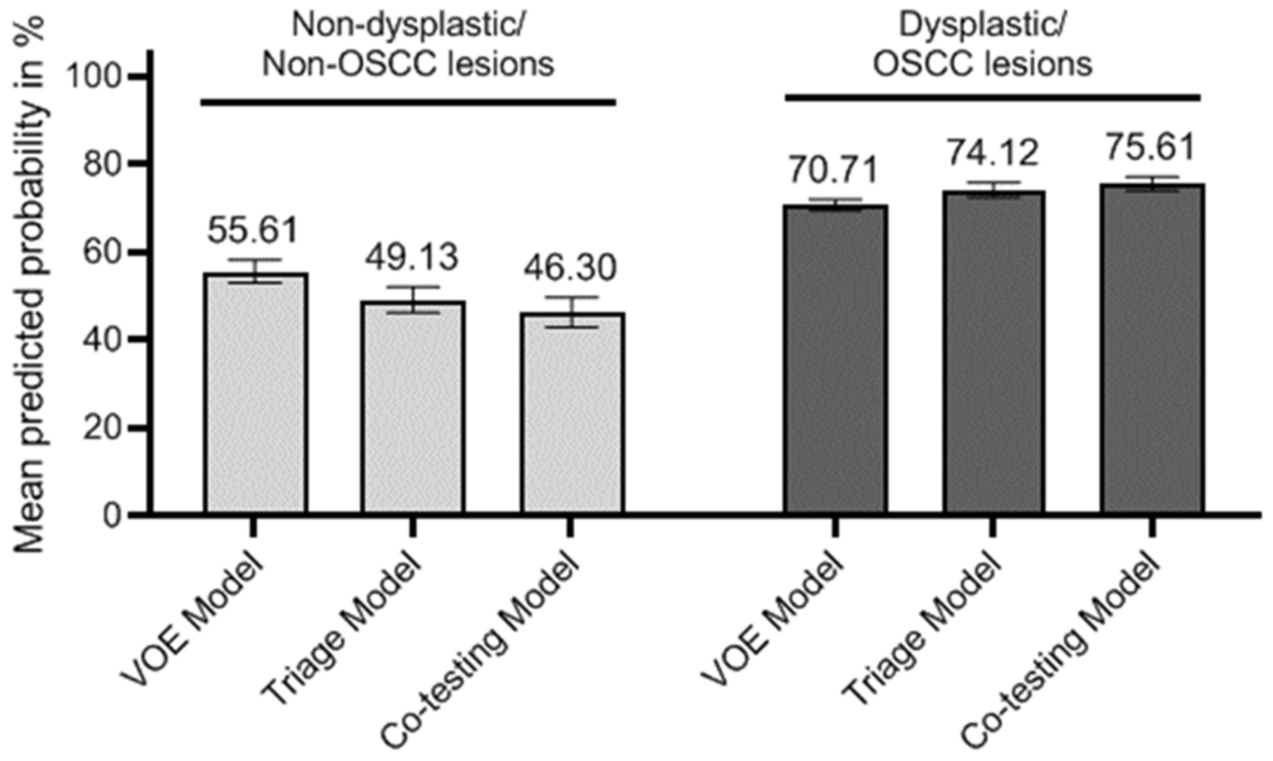
| Model a | VOE Model | MET Model | Triage Model | Co-Testing Model | ||||
|---|---|---|---|---|---|---|---|---|
| Estimate (95% CI) | p-Value | Estimate (95% CI) | p-Value | Estimate (95% CI) | p-Value | Estimate (95% CI) | p-Value | |
| C-index | 0.72 (0.64, 0.80) | <0.001 | 0.81 (0.74, 0.87) | <0.001 | 0.80 (0.73, 0.86) | <0.001 | 0.81 (0.75, 0.88) | <0.001 |
| C-index difference b | 0.03 (−0.02, 0.07) | 0.26 | 0.11 (0.04, 0.18) | 0.001 | 0.10 (0.03, 0.17) | 0.004 | 0.11 (0.04, 0.20) | 0.002 |
| C-index difference c | 0.08 (0.02, 0.13) | 0.01 | 0.09 (0.03, 0.15) | 0.004 | ||||
| Model a | VOE Model | MET Model | Triage Model | Co-Testing Model | ||||
|---|---|---|---|---|---|---|---|---|
| Difference of Mean Predicted Probability | % of Improvement | Difference of Mean Predicted Probability | % of Improvement | Difference of Mean Predicted Probability | % of Improvement | Difference of Mean Predicted Probability | % of Improvement | |
| Compared with reference model | ||||||||
| IDI: events b | 1% | 2% | 6% | 8% | 5% | 7% | 6% | 9% |
| IDI: non-events c | 2% | 4% | 11% | 19% | 9% | 15% | 12% | 20% |
| Overall IDI (95% CI), p-value | 3% (1%, 6%), 0.02 | 17% (11%, 22%), <0.001 | 14% (8%, 18%), <0.001 | 18% (12%, 24%), <0.001 | ||||
| Relative IDI | 0.29 | 1.41 | 1.14 | 1.51 | ||||
| Compared with VOE model | ||||||||
| IDI: events b | 3% | 5% | 5% | 7% | ||||
| IDI: non-events c | 7% | 12% | 9% | 17% | ||||
| Overall IDI (95% CI), p-value | 10% (5%, 14%), <0.001 | 14% (9%, 20%), <0.001 | ||||||
| Relative IDI | 0.65 | 0.94 | ||||||
Publisher’s Note: MDPI stays neutral with regard to jurisdictional claims in published maps and institutional affiliations. |
© 2022 by the authors. Licensee MDPI, Basel, Switzerland. This article is an open access article distributed under the terms and conditions of the Creative Commons Attribution (CC BY) license (https://creativecommons.org/licenses/by/4.0/).
Share and Cite
Yang, C.-C.; Su, Y.-F.; Cheng, H.-C.; Juan, Y.-C.; Chiu, Y.-W.; Wu, C.-H.; Chen, P.-Y.; Lee, Y.-H.; Chen, Y.-L.; Chen, Y.-T.; et al. Improving the Diagnostic Performance by Adding Methylation Marker to Conventional Visual Examination in Identifying Oral Cancer. Diagnostics 2022, 12, 1544. https://doi.org/10.3390/diagnostics12071544
Yang C-C, Su Y-F, Cheng H-C, Juan Y-C, Chiu Y-W, Wu C-H, Chen P-Y, Lee Y-H, Chen Y-L, Chen Y-T, et al. Improving the Diagnostic Performance by Adding Methylation Marker to Conventional Visual Examination in Identifying Oral Cancer. Diagnostics. 2022; 12(7):1544. https://doi.org/10.3390/diagnostics12071544
Chicago/Turabian StyleYang, Cheng-Chieh, Yee-Fun Su, Han-Chieh Cheng, Yi-Chen Juan, Yu-Wei Chiu, Cheng-Hsien Wu, Pei-Yin Chen, Yu-Hsien Lee, Yen-Lin Chen, Yi-Tzu Chen, and et al. 2022. "Improving the Diagnostic Performance by Adding Methylation Marker to Conventional Visual Examination in Identifying Oral Cancer" Diagnostics 12, no. 7: 1544. https://doi.org/10.3390/diagnostics12071544
APA StyleYang, C.-C., Su, Y.-F., Cheng, H.-C., Juan, Y.-C., Chiu, Y.-W., Wu, C.-H., Chen, P.-Y., Lee, Y.-H., Chen, Y.-L., Chen, Y.-T., Peng, C.-Y., Lu, M.-Y., Yu, C.-H., Huang, Y.-F., Kao, S.-Y., Fwu, C.-W., & Liu, C.-J. (2022). Improving the Diagnostic Performance by Adding Methylation Marker to Conventional Visual Examination in Identifying Oral Cancer. Diagnostics, 12(7), 1544. https://doi.org/10.3390/diagnostics12071544