
Diagnostic Performance of Toluidine Blue Stain for Direct Wet Mount Detection of Cryptosporidium Oocysts: Qualitative and Quantitative Comparison to the Modified Ziehl–Neelsen Stain

 , ,
, ,  and
and
Abstract
:1. Introduction
2. Materials and Methods
2.1. The Oocysts Source
2.2. Molecular Identification of Cryptosporidium Oocysts
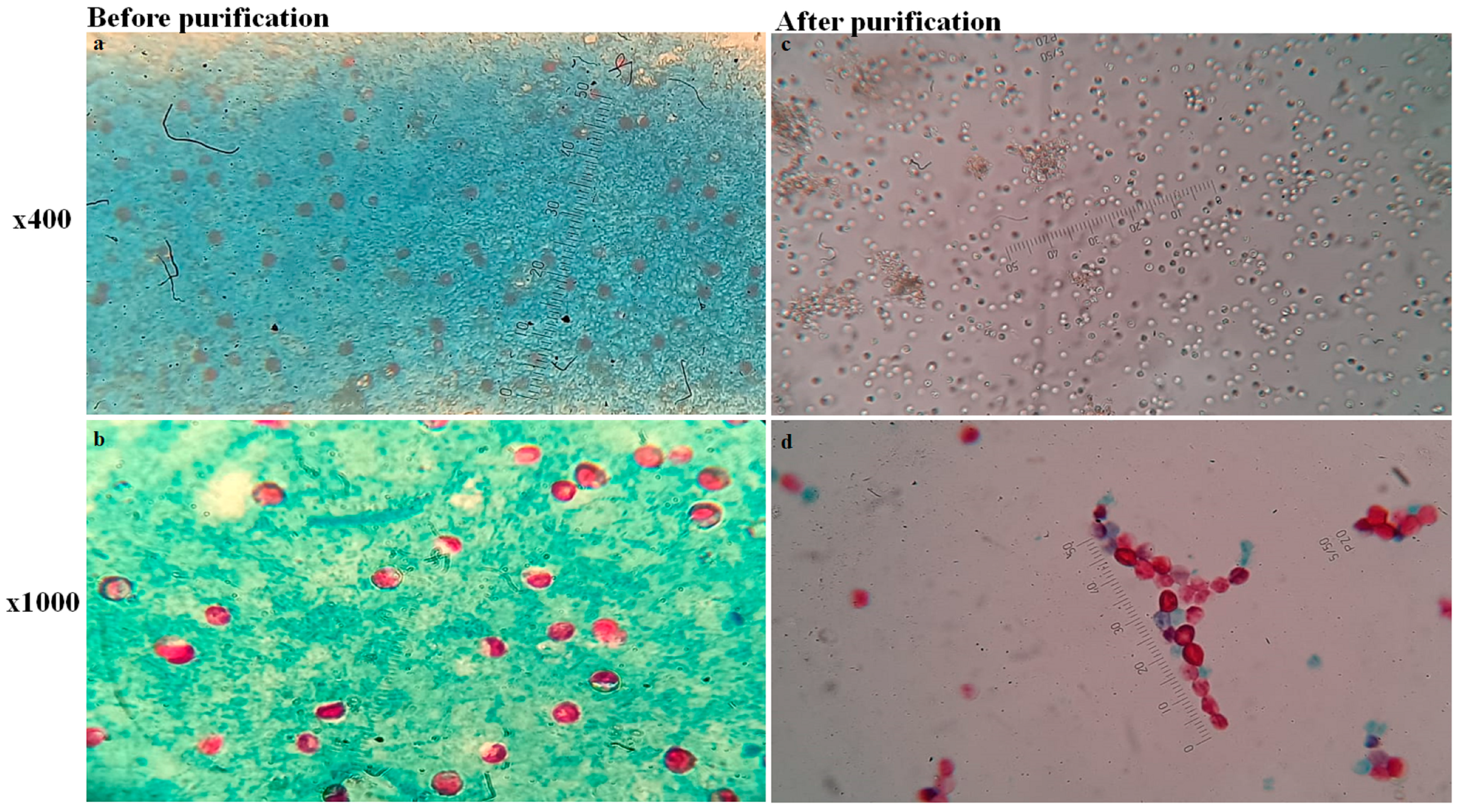
2.3. Concentration and Purification of Cryptosporidium Oocysts
2.4. Counting of Purified Cryptosporidium Oocysts
2.5. Stains Preparation for Qualitative Examination of Cryptosporidium sp. Oocysts
2.6. Sample Size Justification
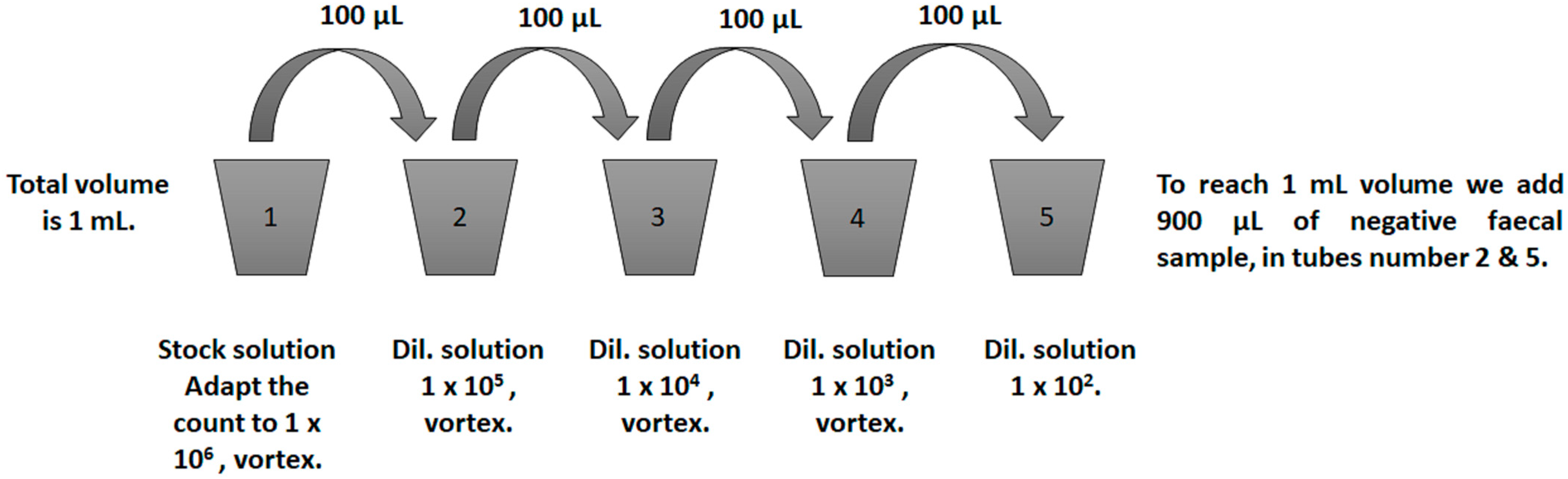
2.7. Negative Faecal Samples Preparation for Oocysts Spiking and Microscopic Examination
2.8. Preparation of Slides for Staining Techniques from Spiked Samples
2.9. Quantitative and Qualitative Assessment of TolB and mZN
2.10. Statistical Analysis
3. Results
3.1. Cryptosporidium Oocysts Purification Load and Molecular Characterization
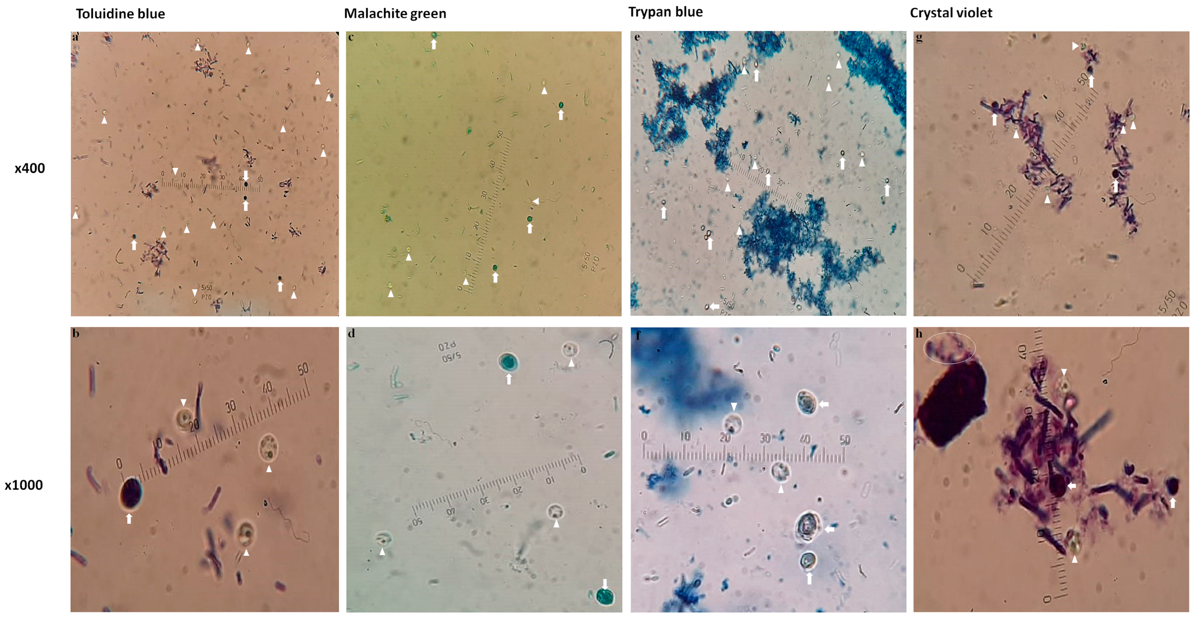
3.2. Qualitative Assessment of Wet Mount Stains in the Detection of Cryptosporidium Oocysts
3.3. Quantitative Assessment of TolB and mZN
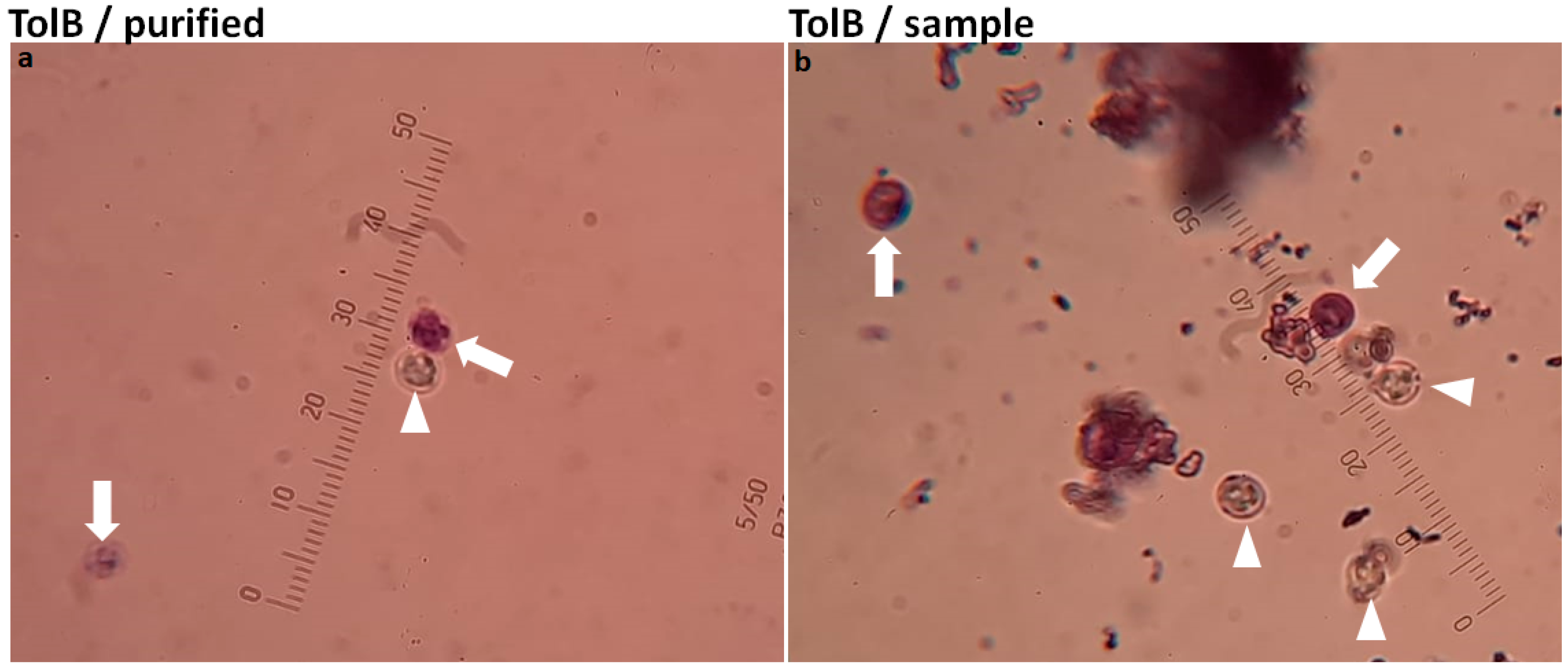
3.4. Qualitative Assessment of TolB and mZN
4. Discussion
5. Conclusions
Author Contributions
Funding
Institutional Review Board Statement
Informed Consent Statement
Data Availability Statement
Acknowledgments
Conflicts of Interest
References
- Ahmed, S.A.; Mohamed, S.F.; Fouad, A.M.; Karanis, P. Gastrointestinal parasites diagnoses at the primary health care units: A comparative analysis of diagnostic abilities of parasitology staff technicians versus medical parasitologists in Ismailia, Egypt. Trans. R. Soc. Trop. Med. Hyg. 2022, 29, trac072. [Google Scholar] [CrossRef] [PubMed]
- Ahmed, S.A.; Kotepui, M.; Masangkay, F.R.; Milanez, G.D.; Karanis, P. Gastrointestinal parasites in Africa: A review. Adv. Parasitol. 2023, 119, 1–64. [Google Scholar]
- Ahmed, S.A.; Guerrero Flórez, M.; Karanis, P. The impact of water crises and climate changes on the transmission of protozoan parasites in Africa. Pathog. Glob. Health 2018, 112, 281. [Google Scholar] [CrossRef] [PubMed]
- Mahmoudi, M.R.; Ongerth, J.E.; Karanis, P. Cryptosporidium and cryptosporidiosis: The Asian perspective. Int. J. Hyg. Environ. Health 2017, 220, 1098–1109. [Google Scholar] [CrossRef]
- Ahmed, S.; Quattrocchi, A.; Karanis, P. Cryptosporidium sp. infection in solid organ transplant recipients: A systematic review and meta-analysis. Pathog. Glob. Health, 2023; in review. [Google Scholar]
- Baldursson, S.; Karanis, P. Waterborne transmission of protozoan parasites: Review of worldwide outbreaks—An update 2004–2010. Water Res. 2011, 45, 6603–6614. [Google Scholar] [CrossRef] [PubMed]
- Ahmed, S.A.; Karanis, P. An overview of methods/techniques for the detection of Cryptosporidium in food samples. Parasitol. Res. 2018, 117, 629–653. [Google Scholar] [CrossRef]
- Efstratiou, A.; Ongerth, J.E.; Karanis, P. Waterborne transmission of protozoan parasites: Review of worldwide outbreaks—An update 2011–2016. Water Res. 2017, 114, 14–22. [Google Scholar] [CrossRef]
- Karanis, P.; Kourenti, C.; Smith, H. Waterborne transmission of protozoan parasites: A worldwide review of outbreaks and lesson learnt. J. Water Health 2007, 5, 1–38. [Google Scholar] [CrossRef]
- Ahmed, S.A.; Karanis, P. Cryptosporidium and cryptosporidiosis: The perspective from the Gulf countries. Int. J. Environ. Res. Public Health 2020, 17, 6824. [Google Scholar] [CrossRef]
- Ježková, J.; Limpouchová, Z.; Prediger, J.; Holubová, N.; Sak, B.; Konečný, R.; Květoňová, D.; Hlásková, L.; Rost, M.; McEvoy, J.; et al. Cryptosporidium myocastoris n. sp. (Apicomplexa: Cryptosporidiidae), the species adapted to the Nutria (Myocastor coypus). Microorganisms 2021, 9, 813. [Google Scholar] [CrossRef]
- Zahedi, A.; Bolland, S.J.; Oskam, C.L.; Ryan, U. Cryptosporidium abrahamseni n. sp. (Apicomplexa: Cryptosporidiiae) from red-eye tetra (Moenkhausia sanctaefilomenae). Exp. Parasitol. 2021, 223, 108089. [Google Scholar] [CrossRef]
- Ryan, U.M.; Feng, Y.; Fayer, R.; Xiao, L. Taxonomy and molecular epidemiology of Cryptosporidium and Giardia—A 50 year perspective (1971–2021). Int. J. Parasitol. 2021, 51, 1099–1119. [Google Scholar] [CrossRef] [PubMed]
- Innes, E.A.; Chalmers, R.M.; Wells, B.; Pawlowic, M.C. A One Health approach to tackle cryptosporidiosis. Trends Parasitol. 2020, 36, 290–303. [Google Scholar] [CrossRef] [Green Version]
- Ahmed, S.A.; Karanis, P. Comparison of current methods used to detect Cryptosporidium oocysts in stools. Int. J. Hyg. Environ. Health 2018, 221, 743–763. [Google Scholar] [CrossRef] [PubMed]
- Ongerth, J.E.; Karanis, P. Cryptosporidium & Giardia in water—Key features and basic principles for monitoring & data analysis. Proceedings 2018, 2, 691. [Google Scholar] [CrossRef]
- Ma, L.; Zhang, X.; Jian, Y.; Li, X.; Wang, G.; Hu, Y.; Karanis, P. Detection of Cryptosporidium and Giardia in the slaughterhouse, sewage and river waters of the Qinghai Tibetan Plateau Area (QTPA), China. Parasitol. Res. 2019, 118, 2041–2051. [Google Scholar] [CrossRef]
- Schou, C.; Filippova, M.; Quattrocchi, A.; Karanis, P. The current status of protozoan parasitic diseases in Cyprus: A narrative literature review. Environ. Sci. Proc. 2020, 2, 61. [Google Scholar] [CrossRef]
- Tahvildar-Biderouni, F.; Salehi, N. Detection of Cryptosporidium infection by modified Ziehl-Neelsen and PCR methods in children with diarrheal samples in pediatric hospitals in Tehran. Gastroenterol. Hepatol. Bed Bench 2014, 7, 125. [Google Scholar] [PubMed]
- Aghamolaie, S.; Rostami, A.; Fallahi, S.; Tahvildar Biderouni, F.; Haghighi, A.; Salehi, N. Evaluation of modified Ziehl–Neelsen, direct fluorescent-antibody and pcr assay for detection of Cryptosporidium spp. in children faecal specimens. J. Parasit. Dis. Off. Organ Indian Soc. Parasitol. 2016, 40, 963. [Google Scholar] [CrossRef] [Green Version]
- Potters, I.; van Esbroeck, M. Negative staining technique of Heine for the detection of Cryptosporidium spp.: A fast and simple screening technique. Open Parasitol. J. 2010, 4, 1–4. [Google Scholar] [CrossRef]
- Chartier, C.; Mallereau-Pellet, M.P.; Mancassola, R.; Nussbaum, D. Détection Des Oocystes de Cryptosporidium Dans Les Fèces de Caprins: Comparaison entre un test d’agglutination au latex et trois autres techniques conventionnelles. Vet. Res. 2002, 33, 169–177. [Google Scholar] [CrossRef]
- Casemore, D.P. Laboratory methods for diagnosing cryptosporidiosis. J. Clin. Pathol. 1991, 44, 445–451. [Google Scholar] [CrossRef] [PubMed] [Green Version]
- Center of Disease and Control (CDC). Laboratory Diagnosis of Cryptosporidiosis. 2021. Available online: https://www.cdc.gov/dpdx/resources/pdf/benchaids/crypto_benchaid.pdf (accessed on 19 June 2023).
- Heine, J. Eine einfache Nachweismethode für Kryptosporidien im Kot. Zent. Für Veterinärmedizin Reihe B 1982, 29, 324–327. [Google Scholar] [CrossRef]
- Rekha, H.K.M.; Puttalakshmamma, G.C.; D’Souza, P.E. Comparison of different diagnostic techniques for the detection of cryptosporidiosis in bovines. Vet. World 2016, 9, 215. [Google Scholar] [CrossRef] [Green Version]
- Khanna, V.; Tilak, K.; Ghosh, A.; Mukhopadhyay, C. Modified negative staining of heine for fast and inexpensive screening of Cryptosporidium, Cyclospora, and Cystoisospora spp. Int. Sch. Res. Not. 2014, 2014, 165424. [Google Scholar] [CrossRef] [PubMed]
- Ahmed, S.A.; El-Mahallawy, H.S.; Karanis, P. Inhibitory activity of chitosan nanoparticles against Cryptosporidium parvum oocysts. Parasitol. Res. 2019, 118, 2053–2063. [Google Scholar] [CrossRef]
- Henriksen, S.A.; Pohlenz, J.F.L. Staining of cryptosporidia by a modified Ziehl-Neelsen technique. Acta Vet. Scand. 1981, 22, 594–596. [Google Scholar] [CrossRef]
- Holzhausen, I.; Lendner, M.; Göhring, F.; Steinhöfel, I.; Daugschies, A. Distribution of Cryptosporidium parvum Gp60 subtypes in calf herds of Saxony, Germany. Parasitol. Res. 2019, 118, 1549–1558. [Google Scholar] [CrossRef]
- Bialek, R.; Binder, N.; Dietz, K.; Joachim, A.; Knobloch, J.; Zelck, U.E. Comparison of fluorescence, antigen and PCR assays to detect Cryptosporidium parvum in fecal specimens. Diagn. Microbiol. Infect. Dis. 2002, 43, 283–288. [Google Scholar] [CrossRef]
- Arrowood, M.; Sterling, C. Isolation of Cryptosporidium oocysts and sporozoites using discontinuous sucrose and isopycnic percoll gradients. J. Parasitol. 1987, 73, 314–319. [Google Scholar] [CrossRef]
- Kourenti, C.; Karanis, P. Evaluation and applicability of a purification method coupled with nested PCR for the detection of Toxoplasma oocysts in water. Lett. Appl. Microbiol. 2006, 43, 475–481. [Google Scholar] [CrossRef] [PubMed]
- Castro-Hermida, J.A.; Pors, I.; Ares-Mazas, E.; Chartier, C. In vitro activity on Cryptosporidium parvum oocyst of different drugs with recognized anti-cryptosporidial efficacy. Rev. Med. Vet. 2004, 155, 453–456. [Google Scholar]
- Finch, G.R.; Daniels, C.W.; Black, E.K.; Schaefer, F.W.; Belosevic, M. Dose response of Cryptosporidium parvum in outbred neonatal CD-1 mice. Appl. Environ. Microbiol. 1993, 59, 3661–3665. [Google Scholar] [CrossRef] [PubMed]
- Center of Disease and Control (CDC). Diagnostic Procedures—Stool Specimens—Staining Procedures; Center of Disease and Control: Atlanta, GA, USA, 2016. Available online: https://www.cdc.gov/dpdx/diagnosticprocedures/stool/staining.html (accessed on 19 June 2023).
- Garcia, L.S.; Bruckner, D.A.; Brewer, T.C.; Shimizu, R.Y. Techniques for the recovery and identification of Cryptosporidium oocysts from stool specimens. J. Clin. Microbiol. 1983, 18, 185–190. [Google Scholar] [CrossRef] [PubMed]
- Buderer, N.M. Statistical methodology: Incorporating the prevalence of disease into the sample size calculation for sensitivity and specificity. Acad. Emerg. Med. 1996, 3, 895–900. [Google Scholar] [CrossRef]
- Youssef, F.G.; Adib, I.; Riddle, M.S.; Schlett, C.D. A review of cryptosporidiosis in Egypt. J. Egypt. Soc. Parasitol. 2008, 38, 9–28. [Google Scholar]
- Elliot, A.; Morgan, U.M.; Thompson, R.C.A. Improved staining method for detecting Cryptosporidium oocysts in stools using malachite green. J. Gen. Appl. Microbiol. 1999, 45, 139–142. [Google Scholar] [CrossRef] [Green Version]
- Karanis, P.; Schoenen, D. Biological test for the detection of low concentrations of infectious Cryptosporidium parvum oocysts in water. Acta Hydrochim. Hydrobiol. 2001, 29, 242. [Google Scholar] [CrossRef]
- Pacheco, F.T.F.; Silva, R.K.N.R.; Martins, A.S.; Oliveira, R.R.; Alcântara-Neves, N.M.; Silva, M.P.; Soares, N.M.; Teixeira, M.C.A. Differences in the detection of Cryptosporidium and Isospora (Cystoisospora) oocysts according to the fecal concentration or staining method used in a clinical laboratory. J. Parasitol. 2013, 99, 1002–1008. [Google Scholar] [CrossRef]
- Feinstein, A.R.; Cicchetti, D.V. High Agreement but Low Kappa: I. The problems of two paradoxes. J. Clin. Epidemiol. 1990, 43, 543–549. [Google Scholar] [CrossRef] [PubMed]
- Klein, D. Implementing a general framework for assessing interrater agreement in stata. Stata J. 2018, 18, 871–901. [Google Scholar] [CrossRef]
- Landis, J.R.; Koch, G. The measurement of observer agreement for categorical data. Biometrics 1977, 33, 159–174. [Google Scholar] [CrossRef]
- Robinson, G.; Chalmers, R.M. Cryptosporidium diagnostic assays: Microscopy. Methods Mol. Biol. 2020, 2052, 1–10. [Google Scholar] [CrossRef]
- Vanathy, K.; Parija, S.C.; Mandal, J.; Hamide, A.; Krishnamurthy, S. Detection of Cryptosporidium in stool samples of immunocompromised patients. Trop. Parasitol. 2017, 7, 46. [Google Scholar]
- Manser, M.; Granlund, M.; Edwards, H.; Saez, A.; Petersen, E.; Evengard, B.; Chiodini, P. Detection of Cryptosporidium and Giardia in clinical laboratories in Europe—A comparative study. Clin. Microbiol. Infect. 2014, 20, O65–O71. [Google Scholar] [CrossRef] [Green Version]
- Center of Disease and Control (CDC). Cryptosporidiosis—Diagnosis & Detection. 2021. Available online: https://www.cdc.gov/parasites/crypto/diagnosis.html (accessed on 19 June 2023).
- Cacciò, S.M.; Chalmers, R.M. Human cryptosporidiosis in Europe. Clin. Microbiol. Infect. 2016, 22, 471–480. [Google Scholar] [CrossRef] [PubMed] [Green Version]
- Microbehunter Microscopy. Making a Wet Mount Microscope Slide. 2010. Available online: https://www.microbehunter.com/making-a-wet-mount-microscope-slide/ (accessed on 19 June 2023).
- Ferrari, E.D.; Nakamura, A.A.; Nardi, A.R.M.; Santana, B.N.; da Silva Camargo, V.; Nagata, W.B.; Bresciani, K.D.S.; Meireles, M.V. Cryptosporidium spp. in caged exotic psittacines from Brazil: Evaluation of diagnostic methods and molecular characterization. Exp. Parasitol. 2018, 184, 109–114. [Google Scholar] [CrossRef] [Green Version]
- Oliveira, B.C.M.; Ferrari, E.D.; da Cruz Panegossi, M.F.; Nakamura, A.A.; Corbucci, F.S.; Nagata, W.B.; dos Santos, B.M.; Gomes, J.F.; Meireles, M.V.; Widmer, G.; et al. First description of Cryptosporidium parvum in carrier pigeons (Columba livia). Vet. Parasitol. 2017, 243, 148–150. [Google Scholar] [CrossRef] [PubMed] [Green Version]
- Ortolani, E. Standardization of the modified Ziehl-Neelsen technique to stain oocysts of Cryptosporidium sp. Rev. Bras. Parasitol. Vet. 2000, 9, 29–30. [Google Scholar]


| ID | Gender | Age in Years | Consistency | ID | Gender | Age in Years | Consistency |
|---|---|---|---|---|---|---|---|
| 1 | Female | 4 | Formed | 23 | Female | 34 | Diarrhoea |
| 2 | Male | 7 | Diarrhoea | 24 | Female | 57 | Formed |
| 3 | Male | 3.5 | Formed | 25 | Female | 56 | Diarrhoea |
| 4 | Female | 8 | Formed | 26 | Female | 51 | Formed |
| 5 | Female | 6 | Diarrhoea | 27 | Female | 40 | Formed |
| 6 | Female | 35 | Formed | 28 | Female | 34 | Formed |
| 7 | Male | 38 | Formed | 29 | Male | 37 | Formed |
| 8 | Male | 9 | Formed | 30 | Male | 62 | Formed |
| 9 | Female | 7 | Formed | 31 | Female | 41 | Formed |
| 10 | Male | 40 | Formed | 32 | Female | 29 | Formed |
| 11 | Male | 4 | Diarrhoea | 33 | Female | 13 | Formed |
| 12 | Female | 8 | Formed | 34 | Female | 26 | Formed |
| 13 | Female | 4.5 | Formed | 35 | Female | 45 | Formed |
| 14 | Male | 3 | Formed | 36 | Male | 9 | Diarrhoea |
| 15 | Male | 10 | Formed | 37 | Male | 4 | Formed |
| 16 | Male | 3 | Formed | 38 | Female | 43 | Formed |
| 17 | Female | 12 | Formed | 39 | Female | 36 | Formed |
| 18 | Female | 42 | Formed | 40 | Male | 6 | Diarrhoea |
| 19 | Female | 22 | Formed | 41 | Female | 8 | Formed |
| 20 | Female | 37 | Formed | 42 | Male | 48 | Formed |
| 21 | Female | 28 | Diarrhoea | 43 | Male | 2.5 | Formed |
| 22 | Female | 44 | Formed |
| Qualitative Variables | Qualitative Assessment | Explanation of Undesirable Characteristics | |
|---|---|---|---|
| Desirable | Undesirable | ||
| Intensity of dye negativity by oocysts | 1 | 0 | Cannot discriminate the oocysts from surrounding areas in faecal smears |
| Distinction of oocysts from the background | 1 | 0 | Dying other faecal elements such as yeast cells, pollen grains, or digested food residues |
| Presence of stained non-relevant residues | 1 | 0 | Produce precipitation of dye particles, which is a main concern for Cryptosporidium diagnosis, especially when sizes and shapes of residues are similar to the oocysts. |
| Background deep staining | 1 | 0 | Background stains light and oocysts detection become difficult |
| Recognition of oocysts easily at ×40 | 1 | 0 | Cannot recognize the oocysts at power ×40. |
| Thickness of the smear | 1 | 0 | Cannot recognize the oocysts in thick smear |
| Faecal material residues | 1 | 0 | Produces distinct outcomes for various faecal materials. |
| Score | TolB | MG | TB | CV |
|---|---|---|---|---|
| Mean (±SD) | 6.8 (±0.6) | 3.9 (±3.6) | 1.9 (±2.8) | 0 |
| Median (IQR) | 7 (7–7) | 7 (0–7) | 0 (0–5) | 0 |
| Total (range 0–140) | 135 | 77 | 38 | 0 |
| % Undesirable | 4 | 45 | 73 | 100 |
| Interpretation | Superior | Regular | Inferior | Inferior |
| Wet Mount Stain | Individual | Average |
|---|---|---|
| TolB | 0.84 * | 0.91 * |
| MG | 0.82 * | 0.90 * |
| TB | 0.94 * | 0.97 * |
| CV | NA | NA |
| Test Comparison | Sensitivity % | Agreement % | Gwet’s Agreement Coefficient | Interpretation |
|---|---|---|---|---|
| (a) TolB × 102 rater 1 vs. rater 2 | 39.5 vs. 48.8 | 48.8 | −0.01 | Poor agreement |
| (b) TolB × 104 rater 1 vs. rater 2 | 93.0 vs. 100 | 93.0 | 0.93 * | Almost perfect |
| (c) mZN × 102 rater 1 vs. rater 2 | 23.3 vs. 23.3 | 81.4 | 0.71 * | Substantial agreement |
| (d) mZN × 104 rater 1 vs. rater 2 | 81.4 vs. 79.1 | 93.0 | 0.90 * | Substantial agreement |
| (e) Rater 1 TolB × 102 vs. ×104 | 39.5 vs. 93.0 | 41.9 | −0.05 | Poor agreement |
| (f) Rater 2 TolB × 102 vs. ×104 | 48.8 vs. 100 | 48.8 | 0.17 | Slight agreement |
| (g) Rater 1 mZN × 102 vs. ×104 | 23.3 vs. 81.4 | 37.2 | −0.25 | Poor agreement |
| (h) Rater 2 mZN × 102 vs. ×104 | 23.3 vs. 79.1 | 44.2 | −0.12 | Poor agreement |
| (i) Rater 1 × 10² TolB vs. mZN | 39.5 vs. 23.3 | 60.5 | 0.31 | Fair agreement |
| (j) Rater 2 × 10² TolB vs. mZN | 48.8 vs. 23.3 | 55.8 | 0.18 | Slight agreement |
| (k) Rater 1 × 10⁴ TolB vs. mZN | 93.0 vs. 81.4 | 79.1 | 0.73 * | Substantial agreement |
| (l) Rater 2 × 10⁴ TolB vs. mZN | 100 vs. 79.1 | 79.1 | 0.74 * | Substantial agreement |
| Items | Variables | Stain | TolB-mZN (Relative Change%) | Favorable Stain | |
|---|---|---|---|---|---|
| TolB (%) | mZN (%) | ||||
| Preparation | Safe | 5 | 3.5 | 1.5 | TolB |
| Practical | 5 | 3.5 | 1.5 | TolB | |
| Time-consuming * | 4.5 | 1.5 | 3 | TolB | |
| Permanent stain | 1.5 | 5 | −3.5 | mZN | |
| Cost-effective | 3.5 | 3 | 0.5 | Both | |
| Wet mount stain | 5 | 1.5 | 3.5 | TolB | |
| Total (range 6–30) | 24.5 (82%) | 18 (60%) | 6.5 (27%) | TolB | |
| Processing | Time-consuming * | 4.5 | 1 | 3.5 | TolB |
| The stain has variable background coloration * | 4.5 | 1 | 3.5 | TolB | |
| The yeast takes up the stain | 5 | 3.5 | 1.5 | TolB | |
| The stain clearly differentiates the yeast from the oocysts | 5 | 2.5 | 2.5 | TolB | |
| The use of microscopic power ×40 is accessible to identify the oocysts | 5 | 2 | 3 | TolB | |
| It is easy to identify the oocysts among faecal sediment | 5 | 3 | 2 | TolB | |
| The oocysts preserve its features | 5 | 3.5 | 1.5 | TolB | |
| The sample sediment characteristics affect the staining * | 4.5 | 1.5 | 3 | TolB | |
| The stain gets affected by the freshness of the faecal sample * | 2 | 3.5 | −1.5 | mZN | |
| The staining technique can be applied in the field | 5 | 3 | 2 | TolB | |
| The technique can be repeated on the same sample | 5 | 3.5 | 1.5 | TolB | |
| Using faecal concentration method affects the result of the staining technique * | 4.5 | 1.5 | 3 | TolB | |
| Total (range 12–60) | 55 (92%) | 29.5 (49%) | 25.5 (47%) | TolB | |
| Diagnosis | Diagnosis will vary with different investigators * | 4.5 | 2 | 2.5 | TolB |
| Diagnosis have sometimes doubt regarding the typical oocysts shape * | 4.5 | 1.5 | 3 | TolB | |
| It is easy to interpret the slide via visual inspection | 5 | 2.5 | 2.5 | TolB | |
| Total (range 3–15) | 14 (93%) | 6 (40%) | 8 (57%) | TolB | |
| Total (range 21–105) | 93.5 (89%) | 53.5 (51%) | 40 (43%) | TolB | |
Disclaimer/Publisher’s Note: The statements, opinions and data contained in all publications are solely those of the individual author(s) and contributor(s) and not of MDPI and/or the editor(s). MDPI and/or the editor(s) disclaim responsibility for any injury to people or property resulting from any ideas, methods, instructions or products referred to in the content. |
© 2023 by the authors. Licensee MDPI, Basel, Switzerland. This article is an open access article distributed under the terms and conditions of the Creative Commons Attribution (CC BY) license (https://creativecommons.org/licenses/by/4.0/).
Share and Cite
Ahmed, S.A.A.; Quattrocchi, A.; Elzagawy, S.M.; Karanis, P.; Gad, S.E.M. Diagnostic Performance of Toluidine Blue Stain for Direct Wet Mount Detection of Cryptosporidium Oocysts: Qualitative and Quantitative Comparison to the Modified Ziehl–Neelsen Stain. Diagnostics 2023, 13, 2557. https://doi.org/10.3390/diagnostics13152557
Ahmed SAA, Quattrocchi A, Elzagawy SM, Karanis P, Gad SEM. Diagnostic Performance of Toluidine Blue Stain for Direct Wet Mount Detection of Cryptosporidium Oocysts: Qualitative and Quantitative Comparison to the Modified Ziehl–Neelsen Stain. Diagnostics. 2023; 13(15):2557. https://doi.org/10.3390/diagnostics13152557
Chicago/Turabian StyleAhmed, Shahira Abdelaziz Ali, Annalisa Quattrocchi, Sherine M. Elzagawy, Panagiotis Karanis, and Samer Eid Mohamed Gad. 2023. "Diagnostic Performance of Toluidine Blue Stain for Direct Wet Mount Detection of Cryptosporidium Oocysts: Qualitative and Quantitative Comparison to the Modified Ziehl–Neelsen Stain" Diagnostics 13, no. 15: 2557. https://doi.org/10.3390/diagnostics13152557
APA StyleAhmed, S. A. A., Quattrocchi, A., Elzagawy, S. M., Karanis, P., & Gad, S. E. M. (2023). Diagnostic Performance of Toluidine Blue Stain for Direct Wet Mount Detection of Cryptosporidium Oocysts: Qualitative and Quantitative Comparison to the Modified Ziehl–Neelsen Stain. Diagnostics, 13(15), 2557. https://doi.org/10.3390/diagnostics13152557