Article

Association of the EPAS1 rs7557402 Polymorphism with Hemodynamically Significant Patent Ductus Arteriosus Closure Failure in Premature Newborns under Pharmacological Treatment with Ibuprofen

by
Diana G. Rogel-Ayala
1,†,‡,
José Esteban Muñoz-Medina
2,
Valeria Dejanira Vicente-Juárez
1,
Patricia Grether-González
3,
Deneb Algedi Morales-Barquet
4,
Alfonso de Jesús Martínez-García
5,
María Olga Leticia Echaniz-Aviles
6,
Rosalba Sevilla-Montoya
1,
Alejandro Martínez-Juárez
7,
Jazmin Artega-Vázquez
8,
Javier Angeles-Martínez
9,
Gilberto Vargas-Alarcón
10,
Alberto Hidalgo-Bravo
11 and
Irma Eloisa Monroy-Muñoz
1,*
1
Reproductive and Perinatal Health Research Department, National Institute of Perinatology, Mexico City 11000, Mexico
2
Quality of Supplies and Specialized Laboratories Coordination, Mexican Social Security Institute, Mexico City 37320, Mexico
3
ABC Medical Center, Santa Fe, Mexico City 05348, Mexico
4
Neonatal Intensive Care Unit, National Institute of Perinatology, Mexico City 11000, Mexico
5
Fetal Maternal Medicine Department, National Institute of Perinatology, Mexico City 11000, Mexico
6
Newborn Intermediate Care Unit, National Institute of Perinatology, Mexico City 11000, Mexico
7
Cellular Physiology Department, National Institute of Perinatology, Mexico City 11000, Mexico
8
Department of Genetics, National Institute of Medical Science and Nutrition, Mexico City 14080, Mexico
9
Specialized Laboratories Division, Mexican Social Security Institute, Mexico City 06700, Mexico
10
Department of Molecular Biology, National Institute of Cardiology, Mexico City 14080, Mexico
11
Genomics Medicine Department, National Institute of Rehabilitation, Mexico City 14610, Mexico
*
Author to whom correspondence should be addressed.
Current address: CNRS, Laboratoire IMoPA, UMR 7365, Université de Lorraine, F-54000 Nancy, France.
Current address: Lung Cancer Epigenetics, Max-Planck-Institute for Heart and Lung Research, 61231 Bad Nauheim, Germany.
Diagnostics 2023, 13(15), 2558; https://doi.org/10.3390/diagnostics13152558
Submission received: 30 June 2023 / Revised: 16 July 2023 / Accepted: 27 July 2023 / Published: 1 August 2023
(This article belongs to the Section Pathology and Molecular Diagnostics)

Abstract

:
Patent ductus arteriosus (PDA) is frequent in preterm newborns, and its incidence is inversely associated with the degree of prematurity. The first choice of pharmacological treatment is ibuprofen. Several genes, including EPAS1, have been proposed as probable markers associated with a genetic predisposition for the development of PDA in preterm infants. EPAS 1 NG_016000.1:g.84131C>G or rs7557402 has been reported to be probably benign and associated with familial erythrocytosis by the Illumina Clinical Services Laboratory. Other variants of EPAS1 have been previously reported to be benign for familial erythrocytosis because they decrease gene function and are positive for familial erythrocytosis because the overexpression of EPAS1 is a key factor in uncontrolled erythrocyte proliferation. However, this could be inconvenient for ductal closure, since for this process to occur, cell proliferation, migration, and differentiation should take place, and a decrease in EPAS1 gene activity would negatively affect these processes. Single-nucleotide polymorphisms (SNPs) in EPAS1 and TFAP2B genes were searched with high-resolution melting and Sanger sequencing in blood samples of preterm infants with hemodynamically significant PDA treated with ibuprofen at the National Institute of Perinatology. The variant rs7557402, present in the EPAS1 gene eighth intron, was associated with a decreased response to treatment (p = 0.007, OR = 3.53). The SNP rs7557402 was associated with an increased risk of pharmacological treatment failure. A probable mechanism involved could be the decreased activity of the product of the EPAS1 gene.

1. Introduction

The ductus arteriosus is a central vascular shunt connecting the pulmonary artery to the aorta, allowing oxygenated blood from the placenta to bypass the uninflated fetal lungs and enter the systemic circulation. The rapid closure of the ductus after birth is essential for vascular transition to the mature divided pattern of arteriovenous circulation. Failed ductus arteriosus closure, termed patent ductus arteriosus (PDA), is frequent in preterm newborns, with up to 64% of infants born at 27 to 28 weeks exhibiting PDA. The incidence of PDA is inversely associated with the degree of prematurity [1,2].
The National Institute of Perinatology is a tertiary-level hospital, where 300 preterm newborns weighing <1500 g are born each year. An observational study performed in 2016 in 295 preterm infants between 27 and 28 weeks found PDA in 21.6% of patients [3]. The diagnostic gold standard is two-dimensional color Doppler echocardiography, which can determine the shape and diameter of the ductus arteriosus at the aortic and pulmonary edges and the degree of hemodynamic burden [4,5,6].
When there is hemodynamically significant PDA, the treatment scheme involves pharmacological treatment as the first choice. When the patient has a condition that contraindicates the use of drugs or if this treatment is not effective, surgical closure may be necessary. Untreated hemodynamically significant PDA can result in life-threatening conditions, such as congestive heart failure, pulmonary artery hypertension, and neonatal necrotizing enterocolitis [7].
Cyclooxygenase (COX) inhibitors are administered as a pharmacological treatment for hemodynamically significant PDA: the most commonly used inhibitors are indomethacin and ibuprofen, although the use of acetaminophen has recently been approved [8,9,10,11,12].
Up to 30% of pharmacological treatment failure has been observed in preterm infants. Both indomethacin and ibuprofen have shown similar efficacy [8,9,10,11,12]. In the National Institute of Perinatology, the most commonly used pharmacological treatment is ibuprofen. In the aforementioned study at the National Institute of Perinatology, 47.7% success was found with 10 mg/kg/d ibuprofen on the first day followed by 5 mg/kg/d ibuprofen on the second and third days, respectively, and 42.8% success was observed during the second cycle [3].
The ductus arteriosus (DA) derives from the left sixth aortic arch, which derives from neural crest cells. To successfully achieve ductal closure, cell growth and differentiation are essential, involving the induction of specific gene expression mediated by the interaction of transcription factors with response elements [13,14]. AP-2 transcription factors play a major role in the cellular differentiation induced by retinoic acid, particularly in neural crest cells. PDA probably results from the abnormal development of the neural crest [15].
Transcription factor AP-2 beta (TFAP2B) expression is enriched in the neural crest and may play an important role in regulating DA closure. This transcription factor has been reported to regulate the expression of EPAS1 (endothelial PAS domain protein 1, also known as hypoxia-inducible factor 2 alpha), which is involved in oxygen sensing [2,16].
The expression of EPAS1 is cell-type-restricted and predominantly occurs in endothelial cells, lung epithelial cells, and cardiac myocytes. EPAS1 trans-activated target genes contain the hypoxia responsive element (HRE) [17,18].
A bioinformatic and statistical study led by Dagle in 2009 reported several genes as probable markers associated with the genetic predisposition to PDA in preterm infants. Of all possible markers analyzed, those that showed a significant association were the polymorphisms rs987237 of TFAP2B (p < 0.005) and rs1867785 of EPAS1 (p < 0.005). These same polymorphisms were studied in relation to the failure of pharmacological treatment with indomethacin, also exhibiting significant association [13].
NG_016000.1:g.84131C>G or rs7557402 has been reported to be probably benign and associated with familial erythrocytosis by the Illumina Clinical Services Laboratory [19]. Other variants of EPAS1 have been previously reported as benign for familial erythrocytosis because they decrease gene function, and they have been revealed to be positive for familial erythrocytosis because the overexpression of EPAS1 is a key factor in uncontrolled erythrocyte proliferation [20]. However, this could be inconvenient for ductal closure, since for this process to occur, cell proliferation, migration, and differentiation should take place, and a decrease in EPAS1 gene activity would negatively affect these processes [21,22,23].
In this study, we explored the association between genetic variants of TFAP2B and EPAS1 and PDA ibuprofen treatment failure.

2. Materials and Methods

2.1. Study Population

A cross-sectional exploratory study including 47 newborns with a diagnosis of PDA treated with ibuprofen was carried out at the National Institute of Perinatology in Mexico City. On behalf of the children enrolled in our study, we obtained written informed consent from their legal guardians. The study complied with the Declaration of Helsinki and was approved by the Institutional Ethics Committee and registered to the National Institute of Perinatology, project number 212250-3140-11105-01-16.
Convenience sampling of consecutive cases was performed. All premature newborns (gestational age between 25 and 36.6 gestation weeks determined by Ballard) with hemodynamically significant PDA diagnosis and ibuprofen treatment born at INPer during the period from 1 January 2017 to 31 December 2019 were included. Oral ibuprofen treatment was used as follows: the first cycle of 10 mg/kg/d on the first day, followed by 5 mg/kg/d on the second and third days, respectively. In the case that no clinical and echocardiographic response was obtained (Supplementary Materials), a second oral ibuprofen cycle was administered (20 mg/kg/d), followed by a second and third dose at 10 mg/kg/dose, with administration intervals of 24 h between the doses [3]. The sample population was separated into two groups. The case group (n = 19) included patients who presented ibuprofen treatment failure, and hemodynamically significant PDA was closed via surgery. The control group (n = 28) included patients with successful pharmacological treatment with ibuprofen.
In addition to the premature newborn samples, we included a family of nine members with a background of PDA. Blood samples from members of this family were used as controls for the techniques employed in this study.

2.2. Genetic Variant Analysis

A sample of peripheral blood was taken with a BD Microtainer® (Franklin Lakes, NJ, USA) blood collection system. DNA was isolated from leukocytes using the Promega Wizard® Genomic DNA Purification Kit (Madison, WI, USA). The DNA samples were quantified with a Thermo Scientific NanoDrop™ 2000 instrument (Waltham, MA, USA) and aliquoted and stored at −20 °C until later use.
The high-resolution melting (HRM) method was used to detect genetic variants of the TFAP2B and EPAS1 genes. The HRM primers were designed using the Primer Select program (DNASTAR lasergene, Maddison, WI, USA), which also ensures that the primer sequences do not form secondary structures during PCR that could increase the complexity of melting profile interpretation. The primer specificity was tested using the Primer Blast platform (NCBI) [24]. Primers were designed to amplify complete exonic sequences and small flanking intronic sequences. Reactions were performed with a total volume of 20 µL (5 µL of Milli-Q water, 1.5 µL of each primer at 20 pmol/µL, 10 µL of Bio-Rad Precision Melt Supermix (Hercules, CA, USA), and 2 µL of DNA at 150 ng/µL). The amplification parameters were 95 °C for 4 min, 30 cycles of 94 °C for 30 s, annealing temperature for 30 s, and 72 °C for 30 s, followed by a final extension step of 72 °C for 5 min. For the melting curve analysis, the parameters were 95 °C for 30 s and 75 °C for 30 s. Data were collected over a temperature range of 75–95 °C in 0.1 °C increments every 10 s using the CFX-96 Touch Real Time PCR System of Bio-Rad (Hercules, CA, USA). Once each HRM reaction was standardized, all samples were tested in triplicate. Melting curve analysis was performed with Bio-Rad Precision Melt Analysis Software v.1.3. (Hercules, CA, USA). All samples with melting curves different from the average underwent capillary sequencing to determine the alteration in the sequence that yielded the difference in the melting curve (see Supplementary Materials). Moreover, three random samples with an average melting curve were capillary sequenced to ensure that the observed curve corresponded to the consensus sequence obtained from the RefSeq database (NCBI) [25].

2.3. DNA Sequencing

After purification with Thermo Fisher Scientific PCR ExoSAP-ITTM (Waltham, MA, USA), HRM products were sequenced using Applied Biosystems Big Dye Terminator v1.1 and v3.1 kits (Applied Biosystems, Foster City, CA, USA) and an ABI PRISM 3130 DNA Analyzer (Applied Biosystems, Foster City, CA, USA). The obtained sequences were analyzed with BioEdit v.7.2 software (Ibis Biosciences, Carlsbad, CA, USA) and the NCBI Nucleotide Blast platform (Blastn) [26]. Nucleotide sequences were translated using the translate tool in the ExPASy Bioinformatics Resource Portal [27]. Whenever a sequence variant was found, the sample was sequenced again from the opposite direction to confirm the nucleotide change.

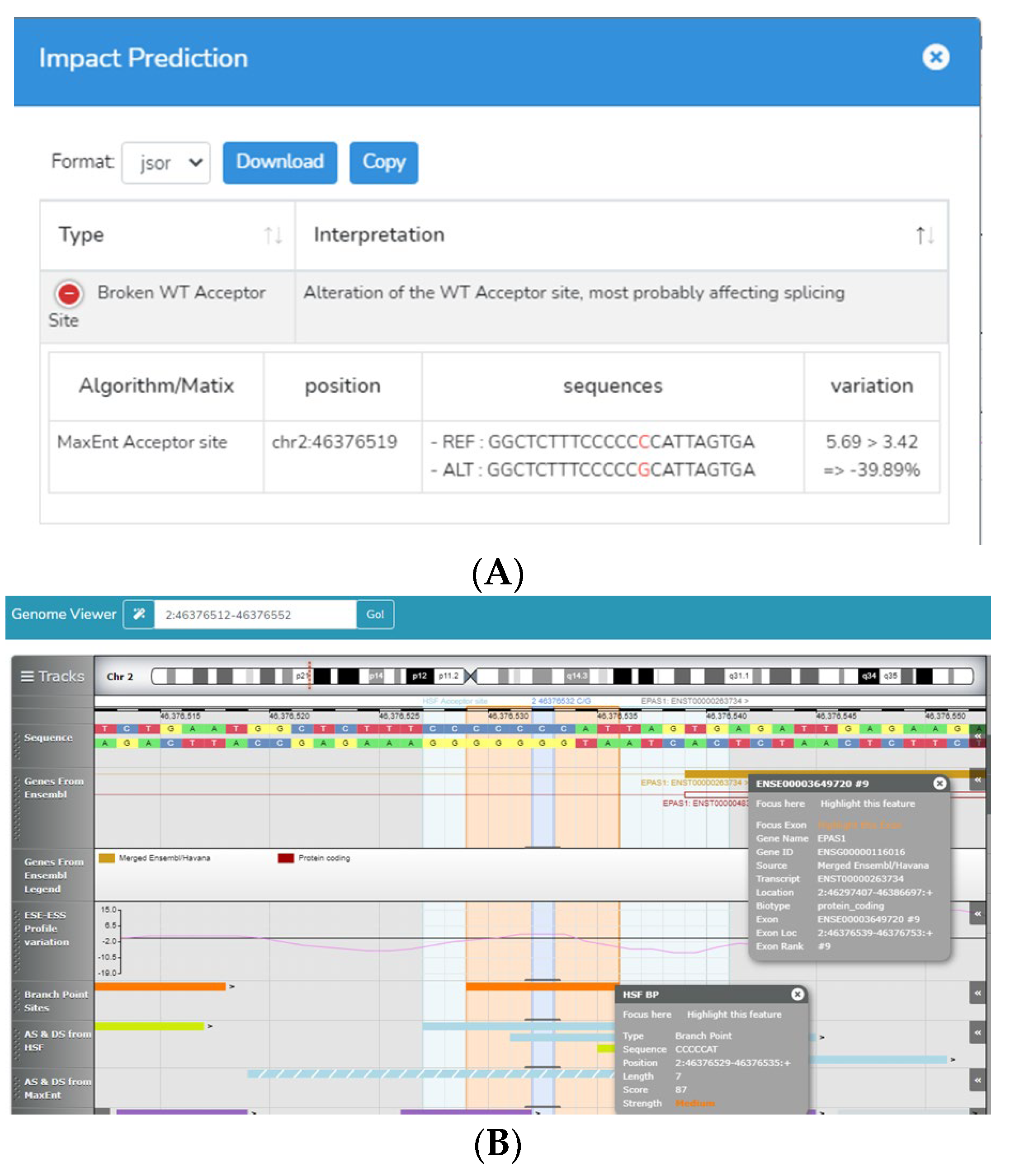
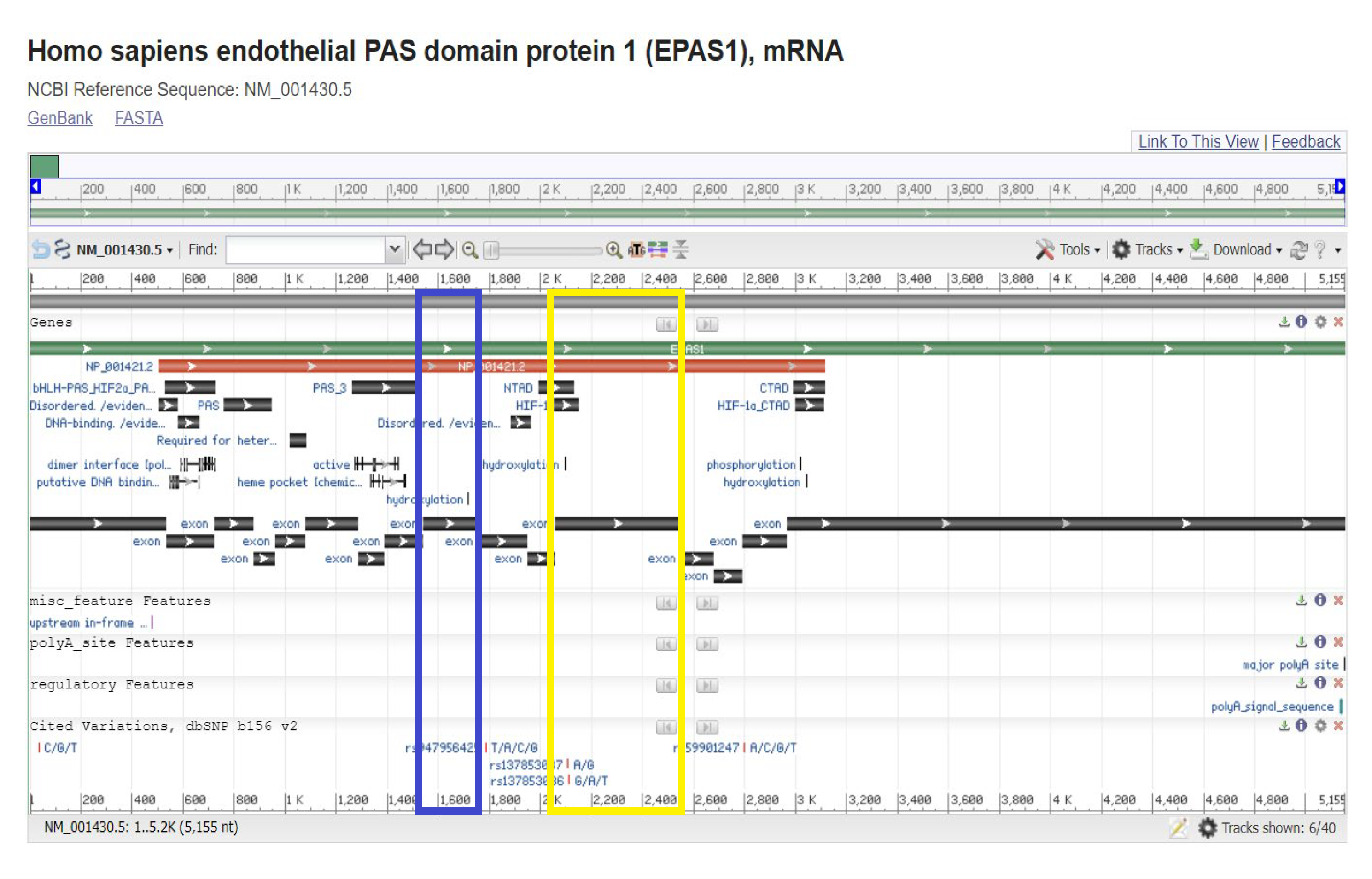
2.4. Bioinformatic Analysis

All variants found were searched in the NCBI databases dbSNP [28] and ClinVar [29] to gather information from previous reports. They were also analyzed with Mutalyzer v.3. software [30] to predict whether they could produce a change in the protein sequence.
The variants that were predicted to alter the protein sequence were analyzed with the PolyPhen v2.0.23 bioinformatic tool [31], which can classify those changes as probably damaging, possibly damaging, or benign. Predictions are based on multiple alignments of proteins that are closely related in function and amino acid sequence to the tested protein.
This tool also considers all available data in the protein databases UniProt [32] and RCSB [33] PDB, such as tridimensional structures and specific protein domain information.
Synonymous variants were analyzed with Human Splicing Finder v.3.0 (HSF) [34], which integrates internal and external detection matrices of splicing sites, branch points, splicing regulatory sequences, etc., to detect site break canonical splicing acceptors and donors, create alternative donor and acceptor sites, analyze branch point breaking, create silencers, and remove enhancers.

2.5. Statistical Analysis

Allele and genotype frequencies of the studied polymorphisms were obtained via direct counting. The Hardy–Weinberg equilibrium (HWE) was calculated using the ꭓ2 test. The significance of the difference between groups was determined using Mantel–Haenzel chi-square analysis. All calculations were performed using SPSS version 18.0 (SPSS, Chicago, IL, USA). Means + SDs and the frequencies of baseline characteristics were calculated. Student’s t-test was performed to compare differences between continuous variables, and the categorical data were analyzed using ꭓ2 and Fisher exact tests. Logistic regression analysis was used to test for the association of polymorphisms with clinical variables under dominant, recessive, codominant, and additive inheritance models in the independent analysis. The most appropriate inheritance model was selected based on Akaike information criteria and was adjusted for gestational age and sex. The statistical power to detect associations with clinical variables was >0.80, as estimated with QUANTO v.1.2.4 software [35]. Pairwise linkage disequilibrium (LD, D’) estimations between polymorphisms and haplotype reconstruction were performed with Haploview version 4.1 [36] (Broad Institute of Massachusetts Institute of Technology and Harvard University, Cambridge, MA, USA).

3. Results

3.1. Population Sample Characteristics

A population sample of 47 patients with a diagnosis of hemodynamically significant PDA participated in the study, and they were divided into two groups: 19 cases (11 males and 8 females) and 28 controls (19 males and 9 females) (Table 1).
Gestational age and birth weight are important risk factors for developing PDA; for this reason, it was necessary to ensure that they were comparable between groups (Table 1). No statistically significant differences were found regarding gestational age (p = 0.112) (Table 1). A significant difference was found with respect to birth weight (p < 0.0001). Therefore, further analysis was adjusted for this variable to avoid spurious results.
We also found statistically significant differences in sepsis development (p < 0.0001), with or without taking the time of development into account, but it was more significant in cases of late development, as observed in Table 1.

3.2. Genetic Variant Detection

We found 63 genetic variants in the population sample: 24 only in the case group, 26 only in the control group, and 13 in both groups (Table 2).
Of the 24 variants found in the case group, 13 were synonymous, 6 were missense, and 5 were INDEL. Only 11 variants resulted in changes in the sequence of amino acid residues in the protein.
In the control group, we found 19 synonymous variants, 7 missense variants, and no INDEL variants (26 altogether, as mentioned before). In this group, only 7 variants affected the protein.
The frequency of genetic variants that modify the sequence of amino acids was higher in the case group, especially considering that this group is smaller. Moreover, this group contained all INDEL variants found. INDEL variants generally cause a shift in the open reading frame, completely changing the amino acid sequence, and can result in premature stop codons.
The statistical analysis of the variants shared by both groups showed no association of TFAP2B variants with treatment response (Table 2).
Only the variant NG_016000.1:g.84131C>G, present in the EPAS1 gene, eighth intron, previously reported as rs7557402, was associated with a risk factor for failure to respond to treatment (p value of 0.007, OR of 3.53, and 95% confidence level). It was found in both groups of the study and in the samples of the PDA family used for the standardized techniques employed in this study.
Because we found the three genotypes present in our study population, it was possible to test haplotype association with treatment response, finding that the recessive model (homozygosity) was strongly associated with treatment failure (p value of 0.017 and 95% confidence level), unlike the dominant and codominant models, which were not statistically associated (Table 3).
According to the Mutalyzer analysis [30], this variant produces no change in amino acid sequence; therefore, it could not be analyzed using the PolyPhen2 bioinformatic tool [31].
Instead, it was analyzed with Human Splicing Finder 3.0 (HSF) [34]. HSF analysis showed that this nucleotide change generates the disruption of the wild-type acceptor splice site located in the intron 8 acceptor splice site (Figure 1).
This variant has been reported as probably benign for familial erythrocytosis by the Illumina Clinical Services Laboratory. The classification does not have a clinical basis, but a criteria classification basis, according to ACGM (American College of Medical Genetics and Genomics) guidance [37].
The analysis of the codifying sequence of EPAS1 showed that exon 9 codes for a hydroxylation site and that it is necessary for the covalent post-translational processing of the transcription factor (Figure 2).

4. Discussion

PDA has a high frequency in the Mexican population, especially in preterm infants [3]. At birth, hemodynamically significant PDA is treated with COX inhibitors, and oral ibuprofen is the treatment of choice at the National Institute of Perinatology, but the use of these types of drugs generates adverse effects that can further complicate patient health. Therefore, the search for biological markers for predicting the efficacy of pharmacological treatment in preterm newborns could avoid unnecessary exposure to the adverse effects of drugs. TFAP2B is the most studied gene in the development of PDA, since it was described as being part of Char syndrome, the phenotype of which may include PCA [38,39]. Nevertheless, the use of massive sequencing and bioinformatic tools has made it possible to identify new target genes for this disease, such as EPAS1. This gene encodes a transcription factor induced when oxygen levels fall. It is part of the molecular pathway necessary to close the ductus arteriosus and is regulated by oxygen levels [40]. A higher number of variants in both genes in the case group and the presence of variants such as rs7557402 indicates a weak but measurable association with a lack of response to pharmacological treatment, highlighting the need for the further and deeper study of this population.
The rs7557402 variant has been reported as probably benign according to the American College of Medical Genetics and Genomics (ACGM) criteria. This variant has been associated with familial erythrocytosis by the Illumina Clinical Services Laboratory [21]. Some other variants in EPAS1 have been previously reported as benign and are also associated with familial erythrocytosis because they decrease gene product function. These variants have a positive effect on familial erythrocytosis since the overexpression of EPAS1 is a key factor for uncontrolled erythrocyte proliferation [20].
It is possible that the variant described as “benign” for familial erythrocytosis may be associated with a decrease in EPAS1 activity. This effect is inconvenient for ductal closure, which requires cell proliferation, migration, and differentiation. A decrease in EPAS1 gene activity would negatively affect all of these processes [22,23]. The activity of EPAS1 could be diminished since the analysis with HSF3 v. 3.0. software showed that the C>G substitution in intron 8 leads to the breakdown of an acceptor site. The ultimate effect is faulty splicing of the mRNA, and therefore a defective protein.
Inside the EPAS1 exon 9 sequence, there is a hydroxylation site. EPAS1 (or HIF-2α) belongs to the family of hypoxia-inducible factors (HIFs). These factors are formed by two subunits, one with nuclear localization HIF-β and the other with cytoplasmic localization HIF-α. HIF-2α possesses 48% amino acid identity with HIF-1α; it is regulated by prolyl hydroxylation, dimerizes with HIF-2β, and binds to the same target DNA sequence (5′-RCGTG-3′) as the HIF-1α:HIF-1β heterodimer. The sets of genes regulated by HIF-1 and HIF-2 overlap, but some are specific and depend on cell type [41]. At physiological oxygen levels (normoxia), HIF-prolyl hydroxylases (PHDs) hydroxylate proline residues on HIF-α subunits, leading to their destabilization by promoting ubiquitination via the von Hippel Lindau (VHL) ubiquitin ligase and subsequent proteasomal degradation [42]. Functional specificity for transactivation via HIF-1α and HIF-2α appears to reside in amino acid residues 415 to 659 and 418 to 619, respectively [41]. HIF-α transactivation is also repressed in an O2-dependent manner due to asparaginyl hydroxylation via the factor-inhibiting HIF (FIH). In hypoxia, the O2-dependent hydroxylation of HIF-α subunits via PHDs and FIH is reduced, resulting in HIF-α accumulation, dimerization with HIF-β, and migration into the nucleus to induce an adaptive transcriptional response [42]. In this case, the covalent modification determines where the protein should be located according to the cellular conditions. HIF-2α hydroxylation sites are located inside exons 9 and 12 (Figure 2). However, how these hydroxylation sites are regulated through space and time is not very clear. One possibility is that the rs7557402 variant disrupts the hydroxylation site encoded in exon 9, leading to the increased translocation of HIF-2α into the nucleus and triggering the expression of genes related to the hypoxia response, which will result in the opposite response as expected. Another possible explanation could be related to a reduced rate of HIF-2α degradation secondary to deficient ubiquitination and degradation by the proteasome. The lack of renewal of HIF-2α could compromise its function. Under normal conditions, HIF-α is continuously synthesized and degraded [42]. In addition to PHD-conferring alterations in protein stability, there is now evidence that hydroxylation can affect protein activity and protein/protein interactions with respect to alternative substrates; therefore, if the hydroxylation sites are lost, HIF-2α activity could be affected [41].
The variant rs7557402 has a CADD score for the G allele of 7.555; this score indicates that the variant is likely benign. In addition, the GERP score is 1.33, which means that the variant is highly conserved among species. We performed an alignment including ten species of mammals, and all of them but the capra hircus showed conservation of the C allele [43]. Furthermore, the analysis with RegulomeDB v.2 data showed that the rs7557402 variant generates more compacted chromatin, resulting in a weaker transcription of the EPAS1 gene in the fetal heart [44].
It is also important to highlight the presence of rs7557402 in the samples of the family used for the standardization of the techniques employed in this study. These samples were not part of the study because we do not have enough information about the treatments employed before closure via catheterization for each case. Nevertheless, we could infer that they ultimately required invasive treatment for the closure of the PDA, as in our case group. Moreover, the presence of this variant in a family case corroborates the effects of genetic factors other than the TFAP2B gene on PDA development and, in this particular case, on the lack of response to pharmacological treatment and the need to close PDA using invasive treatment, such as surgery or catheterization.
These results should be interpreted with caution because of the limited sample size. This study must be replicated in a larger population to confirm or reject the association.

5. Conclusions

The rs7557402 SNP is associated with the failure of pharmacological treatment for PDA closure, specifically with ibuprofen in our study population. A possible explanation could be the decrease in the activity of the EPAS1 gene associated with the variant. Further studies with a larger sample size are needed to strengthen this association and decide whether this polymorphism could be used as a prognostic biomarker for pharmacological response.
Moreover, our findings suggest the need to study other genetic factors involved in PDA development in addition to TFAP2B in preterm newborns.

Supplementary Materials

The following supporting information can be downloaded at: https://www.mdpi.com/article/10.3390/diagnostics13152558/s1. Supplementary Figure S1: Transthoracic echocardiogram with bidimensional and color Doppler modalities that shows a short-axis view of the heart with a hemodynamically significant patent ductus arteriosus. Supplementary Figure S2: Transthoracic echocardiogram with bidimensional and color Doppler modalities that shows a sagittal view of a hemodynamically significant patent ductus arteriosus. Supplementary Figure S3: Transthoracic echocardiogram with bidimensional and color Doppler modalities that shows a short-axis view of the heart of a patient that underwent pharmacologic treatment for the closure of the ductus arteriosus. The image shows laminar flow in the pulmonary artery without any residual shunt.

Author Contributions

Conceptualization, D.G.R.-A., P.G.-G., D.A.M.-B., J.A.-M. and I.E.M.-M.; methodology, D.G.R.-A., V.D.V.-J., D.A.M.-B., A.d.J.M.-G., M.O.L.E.-A., J.A.-M., A.H.-B. and I.E.M.-M.; software, D.G.R.-A., V.D.V.-J., J.A.-M., A.H.-B. and I.E.M.-M.; validation, J.A.-M., J.A.-V., G.V.-A. and I.E.M.-M.; formal analysis, D.G.R.-A., J.E.M.-M., J.A.-M., A.H.-B. and I.E.M.-M.; investigation, D.G.R.-A., J.E.M.-M., V.D.V.-J., D.A.M.-B., R.S.-M. and I.E.M.-M.; resources, P.G.-G., D.A.M.-B. and I.E.M.-M.; data curation, D.G.R.-A., J.E.M.-M., V.D.V.-J., A.M.-J., A.H.-B. and I.E.M.-M.; writing—original draft preparation, D.G.R.-A., J.E.M.-M., V.D.V.-J., D.A.M.-B., A.d.J.M.-G., M.O.L.E.-A., J.A.-M. and I.E.M.-M.; writing—review and editing, R.S.-M., J.A.-V., G.V.-A., A.H.-B. and I.E.M.-M.; visualization, D.G.R.-A., V.D.V.-J. and I.E.M.-M.; supervision, I.E.M.-M.; project administration, I.E.M.-M.; funding acquisition, J.E.M.-M. and I.E.M.-M. All authors have read and agreed to the published version of the manuscript.

Funding

All reagents and consumables required for sample analysis were purchased through institutional funding, with the National Institute of Perinatology project number 212250-3140-11105-01-16.

Institutional Review Board Statement

The study complies with the Declaration of Helsinki and was approved by the Ethics Committee of the National Institute of Perinatology of Mexico City. The study was approved in 2016 with the project number 212250-3140-11105-01-16.

Informed Consent Statement

On behalf of the children enrolled in our study, we obtained written informed consent from their legal guardians.

Data Availability Statement

All data generated or analyzed during this study are included in this published article.

Acknowledgments

We would like to thank all heads and technical teams of the Neonatal Intensive Care Unit and Newborn Intermediate Care Unit of the National Institute of Perinatology.

Conflicts of Interest

The authors declare no conflict of interest.

Sample Availability

No samples are available from the authors.

Abbreviations

The following abbreviations are used in this manuscript:
PDAPatent ductus arteriosus
EPAS1Endothelial PAS domain protein 1
SNPSingle-nucleotide polymorphism
OROdds ratio
HRMHigh-resolution melting
HSFHuman splicing finder
ACGMAmerican College of Medical Genetics and Genomics
COXCyclooxygenase
TFAP2BTranscription factor AP-2 beta
HIFHypoxia-inducible factor
PHDProlyl hydroxylase

References

  1. Gillam-Krakauer, M.; Reese, J. Diagnosis and Management of Patent Ductus Arteriosus. Neoreviews 2018, 19, e394–e402. [Google Scholar] [CrossRef] [PubMed]
  2. Hamrick, S.E.G.; Sallmon, H.; Rose, A.T.; Porras, D.; Shelton, E.L.; Reese, J.; Hansmann, G. Patent Ductus Arteriosus of the Preterm Infant. Pediatrics 2020, 146, e20201209. [Google Scholar] [CrossRef] [PubMed]
  3. Cordero-González, G.; Gómez-Tamayo, T.; Santillán-Briceño, V.; Machuca-Vacac, A.; Fernández-Carrocera, L.A. Experience with ibuprofen in treatment of patent ductus arteriosus in a tertiary hospital in Mexico City. Perinatol. Reprod. Hum. 2016, 30, 115–121. [Google Scholar]
  4. Parkerson, S.; Philip, R.; Talati, A.; Sathanandam, S. Management of Patent Ductus Arteriosus in Premature Infants in 2020. Front. Pediatr. 2021, 8, 590578. [Google Scholar] [CrossRef] [PubMed]
  5. Gonzalez-Luis, G.E.; Borges-Lujan, M.; Villamor, E. Association between endotypes of prematurity and pharmacological closure of patent ductus arteriosus: A systematic review and meta-analysis. Front. Pediatr. 2023, 11, 1078506. [Google Scholar] [CrossRef] [PubMed]
  6. Polania-Rodríquez, M.H.; Rodríguez-Terán, G.; Zamorano-Jiménez, C.; ASánchez-Velázquez, L.D. Diagnóstico ecocardiográfico de persistencia del conducto arterioso en recién nacidos hospitalizados en la Unidad de Cuidados Intensivos Neonatales. An. Médicos 2015, 60, 185–190. [Google Scholar]
  7. Jiang, X.; Rowitch, D.H.; Soriano, P.; McMahon, A.P.; Sucov, H.M. Fate of the mammalian cardiac neural crest. Development 2000, 127, 1607. [Google Scholar] [CrossRef]
  8. Vargas Dornelles, L.; Lúcia-Corso, A.; de-Cássia-Silveira, R.; Soibelmann-Procianoy, R. Comparison of two dose regimens of ibuprofen for the closure of patent ductus arteriosus in preterm newborns. J. Pediatr. 2016, 92, 314–318. [Google Scholar] [CrossRef] [Green Version]
  9. Backes, C.H.; Hill, K.D.; Shelton, E.L.; Slaughter, J.L.; Lewis, T.R.; Weisz, D.E.; Mah, M.L.; Bhombal, S.; Smith, C.V.; McNamara, P.J.; et al. Patent Ductus Arteriosus: A Contemporary Perspective for the Pediatric and Adult Cardiac Care Provider. J Am Heart Assoc. 2022, 11, e025784. [Google Scholar] [CrossRef]
  10. Mitra, S.; de Boode, W.P.; Weisz, D.E.; Shah, P.S. Interventions for patent ductus arteriosus (PDA) in preterm infants: An overview of Cochrane Systematic Reviews. Cochrane Database Syst. Rev. 2023, 4, CD013588. [Google Scholar] [CrossRef]
  11. Mitra, S.; Gardner, C.E.; MacLellan, A.; Disher, T.; Styranko, D.M.; Campbell-Yeo, M.; Kuhle, S.; Johnston, B.C.; Dorling, J. Prophylactic cyclo-oxygenase inhibitor drugs for the prevention of morbidity and mortality in preterm infants: A network meta-analysis. Cochrane Database Syst. Rev. 2022, 4, CD013846. [Google Scholar]
  12. Katsaras, D.N.; Katsaras, G.N.; Chatziravdeli, V.I.; Papavasileiou, G.N.; Touloupaki, M.; Mitsiakos, G.; Doxani, C.; Stefanidis, I.; Dardiotis, E. Comparative safety and efficacy of paracetamol versus non-steroidal anti-inflammatory agents in neonates with patent ductus arteriosus: A systematic review and meta-analysis of randomized controlled trials. Br. J. Clin. Pharmacol. 2022, 88, 3078–3100. [Google Scholar] [CrossRef]
  13. Dagle, J.M.; Lepp, N.T.; Cooper, M.E.; Schaa, L.; Kelsey, K.J.P.; Orr, K.L.; Murray, J.C. Determination of genetic predisposition to patent ductus arteriosus in preterm infants. NIH Public Access 2009, 123, 1116–1123. [Google Scholar] [CrossRef] [Green Version]
  14. Mitra, S.; Weisz, D.; Jain, A.; Jong, G.’t. Management of the patent ductus arteriosus in preterm infants. Paediatr. Child Health 2022, 27, 63–64. [Google Scholar] [CrossRef]
  15. Engbers, A.G.J.; Völler, S.; Flint, R.B.; Goulooze, S.C.; de Klerk, J.; Krekels, E.H.J.; van Dijk, M.; Willemsen, S.P.; Reiss, I.K.M.; Knibbe, C.A.J.; et al. The Effect of Ibuprofen Exposure and Patient Characteristics on the Closure of the Patent Ductus Arteriosus in Preterm Infants. Clin. Pharmacol. Ther. 2022, 112, 307–315. [Google Scholar] [CrossRef]
  16. Ivey, N.K.; Sutcliffe, D.; Richardson, J.; Clyman, R.I.; Garcia, J.A.; Srivastava, D. Transcriptional Regulation During Development of the Ductus Arteriosus. Circ. Res. 2008, 103, 388–395. [Google Scholar] [CrossRef]
  17. Parikh, P.; Bai, H.; Swartz, M.F.; Alfieris, G.M.; Dean, D.A. Identification of differentially regulated genes in human patent ductus arteriosus. Exp. Biol. Med. 2016, 241, 2112–2118. [Google Scholar] [CrossRef]
  18. Chen, Y.W.; Zhao, W.; Zhang, Z.F.; Fu, Q.; Shen, J.; Zhang, Z.; Ji, W.; Wang, J.; Li, F. Familial nonsyndromic patent ductus arteriosus caused by mutations in TFAP2B. Pediatr. Cardiol. 2011, 32, 958. [Google Scholar] [CrossRef]
  19. Illumina Clinical Services Laboratory (Illumina), ICSL. 2018. Available online: https://www.ncbi.nlm.nih.gov/clinvar/submitters/504895/ (accessed on 22 July 2022).
  20. Perrotta SStiehl, P.; Punzo, F.; Scianguetta, S.; Borriello ABencivenga, D.; Casale, M.; Nobili, B.; Fasoli, S.; Balduzzi, A.; Cro, A.; Nytko, K.; et al. Congenital erythrocytosis associated with gain-of-function HIF2A gene mutations and erythropoietin levels in the normal range. Haematologica 2013, 98, 1624. [Google Scholar] [CrossRef]
  21. Hajj, H.; Dagle, J.M. Genetics of Patent Ductus Arteriosus Susceptibility and Treatment to PDA Gene Discovery. YSPER 2012, 36, 98–104. [Google Scholar]
  22. Tanaka, T.; Akiyama, H.; Kanai, H.; Sato, M.; Takeda, S.; Sekiguchi, K.; Yokoyama, T.; Kurabayashi, M. Endothelial PAS Domain Protein 1 (EPAS1) Induces Adrenomedullin Gene Expression in Cardiac Myocytes: Role of EPAS1 in an Inflammatory Response in Cardiac Myocytes. J. Mol. Cell. Cardiol. 2002, 34, 748. [Google Scholar] [CrossRef] [PubMed]
  23. Wu, J.R.; Yeh, J.L.; Liou, S.F.; Dai, Z.K.; Wu, B.N.; Hsu, J.H. Gamma-secretase inhibitor prevents proliferation and migration of ductus arteriosus smooth muscle cells through the Notch3-HES1/2/5 pathway. Int. J. Biol. Sci. 2016, 12, 1063–1073. [Google Scholar] [CrossRef] [PubMed]
  24. National Center for Biotechnology Information. Primer Blast Platform. 2012. Available online: https://www.ncbi.nlm.nih.gov/tools/primer-blast/ (accessed on 22 July 2022).
  25. NCBI Reference Sequence Database. 2000. Available online: https://www.ncbi.nlm.nih.gov/refseq/ (accessed on 22 July 2022).
  26. NCBI. Nucleotide Blast platform. 2022. Available online: https://blast.ncbi.nlm.nih.gov/Blast.cgi?PROGRAM=blastx&PAGE_TYPE=BlastSearch&LINK_LOC=blasthome (accessed on 22 July 2022).
  27. Translate Tool. ExPASy Bioinformatics Resource Portal. 1993. Available online: https://web.expasy.org/translate/ (accessed on 22 July 2022).
  28. NCBI. Single-Nucleotide Polymorphism Database (dbSNP). 1998. Available online: https://www.ncbi.nlm.nih.gov/snp/ (accessed on 22 July 2022).
  29. NCBI. ClinVar Database. 2012. Available online: https://www.ncbi.nlm.nih.gov/clinvar/ (accessed on 22 July 2022).
  30. Mutalyzer Website. 2021. Available online: https://mutalyzer.nl/webservices (accessed on 22 July 2022).
  31. PolyPhen-2 (Polymorphism Phenotyping v2). 2010. Available online: http://genetics.bwh.harvard.edu/pph2/ (accessed on 22 July 2022).
  32. The Uniprot Consortium. 2002. Available online: https://www.uniprot.org/ (accessed on 22 July 2022).
  33. RCSB PDB. 2000. Available online: https://www.rcsb.org (accessed on 22 July 2022).
  34. Human Splicing Finder. 2009. Available online: http://www.umd.be/HSF3/ (accessed on 22 July 2022).
  35. Quanto Software. 2007. Available online: http://hydra.usc.edu/GxE/ (accessed on 22 July 2022).
  36. Haploview Software. 2005. Available online: https://www.broadinstitute.org/haploview/haploview (accessed on 22 July 2022).
  37. Practice Guidelines of the American College of Medical Genetics and Genomics. 2007. Available online: https://www.acmg.net/ACMG/Medical-Genetics-Practice-Resources/Practice-Guidelines.aspx (accessed on 22 July 2022).
  38. Martínez-Roque, A.M. Repercusión hemodinámica en pacientes neonatos con conducto arterioso persistente: Factores asociados. Arch Cardiol Mex. 2017, 87, 248–251. [Google Scholar] [CrossRef] [PubMed]
  39. Wei, J.; Benson, M.A.; Bhattacharya, S.; Chen, Y.; Hu, J.; Li, F. Characterization of transcription factor AP-2 beta mutations involved in familial isolated patent ductus arteriosus suggests haploinsufficiency. J. Surg. Res. 2014, 188, 466–472. [Google Scholar]
  40. Hamrick, S.E.G.; Hansmann, G. Patent Ductus Arteriosus of the Preterm Infant. Pediatrics 2010, 125, 1019–1030. [Google Scholar] [CrossRef] [Green Version]
  41. Semenza, G.L. Hypoxia-inducible factor 1 (HIF-1) pathway. Sci. STKE 2007, 407, cm8. [Google Scholar] [CrossRef]
  42. Strowitzki, M.J.; Cummins, E.P.; Taylor, C.T. Protein Hydroxylation by Hypoxia-Inducible Factor (HIF) Hydroxylases: Unique or Ubiquitous? Cells 2019, 8, 384. [Google Scholar] [CrossRef] [Green Version]
  43. Ensembl Genome Browser. 1999. Available online: https://www.ensembl.org/Homo_sapiens/Variation/Explore?db=core;r=2:46376032-;v=rs7557402;vdb=variation;vf=18484625 (accessed on 13 July 2023).
  44. Regulome DB v.2. 2012. Available online: https://regulomedb.org/regulome-search?regions=chr2%3A46376531-46376532&genome=GRCh38 (accessed on 13 July 2023).
Figure 1. Prediction of the NG_016000.1:g.84131C>G variant effect made with HSF3 v. 3.0. software. (A) Impact prediction showed that this variant alters a wild-type acceptor site by disrupting it. (B) The genome viewer showed that the site where the variant is located is an acceptor site with a medium force, whose disruption most likely affects the splicing of the EPAS1 gene 8th intron.
Diagnostics 13 02558 g001
Figure 2. Analysis of the codifying sequence of EPAS1. The codifying sequence NM_001430.5 showed the presence of two hydroxylation sites, the first one located in exon 9, highlighted with the blue rectangle, and the second one highlighted with the yellow rectangle. Both sites encode a 4-hydroxyproline.
Diagnostics 13 02558 g002
Table 1. Demographic characteristics of the population.
ControlCasep
Mother
Hypertension n (%) 9 (32.1)7 (36.8)0.763
Preeclampsia n (%) 9 (32.1)5 (26.3)0.753
T2D n (%) 4 (14.3)00.137
Prenatal infections n (%) 12(42.9)9 (47.4)0.775
Prenatal treatmentIndomethacin n (%)01 (5.3)0.404
Steroid n (%)20 (76.9)13 (68.4)0.734
Newborn
Sex F/M n 9/198/110.546
Gestational age 30 ± 2.328.8 ± 2.80.112
Birth weight (g) 1229 ± 349928 ± 154<0.0001
Ductal diameter (mm) 2.9 ± 0.92.9 ± 0.80.765
Sepsis n (%) 16 (57.1)1 (5.3)<0.0001
Early n (%)7 (25)00.032
Late n (%)10 (35.7)00.003
Data are expressed as means ± SD or frequencies. The p values were calculated by using Student’s t-test and chi-square/Fisher exact test where appropriate to compare among groups. F: female, M: male. T2D: Type 2 Diabetes. Statistically significant values are highlighted in bold.
Table 2. Comparison of genotype and allele frequencies of variants between the case and control groups.
Genotype Frequency (%)TFAP2BAllelic Frequency (%)
9757ATA/AA/TT/Tp *ATp **ORc(IC 95%)p HW
Control (n = 28)23 (82)5 (18)0 (0) 51 (91)5 (9) 1
Case (n = 19)19 (100)0 (0)0 (0)0.01838 (100)0 (0)0.0032--1
29461ATA/AA/TT/T AT
Control (n = 28)24 (85.7)4 (14.3)0 (0) 52 (93)4 (7) 1
Case (n = 19)17 (89.5)2 (10.5)0 (0)0.7036 (95)2 (5)0.7670.6990.21–2.281
EPAS1
rs7557402C/CC/GG/G CG
Control (n = 28)24 (85.7)4 (14.3)0 (0) 52 (93)4 (7) 1
Case (n = 19)14 (73.7)2 (10.5)3 (15.8)0.0530 (79)8 (21)0.0073.531.42–8.740.0096
54455C>TC/CC/TT/T CT
Control (n = 28)24 (85.7)4 (14.3)0 (0) 52 (93)4 (7) 1
Case (n = 19)17 (89.5)1 (5.3)1 (5.3)0.2535 (92)3 (8)11.150.402–3.310.08
54522G>AG/GG/AA/A GA
Control (n = 28)24 (85.7)1 (3.6)3 (10.7) 49 (88)7 (12) 0.0007
Case (n = 19)16 (84.2)0 (0)3 (15.8)0.5332 (84)6 (16)0.5411.390.62–3.120.0003
rs769385664G/GG/AA/A GA
Control (n = 28)26 (92.6)1 (3.6)1 (3.6) 53 (95)3 (5) 0.055
Case (n = 19)16 (84.2)3 (15.8)0 (0)0.2135 (92)3 (8)0.5671.650.521–5.231
54699C>AC/CC/AA/A CA
Control (n = 28)21 (75)5 (17.9)2 (7.1) 47 (84)9 (16) 0.11
Case (n = 19)13 (68.4)4 (21.1)2 (10.5)0.8730 (79)8 (21)0.4661.390.679–2.860.14
54700A>CA/AA/CC/C AC
Control (n = 28)22 (78.6)5 (17.9)1 (3.6) 49 (88)7 (12) 0.35
Case (n = 19)14 (73.7)4 (21.1)1 (5.3)0.9232 (84)6 (16)0.5411.390.623–3.1270.37
54701G>CG/GG/CC/C GC
Control (n = 28)19 (67.9)8 (28.6)1 (3.6) 46 (82)10 (18) 1
Case (n = 19)14 (73.7)5 (26.3)0 (0)0.5733 (87)5 (13)0.4340.680.313–1.4761
rs1286681984C/CC/AA/A CA
Control (n = 28)24 (85.7)4 (14.3)0 (0) 52 (93)4 (7) 1
Case (n = 19)16 (84.2)2 (10.5)1 (5.3)0.3834 (89)4 (11)0.4591.640.61–4.420.16
54703C>GC/CC/GG/G CG
Control (n = 28)24 (85.7)4 (14.3)0 (0) 52 (93)4 (7) 1
Case (n = 19)17 (89.5)2 (10.5)0 (0)0.736 (95)2 (5)0.7670.6990.21–2.281
54707C>GC/CC/GG/G CG
Control (n = 28)25 (89.3)3 (10.7)0 (0) 53 (95)3 (5) 1
Case (n = 19)17 (89.5)2 (10.5)0 (0)0.9836 (95)2 (5)110.28–3.561
rs762559920G/GG/CC/C GC
Control (n = 28)27 (96.4)1 (3.6)0 (0) 55 (98)1 (2) 1
Case (n = 19)16 (84.2)3 (15.8)0 (0)0.1435 (92)3 (8)0.14.260.88–20.591
Data are expressed as frequencies. The p values were calculated using * ꭓ2 or ** Fisher’s exact test. Statistically significant values are highlighted in bold.
Table 3. rs7557402 association analysis: case-control.
ModelGenotypeCaseControlOR (95% CI)p Value
CodominantC/C14 (73.7%)24 (85.7%) 0.056
C/G2 (10.5%)4 (14.3%)1.17 (0.19–7.21)
G/G3 (15.8%)0 (0%)0.00 (0.00-NA)
DominantC/C14 (73.7%)24 (85.7%) 0.31
C/G-G/G5 (26.3%)4 (14.3%)0.47 (0.11–2.03)
RecessiveC/C-C/G16 (84.2%)28 (100%) 0.017
G/G3 (15.8%)0 (0%)0.00 (0.00-NA)
OverdominantC/C-G/G17 (89.5%)24 (85.7%)10.7
C/G2 (10.5%)4 (14.3%)1.42 (0.23–8.64)
Additive---0.41 (0.13–1.26)0.098
Statistical significant values are highlighted in bold.
Disclaimer/Publisher’s Note: The statements, opinions and data contained in all publications are solely those of the individual author(s) and contributor(s) and not of MDPI and/or the editor(s). MDPI and/or the editor(s) disclaim responsibility for any injury to people or property resulting from any ideas, methods, instructions or products referred to in the content.

