

Computed Tomography-Based Coronary Artery Calcium Score Calculation at a Reduced Tube Voltage Utilizing Iterative Reconstruction and Threshold Modification Techniques: A Feasibility Study
 , , and
Abstract
:1. Introduction
2. Materials and Methods
2.1. Patients
2.2. Data Collection
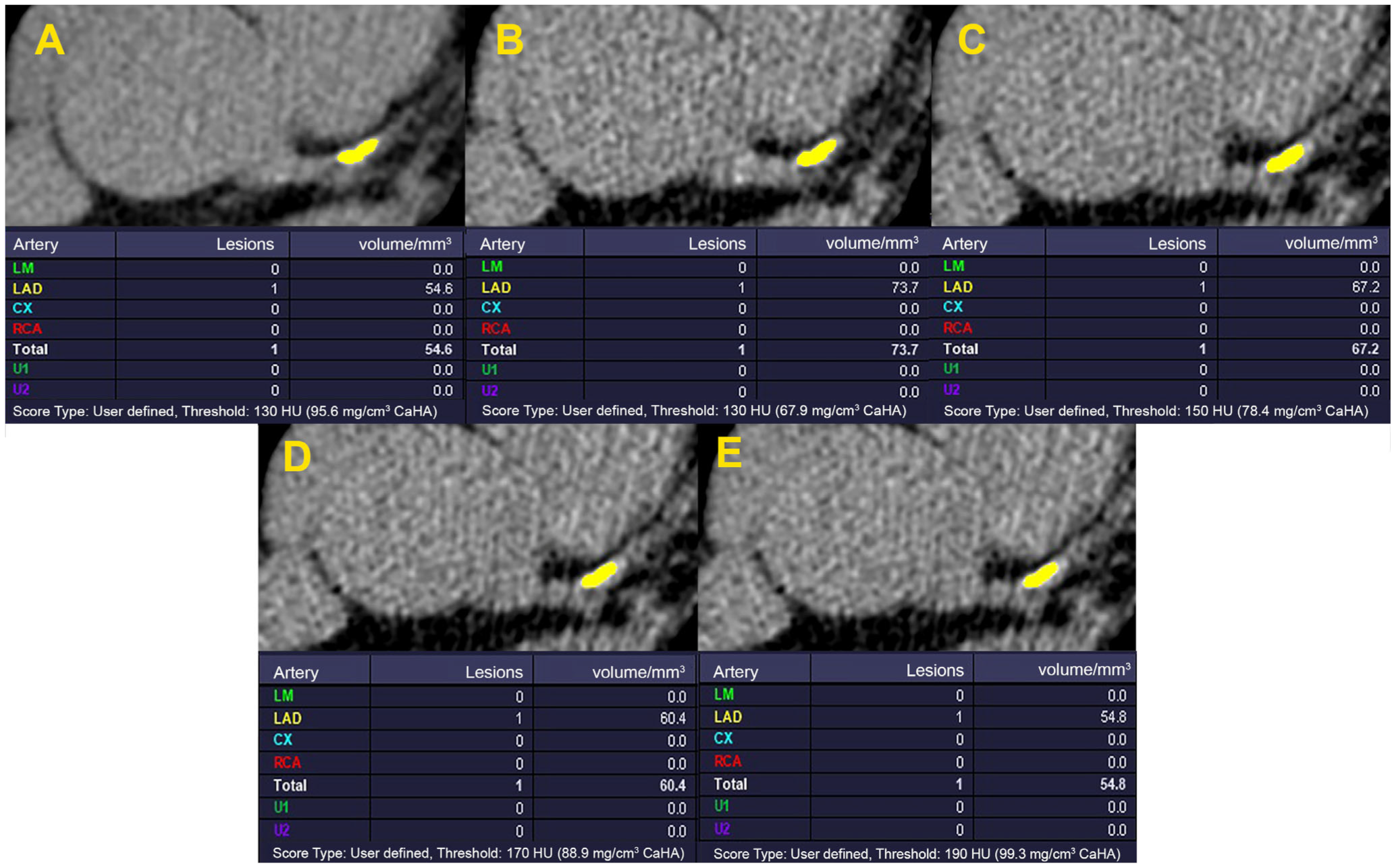
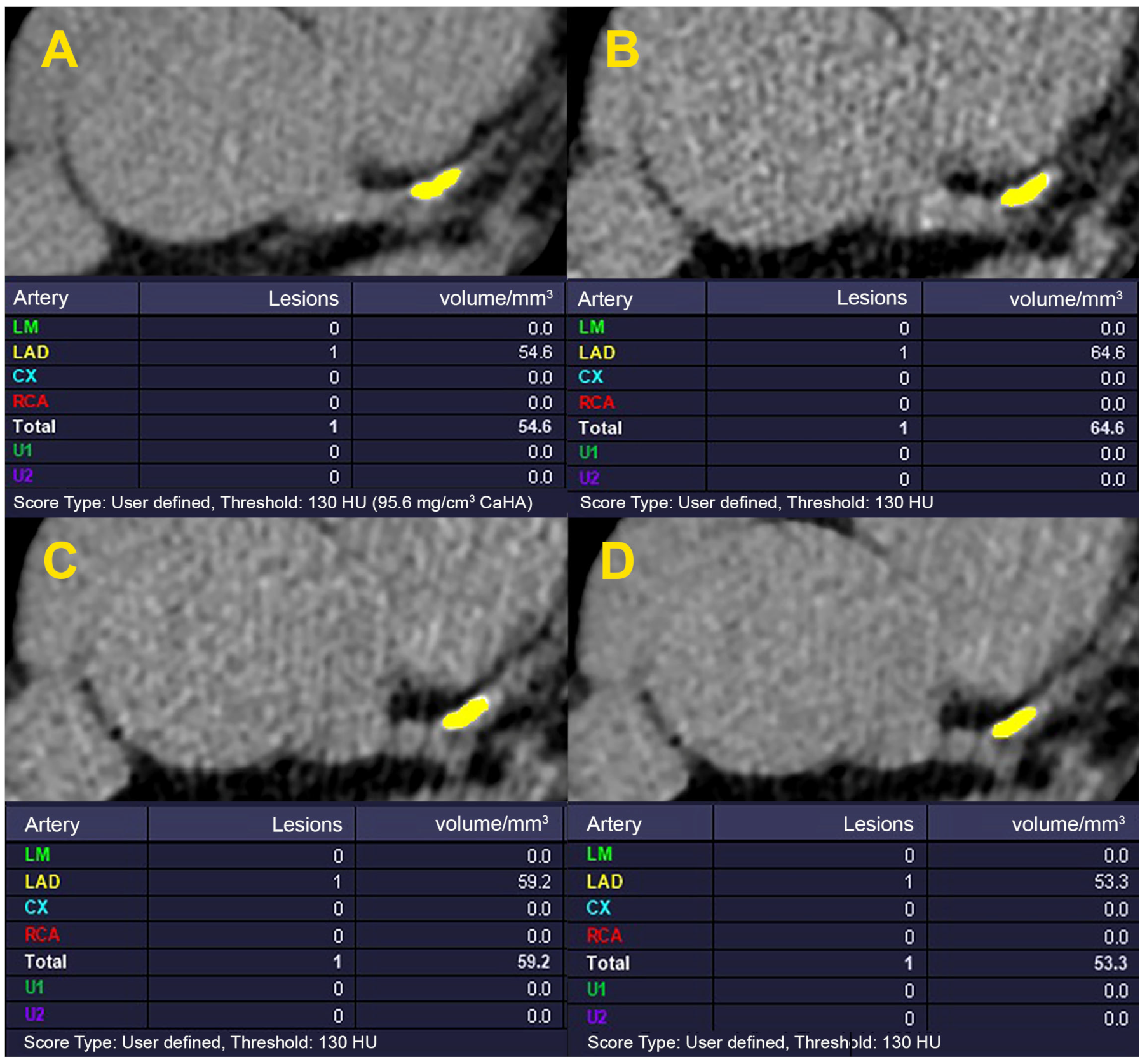
2.3. Classification of CACS
2.4. Statistical Analysis
3. Results
4. Discussion
5. Conclusions
Author Contributions
Funding
Institutional Review Board Statement
Informed Consent Statement
Data Availability Statement
Acknowledgments
Conflicts of Interest
References
- Heron, M.P. Deaths: Leading causes for 2017. NVSS 2019, 68, 6. [Google Scholar]
- Lopez, A.D.; Mathers, C.D. Measuring the global burden of disease and epidemiological transitions: 2002–2030. Ann. Trop. Med. Parasitol. 2006, 100, 481–499. [Google Scholar] [CrossRef]
- Brown, J.C.; Gerhardt, T.E.; Kwon, E. Risk Factors for Coronary Artery Disease; Study Guide from StatPearls Publishing: Treasure Island, FL, USA, 2020. [Google Scholar]
- Sandfort, V.; Bluemke, D.A. CT calcium scoring. History, current status and outlook. Diagn. Interv. Imaging 2017, 98, 3–10. [Google Scholar] [CrossRef] [PubMed]
- Ali, Z.A.; Karimi Galougahi, K.; Mintz, G.S.; Maehara, A.; Shlofmitz, R.A.; Mattesini, A. Intracoronary optical coherence tomography: State of the art and future directions. EuroIntervention 2021, 17, e105–e123. [Google Scholar] [CrossRef] [PubMed]
- Flora, G.D.; Nayak, M.K. A brief review of cardiovascular diseases, associated risk factors and current treatment regimes. Curr. Pharm. Des. 2019, 25, 4063–4084. [Google Scholar] [CrossRef]
- Mahabadi, A.A.; Möhlenkamp, S.; Lehmann, N.; Kälsch, H.; Dykun, I.; Pundt, N.; Moebus, S.; Jöckel, K.-H.; Erbel, R.; Investigators, H.N.R.S. CAC score improves coronary and CV risk assessment above statin indication by ESC and AHA/ACC primary prevention guidelines. JACC Cardiovasc. Imaging 2017, 10, 143–153. [Google Scholar] [CrossRef]
- Mc Namara, K.; Alzubaidi, H.; Jackson, J.K. Cardiovascular disease as a leading cause of death: How are pharmacists getting involved? Integr. Pharm. Res. Pract. 2019, 8, 1–11. [Google Scholar] [CrossRef] [PubMed]
- Roth, G.A.; Mensah, G.A.; Johnson, C.O.; Addolorato, G.; Ammirati, E.; Baddour, L.M.; Barengo, N.C.; Beaton, A.Z.; Benjamin, E.J.; Benziger, C.P. Global burden of cardiovascular diseases and risk factors, 1990–2019: Update from the GBD 2019 study. J. Am. Coll. Cardiol. 2020, 76, 2982–3021. [Google Scholar] [CrossRef]
- Agatston, A.S.; Janowitz, W.R.; Hildner, F.J.; Zusmer, N.R.; Viamonte, M., Jr.; Detrano, R. Quantification of coronary artery calcium using ultrafast computed tomography. J. Am. Coll. Cardiol. 1990, 15, 827–832. [Google Scholar] [CrossRef]
- Rumberger, J.A.; Kaufman, L. A rosetta stone for coronary calcium risk stratification: Agatston, volume, and mass scores in 11,490 individuals. Am. J. Roentgenol. 2003, 181, 743–748. [Google Scholar] [CrossRef]
- Criqui, M.H.; Denenberg, J.O.; Ix, J.H.; McClelland, R.L.; Wassel, C.L.; Rifkin, D.E.; Carr, J.J.; Budoff, M.J.; Allison, M.A. Calcium density of coronary artery plaque and risk of incident cardiovascular events. JAMA 2014, 311, 271–278. [Google Scholar] [CrossRef] [PubMed]
- Adelhoefer, S.; Uddin, S.I.; Osei, A.D.; Obisesan, O.H.; Blaha, M.J.; Dzaye, O. Coronary artery calcium scoring: New insights into clinical interpretation—Lessons from the CAC Consortium. Radiol. Cardiothorac. Imaging 2020, 2, e200281. [Google Scholar] [CrossRef] [PubMed]
- Greenland, P.; Blaha, M.J.; Budoff, M.J.; Erbel, R.; Watson, K.E. Coronary calcium score and cardiovascular risk. J. Am. Coll. Cardiol. 2018, 72, 434–447. [Google Scholar] [CrossRef] [PubMed]
- Kim, K.P.; Einstein, A.J.; De González, A.B. Coronary artery calcification screening: Estimated radiation dose and cancer risk. Arch. Intern. Med. 2009, 169, 1188–1194. [Google Scholar] [CrossRef]
- Cheong, B.; Wilson, J.; Spann, S.; Pettigrew, R.; Preventza, O.; Muthupillai, R. Coronary artery calcium scoring: An evidence-based guide for primary care physicians. J. Intern. Med. 2021, 289, 309–324. [Google Scholar] [CrossRef]
- Matos, D.; Ferreira, A.M.; de Araújo Gonçalves, P.; Gama, F.; Freitas, P.; Guerreiro, S.; Cardoso, G.; Tralhão, A.; Dores, H.; Abecasis, J. Coronary artery calcium scoring and cardiovascular risk reclassification in patients undergoing coronary computed tomography angiography. Rev. Port. Cardiol. 2021, 40, 25–30. [Google Scholar] [CrossRef] [PubMed]
- Nasir, K.; Cainzos-Achirica, M. Role of coronary artery calcium score in the primary prevention of cardiovascular disease. BMJ 2021, 373, n776. [Google Scholar] [CrossRef]
- Rodrigues, M.A.; Williams, M.C.; Fitzgerald, T.; Connell, M.; Weir, N.W.; Newby, D.E.; Van Beek, E.J.; Mirsadraee, S. Iterative reconstruction can permit the use of lower X-ray tube current in CT coronary artery calcium scoring. Br. J. Radiol. 2016, 89, 20150780. [Google Scholar] [CrossRef]
- McCollough, C.H.; Ulzheimer, S.; Halliburton, S.S.; Shanneik, K.; White, R.D.; Kalender, W.A. Coronary artery calcium: A multi-institutional, multimanufacturer international standard for quantification at cardiac CT. Radiology 2007, 243, 527–538. [Google Scholar] [CrossRef]
- Koo, T.K.; Li, M.Y. A guideline of selecting and reporting intraclass correlation coefficients for reliability research. J. Chiropr. Med. 2016, 15, 155–163. [Google Scholar] [CrossRef]
- Marwan, M.; Mettin, C.; Pflederer, T.; Seltmann, M.; Schuhbäck, A.; Muschiol, G.; Ropers, D.; Daniel, W.G.; Achenbach, S. Very low-dose coronary artery calcium scanning with high-pitch spiral acquisition mode: Comparison between 120-kV and 100-kV tube voltage protocols. J. Cardiovasc. Comput. Tomogr. 2013, 7, 32–38. [Google Scholar] [CrossRef] [PubMed]
- Nakazato, R.; Dey, D.; Gutstein, A.; Le Meunier, L.; Cheng, V.Y.; Pimentel, R.; Paz, W.; Hayes, S.W.; Thomson, L.E.; Friedman, J.D. Coronary artery calcium scoring using a reduced tube voltage and radiation dose protocol with dual-source computed tomography. J. Cardiovasc. Comput. Tomogr. 2009, 3, 394–400. [Google Scholar] [CrossRef]
- Shin, J.M.; Kim, T.H.; Kim, J.Y.; Park, C.H. Coronary artery calcium scoring on non-gated, non-contrast chest computed tomography (CT) using wide-detector, high-pitch and fast gantry rotation: Comparison with dedicated calcium scoring CT. J. Thorac. Dis. 2020, 12, 5783. [Google Scholar] [CrossRef] [PubMed]
- Hecht, H.S.; de Siqueira, M.E.M.; Cham, M.; Yip, R.; Narula, J.; Henschke, C.; Yankelevitz, D. Low-vs. standard-dose coronary artery calcium scanning. Eur. Heart J. Cardiovasc. Imaging 2015, 16, 358–363. [Google Scholar] [CrossRef] [PubMed]
- den Harder, A.M.; Wolterink, J.M.; Willemink, M.J.; Schilham, A.M.; de Jong, P.A.; Budde, R.P.; Nathoe, H.M.; Išgum, I.; Leiner, T. Submillisievert coronary calcium quantification using model-based iterative reconstruction: A within-patient analysis. Eur. J. Radiol. 2016, 85, 2152–2159. [Google Scholar] [CrossRef]
- Matsuura, N.; Urashima, M.; Fukumoto, W.; Sunamori, H.; Tatsugami, F.; Toyota, N.; Awai, K. Radiation dose reduction at coronary artery calcium scoring by using a low tube current technique and hybrid iterative reconstruction. J. Comput. Assist. Tomogr. 2015, 39, 119–124. [Google Scholar] [CrossRef]
- Blobel, J.; Mews, J.; Schuijf, J.D.; Overlaet, W. Determining the radiation dose reduction potential for coronary calcium scanning with computed tomography: An anthropomorphic phantom study comparing filtered backprojection and the adaptive iterative dose reduction algorithm for image reconstruction. Investig. Radiol. 2013, 48, 857–862. [Google Scholar] [CrossRef]
- Choi, A.D.; Leifer, E.S.; Yu, J.; Shanbhag, S.M.; Bronson, K.; Arai, A.E.; Chen, M.Y. Prospective evaluation of the influence of iterative reconstruction on the reproducibility of coronary calcium quantification in reduced radiation dose 320 detector row CT. J. Cardiovasc. Comput. Tomogr. 2016, 10, 359–363. [Google Scholar] [CrossRef]
- Allio, I.R.; Caobelli, F.; Popescu, C.E.; Haaf, P.; Alberts, I.; Frey, S.M.; Zellweger, M.J. Low-dose coronary artery calcium scoring compared to the standard protocol. J. Nucl. Cardiol. 2023, 30, 1191–1198. [Google Scholar] [CrossRef]
- Gräni, C.; Vontobel, J.; Benz, D.C.; Bacanovic, S.; Giannopoulos, A.A.; Messerli, M.; Grossmann, M.; Gebhard, C.; Pazhenkottil, A.P.; Gaemperli, O. Ultra-low-dose coronary artery calcium scoring using novel scoring thresholds for low tube voltage protocols—A pilot study. Eur. Heart J. -Cardiovasc. Imaging 2018, 19, 1362–1371. [Google Scholar] [CrossRef]
- Tesche, C.; De Cecco, C.N.; Schoepf, U.J.; Duguay, T.M.; Albrecht, M.H.; Caruso, D.; Varga-Szemes, A.; Lesslie, V.W.; Ebersberger, U.; Canstein, C.; et al. Iterative beam-hardening correction with advanced modeled iterative reconstruction in low voltage CT coronary calcium scoring with tin filtration: Impact on coronary artery calcium quantification and image quality. J Cardiovasc. Comput. Tomogr. 2017, 11, 354–359. [Google Scholar] [CrossRef]
- Schindler, A.; Vliegenthart, R.; Schoepf, U.J.; Blanke, P.; Ebersberger, U.; Cho, Y.J.; Allmendinger, T.; Vogt, S.; Raupach, R.; Fink, C. Iterative image reconstruction techniques for CT coronary artery calcium quantification: Comparison with traditional filtered back projection in vitro and in vivo. Radiology 2014, 270, 387–393. [Google Scholar] [CrossRef] [PubMed]
- Luhur, R.; Schuijf, J.D.; Mews, J.; Blobel, J.; Hamm, B.; Lembcke, A. Accuracy of coronary artery calcium scoring with tube current reduction by 75%, using an adaptive iterative reconstruction algorithm. Br. J. Radiol. 2018, 91, 20170678. [Google Scholar] [CrossRef] [PubMed]
- Bechtiger, F.A.; Grossmann, M.; Bakula, A.; Patriki, D.; von Felten, E.; Fuchs, T.A.; Gebhard, C.; Pazhenkottil, A.P.; Kaufmann, P.A.; Buechel, R.R. Risk stratification using coronary artery calcium scoring based on low tube voltage computed tomography. Int. J. Cardiovasc. Imaging 2022, 38, 2227–2234. [Google Scholar] [CrossRef] [PubMed]
- Van der Werf, N.; Willemink, M.; Willems, T.P.; Vliegenthart, R.; Greuter, M.J.; Leiner, T. Influence of heart rate on coronary calcium scores: A multi-manufacturer phantom study. Int. J. Cardiovasc. Imaging 2018, 34, 959–966. [Google Scholar] [CrossRef] [PubMed]
- Council, N.R. Health Risks from Exposure to Low Levels of Ionizing Radiation: BEIR VII Phase 2; National Academies Press: Washington, DC, USA, 2006. [Google Scholar]
- Lin, E.C. Radiation risk from medical imaging. In Mayo Clinic Proceedings; Elsevier: Amsterdam, The Netherlands, 2010; pp. 1142–1146. [Google Scholar]
- Verdun, F.R.; Bochud, F.; Gundinchet, F.; Aroua, A.; Schnyder, P.; Meuli, R. Quality initiatives radiation risk: What you should know to tell your patient. Radiographics 2008, 28, 1807–1816. [Google Scholar] [CrossRef]

| Characteristic | Measure |
|---|---|
| Age (y) (mean ± SD) | 58.09 ± 10.60 |
| Gender, male, n (%) | 59 (55.1%) |
| Weight (kg) (mean ± SD) | 81.08 ± 13.70 |
| Height (m) (mean ± SD) | 1.68 ± 9.00 |
| Body mass index (kg/m2) | 33.64 ± 4.50 |
| Average heart rate (bpm) | 63.92 ± 7.41 |
| DLP (mGy·cm) in the reduced-dose method and for the full scan length (mean ± SD) | 15.77 ± 8.69 |
| Effective dose (mSv) in the reduced-dose method and for the full scan length (mean ± SD) | 0.21 ± 0.11 |
| DLP (mGy·cm) in the standard method and for the full scan length (mean ± SD) | 44.51 ± 21.67 |
| Effective dose (mSv) in the standard method and for the full scan length (mean ± SD) | 0.60 ± 0.29 |
| CACS Group | ICC | Agreement | p Value | |
|---|---|---|---|---|
| FBP-reconstructed 80 kVp with a threshold of 130 HU | 0 | 0.15 | Poor | 0.3 |
| 1–10 | 0.29 | Poor | 0.04 | |
| 11–100 | 0.67 | Moderate | <0.001 | |
| 101–400 | 0.48 | Poor | 0.003 | |
| >400 | 0.96 | Excellent | <0.001 | |
| FBP-reconstructed 80 kVp with a threshold of 150 HU | 0 | 0.36 | Poor | 0.1 |
| 1–10 | 0.37 | Poor | 0.09 | |
| 11–100 | 0.79 | Good | <0.001 | |
| 101–400 | 0.78 | Good | <0.001 | |
| >400 | 0.97 | Excellent | <0.001 | |
| FBP-reconstructed 80 kVp with a threshold of 170 HU | 0 | 0.60 | Moderate | 0.009 |
| 1–10 | 0.53 | Moderate | 0.03 | |
| 11–100 | 0.89 | Good | <0.001 | |
| 101–400 | 0.87 | Good | <0.001 | |
| >400 | 0.97 | Excellent | <0.001 | |
| FBP-reconstructed 80 kVp with a threshold of 190 HU | 0 | 0.84 | Good | <0.001 |
| 1–10 | 0.65 | Moderate | 0.02 | |
| 11–100 | 0.94 | Excellent | <0.001 | |
| 101–400 | 0.92 | Excellent | <0.001 | |
| >400 | 0.97 | Excellent | <0.001 | |
| IR 80 kVp with a strength level of 1 and threshold of 130 HU | 0 | 0.18 | Poor | 0.3 |
| 1–10 | 0.36 | Poor | 0.02 | |
| 11–100 | 0.76 | Good | <0.001 | |
| 101–400 | 0.74 | Moderate | <0.001 | |
| >400 | 0.97 | Excellent | <0.001 | |
| IR 80 kVp with a strength level of 3 and threshold of 130 HU | 0 | 0.36 | Poor | 0.1 |
| 1–10 | 0.48 | Poor | 0.01 | |
| 11–100 | 0.88 | Good | <0.001 | |
| 101–400 | 0.85 | Good | <0.001 | |
| >400 | 0.98 | Excellent | <0.001 | |
| IR 80 kVp with a strength level of 5 and threshold of 130 HU | 0 | 0.58 | Moderate | 0.01 |
| 1–10 | 0.67 | Moderate | 0.006 | |
| 11–100 | 0.96 | Excellent | <0.001 | |
| 101–400 | 0.93 | Excellent | <0.001 | |
| >400 | 0.99 | Excellent | <0.001 |
| CACS in Standard Protocol | Calculated CACS in FBP-Reconstructed 80 kVp with an HU Threshold of 190 | |||||
|---|---|---|---|---|---|---|
| CACS Group | 0 | 1–10 | 11–100 | 101–400 | >400 | Total |
| 0 | 26 | 26 | ||||
| 1–10 | 3 | 12 | 3 | 18 | ||
| 11–100 | 1 | 25 | 26 | |||
| 101–400 | 1 | 22 | 23 | |||
| >400 | 14 | 14 | ||||
| Total | 29 | 13 | 29 | 22 | 14 | 107 |
| Calculated CACS in IR 80 kVp with the Strength Level of 5 with an HU Threshold of 130 | ||||||
| CACS Group | 0 | 1–10 | 11–100 | 101–400 | >400 | Total |
| 0 | 23 | 3 | 26 | |||
| 1–10 | 14 | 4 | 18 | |||
| 11–100 | 1 | 24 | 1 | 26 | ||
| 101–400 | 23 | 23 | ||||
| >400 | 14 | 14 | ||||
| Total | 23 | 18 | 28 | 24 | 14 | 107 |
Disclaimer/Publisher’s Note: The statements, opinions and data contained in all publications are solely those of the individual author(s) and contributor(s) and not of MDPI and/or the editor(s). MDPI and/or the editor(s) disclaim responsibility for any injury to people or property resulting from any ideas, methods, instructions or products referred to in the content. |
© 2023 by the authors. Licensee MDPI, Basel, Switzerland. This article is an open access article distributed under the terms and conditions of the Creative Commons Attribution (CC BY) license (https://creativecommons.org/licenses/by/4.0/).
Share and Cite
Habibi, S.; Akbarnejad, M.; Rezaeian, N.; Salmanipour, A.; Mohammadzadeh, A.; Rezaei-Kalantari, K.; Chalian, H.; Asadian, S. Computed Tomography-Based Coronary Artery Calcium Score Calculation at a Reduced Tube Voltage Utilizing Iterative Reconstruction and Threshold Modification Techniques: A Feasibility Study. Diagnostics 2023, 13, 3315. https://doi.org/10.3390/diagnostics13213315
Habibi S, Akbarnejad M, Rezaeian N, Salmanipour A, Mohammadzadeh A, Rezaei-Kalantari K, Chalian H, Asadian S. Computed Tomography-Based Coronary Artery Calcium Score Calculation at a Reduced Tube Voltage Utilizing Iterative Reconstruction and Threshold Modification Techniques: A Feasibility Study. Diagnostics. 2023; 13(21):3315. https://doi.org/10.3390/diagnostics13213315
Chicago/Turabian StyleHabibi, Shirin, Mohammad Akbarnejad, Nahid Rezaeian, Alireza Salmanipour, Ali Mohammadzadeh, Kiara Rezaei-Kalantari, Hamid Chalian, and Sanaz Asadian. 2023. "Computed Tomography-Based Coronary Artery Calcium Score Calculation at a Reduced Tube Voltage Utilizing Iterative Reconstruction and Threshold Modification Techniques: A Feasibility Study" Diagnostics 13, no. 21: 3315. https://doi.org/10.3390/diagnostics13213315
APA StyleHabibi, S., Akbarnejad, M., Rezaeian, N., Salmanipour, A., Mohammadzadeh, A., Rezaei-Kalantari, K., Chalian, H., & Asadian, S. (2023). Computed Tomography-Based Coronary Artery Calcium Score Calculation at a Reduced Tube Voltage Utilizing Iterative Reconstruction and Threshold Modification Techniques: A Feasibility Study. Diagnostics, 13(21), 3315. https://doi.org/10.3390/diagnostics13213315