Next Issue
Volume 13, November-2
Previous Issue
Volume 13, October-2


Diagnostics, Volume 13, Issue 21 (November-1 2023) – 103 articles

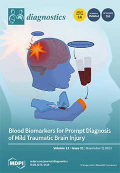
Cover Story (view full-size image): Millions of persons experience mild traumatic brain injuries (mTBI) each year, yet there is no widely accepted systematic method to evaluate this patient population. Individuals experiencing concussion may have long-term symptoms and sequelae. Assessment of mTBI in the Emergency Department varies and follow-up is often limited. Neuroimaging results are frequently normal. There are few effective medical interventions for mTBI. Accurate, sensitive and rapid point-of-care blood biomarker tests for mTBI have the potential to be useful in improving prediction of prognosis, outcome and efficacy of treatment. Further, a large database might be created using biomarkers and other key information gathered on patients with mTBI. Such a database could yield objective, consistent criteria to better understand mTBI. View this paper
  • Issues are regarded as officially published after their release is announced to the table of contents alert mailing list.
  • You may sign up for e-mail alerts to receive table of contents of newly released issues.
  • PDF is the official format for papers published in both, html and pdf forms. To view the papers in pdf format, click on the "PDF Full-text" link, and use the free Adobe Reader to open them.
Order results
Result details
Section
Select all
Export citation of selected articles as:
3 pages, 469 KiB  
Interesting Images
Osmotic Demyelination Syndrome: A Rare Clinical Image
by Prishita Koul, Pallavi Harjpal and Raghuveer Raghumahanti
Diagnostics 2023, 13(21), 3393; https://doi.org/10.3390/diagnostics13213393 - 6 Nov 2023
Viewed by 2130
Abstract
The term “Osmotic Demyelination Syndrome” (ODS) is synonymous with central pontine myelinolysis (CPM), denoting a condition characterised by brain damage, particularly affecting the white matter tracts of the pontine region. This damage arises due to the rapid correction of metabolic imbalances, primarily cases [...] Read more.
The term “Osmotic Demyelination Syndrome” (ODS) is synonymous with central pontine myelinolysis (CPM), denoting a condition characterised by brain damage, particularly affecting the white matter tracts of the pontine region. This damage arises due to the rapid correction of metabolic imbalances, primarily cases of hyponatremia. Noteworthy triggers encompass severe burns, liver transplantations, anorexia nervosa, hyperemesis gravidarum, and hyperglycaemia, all linked to the development of CPM. Clinical manifestations encompass a spectrum of signs and symptoms, including dysphagia, dysarthria, spastic quadriparesis, pseudobulbar paralysis, ataxia, lethargy, tremors, disorientation, catatonia, and, in severe instances, locked-in syndrome and coma. A recent case involving a 45-year-old woman illustrates these complexities. Upon admission to the Medicine Intensive Care Unit, she presented with symptoms indicative of diminished responsiveness and bilateral weakness in the upper and lower limbs. Of significance, the patient had a pre-existing medical history of hyperthyroidism. Extensive diagnostic investigations were undertaken, revealing compelling evidence of profound hyponatremia through blood analyses. Furthermore, magnetic resonance imaging (MRI) was performed, unveiling conspicuous areas of abnormal hyperintensity located in the central pons, intriguingly accompanied by spared peripheral regions. These radiological findings align with the characteristic pattern associated with osmotic demyelination syndrome, illuminating the underlying pathology. Full article
(This article belongs to the Special Issue Neuroimaging: Brain Function and Structure)
Show Figures

Figure 1

10 pages, 229 KiB  
Article
Blunt Trauma in Children: Efficacy and Safety of Transarterial Embolization, 10-Year Experiences in a Single Trauma Center
by Seyoung Ko, Hoon Kwon, Chang Won Kim, Hojun Lee, Jae Hun Kim, Hohyun Kim and Chan Ik Park
Diagnostics 2023, 13(21), 3392; https://doi.org/10.3390/diagnostics13213392 - 6 Nov 2023
Viewed by 1222
Abstract
Background: Transcatheter arterial embolization (TAE) is an established approach for controlling hemorrhage in adults with acute abdominal and pelvic trauma. However, its application in pediatric trauma is not well established. This study aimed to evaluate the safety and effectiveness of TAE in a [...] Read more.
Background: Transcatheter arterial embolization (TAE) is an established approach for controlling hemorrhage in adults with acute abdominal and pelvic trauma. However, its application in pediatric trauma is not well established. This study aimed to evaluate the safety and effectiveness of TAE in a population of pediatric patients with blunt trauma. Methods: This retrospective study was conducted in pediatric patients (<18 years) who underwent TAE for blunt trauma between February 2014 and July 2022. The patients were categorized into subgroups based on age and body weight. Patient demographics, injury severity, transfusion requirements, and clinical outcomes were analyzed. Results: Exactly 73 patients underwent TAE. Technical success was achieved in all patients (100%), and clinical success was achieved in 83.6%. The mortality and complication rates were 4.1% and 1.4%, respectively. The mean duration of hospitalization was 19.3 days. Subgroup analysis showed that age, body weight, and sex did not significantly affect clinical success. The injury severity score and transfusion requirement were predictors of clinical success, with lower values associated with better outcomes. Conclusions: TAE is effective and safe for managing blunt pediatric trauma in younger and lighter patients. Injury severity and transfusion requirement are predictors of clinical success. Full article
(This article belongs to the Special Issue Diagnosis and Management of Traumatic Injury)
18 pages, 2130 KiB  
Article
User Perceptions of Avatar-Based Patient Monitoring for Intensive Care Units: An International Exploratory Sequential Mixed-Methods Study
by Justyna Lunkiewicz, Greta Gasciauskaite, Tadzio Raoul Roche, Samira Akbas, Christoph B. Nöthiger, Michael T. Ganter, Patrick Meybohm, Sebastian Hottenrott, Kai Zacharowski, Florian Jürgen Raimann, Eva Rivas, Manuel López-Baamonde, Elisabeth Anna Beller, David Werner Tscholl and Lisa Bergauer
Diagnostics 2023, 13(21), 3391; https://doi.org/10.3390/diagnostics13213391 - 6 Nov 2023
Viewed by 1901
Abstract
Visual Patient Avatar ICU is an innovative approach to patient monitoring, enhancing the user’s situation awareness in intensive care settings. It dynamically displays the patient’s current vital signs using changes in color, shape, and animation. The technology can also indicate patient-inserted devices, such [...] Read more.
Visual Patient Avatar ICU is an innovative approach to patient monitoring, enhancing the user’s situation awareness in intensive care settings. It dynamically displays the patient’s current vital signs using changes in color, shape, and animation. The technology can also indicate patient-inserted devices, such as arterial lines, central lines, and urinary catheters, along with their insertion locations. We conducted an international, multi-center study using a sequential qualitative-quantitative design to evaluate users’ perception of Visual Patient Avatar ICU among physicians and nurses. Twenty-five nurses and twenty-five physicians from the ICU participated in the structured interviews. Forty of them completed the online survey. Overall, ICU professionals expressed a positive outlook on Visual Patient Avatar ICU. They described Visual Patient Avatar ICU as a simple and intuitive tool that improved information retention and facilitated problem identification. However, a subset of participants expressed concerns about potential information overload and a sense of incompleteness due to missing exact numerical values. These findings provide valuable insights into user perceptions of Visual Patient Avatar ICU and encourage further technology development before clinical implementation. Full article
(This article belongs to the Special Issue Point-of-Care Diagnostics Technology and Applications)
Show Figures

Figure 1

15 pages, 781 KiB  
Article
A Severity Comparison between Italian and Israeli Rett Syndrome Cohorts
by Alberto Romano, Meir Lotan and Rosa Angela Fabio
Diagnostics 2023, 13(21), 3390; https://doi.org/10.3390/diagnostics13213390 - 6 Nov 2023
Cited by 2 | Viewed by 1751
Abstract
Rett syndrome (RTT) is a neurodevelopmental disorder marked by profound cognitive, communication, and motor impairments. Despite identified genotype/phenotype connections, the extent of clinical severity varies even among individuals sharing the same genetic mutation. Diverse sociocultural environments, such as the level of inclusivity of [...] Read more.
Rett syndrome (RTT) is a neurodevelopmental disorder marked by profound cognitive, communication, and motor impairments. Despite identified genotype/phenotype connections, the extent of clinical severity varies even among individuals sharing the same genetic mutation. Diverse sociocultural environments, such as the level of inclusivity of the scholar system, the time spent with family, and the intensity of the rehabilitative intervention provided, might influence their development diversely. This study examines the severity of RTT in people in Italy and Israel, countries with distinct contradictory approaches to caring for those with intricate disabilities, across two age groups. Data from 136 Italian and 59 Israeli girls and women with RTT were assessed and divided into two age categories: above and below 12 years. The RARS, a standardized RTT-specific clinical severity tool, was administered. Despite no differences in age and genetic characteristics, the Italian group showed better scores in the RARS motor and disease-related characteristics areas in both age groups. Moreover, the young Italian participants gathered better total RARS scores and emotional and behavioral characteristics area scores. Furthermore, the young group showed significantly less scoliosis, foot problems, and epilepsy than the older group. These findings endorse the inclusion of girls with RTT in the regular schooling system for a limited daily period, investing in high activity levels within the home and community environments, and suggest continuously providing the person with daily occasions of physical activity and socialization. Full article
(This article belongs to the Section Pathology and Molecular Diagnostics)
Show Figures

Figure 1

22 pages, 3887 KiB  
Article
Application of Machine Learning Strategies to Model the Effects of Sevoflurane on Somatosensory-Evoked Potentials during Spine Surgery
by John Preston Wilson, Jr., Deepak Kumbhare, Charles Ronkon, Bharat Guthikonda and Stanley Hoang
Diagnostics 2023, 13(21), 3389; https://doi.org/10.3390/diagnostics13213389 - 6 Nov 2023
Viewed by 1789
Abstract
In this study, a small sample of patients’ neuromonitoring data was analyzed using machine learning (ML) tools to provide proof of concept for quantifying complex signals. Intraoperative neurophysiological monitoring (IONM) is a valuable asset for monitoring the neurological status of a patient during [...] Read more.
In this study, a small sample of patients’ neuromonitoring data was analyzed using machine learning (ML) tools to provide proof of concept for quantifying complex signals. Intraoperative neurophysiological monitoring (IONM) is a valuable asset for monitoring the neurological status of a patient during spine surgery. Notably, this technology, when operated by neurophysiologists and surgeons familiar with proper alarm criteria, is capable of detecting neurological deficits. However, non-surgical factors, such as volatile anesthetics like sevoflurane, can negatively influence robust IONM signal generation. While sevoflurane has been shown to affect the latency and amplitude of somatosensory evoked potential (SSEP), a more complex and nuanced analysis of the SSEP waveform has not been performed. In this study, signal processing and machine learning techniques were used to more intricately characterize and predict SSEP waveform changes as a function of varying end-tidal sevoflurane concentration. With data from ten patients who underwent spinal procedures, features describing the SSEP waveforms were generated using principal component analysis (PCA), phase space curves (PSC), and time-frequency analysis (TFA). A minimum redundancy maximum relevance (MRMR) feature selection technique was then used to identify the most important SSEP features associated with changing sevoflurane concentrations. Once the features carrying the maximum amount of information about the majority of signal waveform variability were identified, ML models were used to predict future changes in SSEP waveforms. Linear regression, regression trees, support vector machines, and neural network ML models were then selected for testing. Using SSEP data from eight patients, the models were trained using a range of features selected during MRMR calculations. During the training phase of model development, the highest performing models were identified as support vector machines and regression trees. After identifying the highest performing models for each nerve group, we tested these models using the remaining two patients’ data. We compared the models’ performance metrics using the root mean square error values (RMSEs). The feasibility of the methodology described provides a general framework for the applications of machine learning strategies to further delineate the effects of surgical and non-surgical factors affecting IONM signals. Full article
(This article belongs to the Special Issue Applications of Artificial Intelligence in Healthcare Monitoring)
Show Figures

Figure 1

12 pages, 2682 KiB  
Viewpoint
Progress in the Application of Portable Ultrasound Combined with Artificial Intelligence in Pre-Hospital Emergency and Disaster Sites
by Xing Gao, Qi Lv and Shike Hou
Diagnostics 2023, 13(21), 3388; https://doi.org/10.3390/diagnostics13213388 - 6 Nov 2023
Cited by 5 | Viewed by 2816
Abstract
With the miniaturization of ultrasound and the development of artificial intelligence, its application in disaster scenes and pre-hospital emergency care has become more and more common. This study summarizes the literature on portable ultrasound in pre-hospital emergency and disaster scene treatment in the [...] Read more.
With the miniaturization of ultrasound and the development of artificial intelligence, its application in disaster scenes and pre-hospital emergency care has become more and more common. This study summarizes the literature on portable ultrasound in pre-hospital emergency and disaster scene treatment in the past decade and reviews the development and application of portable ultrasound. Portable ultrasound diagnostic equipment can be used to diagnose abdominal bleeding, limb fracture, hemopneumothorax, pericardial effusion, etc., based on which trauma can be diagnosed pre-hospital and provide guiding suggestions for the next triage and rescue; in early rescue, portable ultrasound can guide emergency operations, such as tracheal intubation, pericardial cavity puncture, and thoracic and abdominal puncture as well as improve the accuracy and timeliness of operation techniques. In addition, with the development of artificial intelligence (AI), AI-assisted diagnosis can improve the diagnosis level of ultrasound at disaster sites. The portable ultrasound diagnosis system equipped with an AI robotic arm can maximize the pre-screening classification and fast and concise diagnosis and treatment of batch casualties, thus providing a reliable basis for batch casualty classification and evacuation at disaster accident sites. Full article
(This article belongs to the Special Issue The Use of Portable Devices in Emergency Medicine)
Show Figures

Figure 1

17 pages, 2398 KiB  
Article
Diagnostic Performance of an Artificial Intelligence Model Based on Contrast-Enhanced Ultrasound in Patients with Liver Lesions: A Comparative Study with Clinicians
by Marinela-Cristiana Urhuț, Larisa Daniela Săndulescu, Costin Teodor Streba, Mădălin Mămuleanu, Adriana Ciocâlteu, Sergiu Marian Cazacu and Suzana Dănoiu
Diagnostics 2023, 13(21), 3387; https://doi.org/10.3390/diagnostics13213387 - 5 Nov 2023
Cited by 5 | Viewed by 2240
Abstract
Contrast-enhanced ultrasound (CEUS) is widely used in the characterization of liver tumors; however, the evaluation of perfusion patterns using CEUS has a subjective character. This study aims to evaluate the accuracy of an automated method based on CEUS for classifying liver lesions and [...] Read more.
Contrast-enhanced ultrasound (CEUS) is widely used in the characterization of liver tumors; however, the evaluation of perfusion patterns using CEUS has a subjective character. This study aims to evaluate the accuracy of an automated method based on CEUS for classifying liver lesions and to compare its performance with that of two experienced clinicians. The system used for automatic classification is based on artificial intelligence (AI) algorithms. For an interpretation close to the clinical setting, both clinicians knew which patients were at high risk for hepatocellular carcinoma (HCC), but only one was aware of all the clinical data. In total, 49 patients with 59 liver tumors were included. For the benign and malignant classification, the AI model outperformed both clinicians in terms of specificity (100% vs. 93.33%); still, the sensitivity was lower (74% vs. 93.18% vs. 90.91%). In the second stage of multiclass diagnosis, the automatic model achieved a diagnostic accuracy of 69.93% for HCC and 89.15% for liver metastases. Readers demonstrated greater diagnostic accuracy for HCC (83.05% and 79.66%) and liver metastases (94.92% and 96.61%) compared to the AI system; however, both were experienced sonographers. The AI model could potentially assist and guide less-experienced clinicians to discriminate malignant from benign liver tumors with high accuracy and specificity. Full article
(This article belongs to the Special Issue Diagnostic Imaging of Liver Diseases)
Show Figures

Figure 1

14 pages, 1924 KiB  
Article
Surgical Phase Duration in Robot-Assisted Partial Nephrectomy: A Surgical Data Science Exploration for Clinical Relevance
by Pieter De Backer, Maria Peraire Lores, Meret Demuynck, Federico Piramide, Jente Simoens, Tim Oosterlinck, Wouter Bogaert, Chi Victor Shan, Karel Van Regemorter, Aube Wastyn, Enrico Checcucci, Charlotte Debbaut, Charles Van Praet, Rui Farinha, Ruben De Groote, Anthony Gallagher, Karel Decaestecker and Alexandre Mottrie
Diagnostics 2023, 13(21), 3386; https://doi.org/10.3390/diagnostics13213386 - 5 Nov 2023
Cited by 4 | Viewed by 1870
Abstract
(1) Background: Surgical phases form the basic building blocks for surgical skill assessment, feedback, and teaching. The phase duration itself and its correlation with clinical parameters at diagnosis have not yet been investigated. Novel commercial platforms provide phase indications but have not been [...] Read more.
(1) Background: Surgical phases form the basic building blocks for surgical skill assessment, feedback, and teaching. The phase duration itself and its correlation with clinical parameters at diagnosis have not yet been investigated. Novel commercial platforms provide phase indications but have not been assessed for accuracy yet. (2) Methods: We assessed 100 robot-assisted partial nephrectomy videos for phase durations based on previously defined proficiency metrics. We developed an annotation framework and subsequently compared our annotations to an existing commercial solution (Touch Surgery, Medtronic™). We subsequently explored clinical correlations between phase durations and parameters derived from diagnosis and treatment. (3) Results: An objective and uniform phase assessment requires precise definitions derived from an iterative revision process. A comparison to a commercial solution shows large differences in definitions across phases. BMI and the duration of renal tumor identification are positively correlated, as are tumor complexity and both tumor excision and renorrhaphy duration. (4) Conclusions: The surgical phase duration can be correlated with certain clinical outcomes. Further research should investigate whether the retrieved correlations are also clinically meaningful. This requires an increase in dataset sizes and facilitation through intelligent computer vision algorithms. Commercial platforms can facilitate this dataset expansion and help unlock the full potential, provided that the phase annotation details are disclosed. Full article
(This article belongs to the Section Machine Learning and Artificial Intelligence in Diagnostics)
Show Figures

Figure 1

17 pages, 4647 KiB  
Review
Diagnostic Approach to Lower Limb Entrapment Neuropathies: A Narrative Literature Review
by Nicu Cătălin Drăghici, Vitalie Văcăraș, Roxana Bolchis, Atamyrat Bashimov, Diana Maria Domnița, Silvina Iluț, Livia Livinț Popa, Tudor Dimitrie Lupescu and Dafin Fior Mureșanu
Diagnostics 2023, 13(21), 3385; https://doi.org/10.3390/diagnostics13213385 - 4 Nov 2023
Cited by 4 | Viewed by 6437
Abstract
Entrapment neuropathies of the lower limb are a misunderstood and underdiagnosed group of disorders, characterized by pain and dysesthesia, muscular weakness, and specific provoking movements on physical examination. The most frequent of these syndromes encountered in clinical practice are fibular nerve entrapment, proximal [...] Read more.
Entrapment neuropathies of the lower limb are a misunderstood and underdiagnosed group of disorders, characterized by pain and dysesthesia, muscular weakness, and specific provoking movements on physical examination. The most frequent of these syndromes encountered in clinical practice are fibular nerve entrapment, proximal tibial neuropathy, sural nerve neuropathy, deep gluteal syndrome or sciatic nerve entrapment, and lateral femoral cutaneous nerve entrapment, also known as meralgia paresthetica. These are commonly mistaken for lumbar plexopathies, radiculopathies, and musculotendinous diseases, which appear even more frequently and have overlapping clinical presentations. A comprehensive anamnesis, physical examination, and electrodiagnostic studies should help clarify the diagnosis. If the diagnosis is still unclear or a secondary cause of entrapment is suspected, magnetic resonance neurography, MRI, or ultrasonography should be conducted to clarify the etiology, rule out other diseases, and confirm the diagnosis. The aim of this narrative review was to help clinicians gain familiarity with this disease, with an increase in diagnostic confidence, leading to early diagnosis of nerve damage and prevention of muscle atrophy. We reviewed the epidemiology, anatomy, pathophysiology, etiology, clinical presentation, and EDX technique and interpretation of the entrapment neuropathies of the lower limb, using articles published from 1970 to 2022 included in the Pubmed, MEDLINE, Cochrane Library, Google Scholar, EMBASE, Web of Science, and Scopus databases. Full article
Show Figures

Figure 1

16 pages, 6376 KiB  
Article
Building Automation Pipeline for Diagnostic Classification of Sporadic Odontogenic Keratocysts and Non-Keratocysts Using Whole-Slide Images
by Samahit Mohanty, Divya B. Shivanna, Roopa S. Rao, Madhusudan Astekar, Chetana Chandrashekar, Raghu Radhakrishnan, Shylaja Sanjeevareddygari, Vijayalakshmi Kotrashetti and Prashant Kumar
Diagnostics 2023, 13(21), 3384; https://doi.org/10.3390/diagnostics13213384 - 4 Nov 2023
Viewed by 1606
Abstract
The microscopic diagnostic differentiation of odontogenic cysts from other cysts is intricate and may cause perplexity for both clinicians and pathologists. Of particular interest is the odontogenic keratocyst (OKC), a developmental cyst with unique histopathological and clinical characteristics. Nevertheless, what distinguishes this cyst [...] Read more.
The microscopic diagnostic differentiation of odontogenic cysts from other cysts is intricate and may cause perplexity for both clinicians and pathologists. Of particular interest is the odontogenic keratocyst (OKC), a developmental cyst with unique histopathological and clinical characteristics. Nevertheless, what distinguishes this cyst is its aggressive nature and high tendency for recurrence. Clinicians encounter challenges in dealing with this frequently encountered jaw lesion, as there is no consensus on surgical treatment. Therefore, the accurate and early diagnosis of such cysts will benefit clinicians in terms of treatment management and spare subjects from the mental agony of suffering from aggressive OKCs, which impact their quality of life. The objective of this research is to develop an automated OKC diagnostic system that can function as a decision support tool for pathologists, whether they are working locally or remotely. This system will provide them with additional data and insights to enhance their decision-making abilities. This research aims to provide an automation pipeline to classify whole-slide images of OKCs and non-keratocysts (non-KCs: dentigerous and radicular cysts). OKC diagnosis and prognosis using the histopathological analysis of tissues using whole-slide images (WSIs) with a deep-learning approach is an emerging research area. WSIs have the unique advantage of magnifying tissues with high resolution without losing information. The contribution of this research is a novel, deep-learning-based, and efficient algorithm that reduces the trainable parameters and, in turn, the memory footprint. This is achieved using principal component analysis (PCA) and the ReliefF feature selection algorithm (ReliefF) in a convolutional neural network (CNN) named P-C-ReliefF. The proposed model reduces the trainable parameters compared to standard CNN, achieving 97% classification accuracy. Full article
(This article belongs to the Special Issue Explainable Artificial Intelligence in Medical Imaging)
Show Figures

Figure 1

19 pages, 3485 KiB  
Review
MiRNAs and Their Role in Venous Thromboembolic Complications
by Ilgiz Gareev, Valentin Pavlov, Weijie Du and Baofeng Yang
Diagnostics 2023, 13(21), 3383; https://doi.org/10.3390/diagnostics13213383 - 4 Nov 2023
Cited by 5 | Viewed by 1860
Abstract
Venous thromboembolic complications (VTCs), which include deep vein thrombosis (DVT) and pulmonary embolism (PE), have remained a pressing problem in modern clinical medicine for a long time. Despite the already wide arsenal of modern methods for diagnosing and treating this disease, VTCs rank [...] Read more.
Venous thromboembolic complications (VTCs), which include deep vein thrombosis (DVT) and pulmonary embolism (PE), have remained a pressing problem in modern clinical medicine for a long time. Despite the already wide arsenal of modern methods for diagnosing and treating this disease, VTCs rank third in the structure of causes of death among all cardiovascular diseases, behind myocardial infarction (MI) and ischemic stroke (IS). Numerous studies have confirmed the importance of understanding the molecular processes of VTCs for effective therapy and diagnosis. Significant progress has been made in VTC research in recent years, where the relative contribution of microRNAs (miRNAs) in the mechanism of thrombus formation and their consideration as therapeutic targets have been well studied. In this case, accurate, timely, and as early as possible diagnosis of VTCs is of particular importance, which will help improve both short-term and long-term prognoses of patients. This case accounts for the already well-studied circulating miRNAs as non-invasive biomarkers. This study presents currently available literature data on the role of miRNAs in VTCs, revealing their potential as therapeutic targets and diagnostic and prognostic tools for this terrible disease. Full article
(This article belongs to the Section Pathology and Molecular Diagnostics)
Show Figures

Figure 1

20 pages, 6326 KiB  
Article
A Novel Prognostic Model Using Chaotic CNN with Hybridized Spoofing for Enhancing Diagnostic Accuracy in Epileptic Seizure Prediction
by Preethi Palanisamy, Shabana Urooj, Rajesh Arunachalam and Aime Lay-Ekuakille
Diagnostics 2023, 13(21), 3382; https://doi.org/10.3390/diagnostics13213382 - 3 Nov 2023
Cited by 15 | Viewed by 1246
Abstract
Epileptic seizure detection has undergone progressive advancements since its conception in the 1970s. From proof-of-concept experiments in the latter part of that decade, it has now become a vibrant area of clinical and laboratory research. In an effort to bring this technology closer [...] Read more.
Epileptic seizure detection has undergone progressive advancements since its conception in the 1970s. From proof-of-concept experiments in the latter part of that decade, it has now become a vibrant area of clinical and laboratory research. In an effort to bring this technology closer to practical application in human patients, this study introduces a customized approach to selecting electroencephalogram (EEG) features and electrode positions for seizure prediction. The focus is on identifying precursors that occur within 10 min of the onset of abnormal electrical activity during a seizure. However, there are security concerns related to safeguarding patient EEG recordings against unauthorized access and network-based attacks. Therefore, there is an urgent need for an efficient prediction and classification method for encrypted EEG data. This paper presents an effective system for analyzing and recognizing encrypted EEG information using Arnold transform algorithms, chaotic mapping, and convolutional neural networks (CNNs). In this system, the EEG time series from each channel is converted into a 2D spectrogram image, which is then encrypted using chaotic algorithms. The encrypted data is subsequently processed by CNNs coupled with transfer learning (TL) frameworks. To optimize the fusion parameters of the ensemble learning classifiers, a hybridized spoofing optimization method is developed by combining the characteristics of corvid and gregarious-seeking agents. The evaluation of the model’s effectiveness yielded the following results: 98.9 ± 0.3% accuracy, 98.2 ± 0.7% sensitivity, 98.6 ± 0.6% specificity, 98.6 ± 0.6% precision, and an F1 measure of 98.9 ± 0.6%. When compared with other state-of-the-art techniques applied to the same dataset, this novel strategy demonstrated one of the most effective seizure detection systems, as evidenced by these results. Full article
(This article belongs to the Section Machine Learning and Artificial Intelligence in Diagnostics)
Show Figures

Figure 1

15 pages, 21455 KiB  
Article
Deep Learning Framework with Multi-Head Dilated Encoders for Enhanced Segmentation of Cervical Cancer on Multiparametric Magnetic Resonance Imaging
by Reza Kalantar, Sebastian Curcean, Jessica M. Winfield, Gigin Lin, Christina Messiou, Matthew D. Blackledge and Dow-Mu Koh
Diagnostics 2023, 13(21), 3381; https://doi.org/10.3390/diagnostics13213381 - 3 Nov 2023
Viewed by 1629
Abstract
T2-weighted magnetic resonance imaging (MRI) and diffusion-weighted imaging (DWI) are essential components of cervical cancer diagnosis. However, combining these channels for the training of deep learning models is challenging due to image misalignment. Here, we propose a novel multi-head framework that [...] Read more.
T2-weighted magnetic resonance imaging (MRI) and diffusion-weighted imaging (DWI) are essential components of cervical cancer diagnosis. However, combining these channels for the training of deep learning models is challenging due to image misalignment. Here, we propose a novel multi-head framework that uses dilated convolutions and shared residual connections for the separate encoding of multiparametric MRI images. We employ a residual U-Net model as a baseline, and perform a series of architectural experiments to evaluate the tumor segmentation performance based on multiparametric input channels and different feature encoding configurations. All experiments were performed on a cohort of 207 patients with locally advanced cervical cancer. Our proposed multi-head model using separate dilated encoding for T2W MRI and combined b1000 DWI and apparent diffusion coefficient (ADC) maps achieved the best median Dice similarity coefficient (DSC) score, 0.823 (confidence interval (CI), 0.595–0.797), outperforming the conventional multi-channel model, DSC 0.788 (95% CI, 0.568–0.776), although the difference was not statistically significant (p > 0.05). We investigated channel sensitivity using 3D GRAD-CAM and channel dropout, and highlighted the critical importance of T2W and ADC channels for accurate tumor segmentation. However, our results showed that b1000 DWI had a minor impact on the overall segmentation performance. We demonstrated that the use of separate dilated feature extractors and independent contextual learning improved the model’s ability to reduce the boundary effects and distortion of DWI, leading to improved segmentation performance. Our findings could have significant implications for the development of robust and generalizable models that can extend to other multi-modal segmentation applications. Full article
(This article belongs to the Special Issue Deep Learning for Early Detection of Cancer)
Show Figures

Figure 1

17 pages, 2389 KiB  
Article
Machine Learning and Radiomics of Bone Scintigraphy: Their Role in Predicting Recurrence of Localized or Locally Advanced Prostate Cancer
by Yu-De Wang, Chi-Ping Huang, You-Rong Yang, Hsi-Chin Wu, Yu-Ju Hsu, Yi-Chun Yeh, Pei-Chun Yeh, Kuo-Chen Wu and Chia-Hung Kao
Diagnostics 2023, 13(21), 3380; https://doi.org/10.3390/diagnostics13213380 - 3 Nov 2023
Cited by 2 | Viewed by 1698
Abstract
Background: Machine-learning (ML) and radiomics features have been utilized for survival outcome analysis in various cancers. This study aims to investigate the application of ML based on patients’ clinical features and radiomics features derived from bone scintigraphy (BS) and to evaluate recurrence-free survival [...] Read more.
Background: Machine-learning (ML) and radiomics features have been utilized for survival outcome analysis in various cancers. This study aims to investigate the application of ML based on patients’ clinical features and radiomics features derived from bone scintigraphy (BS) and to evaluate recurrence-free survival in local or locally advanced prostate cancer (PCa) patients after the initial treatment. Methods: A total of 354 patients who met the eligibility criteria were analyzed and used to train the model. Clinical information and radiomics features of BS were obtained. Survival-related clinical features and radiomics features were included in the ML model training. Using the pyradiomics software, 128 radiomics features from each BS image’s region of interest, validated by experts, were extracted. Four textural matrices were also calculated: GLCM, NGLDM, GLRLM, and GLSZM. Five training models (Logistic Regression, Naive Bayes, Random Forest, Support Vector Classification, and XGBoost) were applied using K-fold cross-validation. Recurrence was defined as either a rise in PSA levels, radiographic progression, or death. To assess the classifier’s effectiveness, the ROC curve area and confusion matrix were employed. Results: Of the 354 patients, 101 patients were categorized into the recurrence group with more advanced disease status compared to the non-recurrence group. Key clinical features including tumor stage, radical prostatectomy, initial PSA, Gleason Score primary pattern, and radiotherapy were used for model training. Random Forest (RF) was the best-performing model, with a sensitivity of 0.81, specificity of 0.87, and accuracy of 0.85. The ROC curve analysis showed that predictions from RF outperformed predictions from other ML models with a final AUC of 0.94 and a p-value of <0.001. The other models had accuracy ranges from 0.52 to 0.78 and AUC ranges from 0.67 to 0.84. Conclusions: The study showed that ML based on clinical features and radiomics features of BS improves the prediction of PCa recurrence after initial treatment. These findings highlight the added value of ML techniques for risk classification in PCa based on clinical features and radiomics features of BS. Full article
(This article belongs to the Special Issue Advanced Computer-Aided Diagnosis Using Medical Images)
Show Figures

Figure 1

13 pages, 286 KiB  
Article
Are Tumor Marker Tests Applied Appropriately in Clinical Practice? A Healthcare Claims Data Analysis
by Sabrina M. Stollberg, Markus Näpflin, Michael Nagler and Carola A. Huber
Diagnostics 2023, 13(21), 3379; https://doi.org/10.3390/diagnostics13213379 - 3 Nov 2023
Cited by 1 | Viewed by 1606
Abstract
Tumor markers (TM) are crucial in the monitoring of cancer treatment. However, inappropriate requests for screening reasons have a high risk of false positive and negative findings, which can lead to patient anxiety and unnecessary follow-up examinations. We aimed to assess the appropriateness [...] Read more.
Tumor markers (TM) are crucial in the monitoring of cancer treatment. However, inappropriate requests for screening reasons have a high risk of false positive and negative findings, which can lead to patient anxiety and unnecessary follow-up examinations. We aimed to assess the appropriateness of TM testing in outpatient practice in Switzerland. We conducted a retrospective cohort study based on healthcare claims data. Patients who had received at least one out of seven TM tests (CEA, CA19-9, CA125, CA15-3, CA72-4, Calcitonin, or NSE) between 2018 and 2021 were analyzed. Appropriate determinations were defined as a request with a corresponding cancer-related diagnosis or intervention. Appropriateness of TM determination by patient characteristics and prescriber specialty was estimated by using multivariate analyses. A total of 51,395 TM determinations in 36,537 patients were included. An amount of 41.6% of all TM were determined appropriately. General practitioners most often determined TM (44.3%) and had the lowest number of appropriate requests (27.8%). A strong predictor for appropriate determinations were requests by medical oncologists. A remarkable proportion of TM testing was performed inappropriately, particularly in the primary care setting. Our results suggest that a considerable proportion of the population is at risk for various harms associated with misinterpretations of TM test results. Full article
(This article belongs to the Section Clinical Laboratory Medicine)
17 pages, 2349 KiB  
Article
Successful Implementation of Image-Guided Pencil-Beam Scanning Proton Therapy in Medulloblastomas
by Anindita Das, Utpal Gaikwad, Ganapathy Krishnan, Adhithyan Rajendran, Sushama Patil, Preethi Subramaniam, Uday Krishna, Manoj G. Wakde, Srinivas Chilukuri and Rakesh Jalali
Diagnostics 2023, 13(21), 3378; https://doi.org/10.3390/diagnostics13213378 - 3 Nov 2023
Viewed by 1562
Abstract
Medulloblastoma is the most common malignant brain tumour in children, while much rarer in adults. Although the prognosis and outcomes have greatly improved in the era of modern multidisciplinary management, long-term treatment-induced toxicities are common. Craniospinal irradiation followed by a boost to the [...] Read more.
Medulloblastoma is the most common malignant brain tumour in children, while much rarer in adults. Although the prognosis and outcomes have greatly improved in the era of modern multidisciplinary management, long-term treatment-induced toxicities are common. Craniospinal irradiation followed by a boost to the primary and metastatic tumour sites forms the backbone of treatment. Proton therapy has been endorsed over conventional photon-based radiotherapy due to its superior dosimetric advantages and subsequently lower incidence and severity of toxicities. We report here our experience from South-East Asia’s first proton therapy centre of treating 40 patients with medulloblastoma (38 children and adolescents, 2 adults) who received image-guided, intensity-modulated proton therapy with pencil-beam scanning between 2019 and 2023, with a focus on dosimetry, acute toxicities, and early survival outcomes. All patients could complete the planned course of proton therapy, with mostly mild acute toxicities that were manageable on an outpatient basis. Haematological toxicity was not dose-limiting and did not prolong the overall treatment time. Preliminary data on early outcomes including overall survival and disease-free survival are encouraging, although a longer follow-up and data on long-term toxicities are needed. Full article
(This article belongs to the Special Issue Medulloblastoma—Existing and Evolving Landscape)
Show Figures

Figure 1

7 pages, 1272 KiB  
Interesting Images
Obstructed Hemi-Vagina with Ipsilateral Renal Agenesis Syndrome in Adulthood: A Diagnostic Challenge
by Alexandros Loukas Grammatis, Femi Ajibade, Dinuke Warakaulle and Tunde Dada
Diagnostics 2023, 13(21), 3377; https://doi.org/10.3390/diagnostics13213377 - 3 Nov 2023
Cited by 1 | Viewed by 1360
Abstract
A patient in her early 20s presented with constant and progressive lower abdominal and back pain, mainly on the right side of the abdomen, purulent vaginal discharge and pyrexia. A radiological assessment revealed a possible tubo-ovarian abscess and the incidental diagnosis of ipsilateral [...] Read more.
A patient in her early 20s presented with constant and progressive lower abdominal and back pain, mainly on the right side of the abdomen, purulent vaginal discharge and pyrexia. A radiological assessment revealed a possible tubo-ovarian abscess and the incidental diagnosis of ipsilateral renal agenesis. The patient was treated for pelvic inflammatory disease (PID); however, after antibiotic administration and since the symptoms did not resolve, an abdominal MRI was requested, which revealed uterus didelphys with two cervices, an obstructed haemivagina and evidence of haematocolpos. The diagnosis of Obstructed Hemi-Vagina with Ipsilateral Renal Agenesis (OHVIRA) syndrome was confirmed, and the patient underwent the excision of the vaginal septum, the drainage of the haematopyocolpos and the laparoscopic drainage of the tubo-ovarian abscess. She achieved a good recovery. Full article
Show Figures

Figure 1

15 pages, 9477 KiB  
Article
Structural and Functional Changes in Non-Paraneoplastic Autoimmune Retinopathy
by Amir Akhavanrezayat, Anadi Khatri, Neil Gregory L. Onghanseng, Muhammad Sohail Halim, Christopher Or, Nripun Sredar, Moataz Razeen, Murat Hasanreisoglu, Jonathan Regenold, Zheng Xian Thng, S. Saeed Mohammadi, Tanya Jain, Negin Yavari, Vahid Bazojoo, Ankur Sudhir Gupta, Azadeh Mobasserian, Cigdem Yasar, Ngoc Trong Tuong Than, Gunay Uludag Kirimli, Irmak Karaca, Yong-Un Shin, Woong-Sun Yoo, Hashem Ghoraba, Diana V. Do, Alfredo Dubra and Quan Dong Nguyenadd Show full author list remove Hide full author list
Diagnostics 2023, 13(21), 3376; https://doi.org/10.3390/diagnostics13213376 - 3 Nov 2023
Cited by 1 | Viewed by 1801
Abstract
Background: To describe longitudinal changes in patients with non-paraneoplastic autoimmune retinopathy (npAIR) by utilizing different diagnostic modalities/tests. Methods: The index study is a retrospective longitudinal review of sixteen eyes of eight patients from a tertiary care eye hospital diagnosed with npAIR. Multiple diagnostic [...] Read more.
Background: To describe longitudinal changes in patients with non-paraneoplastic autoimmune retinopathy (npAIR) by utilizing different diagnostic modalities/tests. Methods: The index study is a retrospective longitudinal review of sixteen eyes of eight patients from a tertiary care eye hospital diagnosed with npAIR. Multiple diagnostic modalities such as wide-angle fundus photography (WAFP), WA fundus autofluorescence (WAFAF), spectral-domain optical coherence tomography (SD-OCT), Goldmann visual field (GVF) perimetry, microperimetry (MP), electrophysiologic testing, and adaptive optics scanning laser ophthalmoscopy (AOSLO) were reviewed and analyzed. Results: At the baseline visits, anomalies were detected by multimodal diagnostic tests on all patients. Subjects were followed up for a median duration of 11.5 [3.0–18.7] months. Structural changes at the baseline were detected in 14 of 16 (87.5%) eyes on WAFP and WAFAF and 13 of 16 (81.2%) eyes on SD-OCT. Eight of the ten (80%) eyes that underwent AOSLO imaging depicted structural changes. Functional changes were detected in 14 of 16 (87.5%) eyes on GVF, 15 of 16 (93.7%) eyes on MP, and 11 of 16 (68.7%) eyes on full-field electroretinogram (ff-ERG). Multifocal electroretinogram (mf-ERG) and visual evoked potential (VEP) tests were performed in 14 eyes, of which 12 (85.7%) and 14 (100%) of the eyes demonstrated functional abnormalities, respectively, at baseline. Compared to all the other structural diagnostic tools, AOSLO had a better ability to demonstrate deterioration in retinal microstructures occurring at follow-ups. Functional deterioration at follow-up was detected on GVF in 8 of 10 (80%) eyes, mf-ERG in 4 of 8 (50%) eyes, and MP in 7 of 16 (43.7%) eyes. The ff-ERG and VEP were stable in the majority of cases at follow-up. Conclusions: The utilization of multimodal imaging/tests in the diagnosing and monitoring of npAIR patients can aid in identifying anomalous changes over time. Analysis of both the anatomical and functional aspects by these devices can be supportive of detecting the changes early in such patients. AOSLO shows promise as it enables the capture of high-resolution images demonstrating quantifiable changes to retinal microstructure. Full article
(This article belongs to the Special Issue Diagnostic Imaging of Uveitis and Ocular Inflammation)
Show Figures

Figure 1

13 pages, 4169 KiB  
Article
Prediction of Conversion from Mild Cognitive Impairment to Alzheimer’s Disease Using Amyloid PET and Brain MR Imaging Data: A 48-Month Follow-Up Analysis of the Alzheimer’s Disease Neuroimaging Initiative Cohort
by Do-Hoon Kim, Minyoung Oh and Jae Seung Kim
Diagnostics 2023, 13(21), 3375; https://doi.org/10.3390/diagnostics13213375 - 2 Nov 2023
Cited by 4 | Viewed by 1805
Abstract
We developed a novel quantification method named “shape feature” by combining the features of amyloid positron emission tomography (PET) and brain magnetic resonance imaging (MRI) and evaluated its significance in predicting the conversion from mild cognitive impairment (MCI) to Alzheimer’s disease (AD) in [...] Read more.
We developed a novel quantification method named “shape feature” by combining the features of amyloid positron emission tomography (PET) and brain magnetic resonance imaging (MRI) and evaluated its significance in predicting the conversion from mild cognitive impairment (MCI) to Alzheimer’s disease (AD) in the Alzheimer’s Disease Neuroimaging Initiative (ADNI) cohort. From the ADNI database, 334 patients with MCI were included. The brain amyloid smoothing score (AV45_BASS) and brain atrophy index (MR_BAI) were calculated using the surface area and volume of the region of interest in AV45 PET and MRI. During the 48-month follow-up period, 108 (32.3%) patients converted from MCI to AD. Age, Mini-Mental State Examination (MMSE), cognitive subscale of the Alzheimer’s Disease Assessment Scale (ADAS-cog), apolipoprotein E (APOE), standardized uptake value ratio (SUVR), AV45_BASS, MR_BAI, and shape feature were significantly different between converters and non-converters. Univariate analysis showed that age, MMSE, ADAS-cog, APOE, SUVR, AV45_BASS, MR_BAI, and shape feature were correlated with the conversion to AD. In multivariate analyses, high shape feature, SUVR, and ADAS-cog values were associated with an increased risk of conversion to AD. In patients with MCI in the ADNI cohort, our quantification method was the strongest prognostic factor for predicting their conversion to AD. Full article
Show Figures

Figure 1

16 pages, 1491 KiB  
Article
Pathogenetic Mechanisms Underlying Major Adverse Cardiac Events in Personality Type D Patients after Percutaneous Coronary Intervention: The Roles of Cognitive Appraisal and Coping Strategies
by Alexey N. Sumin and Anna V. Shcheglova
Diagnostics 2023, 13(21), 3374; https://doi.org/10.3390/diagnostics13213374 - 2 Nov 2023
Cited by 2 | Viewed by 1139
Abstract
Background: This paper aimed to study the association of type D personality, coping strategies, and cognitive appraisal with annual prognosis after a percutaneous coronary intervention (PCI) in patients with coronary artery disease (CAD). Methods: The prospective study included 111 CAD patients who underwent [...] Read more.
Background: This paper aimed to study the association of type D personality, coping strategies, and cognitive appraisal with annual prognosis after a percutaneous coronary intervention (PCI) in patients with coronary artery disease (CAD). Methods: The prospective study included 111 CAD patients who underwent a PCI. All participants, before the PCI, completed questionnaires designed to collect information about type D personality, cognitive appraisal, and coping styles. Information was also collected on the clinical and demographic characteristics of the patients. After 1 year of follow-up, the presence of major adverse cardiac events (MACEs) was assessed. Results: The presence of a MACE was noted in 38 patients, and the absence of a MACE was noted in 53 patients. In patients with type D personality, higher incidences of MACEs (54.1% versus 33.3%; p = 0.0489) and hospitalization rates (29.7% versus 7.4%; p = 0.004) were revealed. Patients with poor prognoses preferred a moderate use of the confrontation strategy than patients without a MACE (78.4% vs. 50.9%; p = 0.0082). Patients with MACEs had statistically significantly lower indicators of strong emotions (11.92 ± 5.32 versus 14.62 ± 4.83 points; p = 0.005) and future prospects (11.36 ± 3.81 versus 13.21 ± 3.41 points; p = 0.015) than patients without a MACE. In a multiple binary logistic regression model, the following factors had significant associations with MACE development: type D, moderate use of confrontation coping, moderate use of self-control coping, and strong emotions in cognitive appraisal. Conclusion: This study showed that not only personality type D, but also certain coping strategies and cognitive appraisals increase the likelihood of developing a MACE after a PCI. This provides a theoretical basis for understanding the mechanism underlying type D personality and MACEs in patients after a PCI. Full article
(This article belongs to the Section Pathology and Molecular Diagnostics)
Show Figures

Figure 1

12 pages, 15660 KiB  
Article
Comparison of Bleb Morphology following PRESERFLO® MicroShunt and Trabeculectomy Using Anterior Segment OCT
by Somar M. Hasan, Theresa Theilig and Daniel Meller
Diagnostics 2023, 13(21), 3373; https://doi.org/10.3390/diagnostics13213373 - 2 Nov 2023
Cited by 2 | Viewed by 1720
Abstract
Trabeculectomy has traditionally been upheld as the gold standard in glaucoma surgery, but recent advancements, including the PRESERFLO® MicroShunt, have introduced less invasive techniques to mitigate complications and reduce intraocular pressure (IOP). The examination of bleb morphology post-filtering surgery is critical for [...] Read more.
Trabeculectomy has traditionally been upheld as the gold standard in glaucoma surgery, but recent advancements, including the PRESERFLO® MicroShunt, have introduced less invasive techniques to mitigate complications and reduce intraocular pressure (IOP). The examination of bleb morphology post-filtering surgery is critical for modulating wound healing and ensuring long-term success. While previous evaluations of PRESERFLO-generated blebs have relied on data from trabeculectomy blebs, the differing surgical techniques used in PRESERFLO and trabeculectomy surgeries suggest potential variations in bleb morphologies. This study conducted a comparative analysis of blebs resulting from PRESERFLO and trabeculectomy procedures. Retrospective descriptive assessments using the Jenaer Bleb Grading System were performed, along with quantitative evaluation using eight-dimensional parameters utilizing anterior segment OCT. We included 93 eyes (57 following PRESERFLO, 36 following trabeculectomy). In the descriptive assessment, PRESERFLO-generated blebs exhibited fewer conjunctival cysts (3.5% vs. 22.2%, p = 0.007) and cavernous changes (10.5% vs. 30.5%, p = 0.021) compared to trabeculectomy. Quantitatively, overall bleb dimensions were comparable (maximal bleb height, width, and length, p > 0.05 for all). However, PRESERFLO blebs displayed a shallower (0.52 ± 0.24 vs. 0.67 ± 0.3 mm, p = 0.017) yet longer (4.12 ± 1.54 vs. 3.23 ± 1.64 mm, p = 0.024) episcleral lake. A thicker bleb wall (1.52 ± 0.46 vs. 1.10 ± 0.37 mm, p < 0.001) along with more posteriorly positioned blebs (distance to limbus = 6.16 ± 1.36 vs. 4.87 ± 1.34 mm, p < 0.001) were observed following PRESERFLO. This study illuminates the nuanced morphological differences between PRESERFLO and trabeculectomy blebs. Understanding these distinctions is vital, empowering clinicians to make informed postoperative decisions and avoid misinterpretation of bleb morphology. Full article
(This article belongs to the Section Biomedical Optics)
Show Figures

Figure 1

17 pages, 686 KiB  
Systematic Review
Current Status and Future of Artificial Intelligence in MM Imaging: A Systematic Review
by Ehsan Alipour, Atefe Pooyan, Firoozeh Shomal Zadeh, Azad Duke Darbandi, Pietro Andrea Bonaffini and Majid Chalian
Diagnostics 2023, 13(21), 3372; https://doi.org/10.3390/diagnostics13213372 - 2 Nov 2023
Viewed by 2353
Abstract
Artificial intelligence (AI) has attracted increasing attention as a tool for the detection and management of several medical conditions. Multiple myeloma (MM), a malignancy characterized by uncontrolled proliferation of plasma cells, is one of the most common hematologic malignancies, which relies on imaging [...] Read more.
Artificial intelligence (AI) has attracted increasing attention as a tool for the detection and management of several medical conditions. Multiple myeloma (MM), a malignancy characterized by uncontrolled proliferation of plasma cells, is one of the most common hematologic malignancies, which relies on imaging for diagnosis and management. We aimed to review the current literature and trends in AI research of MM imaging. This study was performed according to the PRISMA guidelines. Three main concepts were used in the search algorithm, including “artificial intelligence” in “radiologic examinations” of patients with “multiple myeloma”. The algorithm was used to search the PubMed, Embase, and Web of Science databases. Articles were screened based on the inclusion and exclusion criteria. In the end, we used the checklist for Artificial Intelligence in Medical Imaging (CLAIM) criteria to evaluate the manuscripts. We provided the percentage of studies that were compliant with each criterion as a measure of the quality of AI research on MM. The initial search yielded 977 results. After reviewing them, 14 final studies were selected. The studies used a wide array of imaging modalities. Radiomics analysis and segmentation tasks were the most popular studies (10/14 studies). The common purposes of radiomics studies included the differentiation of MM bone lesions from other lesions and the prediction of relapse. The goal of the segmentation studies was to develop algorithms for the automatic segmentation of important structures in MM. Dice score was the most common assessment tool in segmentation studies, which ranged from 0.80 to 0.97. These studies show that imaging is a valuable data source for medical AI models and plays an even greater role in the management of MM. Full article
(This article belongs to the Special Issue Advances in Multiple Myeloma Imaging)
Show Figures

Figure 1

30 pages, 2149 KiB  
Systematic Review
Oral Complications Associated with the Piercing of Oral and Perioral Tissues and the Corresponding Degree of Awareness among Public and Professionals: A Systematic Review
by Seyed Ali Mosaddad, Sahar Talebi, Maryam Hemmat, Mohammadreza Karimi, Alireza Jahangirnia, Mostafa Alam, Kamyar Abbasi, Mohsn Yazadaniyan, Ahmed Hussain, Hamid Tebyaniyan and Reza Abdollahi Namanloo
Diagnostics 2023, 13(21), 3371; https://doi.org/10.3390/diagnostics13213371 - 2 Nov 2023
Cited by 10 | Viewed by 3946
Abstract
This study systematically reviews the literature to evaluate the potential relationships between oral/perioral piercing and consequent oral complications in the corresponding society. The second objective was determining public/professional sectors’ awareness of the subject. This research followed PRISMA and Cochrane guidelines for conducting systematic [...] Read more.
This study systematically reviews the literature to evaluate the potential relationships between oral/perioral piercing and consequent oral complications in the corresponding society. The second objective was determining public/professional sectors’ awareness of the subject. This research followed PRISMA and Cochrane guidelines for conducting systematic reviews and searching scientific databases, including PubMed, Scopus, Cochrane, and Google Scholar, until April 2023. Cross-sectional, cohort, and case–control studies in English were deemed eligible. The methodological quality of the included studies was assessed using proper quality assessment guidelines. Of the 965 initial articles retrieved, 34 were considered suitable for qualitative synthesis after screening procedures and removing duplicates and irrelevant records. There appears to be an imbalance between the general public’s low and dentists’ high awareness. This draws attention to the shortage of professional and societal knowledge-sharing and education initiatives. Women were more than twice as likely as men to have oral piercings. Piercing usage had a low incidence among a cohort of students with a mean age of 16. Merely circumstantial evidence has indicated a plausible correlation between oral and perioral piercings and the emergence of secondary bacterial and fungal colonization, particularly periodontopathogenic bacteria and Candida albicans. Furthermore, several adverse consequences have been observed linked to various piercings—such as lip and tongue piercings. These include caries, gingivitis/periodontitis, dental fractures, enamel chipping/cracks, plaque buildup, bone loss, bleeding, inflammation, and swelling. Given the risks involved and the complications that might impair oral health, the prevalence of oral piercings is alarming. As a result, public health authorities need to firmly support initiatives to raise awareness of the risks associated with oral/perioral piercings. For piercers to enhance their expertise in this field, professional training is necessary because there is a shortage of knowledge on the possible adverse effects of piercings. Full article
(This article belongs to the Section Pathology and Molecular Diagnostics)
Show Figures

Figure 1

14 pages, 5352 KiB  
Article
Percutaneous Computed Tomography-Guided Oxygen-Ozone (O2O3) Injection Therapy in Patients with Lower Back Pain—An Interventional Two-Year Follow-Up Study of 321 Patients
by Kristina Davidovic, Sebastian Cotofana, Stephan Heisinger, Slavica Savic, Michael Alfertshofer, Tatjana Antonić, Sanja Jovanović, Marko Ercegovac, Mario Muto, Danilo Jeremić, Aleksandar Janićijević, Lukas Rasulić, Vesna Janošević, Lidija Šarić, Deborah Chua, Dragan Masulovic and Ružica Maksimović
Diagnostics 2023, 13(21), 3370; https://doi.org/10.3390/diagnostics13213370 - 2 Nov 2023
Viewed by 2503
Abstract
Objectives: To assess the effect of oxygen-ozone therapy guided by percutaneous Computed Tomography (CT) compared to corticosteroids in individuals experiencing lower back pain (LBP) not attributed to underlying bone-related issues. Methods: A total of 321 patients (192 males and 129 females, mean age: [...] Read more.
Objectives: To assess the effect of oxygen-ozone therapy guided by percutaneous Computed Tomography (CT) compared to corticosteroids in individuals experiencing lower back pain (LBP) not attributed to underlying bone-related issues. Methods: A total of 321 patients (192 males and 129 females, mean age: 51.5 ± 15.1 years) with LBP were assigned to three treatment groups: group A) oxygen-ozone only, group B) corticosteroids only, group C) oxygen-ozone and corticosteroids. Treatment was administered via CT-guided injections to the intervertebral disc (i.e., intradiscal location). Clinical improvement of pain and functionality was assessed via self-reported pain scales and magnetic resonance (MR) and CT imaging. Results: At all follow-up times, the mean score of the numeric rating scale and the total global pain scale (GPS) of study groups receiving oxygen-ozone (groups A and C) were statistically significantly lower than the study group receiving corticosteroids only (group B), with p < 0.001. There was a statistically significant difference between groups A and C at 30 days for the numeric rating scale. Conclusions: The percutaneous application of oxygen-ozone in patients with LBP due to degeneration of the lumbosacral spine showed long-lasting significant pain reduction of up to two years post-treatment when compared to corticosteroids alone. Combination therapy of oxygen-ozone and corticosteroids can be useful as corticosteroids showed statistically significant improvement in LBP earlier than the oxygen-ozone-only treatment. Full article
Show Figures

Figure 1

18 pages, 1462 KiB  
Article
Validation of Machine Learning Models for Craniofacial Growth Prediction
by Eungyeong Kim, Yasuhiro Kuroda, Yoshiki Soeda, So Koizumi and Tetsutaro Yamaguchi
Diagnostics 2023, 13(21), 3369; https://doi.org/10.3390/diagnostics13213369 - 2 Nov 2023
Cited by 3 | Viewed by 1839
Abstract
This study identified the most accurate model for predicting longitudinal craniofacial growth in a Japanese population using statistical methods and machine learning. Longitudinal lateral cephalometric radiographs were collected from 59 children (27 boys and 32 girls) with no history of orthodontic treatment. Multiple [...] Read more.
This study identified the most accurate model for predicting longitudinal craniofacial growth in a Japanese population using statistical methods and machine learning. Longitudinal lateral cephalometric radiographs were collected from 59 children (27 boys and 32 girls) with no history of orthodontic treatment. Multiple regression analysis, least absolute shrinkage and selection operator, radial basis function network, multilayer perceptron, and gradient-boosted decision tree were used. The independent variables included 26 coordinated values of skeletal landmarks, 13 linear skeletal parameters, and 17 angular skeletal parameters in children ages 6 to 12 years. The dependent variables were the values of the 26 coordinated skeletal landmarks, 13 skeletal linear parameters, and 17 skeletal angular parameters at 13 years of age. The difference between the predicted and actual measured values was calculated using the root-mean-square error. The prediction model for craniofacial growth using the least absolute shrinkage and selection operator had the smallest average error for all values of skeletal landmarks, linear parameters, and angular parameters. The highest prediction accuracies when predicting skeletal linear and angular parameters for 13-year-olds were 97.87% and 94.45%, respectively. This model incorporates several independent variables and is useful for future orthodontic treatment because it can predict individual growth. Full article
(This article belongs to the Section Machine Learning and Artificial Intelligence in Diagnostics)
Show Figures

Figure 1

22 pages, 1223 KiB  
Review
Unmasking Pandemic Echoes: An In-Depth Review of Long COVID’s Unabated Cardiovascular Consequences beyond 2020
by Maria-Luiza Luchian, Julien Higny, Martin Benoit, Benoit Robaye, Yannick Berners, Jean-Philippe Henry, Benjamin Colle, Olivier Xhaët, Dominique Blommaert, Steven Droogmans, Andreea Iulia Motoc, Bernard Cosyns, Laurence Gabriel, Antoine Guedes and Fabian Demeure
Diagnostics 2023, 13(21), 3368; https://doi.org/10.3390/diagnostics13213368 - 2 Nov 2023
Cited by 3 | Viewed by 3122
Abstract
At the beginning of 2020, coronavirus disease 2019 (COVID-19) emerged as a new pandemic, leading to a worldwide health crisis and overwhelming healthcare systems due to high numbers of hospital admissions, insufficient resources, and a lack of standardized therapeutic protocols. Multiple genetic variants [...] Read more.
At the beginning of 2020, coronavirus disease 2019 (COVID-19) emerged as a new pandemic, leading to a worldwide health crisis and overwhelming healthcare systems due to high numbers of hospital admissions, insufficient resources, and a lack of standardized therapeutic protocols. Multiple genetic variants of severe acute respiratory syndrome coronavirus 2 (SARS-CoV-2) have been detected since its first public declaration in 2020, some of them being considered variants of concern (VOCs) corresponding to several pandemic waves. Nevertheless, a growing number of COVID-19 patients are continuously discharged from hospitals, remaining symptomatic even months after their first episode of COVID-19 infection. Long COVID-19 or ‘post-acute COVID-19 syndrome’ emerged as the new pandemic, being characterized by a high variability of clinical manifestations ranging from cardiorespiratory and neurological symptoms such as chest pain, exertional dyspnoea or cognitive disturbance to psychological disturbances, e.g., depression, anxiety or sleep disturbance with a crucial impact on patients’ quality of life. Moreover, Long COVID is viewed as a new cardiovascular risk factor capable of modifying the trajectory of current and future cardiovascular diseases, altering the patients’ prognosis. Therefore, in this review we address the current definitions of Long COVID and its pathophysiology, with a focus on cardiovascular manifestations. Furthermore, we aim to review the mechanisms of acute and chronic cardiac injury and the variety of cardiovascular sequelae observed in recovered COVID-19 patients, in addition to the potential role of Long COVID clinics in the medical management of this new condition. We will further address the role of future research for a better understanding of the actual impact of Long COVID and future therapeutic directions. Full article
Show Figures

Figure 1

15 pages, 797 KiB  
Article
Point-of-Care Testing to Differentiate Various Acid–Base Disorders in Chronic Kidney Disease
by Tomasz Gołębiowski, Sławomir Zmonarski, Wiktoria Rożek, Mateusz Powązka, Patryk Jerzak, Maciej Gołębiowski, Mariusz Kusztal, Piotr Olczyk, Jakub Stojanowski, Krzysztof Letachowicz, Mirosław Banasik, Andrzej Konieczny and Magdalena Krajewska
Diagnostics 2023, 13(21), 3367; https://doi.org/10.3390/diagnostics13213367 - 2 Nov 2023
Cited by 1 | Viewed by 1554
Abstract
Background: Normal-anion-gap metabolic acidosis (AGMA) and high-anion-gap metabolic acidosis (HAGMA) are two forms of metabolic acidosis, which is a common complication in patients with chronic kidney disease (CKD). The aim of this study is to identify the prevalence of various acid–base disorders in [...] Read more.
Background: Normal-anion-gap metabolic acidosis (AGMA) and high-anion-gap metabolic acidosis (HAGMA) are two forms of metabolic acidosis, which is a common complication in patients with chronic kidney disease (CKD). The aim of this study is to identify the prevalence of various acid–base disorders in patients with advanced CKD using point-of-care testing (POCT) and to determine the relationship between POCT parameters. Methods: In a group of 116 patients with CKD in stages G4 and G5, with a mean age of 62.5 ± 17 years, a sample of arterial blood was taken during the arteriovenous fistula procedure for POCT, which enables an assessment of the most important parameters of acid–base balance, including: pH, base excess (BE), bicarbonate (HCO3), chloride(Cl), anion gap (AG), creatinine and urea concentration. Based on this test, patients were categorized according to the type of acidosis-base disorder. Results: Decompensate acidosis with a pH < 7.35 was found in 68 (59%) patients. Metabolic acidosis (MA), defined as the concentration of HCO3 ≤ 22 mmol/L, was found in 92 (79%) patients. In this group, significantly lower pH, BE, HCO3 and Cl concentrations were found. In group of MA patients, AGMA and HAGMA was observed in 48 (52%) and 44 (48%) of patients, respectively. The mean creatinine was significantly lower in the AGMA group compared to the HAGMA group (4.91 vs. 5.87 mg/dL, p < 0.05). The AG correlated positively with creatinine (r = 0.44, p < 0.01) and urea (r = 0.53, p < 0.01), but there was no correlation between HCO3 and both creatinine (r = −0.015, p > 0.05) and urea (r = −0.07, p > 0.05). The Cl concentrations correlated negatively with HCO3 (r = −0.8, p < 0.01). Conclusions: The most common type of acid–base disturbance in CKD patients in stages 4 and 5 is AGMA, which is observed in patients with better kidney function and is associated with compensatory hyperchloremia. The initiation of renal replacement therapy was significantly earlier for patients diagnosed with HAGMA compared to those diagnosed with AGMA. The more advanced the CKD, the higher the AG. Full article
(This article belongs to the Special Issue Recent Advances in Diagnosis and Treatment of Kidney Diseases)
Show Figures

Figure 1

11 pages, 1122 KiB  
Article
Albuminuria Is Affected by Urinary Tract Infection: A Comparison between Biochemical Quantitative Method and Automatic Urine Chemistry Analyzer UC-3500
by Yi-Ju Chou, Chun-Chun Yang, Shang-Jen Chang and Stephen Shei-Dei Yang
Diagnostics 2023, 13(21), 3366; https://doi.org/10.3390/diagnostics13213366 - 2 Nov 2023
Cited by 1 | Viewed by 2087
Abstract
The automated urine reagent strip test is a cost-effective tool for detecting albuminuria in patients. However, prior research has not investigated how urinary tract infections (UTIs) affect the test’s accuracy. Therefore, this study aims to assess the impact of UTIs on albuminuria diagnosis [...] Read more.
The automated urine reagent strip test is a cost-effective tool for detecting albuminuria in patients. However, prior research has not investigated how urinary tract infections (UTIs) affect the test’s accuracy. Therefore, this study aims to assess the impact of UTIs on albuminuria diagnosis using both the biochemical quantitative method and the test strip method of the Fully Automatic Urine Chemistry Analyzer, UC-3500 (Sysmex, Kobe, Japan). From March to December 2019, we prospectively collected midstream urine from adult female UTI patients before and after one week of cephalexin treatment. The urine samples were subjected to culture, routine urinalysis, and albuminuria diagnosis using the biochemical quantitative method and UC-3500. Albuminuria was defined as a urine albumin to creatinine ratio (UACR) ≥ 30 mg/g in the biochemical quantitative method. The results were compared between the two methods. Among fifty-four female patients (average age: 50.5 ± 4.4 years) with UTIs, 24 (44.44%) had transient albuminuria. The quantitative UACR significantly decreased after one week of antibiotic treatment (median: 53 mg/g to 9 mg/g; median difference: −0.54, p < 0.0001). UC-3500 exhibited a higher false positive rate for diagnosing albuminuria during UTIs (42%) compared to after treatment (19%). Its agreement with the biochemical quantitative method was moderate during UTI (κ = 0.49, 95% confidence interval [CI]: 0.24–0.73) and good after treatment (κ = 0.65, 95% CI: 0.45–0.86). UC-3500’s accuracy in diagnosing albuminuria is influenced by UTIs, leading to either transient albuminuria or a false positive reaction of the test strip. UTI should be excluded or treated before its application in albuminuria screening. Full article
(This article belongs to the Special Issue Advances in the Diagnosis and Management of Kidney Diseases)
Show Figures

Figure 1

10 pages, 638 KiB  
Article
Genetic Association Studies of MICB and PLCE1 with Severity of Dengue in Indonesian and Taiwanese Populations
by Imaniar Noor Faridah, Haafizah Dania, Rita Maliza, Wan-Hsuan Chou, Wen-Hung Wang, Yen-Hsu Chen, Dyah Aryani Perwitasari and Wei-Chiao Chang
Diagnostics 2023, 13(21), 3365; https://doi.org/10.3390/diagnostics13213365 - 1 Nov 2023
Cited by 1 | Viewed by 1558
Abstract
Dengue is an arboviral disease that has spread globally and become a major public health concern. A small proportion of patients may progress from symptomatic dengue fever (DF) to dengue hemorrhagic fever (DHF) or dengue shock syndrome (DSS). Findings from a previous genome-wide [...] Read more.
Dengue is an arboviral disease that has spread globally and become a major public health concern. A small proportion of patients may progress from symptomatic dengue fever (DF) to dengue hemorrhagic fever (DHF) or dengue shock syndrome (DSS). Findings from a previous genome-wide association study (GWAS) demonstrated that variations in the major histocompatibility complex (MHC) class I chain-related B (MICB) and the phospholipase C epsilon 1 (PLCE1) genes were related to DSS in a Vietnamese population. This study investigated associations of variations in MICB (rs3132468) and PLCE1 (rs3740360, rs3765524) with dengue severity and thrombocytopenia in both the Indonesian and Taiwanese populations. We sampled 160 patients from the Indonesian population and 273 patients from the Taiwanese population. None of the patients had DSS in the Taiwanese population. Based on age demographics, we found that dengue is more prevalent among younger individuals in the Indonesian population, whereas it has a greater impact on adults in the Taiwanese population. Our results showed the association between MICB rs3132468 and DSS. In addition, an association was identified between PLCE1 rs3740360 and DHF in secondary dengue in Indonesian patients. However, there is no association of MICB or PLCE1 variants with thrombocytopenia. This study highlights the value of genetic testing, which might be included in the clinical pathway for specific patients who can be protected from severe dengue. Full article
(This article belongs to the Special Issue Genomic Analysis of Infectious Diseases)
Show Figures

Figure 1

20 pages, 2974 KiB  
Article
Multi-Layer Preprocessing and U-Net with Residual Attention Block for Retinal Blood Vessel Segmentation
by Ahmed Alsayat, Mahmoud Elmezain, Saad Alanazi, Meshrif Alruily, Ayman Mohamed Mostafa and Wael Said
Diagnostics 2023, 13(21), 3364; https://doi.org/10.3390/diagnostics13213364 - 1 Nov 2023
Cited by 3 | Viewed by 2387
Abstract
Retinal blood vessel segmentation is a valuable tool for clinicians to diagnose conditions such as atherosclerosis, glaucoma, and age-related macular degeneration. This paper presents a new framework for segmenting blood vessels in retinal images. The framework has two stages: a multi-layer preprocessing stage [...] Read more.
Retinal blood vessel segmentation is a valuable tool for clinicians to diagnose conditions such as atherosclerosis, glaucoma, and age-related macular degeneration. This paper presents a new framework for segmenting blood vessels in retinal images. The framework has two stages: a multi-layer preprocessing stage and a subsequent segmentation stage employing a U-Net with a multi-residual attention block. The multi-layer preprocessing stage has three steps. The first step is noise reduction, employing a U-shaped convolutional neural network with matrix factorization (CNN with MF) and detailed U-shaped U-Net (D_U-Net) to minimize image noise, culminating in the selection of the most suitable image based on the PSNR and SSIM values. The second step is dynamic data imputation, utilizing multiple models for the purpose of filling in missing data. The third step is data augmentation through the utilization of a latent diffusion model (LDM) to expand the training dataset size. The second stage of the framework is segmentation, where the U-Nets with a multi-residual attention block are used to segment the retinal images after they have been preprocessed and noise has been removed. The experiments show that the framework is effective at segmenting retinal blood vessels. It achieved Dice scores of 95.32, accuracy of 93.56, precision of 95.68, and recall of 95.45. It also achieved efficient results in removing noise using CNN with matrix factorization (MF) and D-U-NET according to values of PSNR and SSIM for (0.1, 0.25, 0.5, and 0.75) levels of noise. The LDM achieved an inception score of 13.6 and an FID of 46.2 in the augmentation step. Full article
(This article belongs to the Special Issue Medical Data Processing and Analysis—2nd Edition)
Show Figures

Figure 1

Previous Issue
Back to TopTop