

High-Frequency Ultrasound in Diagnosis and Treatment of Non-Melanoma Skin Cancer in the Head and Neck Region

 , , ,
, , ,  ,
,
Abstract
:1. Introduction
2. Materials and Methods
Statistical Analyses
3. Results
4. Discussion
5. Conclusions
Author Contributions
Funding
Institutional Review Board Statement
Informed Consent Statement
Data Availability Statement
Conflicts of Interest
References
- Altamura, D.; Menzies, S.W.; Argenziano, G.; Zalaudek, I.; Soyer, H.P.; Sera, F.; Avramidis, M.; DeAmbrosis, K.; Fargnoli, M.C.; Peris, K. Dermatoscopy of basal cell carcinoma: Morphologic variability of global and local features and accuracy of diagnosis. J. Am. Acad. Dermatol. 2010, 62, 67–75. [Google Scholar] [CrossRef] [PubMed]
- Didona, D.; Paolino, G.; Bottoni, U.; Cantisani, C. Non Melanoma Skin Cancer Pathogenesis Overview. Biomedicines 2018, 6, 6. [Google Scholar] [CrossRef] [PubMed] [Green Version]
- Nassiri-Kashani, M.; Sadr, B.; Fanian, F.; Kamyab, K.; Noormohammadpour, P.; Shahshahani, M.M.; Zartab, H.; Naghizadeh, M.M.; Sarraf-Yazdy, M.; Firooz, A. Pre-operative assessment of basal cell carcinoma dimensions using high frequency ultrasonography and its correlation with histopathology. Skin Res. Technol. 2013, 19, e132–e138. [Google Scholar] [CrossRef] [PubMed]
- Leiter, U.; Eigentler, T.; Garbe, C. Epidemiology of skin cancer. Adv. Exp. Med. Biol. 2014, 810, 120–140. [Google Scholar] [CrossRef] [PubMed]
- Katalinic, A.; Kunze, U.; Schäfer, T. Epidemiology of cutaneous melanoma and non-melanoma skin cancer in Schleswig-Holstein, Germany: Incidence, clinical subtypes, tumour stages and localization (epidemiology of skin cancer). Br. J. Dermatol. 2003, 149, 1200–1206. [Google Scholar] [CrossRef]
- Tamas, T.; Dinu, C.; Lenghel, M.; Băciuț, G.; Bran, S.; Stoia, S.; Băciuț, M. The role of ultrasonography in head and neck Non-Melanoma Skin Cancer approach: An update with a review of the literature. Med. Ultrason. 2021, 23, 83–88. [Google Scholar] [CrossRef]
- Barcaui, E.d.O.; Carvalho, A.C.; Valiante, P.M.; Barcaui, C.B. High-frequency ultrasound associated with dermoscopy in pre-operative evaluation of basal cell carcinoma. An. Bras. Dermatol. 2014, 89, 828–831. [Google Scholar] [CrossRef] [Green Version]
- Calzavara-Pinton, P.; Ortel, B.; Venturini, M. Non-melanoma skin cancer, sun exposure and sun protection. G. Ital. Di Dermatol. E Venereol. Organo Uff. Soc. Ital. Di Dermatol. E Sifilogr. 2015, 150, 369–378. [Google Scholar]
- Crişan, D.; Badea, A.F.; Crişan, M.; Rastian, I.; Gheuca Solovastru, L.; Badea, R. Integrative analysis of cutaneous skin tumours using ultrasonogaphic criteria. Preliminary results. Med. Ultrason. 2014, 16, 285–290. [Google Scholar] [CrossRef] [Green Version]
- Crisan, M.; Crisan, D.; Sannino, G.; Lupsor, M.; Badea, R.; Amzica, F. Ultrasonographic staging of cutaneous malignant tumors: An ultrasonographic depth index. Arch. Dermatol. Res. 2013, 305, 305–313. [Google Scholar] [CrossRef]
- Badea, R.; Crişan, M.; Lupşor, M.; Fodor, L. Diagnosis and characterization of cutaneous tumors using combined ultrasonographic procedures (conventional and high resolution ultrasonography). Med. Ultrason. 2010, 12, 317–322. [Google Scholar]
- Alfageme Roldán, F. Elastography in Dermatology. Elastografía en dermatología. Actas Dermo-Sifiliogr. 2016, 107, 652–660. [Google Scholar] [CrossRef]
- Wortsman, X. Sonography of facial cutaneous basal cell carcinoma: A first-line imaging technique. J. Ultrasound Med. 2013, 32, 567–572. [Google Scholar] [CrossRef]
- Song, W.J.; Choi, H.J.; Lee, Y.M.; Tark, M.S.; Nam, D.H.; Han, J.K.; Cho, H.D. Clinical analysis of an ultrasound system in the evaluation of skin cancers: Correlation with histology. Ann. Plast. Surg. 2014, 73, 427–433. [Google Scholar] [CrossRef]
- Wortsman, X.; Vergara, P.; Castro, A.; Saavedra, D.; Bobadilla, F.; Sazunic, I.; Zemelman, V.; Wortsman, J. Ultrasound as predictor of histologic subtypes linked to recurrence in basal cell carcinoma of the skin. J. Eur. Acad. Dermatol. Venereol. JEADV 2015, 29, 702–707. [Google Scholar] [CrossRef]
- Schmid-Wendtner, M.H.; Burgdorf, W. Ultrasound scanning in dermatology. Arch. Dermatol. 2005, 141, 217–224. [Google Scholar] [CrossRef]
- Bhatt, K.D.; Tambe, S.A.; Jerajani, H.R.; Dhurat, R.S. Utility of high-frequency ultrasonography in the diagnosis of benign and malignant skin tumors. Indian J. Dermatol. Venereol. Leprol. 2017, 83, 162–182. [Google Scholar] [CrossRef]
- Jasaitiene, D.; Valiukeviciene, S.; Linkeviciute, G.; Raisutis, R.; Jasiuniene, E.; Kazys, R. Principles of high-frequency ultrasonography for investigation of skin pathology. J. Eur. Acad. Dermatol. Venereol. JEADV 2011, 25, 375–382. [Google Scholar] [CrossRef]
- Bezugly, A. High frequency ultrasound study of skin tumors in dermatological and aesthetic practice. Med. Ultrason. 2015, 17, 541–544. [Google Scholar] [CrossRef] [Green Version]
- Dasgeb, B.; Morris, M.A.; Mehregan, D.; Siegel, E.L. Quantified ultrasound elastography in the assessment of cutaneous carcinoma. Br. J. Radiol. 2015, 88, 20150344. [Google Scholar] [CrossRef] [Green Version]
- Markowitz, O.; Schwartz, M.; Feldman, E.; Bienenfeld, A.; Bieber, A.K.; Ellis, J.; Alapati, U.; Lebwohl, M.; Siegel, D.M. Evaluation of Optical Coherence Tomography as a Means of Identifying Earlier Stage Basal Cell Carcinomas while Reducing the Use of Diagnostic Biopsy. J. Clin. Aesthetic Dermatol. 2015, 8, 14–20. [Google Scholar]
- Olmedo, J.M.; Warschaw, K.E.; Schmitt, J.M.; Swanson, D.L. Optical coherence tomography for the characterization of basal cell carcinoma in vivo: A pilot study. J. Am. Acad. Dermatol. 2006, 55, 408–412. [Google Scholar] [CrossRef] [PubMed]
- Gambichler, T.; Orlikov, A.; Vasa, R.; Moussa, G.; Hoffmann, K.; Stücker, M.; Altmeyer, P.; Bechara, F.G. In vivo optical coherence tomography of basal cell carcinoma. J. Dermatol. Sci. 2007, 45, 167–173. [Google Scholar] [CrossRef] [PubMed]
- Sauermann, K.; Gambichler, T.; Wilmert, M.; Rotterdam, S.; Stücker, M.; Altmeyer, P.; Hoffmann, K. Investigation of basal cell carcinoma [correction of carcionoma] by confocal laser scanning microscopy in vivo. Skin Res. Technol. 2002, 8, 141–147. [Google Scholar] [CrossRef]
- Nori, S.; Rius-Díaz, F.; Cuevas, J.; Goldgeier, M.; Jaen, P.; Torres, A.; González, S. Sensitivity and specificity of reflectance-mode confocal microscopy for in vivo diagnosis of basal cell carcinoma: A multicenter study. J. Am. Acad. Dermatol. 2004, 51, 923–930. [Google Scholar] [CrossRef] [PubMed]
- Patalay, R.; Talbot, C.; Alexandrov, Y.; Lenz, M.O.; Kumar, S.; Warren, S.; Munro, I.; Neil, M.A.; König, K.; French, P.M.; et al. Multiphoton multispectral fluorescence lifetime tomography for the evaluation of basal cell carcinomas. PLoS ONE 2012, 7, e43460. [Google Scholar] [CrossRef] [PubMed] [Green Version]
- Koo, T.K.; Li, M.Y. A Guideline of Selecting and Reporting Intraclass Correlation Coefficients for Reliability Research. J. Chiropr. Med. 2016, 15, 155–163. [Google Scholar] [CrossRef] [Green Version]
- Portney, L.G.; Watkins, M.P. Foundations of Clinical Research: Applications to Practice, 3rd ed.; Prentice Hall: Upper Saddle River, NJ, USA, 2009; p. 892. [Google Scholar]
- Gamer, M.; Lemon, J.; Singh, I.F.P. Various Coefficients of Interrater Reliability and Agreement. 2019. Available online: https://CRAN.R-project.org/package=irr (accessed on 12 October 2022).
- McHugh, M.L. Interrater reliability: The kappa statistic. Biochem. Med. 2012, 22, 276–282. [Google Scholar] [CrossRef]
- Altman, D.G. Practical Statistics for Medical Research, 1st ed.; Chapman and Hall/CRC: Boca Raton, FL, USA, 1990; 624p. [Google Scholar]
- Sim, J.; Wright, C.C. The kappa statistic in reliability studies: Use, interpretation, and sample size requirements. Phys. Ther. 2005, 85, 257–268. [Google Scholar] [CrossRef] [Green Version]
- Tamas, T.; Baciut, M.; Nutu, A.; Bran, S.; Armencea, G.; Stoia, S.; Manea, A.; Crisan, L.; Opris, H.; Onisor, F.; et al. Is miRNA Regulation the Key to Controlling Non-Melanoma Skin Cancer Evolution? Genes 2021, 12, 1929. [Google Scholar] [CrossRef]
- Pampena, R.; Parisi, G.; Benati, M.; Borsari, S.; Lai, M.; Paolino, G.; Cesinaro, A.M.; Ciardo, S.; Farnetani, F.; Bassoli, S.; et al. Clinical and Dermoscopic Factors for the Identification of Aggressive Histologic Subtypes of Basal Cell Carcinoma. Front. Oncol. 2021, 10, 630458. [Google Scholar] [CrossRef]
- Uhara, H.; Hayashi, K.; Koga, H.; Saida, T. Multiple hypersonographic spots in basal cell carcinoma. Dermatol. Surg. Off. Publ. Am. Soc. Dermatol. Surg. 2007, 33, 1215–1219. [Google Scholar] [CrossRef]
- Alfageme Roldán, F. Ultrasound skin imaging. Actas Dermo-Sifiliogr. 2014, 105, 891–899. [Google Scholar] [CrossRef]
- Morris, M.A.; Ring, C.M.; Managuli, R.; Saboury, B.; Mehregan, D.; Siegel, E.; Dasgeb, B. Feature analysis of ultrasound elastography image for quantitative assessment of cutaneous carcinoma. Skin Res. Technol. 2018, 24, 242–247. [Google Scholar] [CrossRef]
- Alfageme, F.; Salgüero, I.; Nájera, L.; Suarez, M.L.; Roustan, G. Increased Marginal Stiffness Differentiates Infiltrative from Noninfiltrative Cutaneous Basal Cell Carcinomas in the Facial Area: A Prospective Study. J. Ultrasound Med. 2019, 38, 1841–1845. [Google Scholar] [CrossRef]
- Tanaka, T.; Tada, Y.; Ohnishi, T.; Watanabe, S. Usefulness of real-time tissue elastography for detecting the border of basal cell carcinomas. J. Dermatol. 2017, 44, 438–443. [Google Scholar] [CrossRef]
- National Comprehensive Cancer Network. NCCN Clinical Practice Guidelines in Oncology; Merkel Cell Carcinoma. Available online: http://www.nccn.org (accessed on 15 January 2017).
- Tai, P. A practical update of surgical management of Merkel cell carcinoma of the skin. ISRN Surg. 2013, 2013, 850797. [Google Scholar] [CrossRef] [Green Version]
- European Dermatology Forum. Guideline on the Diagnosis and Treatment of Merkel Cell Carcinoma. Available online: http://www.euroderm.org (accessed on 3 January 2016).
- Prickett, K.A.; Ramsey, M.L. Mohs Micrographic Surgery. In StatPearls; StatPearls Publishing: Treasure Island, FL, USA, 2022. Available online: https://www.ncbi.nlm.nih.gov/books/NBK441833/ (accessed on 25 July 2022).
- Nolan, G.S.; Wormald, J.C.R.; Kiely, A.L.; Totty, J.P.; Jain, A. Global incidence of incomplete surgical excision in adult patients with non-melanoma skin cancer: Study protocol for a systematic review and meta-analysis of observational studies. Syst. Rev. 2020, 9, 83. [Google Scholar] [CrossRef]
- Vilas-Sueiro, A.; Alfageme, F.; Salgüero, I.; De Las Heras, C.; Roustan, G. Ex Vivo High-Frequency Ultrasound for Assessment of Basal Cell Carcinoma. J. Ultrasound Med. 2019, 38, 529–531. [Google Scholar] [CrossRef] [Green Version]


| Ultrasonography | Dermoscopy | OCT | Confocal Microscopy | |
|---|---|---|---|---|
| Assessing the size of the tumor | It can assess the size in all three dimensions including the thickness [14] | It can assess the size except the thickness [1,6] | It can assess the size in all three dimensions for tumors with a thickness less than 2 mm [21,22,23,24] | It can assess the size in all three dimensions for tumors with a thickness less than 3 mm [25,26] |
| Type of subjacent tissue involvement | It can precisely identify the type of subjacent tissue involved (muscle, fascia, perichondrium) | It cannot precisely identify the type of subjacent tissue involved | Because the depth of penetration is less than 2 mm, we do not recommend using it for evaluation of subjacent tissue involvement | Because the depth of penetration is less than 3 mm, we do not recommend to using it for evaluation of subjacent tissue involvement |
| Surgical margins involvement detection | Yes (postoperative evaluation of the excised specimen) | Only preoperatively | Only preoperatively. Very limited data in the literature | Yes (Ex vivo confocal microscopy) |
| Vascularity | Yes | Yes | Yes (Dynamic Optical Coherence Tomography) | Yes |
| Specificity is diagnosis | 85–90% for both BCC and SCC | 95–99% for BCC Insufficient data for SCC | 80–100% for SCC 70–80% for BCC | 92–98% for SCC 93% for BCC |
| Sensitivity is diagnosis | 93–95% for both BCC and SCC | 90–93% for BCC 75–77% for SCC | 92–93% for SCC 92–95% for BCC | 74–77% for SCC 92% for BCC |
| Method | Tumor Thickness (mm), Median (IQR) | Difference (95% CI) | p-Value | ICC Consistency (95% CI) | p-Value | ICC Agreement (95% CI) | p-Value |
|---|---|---|---|---|---|---|---|
| Pathology | 2.5 (1.98–4.5) | ||||||
| Preoperative | |||||||
| 13 MHz | 2.5 (1.75–4.4) | 0 (0–0.32) | 0.063 | 0.982 (0.962–0.991) | <0.001 | 0.98 (0.957–0.99) | <0.001 |
| 20 MHz | 2.3 (1.75–4.4) | 0.2 (−0.09–0.18) | 0.574 | 0.981 (0.96–0.991) | <0.001 | 0.981 (0.961–0.991) | <0.001 |
| 40 MHz | 2.2 (1.9–4.5) | 0.3 (−0.07–0.15) | 0.394 | 0.992 (0.984–0.996) | <0.001 | 0.992 (0.985–0.996) | <0.001 |
| Postoperative | |||||||
| 13 MHz | 2.3 (1.45–4) | 0.2 (0.13–0.65) | 0.008 | 0.927 (0.853–0.964) | <0.001 | 0.909 (0.776–0.96) | <0.001 |
| 20 MHz | 2.35 (1.6–4.05) | 0.15 (0.1–0.6) | 0.011 | 0.927 (0.855–0.964) | <0.001 | 0.911 (0.787–0.96) | <0.001 |
| 40 MHz | 2.4 (1.6–3.95) | 0.1 (0.07–0.58) | 0.012 | 0.93 (0.859–0.965) | <0.001 | 0.916 (0.806–0.962) | <0.001 |
| Histopathological Diagnosis | Basal Cell Carcinoma | Squamous Cell Carcinoma | Total |
|---|---|---|---|
| Echographic diagnosis | |||
| Basal cell carcinoma | 25 | 0 | 25 |
| Squamous carcinoma | 1 | 5 | 6 |
| Total | 26 | 5 | 31 |
| Hyperechoic spots | |||
| Present | 25 | 0 | 25 |
| Absent | 1 | 5 | 6 |
| Total | 26 | 5 | 31 |
| Diagnostic Histopathologic | Cc Basal cell (n = 25) | Cc Squamous (n = 5) | Difference (95% CI) | p |
|---|---|---|---|---|
| Pulsatility index, median (IQR) | 0.75 (0.6–0.94) | 0.82 (0.54–0.95) | 0.07 (−0.3–0.31) | 0.889●[n1 = 25, n2 = 5] |
| Resistive index, median (IQR) | 0.5 (0.4–0.6) | 0.52 (0.42–0.63) | 0.02 (−0.13–0.18) | 0.933●[n1 = 25, n2 = 5] |
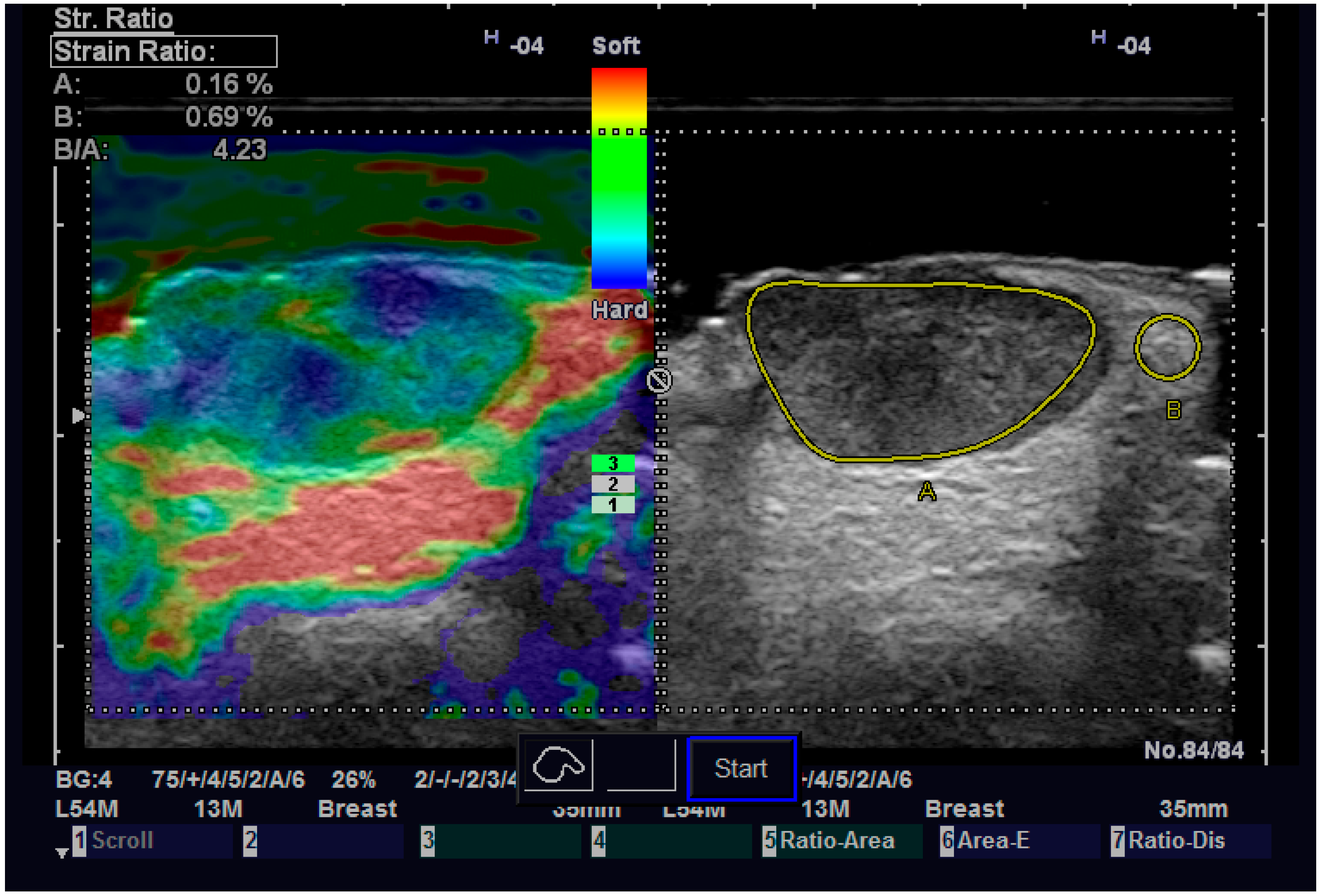
| Strain ratio, median (IQR) | 1.94 (1.38–2.62) | 3.2 (2.9–3.6) | 1.26 (−2.98–0.35) | 0.126●[n1 = 26, n2 = 5] |
| Diastolic speed, median (IQR) | 3.9 (3.9–4.1) | 4.3 (2.5–4.9) | 0.4 (−1–1.6) | 0.822●[n1 = 25, n2 = 5] |
| Systolic speed, median (IQR) | 6.3 (6.1–9) | 6.8 (6.4–8.2) | 0.5 (−1.9–2.2) | 0.758●[n1 = 25, n2 = 5] |
| Method | Tumor Margin (mm), Median (IQR) | Difference (95% CI) | p-Value | ICC Consistency (95% CI) | p-Value | ICC Agreement (95% CI) | p-Value |
|---|---|---|---|---|---|---|---|
| Pathology | 2 (1–2.75) | ||||||
| 13 MHz | 2 (1–2.65) | 0 (−0.05–0.15) | 0.367 | 0.993 (0.985–0.997) | <0.001 | 0.993 (0.985–0.997) | <0.001 |
| 20 MHz | 1.9 (1.15–3) | −0.1 (−0.05–0.25) | 0.203 | 0.961 (0.921–0.981) | <0.001 | 0.96 (0.92–0.981) | <0.001 |
| 40 MHz | 2 (1.1–2.8) | 0 (−0.05–0.27) | 0.195 | 0.972 (0.942–0.986) | <0.001 | 0.971 (0.941–0.986) | <0.001 |
Disclaimer/Publisher’s Note: The statements, opinions and data contained in all publications are solely those of the individual author(s) and contributor(s) and not of MDPI and/or the editor(s). MDPI and/or the editor(s) disclaim responsibility for any injury to people or property resulting from any ideas, methods, instructions or products referred to in the content. |
© 2023 by the authors. Licensee MDPI, Basel, Switzerland. This article is an open access article distributed under the terms and conditions of the Creative Commons Attribution (CC BY) license (https://creativecommons.org/licenses/by/4.0/).
Share and Cite
Tamas, T.; Dinu, C.; Lenghel, L.M.; Boțan, E.; Tamas, A.; Stoia, S.; Leucuta, D.C.; Bran, S.; Onisor, F.; Băciuț, G.; et al. High-Frequency Ultrasound in Diagnosis and Treatment of Non-Melanoma Skin Cancer in the Head and Neck Region. Diagnostics 2023, 13, 1002. https://doi.org/10.3390/diagnostics13051002
Tamas T, Dinu C, Lenghel LM, Boțan E, Tamas A, Stoia S, Leucuta DC, Bran S, Onisor F, Băciuț G, et al. High-Frequency Ultrasound in Diagnosis and Treatment of Non-Melanoma Skin Cancer in the Head and Neck Region. Diagnostics. 2023; 13(5):1002. https://doi.org/10.3390/diagnostics13051002
Chicago/Turabian StyleTamas, Tiberiu, Cristian Dinu, Lavinia Manuela Lenghel, Emil Boțan, Adela Tamas, Sebastian Stoia, Daniel Corneliu Leucuta, Simion Bran, Florin Onisor, Grigore Băciuț, and et al. 2023. "High-Frequency Ultrasound in Diagnosis and Treatment of Non-Melanoma Skin Cancer in the Head and Neck Region" Diagnostics 13, no. 5: 1002. https://doi.org/10.3390/diagnostics13051002
APA StyleTamas, T., Dinu, C., Lenghel, L. M., Boțan, E., Tamas, A., Stoia, S., Leucuta, D. C., Bran, S., Onisor, F., Băciuț, G., Armencea, G., & Băciuț, M. (2023). High-Frequency Ultrasound in Diagnosis and Treatment of Non-Melanoma Skin Cancer in the Head and Neck Region. Diagnostics, 13(5), 1002. https://doi.org/10.3390/diagnostics13051002