Abstract
A 54-year-old patient presented with progressive pain for one month in the second finger of the right hand with an emphasis on the proximal interphalangeal (PIP) joint. Subsequent magnetic resonance imaging (MRI) showed a diffuse intraosseous lesion at the base of the middle phalanx with destruction of the cortical bone and extraosseous soft tissue. An expansively growing chondromatous bone tumor, e.g., a chondrosarcoma, was suspected. After incisional biopsy, the pathologic findings finally revealed, surprisingly, a metastasis of a poorly differentiated non-small cell adenocarcinoma of the lung. This case illustrates a rare but important differential diagnosis for painful finger lesions.
Detailed Figure Legend
A 54-year-old patient presented with progressive pain for one month and mild swelling in the second finger of the right hand with an emphasis on the proximal interphalangeal (PIP) joint. Subsequent 3T magnetic resonance imaging (MRI) of the right hand in T1-weighted fat-saturated sequence after administration of 20 mL Gd-DOTA showed a contrast-enhancing diffuse intraosseous lesion at the base of the middle phalanx with destruction of the cortical bone and surrounding extraosseous soft tissue (Figure 1a). The axial view (Figure 1b) revealed ulnar infiltration of the ligaments of the articular capsule (arrow). Based on the imaging findings, an expansively growing chondromatous bone tumor, e.g., a chondrosarcoma, was suspected. After incisional biopsy, the pathologic findings finally revealed, surprisingly, a metastasis of a poorly differentiated non-small cell adenocarcinoma of the lung.
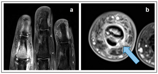
Figure 1.
T1-weighted fat-saturated, contrast-enhanced MRI. (a) coronal view. (b) axial view.
The patient stopped smoking 12 years ago (after 20–30 pack years); there was no positive family history, no B symptoms (i.e., fever, night sweats, unintentional weight loss), occasional cough, no sputum, and no hemoptysis. Staging by positron emission tomography/computed tomography (PET/CT) with 316 MBq [18F]FDG revealed a primary tumor in the upper lobe of the right lung (Figure 2a) and multiple lymph node, soft tissue, and bone metastases (Figure 2b); cranial MRI revealed a brain metastasis.
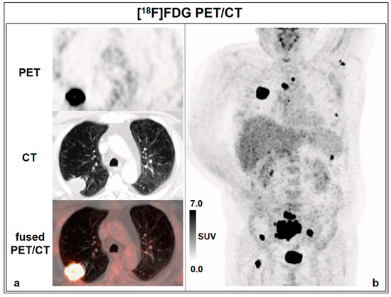
Figure 2.
[18F]FDG PET/CT. (a) axial view. (b) maximum intensity projection (MIP).
In contrast to this case, typical initial symptoms of lung carcinoma are respiratory symptoms including cough, dyspnoea or haemoptysis, but not bone pain [1]. Overall, the occurrence of metastases in the acral parts of the body is a rare condition [2,3]. Amongst acro-metastases, lung cancer was reported to be the most common cause, with one third of cases [4]. The imaging findings of peripheral bone metastases, however, are variable and a robust differentiation from chondrosarcoma or other malignant bone and soft tissue tumors is not regularly possible using MRI alone [5]. Typically, medullary chondrosarcomas present with a lobular growth pattern at the margins and display a heterogeneous signal behavior across the sequences in MRI, partly due to diverse matrix mineralization and areas of entrapped yellow marrow [6].
These images illustrate the findings in a rare but important differential diagnosis for painful finger lesions and underline the value of timely histological confirmation, especially in the case of inconclusive imaging findings. Further, they recall that patients with advanced stage cancer may be either asymptomatic or symptomatic with an unusual pattern.
Author Contributions
A.H.: clinical management, manuscript draft. H.R.D. and L.M.U.: clinical management, revision of the manuscript, increased intellectual content. A.S. and M.K.: provision of the MR images, increased intellectual content. A.T., F.M. and W.G.K.: revision of the manuscript, increased intellectual content. M.U.: manuscript draft, supervision. All authors have read and agreed to the published version of the manuscript.
Funding
L.M.U. was funded by the Munich-Clinician-Scientist-Program (LMU Munich).
Informed Consent Statement
The Ethics Committee waived additional approval for case reports.
Conflicts of Interest
The authors declare no conflict of interest.
References
- Rudin, C.M.; Brambilla, E.; Faivre-Finn, C.; Sage, J. Small-cell lung cancer. Nat. Rev. Dis. Prim. 2021, 7, 3. [Google Scholar] [CrossRef] [PubMed]
- Afshar, A.; Farhadnia, P.; Khalkhali, H. Metastases to the hand and wrist: An analysis of 221 cases. J. Hand Surg. 2014, 39, 923–932. [Google Scholar] [CrossRef] [PubMed]
- Sur, Y.J.; Kang, Y.K.; Bahk, W.J.; Chang, D.K.; Rhee, S.K. Metastatic malignant tumour in the hand. J. Plast. Surg. Hand Surg. 2011, 45, 90–95. [Google Scholar] [CrossRef] [PubMed]
- Stomeo, D.; Tulli, A.; Ziranu, A.; Perisano, C.; De Santis, V.; Maccauro, G. Acrometastasis: A literature review. Eur. Rev. Med. Pharmacol. Sci. 2015, 19, 2906–2915. [Google Scholar] [PubMed]
- Ma, L.D. Magnetic resonance imaging of musculoskeletal tumors: Skeletal and soft tissue masses. Curr. Probl. Diagn. Radiol. 1999, 28, 29–62. [Google Scholar] [CrossRef] [PubMed]
- Douis, H.; Saifuddin, A. The imaging of cartilaginous bone tumours. II. Chondrosarcoma. Skelet. Radiol. 2013, 42, 611–626. [Google Scholar] [CrossRef]


Disclaimer/Publisher’s Note: The statements, opinions and data contained in all publications are solely those of the individual author(s) and contributor(s) and not of MDPI and/or the editor(s). MDPI and/or the editor(s) disclaim responsibility for any injury to people or property resulting from any ideas, methods, instructions or products referred to in the content. |
© 2023 by the authors. Licensee MDPI, Basel, Switzerland. This article is an open access article distributed under the terms and conditions of the Creative Commons Attribution (CC BY) license (https://creativecommons.org/licenses/by/4.0/).