Interesting Images

Dynamic Ultrasound Examination for Extensor Pollicis Longus Tendon Rupture after Palpation-Guided Corticosteroid Injection

1
Department of Physical Medicine and Rehabilitation, College of Medicine, National Taiwan University, Taipei 10048, Taiwan
2
Department of Physical Medicine and Rehabilitation, National Taiwan University Hospital, Bei-Hu Branch, Taipei 10845, Taiwan
3
Department of Rehabilitation Medicine, Charles University, First Faculty of Medicine and General University Hospital in Prague, 12800 Prague, Czech Republic
4
Physical and Rehabilitation Medicine Unit, Luigi Sacco University Hospital, ASST Fatebenefratelli-Sacco, 20157 Milan, Italy
5
Department of Physical and Rehabilitation Medicine, Hacettepe University Medical School, Ankara 06100, Turkey
6
Center for Regional Anesthesia and Pain Medicine, Wang-Fang Hospital, Taipei Medical University, Taipei 11600, Taiwan
*
Author to whom correspondence should be addressed.
Diagnostics 2023, 13(5), 959; https://doi.org/10.3390/diagnostics13050959
Submission received: 28 January 2023 / Revised: 28 February 2023 / Accepted: 1 March 2023 / Published: 3 March 2023
(This article belongs to the Section Pathology and Molecular Diagnostics)

Abstract

:
This report aimed to present a case of wrist-tendon rupture and to discuss a rare complication after corticosteroid injection. A 67-year-old woman had difficulty extending her left-thumb interphalangeal joint several weeks after a palpation-guided local corticosteroid injection. Passive motions remained intact without sensory abnormalities. Ultrasound examination showed hyperechoic tissues at the site of the extensor pollicis longus (EPL) tendon at the wrist level and an atrophic EPL muscle stump at the forearm level. Dynamic imaging demonstrated no motion in the EPL muscle during passive thumb flexion/extension. The diagnosis of complete EPL rupture, possibly due to inadvertent intratendinous corticosteroid injection, was therefore confirmed.

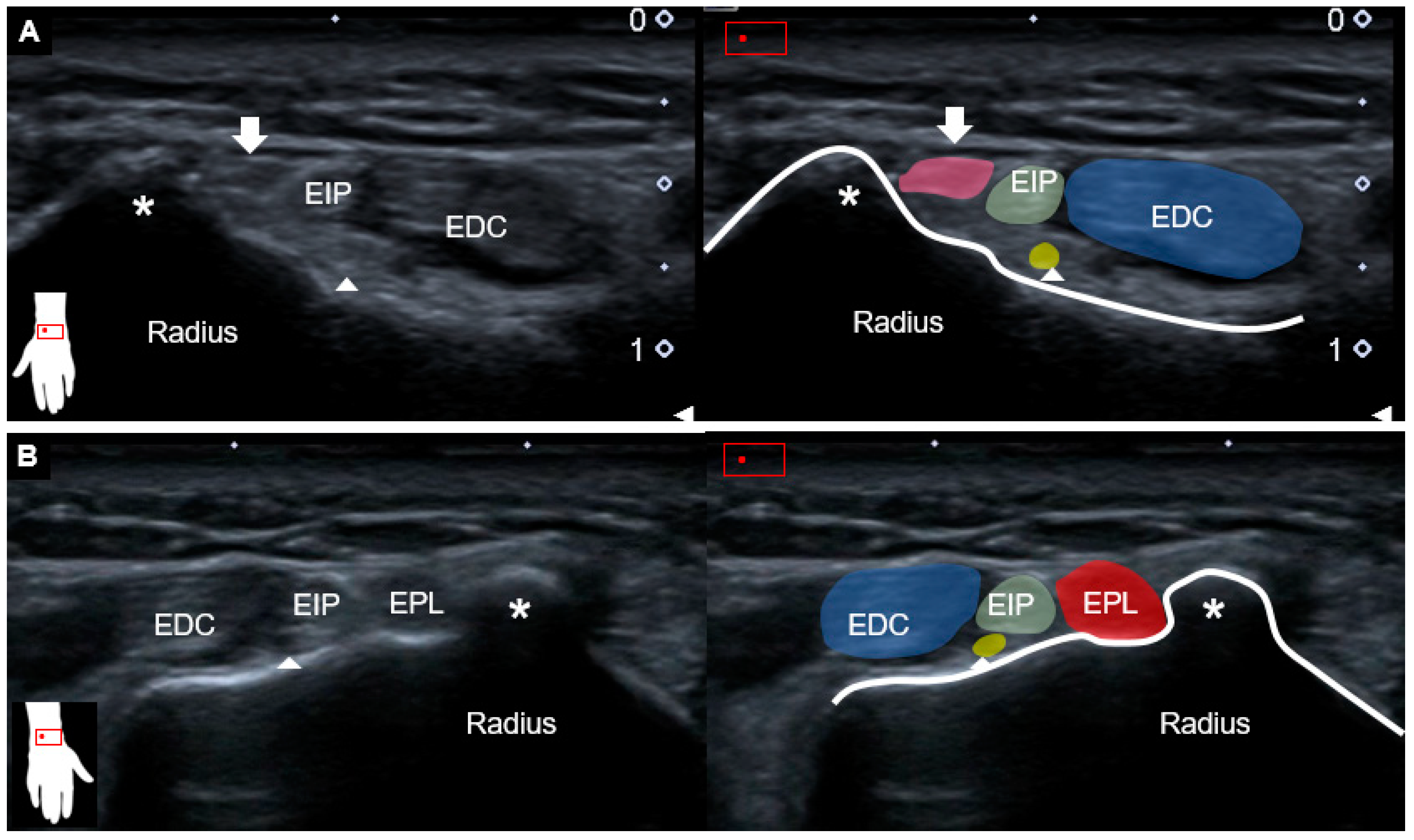
This report aimed to present a case of wrist-tendon rupture and to discuss a rare complication after corticosteroid injection. A 67-year-old female complained of difficulty in extending the left thumb for several months. The medical history comprised radial wrist pain after a contusion injury one year ago, for which corticosteroid injection under palpation guidance had also been performed, with gradual symptom relief. The physical examination revealed loss of the active extension of the left-thumb interphalangeal joint with normal passive motions (Figure 1, Video S1). Other finger joints were not affected, nor were there any sensory abnormalities over the distal upper extremity. Plain radiographs of the left wrist showed no evidence of fracture or joint dislocation. The ultrasound examination of the left wrist showed hyperechoic tissue between the Lister’s tubercle and the extensor digitorum communis tendons where the extensor pollicis longus (EPL) tendon should normally reside (Figure 2A). Herewith, the EPL tendon was clearly visualized at the right wrist (Figure 2B).
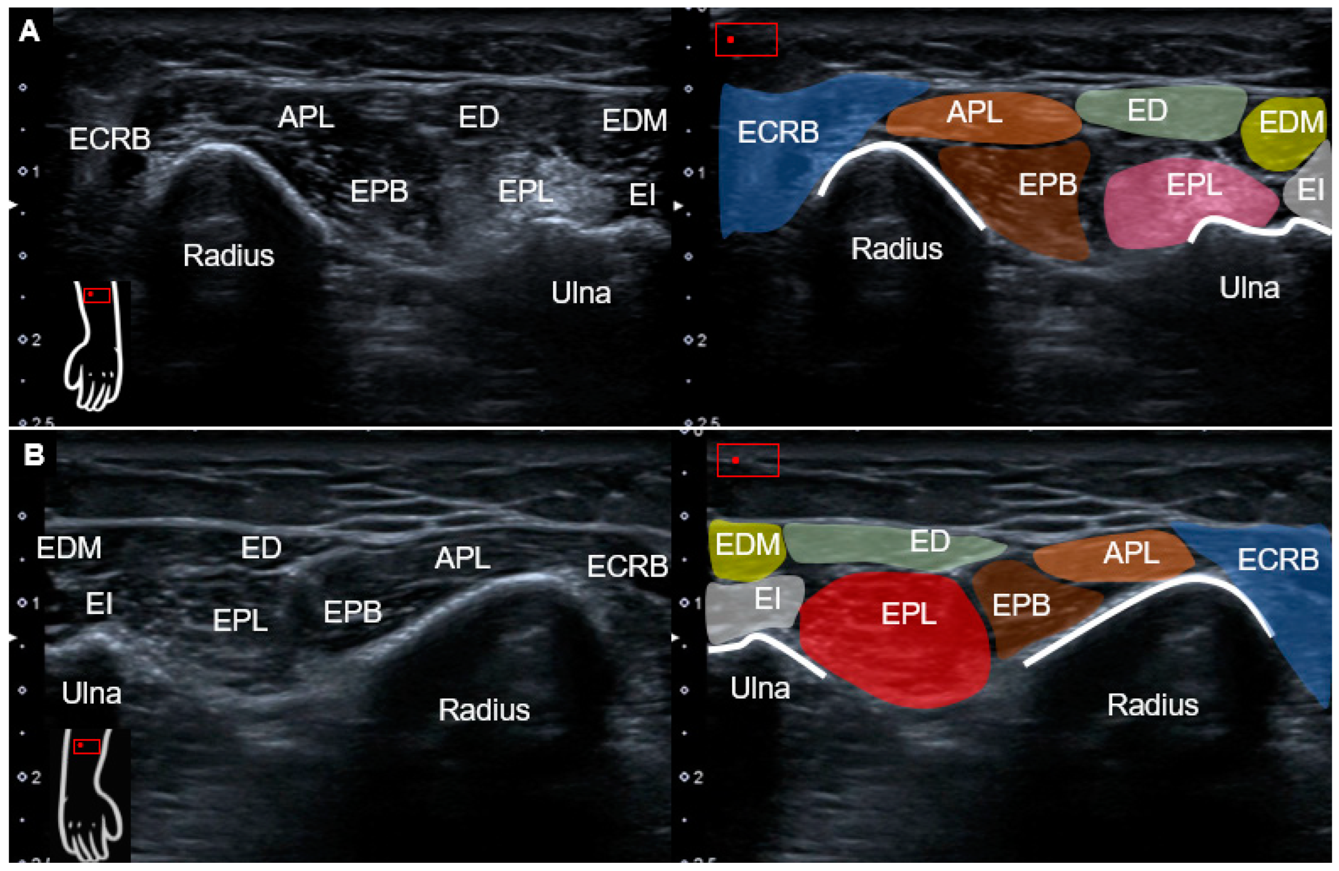
The posterior interosseous nerve (PIN) remained bilaterally intact based on dynamic tracking from the level of the supinator tunnel to the dorsal wrist [1]. Moving the transducer more proximally toward the distal forearm, a hyperechoic region was seen at the ulnar aspect of the first extensor compartment, representing the atrophic EPL muscle (Figure 3A). On the normal side, the intact EPL muscle could be explicitly recognized (Figure 3B). Dynamic ultrasound was applied to trace the left EPL tendon distally to proximally, whereby no excursion was observed for the EPL muscle during passive thumb flexion/extension (Video S2). As a complete rupture of the left EPL tendon was diagnosed/confirmed, the patient was referred for surgical repair.
Finger drop can ensue due to neurological and orthopedic causes [2]. In cases with more generalized weakness or sensory signs, neurological causes, such as PIN palsy, cervical radiculopathy or multifocal neuropathy, should be considered [3]. More specifically, PIN palsy may result in the loss of finger and thumb extension, without wrist drop. Four types of PIN palsy have been reported. Among them, (although uncommon) type 4 exclusively involves the branch supplying the EPL, with isolated difficulty in extending the IP joint of the thumb [4]. Orthopedic causes, on the other hand, include rupture, subluxation or entrapment of the finger extensor tendon. The absence of the tenodesis effect, i.e., finger automatic extension during wrist passive flexion, would indicate an abnormality of the tendons instead of nerves [2]. In patients with an isolated loss of thumb extension, the EPL tendon (responsible for the extension of the thumb interphalangeal joint) should be investigated.
Static ultrasound examination can serve as a helpful tool for scrutinizing tendon injury, whereas dynamic imaging can be employed to validate motion problems. The most common sonographic finding of tendon rupture is loss of fiber continuity with the formation of a retracted stump. The absence of normal tendon gliding during passive motion further confirms the diagnosis [5]. In chronic cases, the proximal muscle part would become thinned and hyperechoic due to fat infiltration and disuse atrophy. In the case of a ruptured EPL tendon, the tubular-shaped hypoechoic area might be seen, corresponding to fluid, hemorrhage, and scar tissue [6]. In addition, a bulging contoured hypoechoic lesion (also referred to as a pseudo-mass), located between the Lister’s tubercle and the carpometacarpal joint, has been reported as being more frequent over the ruptured EPL than other finger tendons [5].
One of the important etiologies of spontaneous EPL tendon rupture is intratendinous corticosteroid injection. Other causes include distal radial fracture, repetitive friction against metal implants, and contusions [7]. Local corticosteroid injection is associated with rupture, not only in the EPL tendon, but also in other locations, such as the rotator cuff, common extensor, patellar, and Achilles tendons [8,9,10,11,12]. Intratendinous corticosteroid injection in animal models was found to inhibit connective-tissue formation and lead to a reduction of tendon mass and biomechanical integrity [13,14]. Lu et al. [15] reported 13 patients with tendon rupture after corticosteroid injection on the wrist region. The most common injected site was the radial styloid under the clinical impression of De Quervain’s tenosynovitis. The injection times over the same sites ranged from 1 to 4, whereas the periods from the last injection ranged from 3 to 32 weeks.
Needless to say, ultrasound is a preferred tool for guiding the needle, in order to avoid unwanted intratendinous corticosteroid injection [16,17]. Peritendinous or intrabursal corticosteroid injection, even in the highest dose, do not affect the tendon’s elasticity when properly performed under ultrasound guidance [18]. Compared to palpation guidance, ultrasound guidance provides fewer local complications, e.g., fat atrophy and depigmentation [19]. Furthermore, ultrasound- vs. palpation-guided corticosteroid injection has been proven to have better accuracy and clinical outcome in bicipital tendinosis [20]. In short, this case of ours revealed that static/dynamic ultrasound played a vital role in the management of (thumb) tendon disorders. This is true not only for a precise diagnosis or targeting during an intervention, but is therefore also paramount to the indirect avoidance of adverse effects, e.g., an intratendinous injection.

Supplementary Materials

The following supporting information can be downloaded at: https://www.mdpi.com/article/10.3390/diagnostics13050959/s1, Video S1: Physical examination of bilateral thumb motions. Video S2: Dynamic tracking of EPL tendons and muscles.

Author Contributions

Conceptualization, Y.-C.C. and K.-V.C.; methodology, K.-V.C.; validation, K.M., V.R. and L.Ö.; writing—original draft preparation, W.-T.W.; writing—review and editing, K.-V.C. and L.Ö.; funding acquisition, K.-V.C. All authors have read and agreed to the published version of the manuscript.

Funding

This study was made possible by (1) research funding from the Community and Geriatric Medicine Research Center, National Taiwan University Hospital, Bei-Hu Branch, Taipei, Taiwan; (2) Ministry of Science and Technology (MOST 106-2314-B-002-180-MY3, 109-2314-B-002-114-MY3, and 109-2314-B-002-127), and (3) the Taiwan Society of Ultrasound in Medicine.

Institutional Review Board Statement

Not applicable.

Informed Consent Statement

Patient consent was obtained.

Data Availability Statement

Data are contained within the main text of the manuscript.

Conflicts of Interest

The authors declare no conflict of interest.

References

  1. Wu, C.H.; Chang, K.V.; Özçakar, L.; Hsiao, M.Y.; Hung, C.Y.; Shyu, S.G.; Wang, T.G.; Chen, W.S. Sonographic tracking of the upper limb peripheral nerves: A pictorial essay and video demonstration. Am. J. Phys. Med. Rehabil. 2015, 94, 740–747. [Google Scholar] [CrossRef] [PubMed]
  2. Varatharaj, A.; Pinto, A.; Manning, M. Differential diagnosis of finger drop. Neurologist 2015, 19, 128–131. [Google Scholar] [CrossRef] [PubMed]
  3. Kudlac, M.; Cummings, R.; Finocchiaro, J. Finger Drop: Cervical Radiculopathy, Peripheral Nerve Lesion, or Multifocal Neuropathy? A Case Report. JOSPT Cases 2022, 2, 112–116. [Google Scholar]
  4. Horton, T. Isolated paralysis of the extensor pollicis longus muscle: A further variation of posterior interosseous nerve palsy. J. Hand Surg. 2000, 25, 225–226. [Google Scholar] [CrossRef] [PubMed]
  5. Lee, S.M.; Ha, D.H.; Han, S.H. Differential sonographic features of the extensor pollicis longus tendon rupture and other finger tendons rupture in the setting of hand and wrist trauma. PLoS ONE 2018, 13, e0205111. [Google Scholar] [CrossRef] [Green Version]
  6. De Maeseneer, M.; Marcelis, S.; Osteaux, M.; Jager, T.; Machiels, F.; Van Roy, P. Sonography of a rupture of the tendon of the extensor pollicis longus muscle: Initial clinical experience and correlation with findings at cadaveric dissection. Am. J. Roentgenol. 2005, 184, 175–179. [Google Scholar] [CrossRef]
  7. Björkman, A.; Jörgsholm, P. Rupture of the extensor pollicis longus tendon: A study of aetiological factors. Scand. J. Plast. Reconstr. Surg. Hand Surg. 2004, 38, 32–35. [Google Scholar] [CrossRef]
  8. Chen, S.-K.; Lu, C.-C.; Chou, P.-H.; Guo, L.-Y.; Wu, W.-L. Patellar tendon ruptures in weight lifters after local steroid injections. Arch. Orthop. Trauma Surg. 2009, 129, 369–372. [Google Scholar] [CrossRef] [PubMed]
  9. Lin, C.-Y.; Huang, S.-C.; Tzou, S.-J.; Yin, C.-H.; Chen, J.-S.; Chen, Y.-S.; Chang, S.-T. A Positive Correlation between Steroid Injections and Cuff Tendon Tears: A Cohort Study Using a Clinical Database. Int. J. Environ. Res. Public Health 2022, 19, 4520. [Google Scholar] [CrossRef]
  10. Newnham, D.; Douglas, J.; Legge, J.; Friend, J. Achilles tendon rupture: An underrated complication of corticosteroid treatment. Thorax 1991, 46, 853–854. [Google Scholar] [CrossRef] [PubMed] [Green Version]
  11. Smith, A.G.; Kosygan, K.; Williams, H.; Newman, R.J. Common extensor tendon rupture following corticosteroid injection for lateral tendinosis of the elbow. Br. J. Sport. Med. 1999, 33, 423–424. [Google Scholar] [CrossRef] [PubMed] [Green Version]
  12. Speed, C. Corticosteroid injections in tendon lesions. BMJ 2001, 323, 382–386. [Google Scholar] [CrossRef] [PubMed]
  13. Kapetanos, G. The effect of the local corticosteroids on the healing and biomechanical properties of the partially injured tendon. Clin. Orthop. Relat. Res. 1982, 163, 170–179. [Google Scholar] [CrossRef]
  14. Ketchum, L.D. Effects of triamcinolone on tendon healing and function: A laboratory study. Plast. Reconstr. Surg. 1971, 47, 471–482. [Google Scholar] [CrossRef] [PubMed]
  15. Lu, H.; Yang, H.; Shen, H.; Ye, G.; Lin, X.J. The clinical effect of tendon repair for tendon spontaneous rupture after corticosteroid injection in hands: A retrospective observational study. Medicine 2016, 95, e5145. [Google Scholar] [CrossRef] [PubMed]
  16. Chang, K.V.; Wu, W.T.; Han, D.S.; Özçakar, L. Static and Dynamic Shoulder Imaging to Predict Initial Effectiveness and Recurrence After Ultrasound-Guided Subacromial Corticosteroid Injections. Arch. Phys. Med. Rehabil. 2017, 98, 1984–1994. [Google Scholar] [CrossRef] [PubMed]
  17. Wang, J.C.; Chang, K.V.; Wu, W.T.; Han, D.S.; Özçakar, L. Ultrasound-Guided Standard vs Dual-Target Subacromial Corticosteroid Injections for Shoulder Impingement Syndrome: A Randomized Controlled Trial. Arch. Phys. Med. Rehabil. 2019, 100, 2119–2128. [Google Scholar] [CrossRef] [PubMed]
  18. Hsu, P.-C.; Chang, K.-V.; Wu, W.-T.; Wang, J.-C.; Özçakar, L. Effects of ultrasound-guided peritendinous and intrabursal corticosteroid injections on shoulder tendon elasticity: A post hoc analysis of a randomized controlled trial. Arch. Phys. Med. Rehabil. 2021, 102, 905–913. [Google Scholar] [CrossRef] [PubMed]
  19. Jeyapalan, K.; Choudhary, S. Ultrasound-guided injection of triamcinolone and bupivacaine in the management of de Quervain’s disease. Skelet. Radiol. 2009, 38, 1099–1103. [Google Scholar] [CrossRef] [PubMed]
  20. Yiannakopoulos, C.K.; Megaloikonomos, P.D.; Foufa, K.; Gliatis, J. Ultrasound-guided versus palpation-guided corticosteroid injections for tendinosis of the long head of the biceps: A randomized comparative study. Skelet. Radiol. 2020, 49, 585–591. [Google Scholar] [CrossRef] [PubMed]
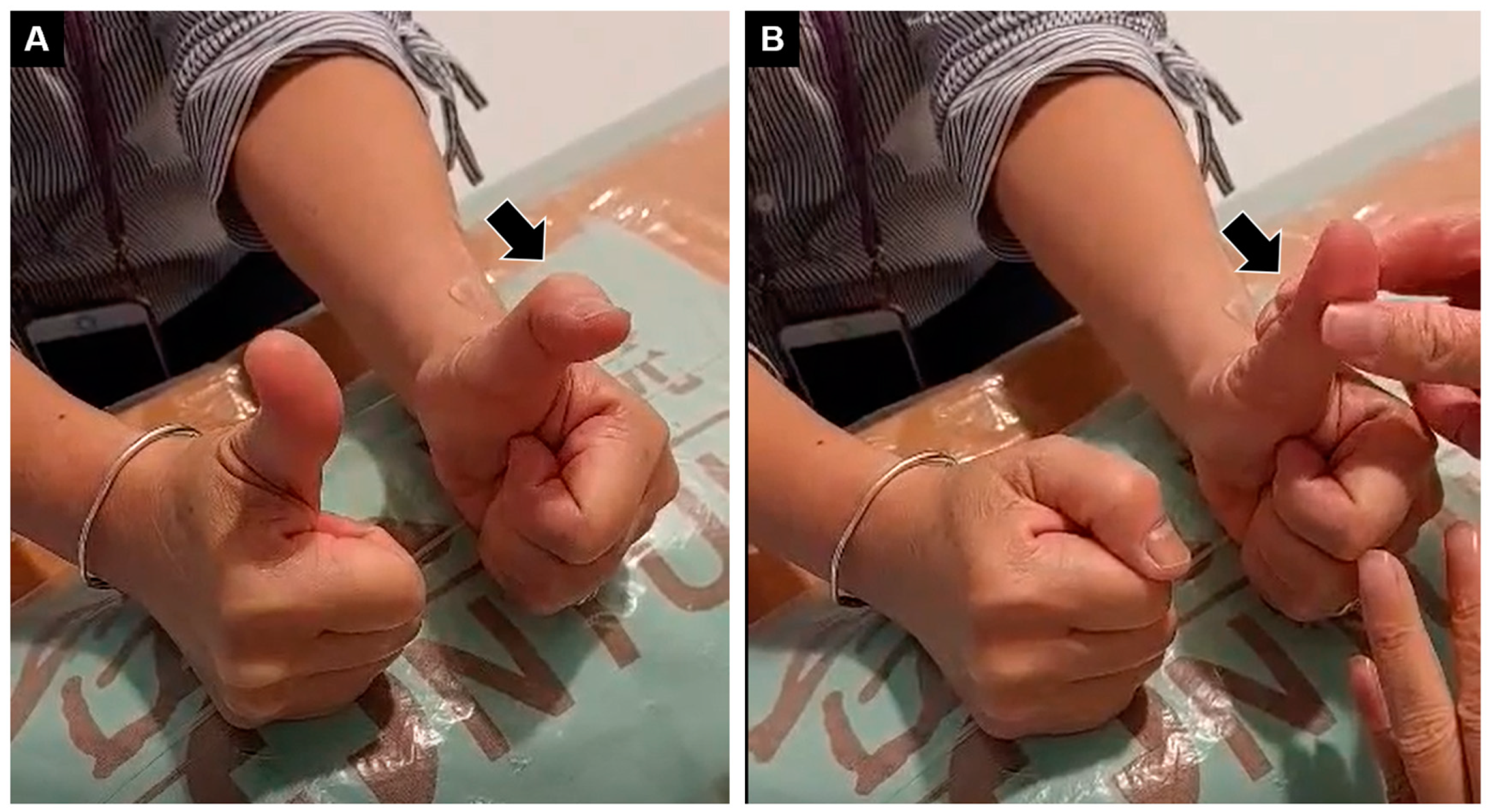
Figure 1. (A) Inability to extend the interphalangeal joint of the left thumb (black arrow), with the normal right side. (B) Normal passive movement of the same joint.
Diagnostics 13 00959 g001
Figure 2. Ultrasound imaging (transverse view) and schematic drawing of (A) left and (B) right wrists at the level of Lister’s tubercle. Compared to the right side, EPL tendon was absent with hyperechoic fibrofatty tissues (white arrow and pink shade) instead. Asterisks, Lister’s tubercle; EPL and red shade, extensor pollicis longus tendon; EDC and blue shade, extensor digitorum communis tendon; EIP and green shade, extensor indicis proprius tendon; arrowheads and yellow shade, posterior interosseous nerve.
Diagnostics 13 00959 g002
Figure 3. Ultrasound imaging (transverse view) and schematic drawing of the (A) left and (B) right distal forearm. Compared with the normal side, the left EPL muscle, located at the ulnar aspect of EPB muscle, was hyperechoic and atrophic. APL and orange shade, abductor pollicis longus; ECRB and blue shade, extensor carpi radialis brevis; EPB and brown shade, extensor pollicis brevis; ED and green shade, extensor digitorum communis; EDM and yellow shade, extensor digiti minimi; EI and gray shade, extensor indices; EPL and pink/red shade, extensor pollicis longus.
Diagnostics 13 00959 g003
Disclaimer/Publisher’s Note: The statements, opinions and data contained in all publications are solely those of the individual author(s) and contributor(s) and not of MDPI and/or the editor(s). MDPI and/or the editor(s) disclaim responsibility for any injury to people or property resulting from any ideas, methods, instructions or products referred to in the content.