
YOLOv5s-SA: Light-Weighted and Improved YOLOv5s for Sperm Detection

 , ,
, ,
Abstract
:1. Introduction
- We propose a novel YOLOv5s algorithm and introduce Shuffle Attention (SA) mechanism [30] to enhance the model’s attention to small target sperm.
- We propose to replace partial convolution in the backbone network with depthwise separable convolution (DWConv) [31] to improve the convergence speed of the model.
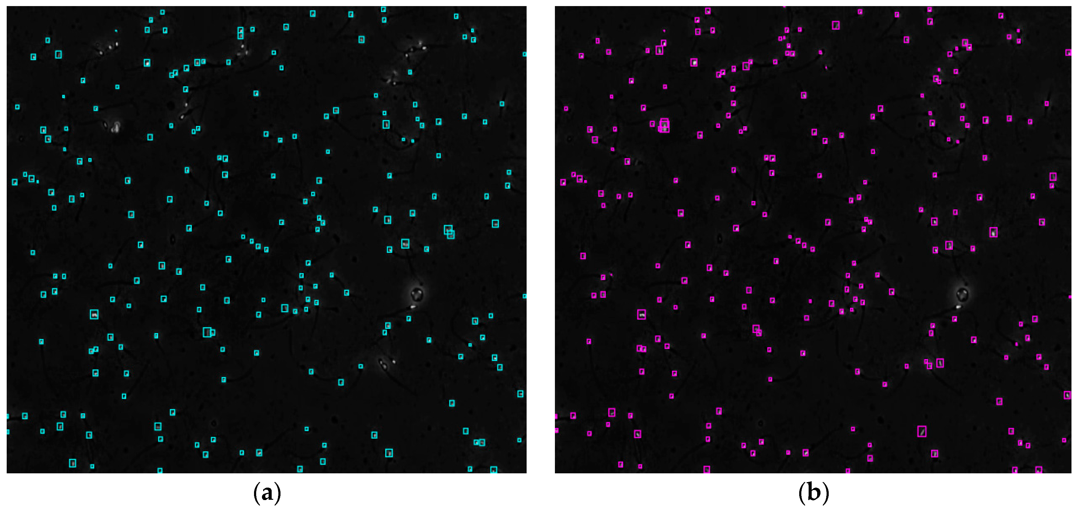
- The proposed method can effectively process semen images with low quality and a complex background, improve the detection performance of sperm and provide the feasibility of accurate detection using human or other animal sperm cells.
2. Materials and Methods
2.1. Date Set
2.2. YOLOv5s-SA Architecture
2.2.1. Lightweight Backbone Feature Extraction Module
2.2.2. Multi-Scale Feature Fusion Enhancement Module
2.3. Parameter Setting and Experimental Environment
3. Experiments
3.1. Evaluation Metrics
3.2. YOLOv5s Ablation Study
3.3. Evaluation of Sperm Detection Methods
3.4. Partial Occlusion Handling
3.5. Failure Case Analysis
4. Conclusions
Author Contributions
Funding
Institutional Review Board Statement
Informed Consent Statement
Data Availability Statement
Conflicts of Interest
References
- Zauner, G.; Girardi, G. Potential causes of male and female infertility in Qatar. J. Reprod. Immunol. 2020, 141, 103173. [Google Scholar] [CrossRef] [PubMed]
- Selvam, M.K.P.; Ambar, R.F.; Agarwal, A.; Henkel, R. Etiologies of sperm DNA damage and its impact on male infertility. Andrologia 2021, 53, 1. [Google Scholar]
- Matson, P.; Kitson, M.; Zuvela, E. Human sperm morphology assessment since 2010: Experience of an Australian external quality assurance programme. Reprod. Biomed. Online 2022, 44, 340–348. [Google Scholar] [CrossRef] [PubMed]
- Miahi, E.; Mirroshandel, S.A.; Nasr, A. Genetic Neural Architecture Search for automatic assessment of human sperm images. Expert Syst. Appl. 2022, 188, 115937. [Google Scholar] [CrossRef]
- World Health Organization. WHO Laboratory Manual for the Examination and Processing of Human Semen, 5th ed.; World Health Organization: Geneva, Switzerland, 2010; pp. 280–286. [Google Scholar]
- Valiuskaite, V.; Raudonis, V.; Maskeliunas, R.; Damasevicius, R.; Krilavicious, T. Deep Learning Based Evaluation of Spermatozoid Motility for Artificial Insemination. Sensors 2020, 21, 72. [Google Scholar] [CrossRef]
- Ilhan, H.O.; Yuzkat, M.; Aydin, N. Sperm Motility Analysis by using Recursive Kalman Filters with the smartphone based data acquisition and reporting approach. Export Syst. Appl. 2021, 186, 115774. [Google Scholar] [CrossRef]
- Garcia-Grau, E.; Lleberia, J.; Costa, L.; Guitart, M.; Yeste, M.; Benet, J.; Amengual, M.J.; Ribas-Maynou, J. Decline of Sperm Quality over the Last Two Decades in the South of Europe: A Retrospective Study in Infertile Patients. Biology 2023, 12, 70. [Google Scholar] [CrossRef]
- Ozyer, G.T.; Ozyer, B.; Negin, F.; Alarabi, I.; Agahian, S. A hybrid IMM-JPDAF algorithm for tracking multiple sperm targets and motility analysis. Neural Comput. Appl. 2022, 34, 17407–17421. [Google Scholar] [CrossRef]
- Liu, G.L.; Shi, H.; Zhang, H.; Zhou, Y.T.; Sun, Y.J.; Li, W.; Huang, X.F.; Jiang, Y.Q.; Fang, Y.L.; Yang, G. Fast Noninvasive Morphometric Characterization of Free Human Sperms Using Deep Learning. Microsc. Microanal. 2022, 28, 1767–1779. [Google Scholar] [CrossRef]
- Ilhan, H.O.; Serbes, G. Sperm morphology analysis by using the fusion of two-stage fine-tuned deep networks. Biomed. Signal Process. Control 2022, 7, 103246. [Google Scholar] [CrossRef]
- Spencer, L.; Fernando, J.; Akbaridoust, F.; Ackermann, K.; Nosrati, R. Ensembled Deep Learning for the Classification of Human Sperm Head Mophology. Adv. Intell. Syst. 2022, 4, 2200111. [Google Scholar] [CrossRef]
- Mahmoud, A.M.; Depoorter, B.; Piens, N.; Comhaire, F.H. The performance of 10 different methods for the estimation of sperm concentration. Fertil. Steril. 1997, 68, 345. [Google Scholar] [CrossRef] [PubMed]
- Davis, R.O.; Rothmann, S.A.; Overstreet, J.W. Accuracy and precision of computer-aided sperm analysis in multicenter studies. Fertil. Steril. 1992, 57, 648–653. [Google Scholar] [CrossRef]
- Talarczyk-Desole, J.; Berger, A.; Taszarek, G.; Hauke, J.; Pawelczyk, L.; Jedrzejczak, P. Manual vs. computer-assisted sperm analysis: Can CASA replace manual assessment of human semen in clinical practice? Ginekol. Pol. 2017, 88, 56–60. [Google Scholar] [CrossRef] [PubMed] [Green Version]
- Larsen, L.; Scheike, T.; Jensen, T.K.; Bonde, J.P.; Ernst, E.; Hjollund, N.H.; Zhou, Y.; Skakkebaek, N.E.; Giwercman, A. Computer-assisted semen analysis parameters as predictors for fertility of men from the general population. Human Reprod. 2000, 15, 1562–1567. [Google Scholar] [CrossRef] [Green Version]
- Tomlinson, M.J.; Pooley, K.; Simpson, T.; Newton, T.; Hopkisson, J.; Jayaprakasan, K.; Jayaprakasan, R.; Naeem, A.; Pridmore, T. Validation of a novel computer-assisted sperm analysis (CASA) system using multitarget-tracking algorithms. Fertil. Steril. 2010, 93, 1911–1920. [Google Scholar] [CrossRef]
- Hicks, S.A.; Andersen, J.M.; Witczak, O.; Thambawita, V.; Halvorsen, P.; Hammer, H.L.; Haugen, T.B.; Riegler, M.A. Machine learning-based analysis of sperm videos and participant data for male fertility prediction. Sci. Rep. 2019, 9, 16770. [Google Scholar] [CrossRef] [Green Version]
- Jati, G.; Gunawan, A.A.; Lestari, S.W. Multi-sperm tracking using Hungarian kalman filter on low frame rate video. In Proceedings of the Advanced Computer Science and Information Systems (ICACSIS), Malang, Indonesia, 15–16 October 2016; pp. 530–535. [Google Scholar]
- Hasikin, K.; Ii, N.A.M.; Mohamed, M. A new region-based adaptive thresholding for sperm motility segmentation. Malays. J. Comput. Sci. 2016, 29, 272–286. [Google Scholar] [CrossRef]
- Alameri, M.; Hasikin, K.; Kadri, N.A. Multistage Optimization Using a Modified Gaussian Mixture Model in Sperm Motility Tracking. Comput. Math. Methods Med. 2021, 8, 14. [Google Scholar] [CrossRef]
- Qi, S.; Nie, T.; Li, Q.; He, Z.; Xu, D.; Chen, Q. A Sperm Cell Tracking Recognition and Classification Method. In Proceedings of the 2019 International Conference on Systems, Signals and Image Processing (IWSSIP), Osijek, Croatia, 5–7 June 2019; pp. 163–167. [Google Scholar] [CrossRef]
- Alabdulla, A.A.A.; Haşıloğlu, A.; Aksu, E.H. A robust sperm cell tracking algorithm using uneven lighting image fixing and improved branch and bound algorithm. IET Image Process. 2021, 15, 2068–2079. [Google Scholar] [CrossRef]
- Somasundaram, D.; Nirmala, M. Faster region convolutional neural network and semen tracking algorithm for sperm analysis. Comput. Methods Programs Biomed. 2021, 200, 105918. [Google Scholar] [CrossRef] [PubMed]
- Prabaharan, L.; Raghunathan, A. An improved convolutional neural network for abnormality detection and segmentation from human sperm images. J. Ambient Intell. Humaniz. Comput. 2021, 12, 3341–3352. [Google Scholar] [CrossRef]
- Abbasi, A.; Miahi, E.; Mirroshandel, S.A. Effect of Deep Transfer and Multi task Learning on Sperm Abnormality Detection. Comput. Biol. Med. 2021, 128, 104121. [Google Scholar] [CrossRef]
- Nissen, S.M.; Krause, O.; Almstrup, K.; Kjærulff, S.; Nielsen, T.T.; Nielsen, M. Convolutional neural networks for segmentation and object detection of human semen. In Proceedings of the 20th Scandinavian Conference on Image Analysis (SCIA), Tromsø, Norway, 12–14 June 2017; pp. 397–406. [Google Scholar]
- Qixian, L.V.; Yuan, X.R.; Qian, J.Z.; Li, X.K.; Zhang, H.Y.; Zhan, S. An Improved U-Net for Human Sperm Head Segmentation. Neural Process. Lett. 2022, 54, 537–557. [Google Scholar]
- Qiao, G.C.; Yang, M.X.; Wang, H. A water level measurement approach based on YOlOv5s. Sensors 2022, 10, 3714. [Google Scholar] [CrossRef]
- Zhang, Q.L.; Yang, Y.B. SA-Net: Shuffle attention for deep convolutional neural networks. In Proceedings of the IEEE International Conference on Acoustics, Speech and Signal Processing (ICASSP), Toronto, ON, Canada, 6–11 June 2021; pp. 2235–2239. [Google Scholar]
- Howard, A.; Sandler, M.; Chen, B. Searching for MobileNet V3. In Proceedings of the 2019 IEEE/CVF International Conference on Computer Vision, Seoul, Republic of Korea, 27–28 October 2019; pp. 1314–1324. [Google Scholar]
- Haugen, T.B.; Hicks, S.A.; Andersen, J.M.; Witczak, O.; Hammer, H.L.; Borgli, R.; Halvorsen, P.; Riegler, M.A. Visem: A multimodal video dataset of human spermatozoa. In Proceedings of the 10th ACM on Multimedia Systems Conference, ACM, Amherst, MA, USA, 18–21 June 2019. [Google Scholar]
- Huang, Z.C.; Wang, J.L. DC- SPP- YOLO: Dense connection and spatial pyramid pooling based YOLO for object detection. Inf. Sci. 2020, 522, 241–258. [Google Scholar] [CrossRef] [Green Version]
- Lin, T.Y.; Dollar, P.; Girshick, R. Feature pyramid networks for object detection. In Proceedings of the Computer Vision and Pattern Recognition (CVPR), Honolulu, HI, USA, 21–26 July 2017; pp. 2117–2125. [Google Scholar]
- Liu, S.; Qi, L.; Qin, H.F.; Shi, J.; Jia, J. Path aggregation network for instance segmentation. In Proceedings of the Computer Vision and Pattern Recognition (CVPR), Salt Lake City, UT, USA, 21–23 June 2018; pp. 8759–8768. [Google Scholar]
- Zheng, Z.; Wang, P.; Liu, W.; Li, J.; Ye, R.; Ren, D. Distance-IoU loss: Faster and better learning for bounding box regression. In Proceedings of the AAAI Conference on Artificial Intelligence, New York, NY, USA, 7–12 February 2020; Volume 34, pp. 12993–13000. [Google Scholar]
- Murtaza, M.; Sharif, M.; Yasmin, M.; Fayyaz, M.; Kadry, S.; Lee, M.Y. Clothes Retrieval Using M-AlexNet with Mish Function and Feature Selection Using Joint Shannon’s Entropy Pearson’s Correlation Coefficient. IEEE Access 2022, 2, 115469–115490. [Google Scholar] [CrossRef]
- Glorot, X.; Bordes, A.; Bengio, Y. Deep sparse rectifier neural networks. In Proceedings of the Fourteenth International Conference on Artificial Intelligence and Statistics, JMLR Workshop and Conference Proceedings, Fort Lauderdale, FL, USA, 11–13 April 2011; pp. 315–323. [Google Scholar]
- Elfwing, S.; Uchibe, E.; Doya, K. Sigmoidweighted linear units for neural network function approximation in reinforcement learning. Neural Netw. 2018, 107, 3–11. [Google Scholar] [CrossRef]


| Samples | Video1∼Video6 | Video7 | Video8 | Video9 | Video10 | |
|---|---|---|---|---|---|---|
| Number of frames | Training | Validation | Testing | |||
| 480 | 120 | 30 | 30 | 30 | 30 | |
| Model | DWConv | SA | P (%) | R (%) | AP (%) | Params (M) | FPS |
|---|---|---|---|---|---|---|---|
| YOLOv5s | ○ | ○ | 77.4 | 79.6 | 80.6 | 7.19 | 52.5 |
| YOLOv5s + DWConv | ● | ○ | 76.3 | 81.1 | 80.6 | 7.06 | 55.8 |
| YOLOv5s + SA | ○ | ● | 76.8 | 81.5 | 81.3 | 7.27 | 53.0 |
| YOLOv5s + DWConv + SA | ● | ● | 83.1 | 88.2 | 87.5 | 7.14 | 57.3 |
| Model | Validation Set AP (%) | Test Set | Params (M) | FPS | |||
|---|---|---|---|---|---|---|---|
| P (%) | R (%) | AP (%) | F1 | ||||
| Digital image processing | 79.2 | 55.7 | 55.9 | 60.1 | 55.8 | n/a | 1.59 |
| YOLOv3 | 83.0 | 67.3 | 66.0 | 69.4 | 66.6 | 61.85 | 19.6 |
| YOLOv3-spp | 86.9 | 75.0 | 71.4 | 72.3 | 73.2 | 63.53 | 17.2 |
| YOLOv5s | 89.4 | 77.4 | 79.6 | 80.6 | 78.5 | 7.19 | 52.5 |
| YOLOv5m | 91.2 | 82.2 | 85.4 | 85.6 | 83.8 | 21.07 | 46.1 |
| YOLOv5s-SA | 93.1 | 83.1 | 88.2 | 87.5 | 85.6 | 7.14 | 57.3 |
| Case | Conventional Image Processing | YOLOv3-spp | YOLOv5s | YOLOv5s-SA |
|---|---|---|---|---|
| 1 |  |  | 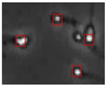 |  |
| 2 |  |  |  |  |
| Case | Original Frame | Detection Result |
|---|---|---|
| 1. Failure example from the validation dataset (false positive) |  |  |
| 2. Failure example from the test dataset (false positive) | 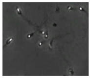 |  |
| 3. Failure example from the test dataset (false negative) |  |  |
Disclaimer/Publisher’s Note: The statements, opinions and data contained in all publications are solely those of the individual author(s) and contributor(s) and not of MDPI and/or the editor(s). MDPI and/or the editor(s) disclaim responsibility for any injury to people or property resulting from any ideas, methods, instructions or products referred to in the content. |
© 2023 by the authors. Licensee MDPI, Basel, Switzerland. This article is an open access article distributed under the terms and conditions of the Creative Commons Attribution (CC BY) license (https://creativecommons.org/licenses/by/4.0/).
Share and Cite
Zhu, R.; Cui, Y.; Huang, J.; Hou, E.; Zhao, J.; Zhou, Z.; Li, H. YOLOv5s-SA: Light-Weighted and Improved YOLOv5s for Sperm Detection. Diagnostics 2023, 13, 1100. https://doi.org/10.3390/diagnostics13061100
Zhu R, Cui Y, Huang J, Hou E, Zhao J, Zhou Z, Li H. YOLOv5s-SA: Light-Weighted and Improved YOLOv5s for Sperm Detection. Diagnostics. 2023; 13(6):1100. https://doi.org/10.3390/diagnostics13061100
Chicago/Turabian StyleZhu, Ronghua, Yansong Cui, Jianming Huang, Enyu Hou, Jiayu Zhao, Zhilin Zhou, and Hao Li. 2023. "YOLOv5s-SA: Light-Weighted and Improved YOLOv5s for Sperm Detection" Diagnostics 13, no. 6: 1100. https://doi.org/10.3390/diagnostics13061100