Hybridization of Deep Learning Pre-Trained Models with Machine Learning Classifiers and Fuzzy Min–Max Neural Network for Cervical Cancer Diagnosis
Abstract
:1. Introduction
2. Literature Review
3. Proposed Methodology
3.1. Module 1
3.1.1. Feature Extraction Using Pre-Trained Models
3.1.2. Min–Max Normalization
- is minimum value in , and
- is maximum value in .
3.2. Module 2
3.2.1. Machine Learning Classifiers
3.2.2. Fuzzy Min–Max Neural Network
Expansion
Overlap Test
- Case 1
- Case 2
- Case 3
- Case 4
Contraction
- Case 1
- Case 2
- Case 3(a)
- Case 3(b)
- Case 4(a)
- Case 4(b)
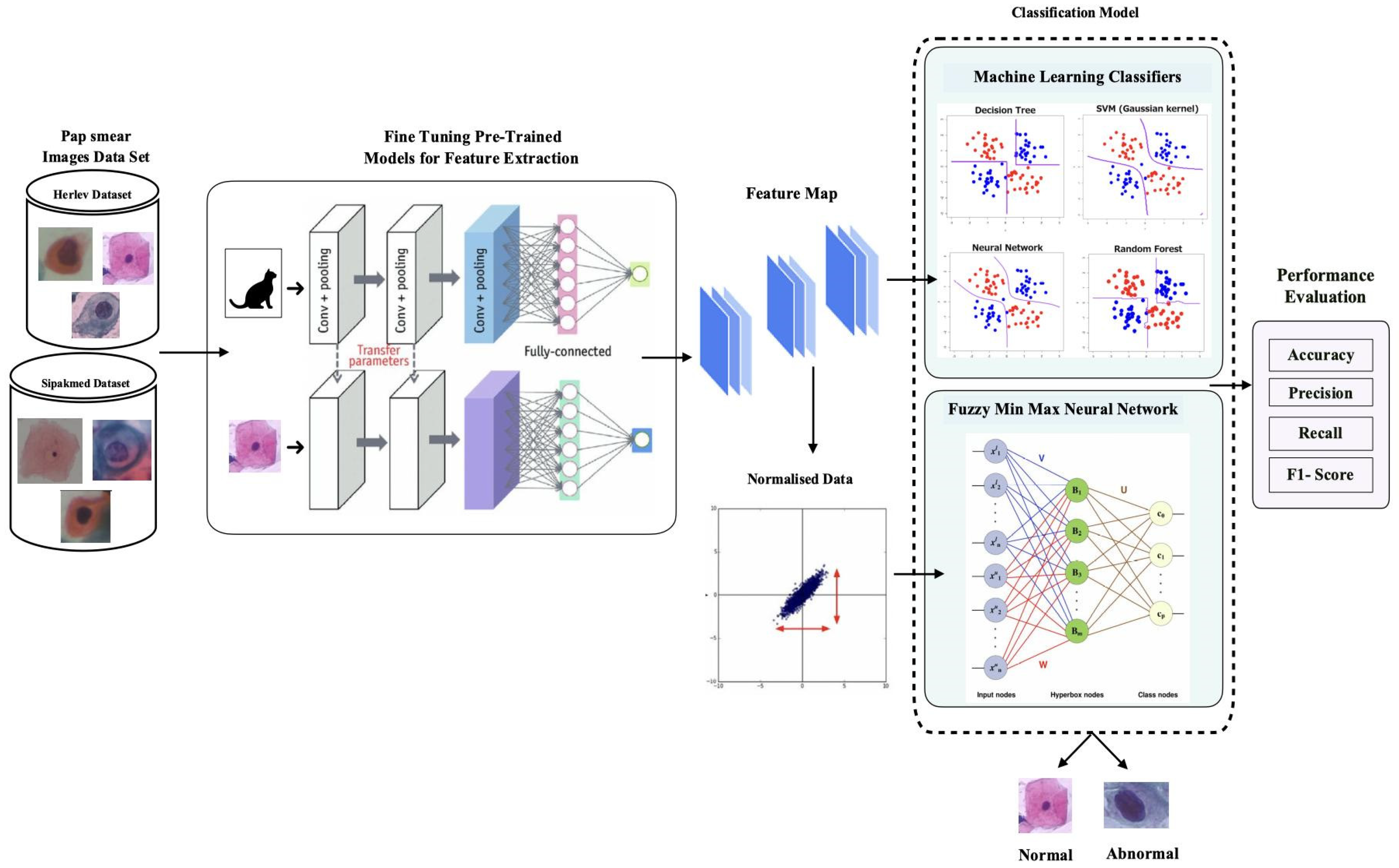
3.3. Algorithm 1
| Algorithm 1: Algorithm for cervical cancer classification |
| Input: Herlev dataset, Sipakmed dataset of Pap-smear images |
| Output: Prediction of classes—normal or abnormal |
| Begin |
| Step 1: Pre-process the images |
| Step 2: Split the dataset into training and testing datasets |
| Step 3: Pre-trained models= {AlextNet, GoogleNet, ResNet18, ResNet50} |
| Step 4: For each model in Step 3 |
| Train the model |
| Extract the feature vector |
| Step 5: Classifiers = {{machine learning classifiers: simple logistic, Naive Bays, Bayes Net, decision table, random forest, random tree, PART}, {fuzzy min–max neural network}} |
| Step 6: For each classifier in Step 5 |
| Train with the feature vector |
| Evaluate with Testing Set |
| End |
4. Experimentation Environment
4.1. Herlev Dataset
4.2. Sipakmed
4.3. Performance Measures
5. Experiments and Results
Performance Analysis
6. Conclusions
Author Contributions
Funding
Institutional Review Board Statement
Informed Consent Statement
Data Availability Statement
Conflicts of Interest
References
- Sung, H.; Ferlay, J.; Siegel, R.L.; Laversanne, M.; Soerjomataram, I.; Jemal, A.; Bray, F. Global Cancer Statistics 2020: GLOBOCAN Estimates of Incidence and Mortality Worldwide for 36 Cancers in 185 Countries. CA Cancer J. Clin. 2021, 71, 209–249. [Google Scholar] [CrossRef] [PubMed]
- Vaccarella, S.; Laversanne, M.; Ferlay, J.; Bray, F. Cervical cancer in Africa, Latin America and the Caribbean and Asia: Regional inequalities and changing trends. Int. J. Cancer 2017, 141, 1997–2001. [Google Scholar] [CrossRef] [Green Version]
- Chandran, V.; Sumithra, M.G.; Karthick, A.; George, T.; Deivakani, M.; Elakkiya, B.; Subramaniam, U.; Manoharan, S. Diagnosis of Cervical Cancer based on Ensemble Deep Learning Network using Colposcopy Images. BioMed Res. Int. 2021, 2021, 5584004. [Google Scholar] [CrossRef]
- Gençtav, A.; Aksoy, S.; Önder, S. Unsupervised segmentation and classification of cervical cell images. Pattern Recognit. 2012, 45, 4151–4168. [Google Scholar] [CrossRef] [Green Version]
- Jantzen, J.; Norup, J.; Dounias, G.; Bjerregaard, B. Pap-smear benchmark data for pattern classification. In Proceedings of the NiSIS 2005: Nature Inspired Smart Information Systems (NiSIS), EU Co-Ordination Action, Albufeira, Portugal, 1 January 2005; pp. 1–9. [Google Scholar]
- Marinakis, Y.; Dounias, G.; Jantzen, J. Pap smear diagnosis using a hybrid intelligent scheme focusing on genetic algorithm based feature selection and nearest neighbor classification. Comput. Biol. Med. 2009, 39, 69–78. [Google Scholar] [CrossRef] [PubMed]
- LeCun, Y.; Bengio, Y.; Hinton, G. Deep learning. Nature 2015, 521, 436–444. [Google Scholar] [CrossRef]
- Krizhevsky, A.; Sutskever, I.; Hinton, G.E. ImageNet classification with deep convolutional neural networks. Adv. Neural Inf. Process. Syst. 2017, 60, 84–90. [Google Scholar] [CrossRef] [Green Version]
- Sutskever, I.; Vinyals, O.; Le, Q. Sequence to sequence learning with neural networks. Adv. Neural Inf. Process. Syst. 2014, 27, 3104–3112. [Google Scholar] [CrossRef]
- Hinton, G.; Deng, L.; Yu, D.; Dahl, G.E.; Mohamed, A.-R.; Jaitly, N.; Senior, A.; Vanhoucke, V.; Nguyen, P.; Sainath, T.N.; et al. Deep Neural Networks for Acoustic Modeling in Speech Recognition: The Shared Views of Four Research Groups. IEEE Signal Process. Mag. 2012, 29, 82–97. [Google Scholar] [CrossRef]
- Yan, Z.; Zhan, Y.; Zhang, S.; Metaxas, D.; Zhou, X.S. Chapter 4—Multi-Instance Multi-Stage Deep Learning for Medical Image Recognition, Deep Learning for Medical Image Analysis; Academic Press: Cambridge, MA, USA, 2017; pp. 83–104. ISBN 9780128104088. [Google Scholar] [CrossRef]
- Zadeh, L.A. Fuzzy sets. Inf. Control 1965, 8, 338–353. [Google Scholar] [CrossRef] [Green Version]
- Ahmed, A.A.; Mohammed, M.F. SAIRF: A similarity approach for attack intention recognition using fuzzy min-max neural network. J. Comput. Sci. 2018, 25, 467–473. [Google Scholar] [CrossRef]
- Deshmukh, S.; Shinde, S. Diagnosis of lung cancer using pruned fuzzy min-max neural network. In Proceedings of the 2016 International Conference on Automatic Control and Dynamic Optimization Techniques ICACDOT, Pune, India, 9–10 September 2016; pp. 398–402. [Google Scholar] [CrossRef]
- Quteishat, A.; Lim, C.P. Application of the fuzzy min-max neural networks to medical diagnosis. In Proceedings of the 12th International Conference on Knowledge-Based Intelligent Information and Engineering Systems, Part III, Zagreb, Croatia, 3–5 September 2008; pp. 548–555. [Google Scholar] [CrossRef]
- Simpson, P. Fuzzy min-max neural networks. I. Classification. IEEE Trans. Neural Netw. 1992, 3, 776–786. [Google Scholar] [CrossRef]
- Sukumar, P.; Gnanamurthy, R. Computer aided detection of cervical cancer using pap smear images based on hybrid classifiers. Int. J. Appl. Eng. Res. Res. India Publ. 2015, 10, 21021–21032. [Google Scholar]
- Alaslani, M.G.; Elrefaei, L. Convolutional Neural Network Based Feature Extraction for IRIS Recognition. Int. J. Comput. Sci. Inf. Technol. 2018, 10, 65–78. [Google Scholar] [CrossRef] [Green Version]
- Athinarayanan, S.; Srinath, M.V.; Kavitha, R. Multi Class Cervical Cancer Classification by using ERSTCM, EMSD & CFE methods based Texture Features and Fuzzy Logic based Hybrid Kernel Support Vector Machine Classifier. IOSR J. Comput. Eng. 2017, 19, 23–34. [Google Scholar] [CrossRef]
- Priyankaa, J.; Bhadri Rajub, M.S.V.S. Machine Learning Approach for Prediction of Cervical Cancer. Turk. J. Comput. Math. Educ. 2021, 12, 3050–3058. [Google Scholar]
- Tripathi, A. Classification of cervical cancer using Deep Learning Algorithm. In Proceedings of the Fifth International Conference on Intelligent Computing and Control Systems (ICICCS 2021), Madurai, India, 6–8 May 2021. pp. 1210–1218. [CrossRef]
- Mousser, W.; Ouadfel, S. Deep Feature Extraction for Pap-Smear Image Classification: A Comparative Study. In Proceedings of the ICCTA 2019: 2019 5th International Conference on Computer and Technology Applications, Istanbul, Turkey, 16–17 April 2019; pp. 6–10. [Google Scholar] [CrossRef]
- Kurnianingsih; Allehaibi, K.H.S.; Nugroho, L.E.; Widyawan; Lazuardi, L.; Prabuwono, A.S.; Mantoro, T. Segmentation and Classification of Cervical Cells Using Deep Learning. IEEE Access 2019, 7, 116925–116941. [Google Scholar] [CrossRef]
- Sornapudi, S.; Brown, G.T.; Xue, Z.; Long, R.; Allen, L.; Antani, S. Comparing Deep Learning Models for Multi-cell Classification in Liquid- based Cervical Cytology Image. AMIA Annu. Symp. Proc. AMIA Symp. 2020, 2019, 820–827. [Google Scholar]
- Shinde, S.; Kalbhor, M.; Wajire, P. DeepCyto: A hybrid framework for cervical cancer classification by using deep feature fusion of cytology images. Math. Biosci. Eng. 2022, 19, 6415–6434. [Google Scholar] [CrossRef]
- Kalbhor, M.; Shinde, S.V.; Jude, H. Cervical cancer diagnosis based on cytology pap smear image classification using fractional coefficient and machine learning classifiers. TELKOMNIKA Telecommun. Comput. Electron. Control 2022, 20, 1091–1102. [Google Scholar] [CrossRef]
- Kalbhor, M.; Shinde, S. ColpoClassifier: A Hybrid Framework for Classification of the Cervigrams. Diagnostics 2023, 13, 1103. [Google Scholar] [CrossRef] [PubMed]
- Sokolova, M.; Lapalme, G. A systematic analysis of performance measures for classification tasks. Inf. Process. Manag. Int. J. 2009, 45, 427–437. [Google Scholar] [CrossRef]
- Raghu, M.; Zhang, C.; Kleinberg, J.; Bengio, S. Transfusion: Understanding transfer learning for medical imaging. In Proceedings of the 33rd Conference on Neural Information Processing Systems (NeurIPS 2019), Vancouver, BC, Canada, 8–14 December 2019; pp. 3347–3357. [Google Scholar] [CrossRef]
- Lin, H.; Hu, Y.; Chen, S.; Yao, J.; Zhang, L. Fine-Grained Classification of Cervical Cells Using Morphological and Appearance Based Convolutional Neural Networks. IEEE Access 2019, 7, 71541–71549. [Google Scholar] [CrossRef]
- Kalbhor, M.; Shinde, S.; Joshi, H.; Wajire, P. Pap smear-based cervical cancer detection using hybrid deep learning and performance evaluation. Comput. Methods Biomech. Biomed. Eng. Imaging Vis. 2023, 19, 6415–6434. [Google Scholar] [CrossRef]
- Mbaga, A.H.; Zhijun, P. Pap Smear Images Classification for Early Detection of Cervical Cancer. Int. J. Comput. Appl. 2015, 118, 10–16. [Google Scholar] [CrossRef]
- Shanthi, P.B.; Hareesha, K.S.; Kudva, R. Automated Detection and Classification of Cervical Cancer Using Pap Smear Microscopic Images: A Comprehensive Review and Future Perspectives. Eng. Sci. 2022, 19, 20–41. [Google Scholar] [CrossRef]
- Plissiti, M.E.; Dimitrakopoulos, P.; Sfikas, G.; Nikou, C.; Krikoni, O.; Charchanti, A. Sipakmed: A new dataset for feature and image based classification of normal and pathological cervical cells in Pap smear images. IEEE Int. Conf. Image Process. 2018, 2018, 3144–3148. [Google Scholar] [CrossRef]
- Pan, S.J.; Yang, Q. A survey on transfer learning. IEEE Trans. Knowl. Data Eng. 2009, 22, 1345–1359. [Google Scholar] [CrossRef]


| Paper | Data Set | Pre-Processing | Feature Extraction/ Classification | Results |
|---|---|---|---|---|
| [20] | Herlev University Hospital | Resize, Color to Grey, Expansion of dimensions | RESNET-50 | Accuracy 74.04% |
| [21] | SIPAKMED | Resize 244 × 244 | RESNET-50, RESNET-152, VGG-16, VGG-19 | Highest 94.89% accuracy was obtained with ResNet-152 |
| [22] | Herlev University Hospital | Data Augmentation | VGG16. InceptionV3 VGG19, ResNet50 Classification—MLP classifier | ResNet-50 89% |
| [23] | Herlev University Hospital | Data Augmentation Segmentation—Mask R-CNN | VGGNet | Mask R-CNN segmentation produces the best average performance, i.e., 0.92 ± 0.06 precision, 0.91 ± 0.05 recall and 0.91 ± 0.04 ZSI and 0.83 ± 0.10 Binary classification problem 98.1% accuracy Seven-class problem high accuracy of 95.9% |
| [24] | Herlev University Hospital | Subtraction of blue color space from red color space, skeletonizing and refining boundaries | VGG-19, ResNet-50, DenseNet-120, and Inception_v3 | VGG-19—88% Accuracy |
| [25] | Herlev University Hospital, SIPAKMED, LBC | Data Augmentation | XceptionNet, VGGNet, ResNet50 and Ensemble of classifiers | Accuracy 97%, 99%, and 100% |
| [26] | Herlev University Hospital | Resize 256 × 256 | DCT and Haar transform | Highest 81.11% accuracy was obtained with DCT |
| Pre-Trained Model | Alexnet | Googlenet | Resnet-18 | Resnet-50 |
|---|---|---|---|---|
| Number of Features | 4096 | 1000 | 512 | 1000 |
| Cell Category | Number of Cells | |
|---|---|---|
| Normal squamous | Normal | 74 |
| Intermediate squamous | 70 | |
| Columnar | 98 | |
| Mild dysplasia | Abnormal | 182 |
| Moderate dysplasia | 146 | |
| Severe dysplasia | 197 | |
| Carcinoma in situ | 150 | |
| Total | 917 |
| Cell Category | Number of Cells | |
|---|---|---|
| Superficial | Normal | 831 |
| Parabasal | 787 | |
| Koilocytotic | Abnormal | 825 |
| Dyskeratotic | 813 | |
| Metaplastic | Benign | 793 |
| Total | 4049 |
| Assessments | Formula |
|---|---|
| Accuracy | |
| Sensitivity/Recall | |
| Specificity | |
| Precision | |
| F1 Score |
| AlexNet | ||||||||
|---|---|---|---|---|---|---|---|---|
| Dataset | Classifier | Bayes Net | Navie Bayes | Random Forest | Random Tree | Decision Table | Part | Simple Logistic |
| Herlev | Testing Accuracy (%) | 83.33 | 82.24 | 87.68 | 81.8 | 88.04 | 86.59 | 88.6 |
| Sipakmed | 91. 2 | 91.6 | 91.2 | 90.70 | 93.23 | 89.5 | 95.14 | |
| GoogleNet | ||||||||
|---|---|---|---|---|---|---|---|---|
| Dataset | Classifier | BayeNet | Navie Bayes | Random Forest | Random Tree | Decision Table | Part | Simple Logistic |
| Herlev | Testing Accuracy (%) | 83.70 | 82.97 | 86.96 | 81.88 | 84.06 | 86.59 | 87.32 |
| Sipakmed | 87.37 | 85.24 | 90.24 | 83.11 | 87.62 | 89.75 | 92.21 | |
| ResNet-18 | ||||||||
|---|---|---|---|---|---|---|---|---|
| Dataset | Classifier | BayeNet | Naive Bayes | Random Forest | Random Tree | Decision Table | Part | Simple Logistic |
| Herlev | Testing Accuracy (%) | 86.59 | 86.59 | 87.68 | 82.6 | 84.42 | 79.71 | 88.76 |
| Sipakmed | 90.9 | 89.26 | 88.36 | 80.49 | 84.75 | 88.42 | 93.85 | |
| ResNet-50 | ||||||||
|---|---|---|---|---|---|---|---|---|
| Dataset | Classifier | BayeNet | Naive Bayes | Random Forest | Random Tree | Decision Table | Part | Simple Logistic |
| Herlev | Testing Accuracy (%) | 88.04 | 89.13 | 88.04 | 78.62 | 86.23 | 81.88 | 92.03 |
| Sipakmed | 89.67 | 88.19 | 89.83 | 81.8 | 84.75 | 90 | 93.60 | |
| Theta | 0 | 0.1 | 0.2 | 0.3 | 0.4 | 0.5 | 0.6 | 0.7 | 0.8 | 0.9 | 1 | ||
|---|---|---|---|---|---|---|---|---|---|---|---|---|---|
| Alexnet | Herlev Dataset | Accuracy | 87.32 | 84.06 | 84.06 | 90.22 | 82.97 | 84.78 | 85.14 | 88.04 | 84.78 | 39.86 | 34.78 |
| Sensitivity | 0.90 | 0.94 | 0.86 | 0.95 | 0.85 | 0.90 | 0.91 | 0.97 | 0.91 | 0.19 | 0.11 | ||
| Specificity | 0.81 | 0.58 | 0.78 | 0.77 | 0.77 | 0.70 | 0.70 | 0.64 | 0.68 | 0.99 | 1.00 | ||
| Precision | 0.93 | 0.86 | 0.92 | 0.92 | 0.91 | 0.89 | 0.89 | 0.88 | 0.89 | 0.97 | 1.00 | ||
| F1 Score | 0.91 | 0.90 | 0.89 | 0.93 | 0.88 | 0.90 | 0.90 | 0.92 | 0.90 | 0.31 | 0.20 | ||
| Sipakmed Dataset | Accuracy | 92.62 | 93.20 | 95.08 | 95.00 | 93.93 | 95.33 | 94.92 | 93.69 | 90.82 | 80.66 | 80.00 | |
| Sensitivity | 0.95 | 0.93 | 0.94 | 0.94 | 0.93 | 0.95 | 0.95 | 0.94 | 0.95 | 0.99 | 0.99 | ||
| Specificity | 0.90 | 0.93 | 0.96 | 0.97 | 0.95 | 0.96 | 0.95 | 0.93 | 0.85 | 0.54 | 0.52 | ||
| Precision | 0.93 | 0.95 | 0.97 | 0.98 | 0.97 | 0.97 | 0.97 | 0.95 | 0.90 | 0.76 | 0.76 | ||
| F1 Score | 0.94 | 0.94 | 0.96 | 0.96 | 0.95 | 0.96 | 0.96 | 0.95 | 0.93 | 0.86 | 0.86 |
| Theta | 0 | 0.1 | 0.2 | 0.3 | 0.4 | 0.5 | 0.6 | 0.7 | 0.8 | 0.9 | 1 | ||
|---|---|---|---|---|---|---|---|---|---|---|---|---|---|
| Googlenet | Herlev Dataset | Accuracy | 82.25 | 86.23 | 83.70 | 84.78 | 86.96 | 88.41 | 89.49 | 88.04 | 86.96 | 82.25 | 82.25 |
| Sensitivity | 0.87 | 0.93 | 0.89 | 0.89 | 0.92 | 0.98 | 0.97 | 0.97 | 0.95 | 0.87 | 0.87 | ||
| Specificity | 0.68 | 0.67 | 0.70 | 0.74 | 0.74 | 0.62 | 0.70 | 0.63 | 0.64 | 0.70 | 0.70 | ||
| Precision | 0.89 | 0.89 | 0.89 | 0.90 | 0.91 | 0.88 | 0.90 | 0.88 | 0.88 | 0.89 | 0.89 | ||
| F1 Score | 0.88 | 0.91 | 0.89 | 0.90 | 0.91 | 0.93 | 0.93 | 0.92 | 0.91 | 0.88 | 0.88 | ||
| Sipakmed Dataset | Accuracy | 89.34 | 90.66 | 90.66 | 92.13 | 91.15 | 91.80 | 91.15 | 88.52 | 85.16 | 83.03 | 82.79 | |
| Sensitivity | 0.91 | 0.91 | 0.92 | 0.91 | 0.89 | 0.91 | 0.90 | 0.86 | 0.86 | 0.96 | 0.93 | ||
| Specificity | 0.86 | 0.90 | 0.89 | 0.94 | 0.94 | 0.92 | 0.93 | 0.92 | 0.84 | 0.64 | 0.68 | ||
| Precision | 0.91 | 0.93 | 0.93 | 0.96 | 0.96 | 0.95 | 0.95 | 0.94 | 0.89 | 0.80 | 0.81 | ||
| F1 Score | 0.91 | 0.92 | 0.92 | 0.93 | 0.92 | 0.93 | 0.92 | 0.90 | 0.87 | 0.87 | 0.87 |
| Theta | 0 | 0.1 | 0.2 | 0.3 | 0.4 | 0.5 | 0.6 | 0.7 | 0.8 | 0.9 | 1 | ||
|---|---|---|---|---|---|---|---|---|---|---|---|---|---|
| ResNet-18 | Herlev | Accuracy | 88.77 | 75.00 | 89.49 | 89.13 | 91.30 | 91.67 | 88.04 | 86.96 | 86.23 | 86.96 | 86.96 |
| Sensitivity | 0.92 | 0.92 | 0.91 | 0.91 | 0.97 | 0.99 | 0.97 | 0.94 | 0.94 | 0.95 | 0.95 | ||
| Specificity | 0.81 | 0.27 | 0.86 | 0.85 | 0.75 | 0.73 | 0.64 | 0.67 | 0.64 | 0.66 | 0.66 | ||
| Precision | 0.93 | 0.78 | 0.95 | 0.94 | 0.92 | 0.91 | 0.88 | 0.89 | 0.88 | 0.88 | 0.88 | ||
| F1 Score | 0.92 | 0.84 | 0.93 | 0.92 | 0.94 | 0.95 | 0.92 | 0.91 | 0.91 | 0.91 | 0.91 | ||
| Sipakmed | Accuracy | 91.48 | 90.82 | 91.31 | 92.79 | 92.87 | 93.77 | 90.90 | 86.80 | 81.72 | 77.21 | 72.46 | |
| Sensitivity | 0.93 | 0.92 | 0.92 | 0.92 | 0.93 | 0.93 | 0.93 | 0.92 | 0.91 | 0.93 | 0.96 | ||
| Specificity | 0.89 | 0.88 | 0.90 | 0.94 | 0.93 | 0.95 | 0.87 | 0.79 | 0.67 | 0.53 | 0.36 | ||
| Precision | 0.93 | 0.92 | 0.93 | 0.96 | 0.95 | 0.96 | 0.92 | 0.87 | 0.81 | 0.75 | 0.70 | ||
| F1 Score | 0.93 | 0.92 | 0.93 | 0.94 | 0.94 | 0.95 | 0.93 | 0.89 | 0.86 | 0.83 | 0.81 |
| Theta | 0 | 0.1 | 0.2 | 0.3 | 0.4 | 0.5 | 0.6 | 0.7 | 0.8 | 0.9 | 1 | ||
|---|---|---|---|---|---|---|---|---|---|---|---|---|---|
| ResNet50 | Herlev | Accuracy | 88.77 | 86.23 | 87.32 | 88.04 | 87.32 | 87.32 | 85.87 | 87.32 | 86.96 | 82.25 | 81.88 |
| Sensitivity | 0.91 | 0.93 | 0.91 | 0.90 | 0.90 | 0.93 | 0.89 | 0.93 | 0.91 | 0.83 | 0.85 | ||
| Specificity | 0.84 | 0.68 | 0.78 | 0.82 | 0.79 | 0.73 | 0.77 | 0.73 | 0.77 | 0.81 | 0.73 | ||
| Precision | 0.94 | 0.89 | 0.92 | 0.93 | 0.92 | 0.90 | 0.91 | 0.90 | 0.92 | 0.92 | 0.90 | ||
| F1 Score | 0.92 | 0.91 | 0.91 | 0.92 | 0.91 | 0.91 | 0.90 | 0.91 | 0.91 | 0.87 | 0.87 | ||
| Sipakmed | Accuracy | 92.05 | 92.62 | 92.70 | 94.18 | 95.25 | 95.33 | 94.18 | 89.10 | 84.02 | 80.82 | 72.70 | |
| Sensitivity | 0.93 | 0.93 | 0.94 | 0.95 | 0.94 | 0.95 | 0.94 | 0.85 | 0.82 | 0.95 | 0.99 | ||
| Specificity | 0.90 | 0.92 | 0.91 | 0.93 | 0.97 | 0.96 | 0.95 | 0.96 | 0.87 | 0.60 | 0.32 | ||
| Precision | 0.93 | 0.95 | 0.94 | 0.95 | 0.98 | 0.97 | 0.96 | 0.97 | 0.91 | 0.78 | 0.69 | ||
| F1 Score | 0.93 | 0.94 | 0.94 | 0.95 | 0.96 | 0.96 | 0.95 | 0.90 | 0.86 | 0.86 | 0.81 |
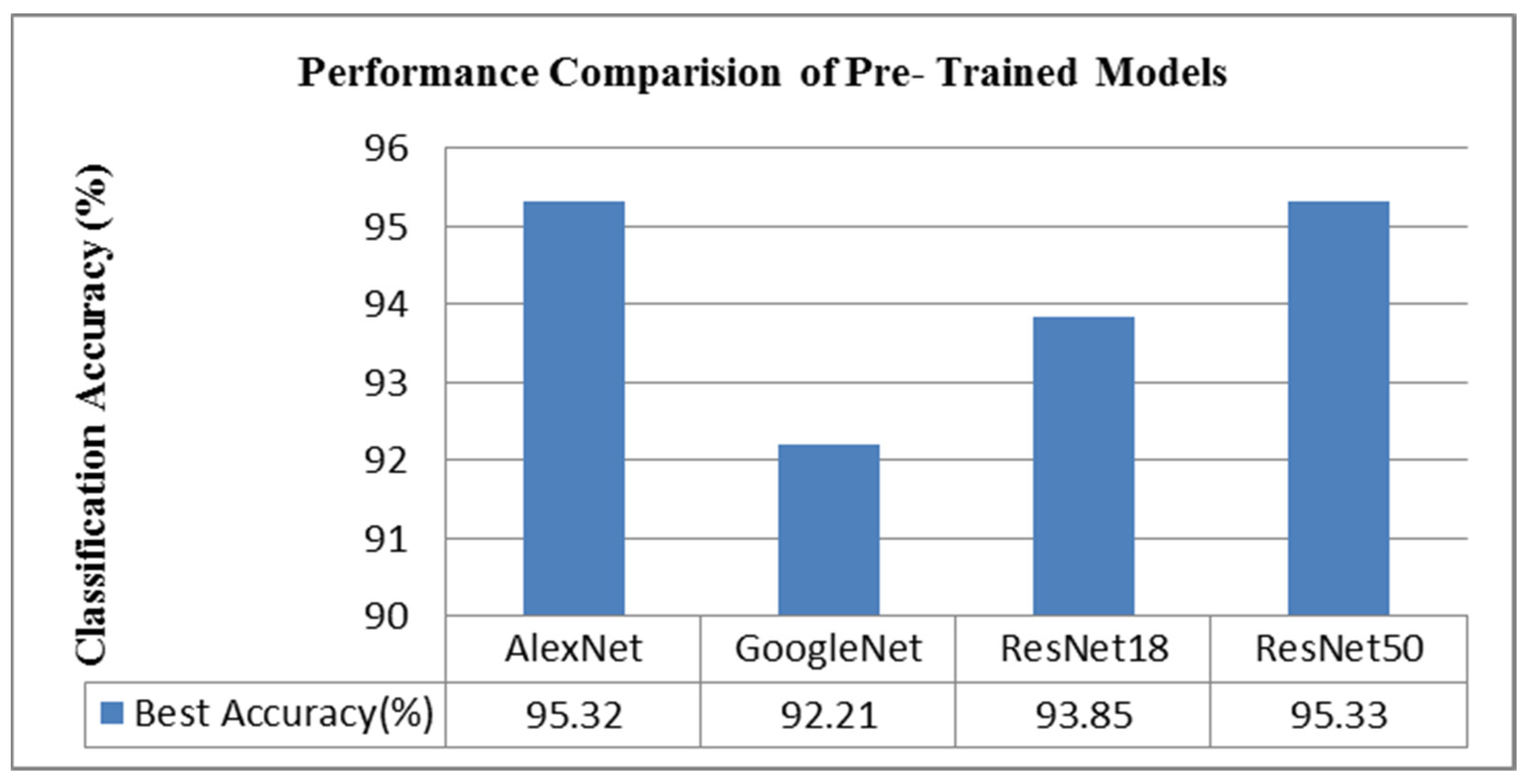
| AlexNet | GoogleNet | ResNet18 | ResNet50 | |
|---|---|---|---|---|
| Herlev | 90.22 (FMMN) | 89.49 (FMMN) | 91.67 (FMMN) | 92.03 (Simple logistic) |
| Sipakmed | 95.32 (FMMN) | 92.21 (Simple logistic) | 93.85 (Simple logistic) | 95.33 (FMMN) |
Disclaimer/Publisher’s Note: The statements, opinions and data contained in all publications are solely those of the individual author(s) and contributor(s) and not of MDPI and/or the editor(s). MDPI and/or the editor(s) disclaim responsibility for any injury to people or property resulting from any ideas, methods, instructions or products referred to in the content. |
© 2023 by the authors. Licensee MDPI, Basel, Switzerland. This article is an open access article distributed under the terms and conditions of the Creative Commons Attribution (CC BY) license (https://creativecommons.org/licenses/by/4.0/).
Share and Cite
Kalbhor, M.; Shinde, S.; Popescu, D.E.; Hemanth, D.J. Hybridization of Deep Learning Pre-Trained Models with Machine Learning Classifiers and Fuzzy Min–Max Neural Network for Cervical Cancer Diagnosis. Diagnostics 2023, 13, 1363. https://doi.org/10.3390/diagnostics13071363
Kalbhor M, Shinde S, Popescu DE, Hemanth DJ. Hybridization of Deep Learning Pre-Trained Models with Machine Learning Classifiers and Fuzzy Min–Max Neural Network for Cervical Cancer Diagnosis. Diagnostics. 2023; 13(7):1363. https://doi.org/10.3390/diagnostics13071363
Chicago/Turabian StyleKalbhor, Madhura, Swati Shinde, Daniela Elena Popescu, and D. Jude Hemanth. 2023. "Hybridization of Deep Learning Pre-Trained Models with Machine Learning Classifiers and Fuzzy Min–Max Neural Network for Cervical Cancer Diagnosis" Diagnostics 13, no. 7: 1363. https://doi.org/10.3390/diagnostics13071363
APA StyleKalbhor, M., Shinde, S., Popescu, D. E., & Hemanth, D. J. (2023). Hybridization of Deep Learning Pre-Trained Models with Machine Learning Classifiers and Fuzzy Min–Max Neural Network for Cervical Cancer Diagnosis. Diagnostics, 13(7), 1363. https://doi.org/10.3390/diagnostics13071363


