Patient-Reported Outcome Measures in Patients with and without Non-Expandable Lung Secondary to Malignant Pleural Effusion—A Single-Centre Observational Study

 , ,
, ,
Abstract
1. Introduction
2. Materials and Methods
2.1. Non-Expandable Lung (NEL) Definition
2.2. Baseline Data
2.3. Patient-Reported Outcome Measures (PROMs)
2.3.1. Pre-Thoracentesis PROMs
2.3.2. Post-Thoracentesis PROMs
2.4. Thoracentesis and TUS-Protocol
2.5. Follow-Up Data
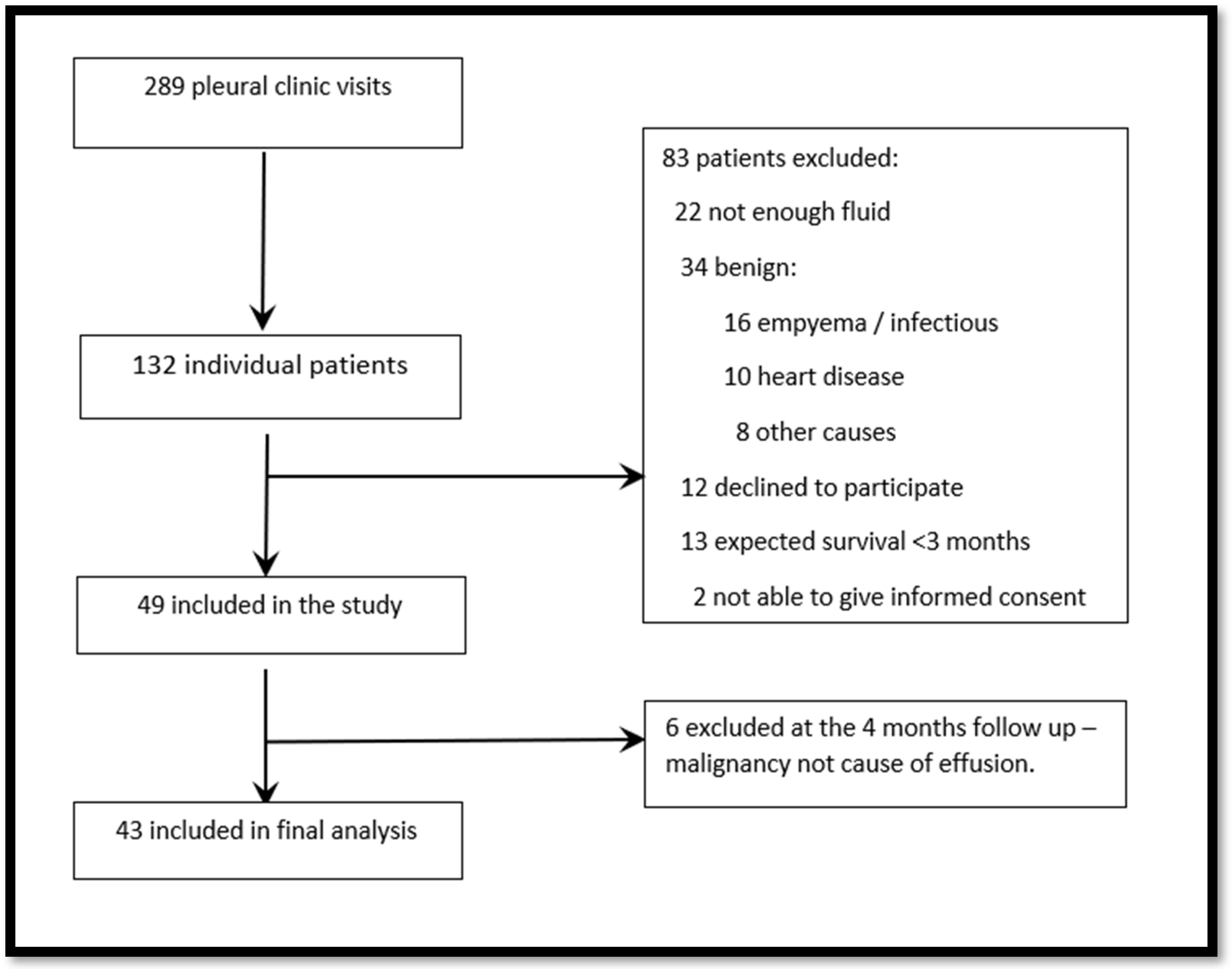
3. Results
3.1. Primary Outcome
3.2. Secondary Outcomes
4. Discussion
5. Conclusions
Supplementary Materials
Author Contributions
Funding
Institutional Review Board Statement
Informed Consent Statement
Data Availability Statement
Acknowledgments
Conflicts of Interest
References
- Porcel, J.M.; Gasol, A.; Bielsa, S.; Civit, C.; Light, R.W.; Salud, A. Clinical features and survival of lung cancer patients with pleural effusions. Respirology 2015, 20, 654–659. [Google Scholar] [CrossRef] [PubMed]
- Arnold, D.T.; De Fonseka, D.; Perry, S.; Morley, A.; Harvey, J.E.; Medford, A.; Brett, M.; Maskell, N.A. Investigating unilateral pleural effusions: The role of cytology. Eur. Respir. J. 2018, 52, 1801254. [Google Scholar] [CrossRef] [PubMed]
- Bibby, A.C.; Dorn, P.; Psallidas, I.; Porcel, J.M.; Janssen, J.; Froudarakis, M.; Subotic, D.; Astoul, P.; Licht, P.; Schmid, R.; et al. ERS/EACTS statement on the management of malignant pleural effusions. Eur. Respir. J. 2018, 52, 1800349. [Google Scholar] [CrossRef] [PubMed]
- Bodtger, U.; Hallifax, R.J. Epidemiology: Why is pleural disease becoming more common? In Pleural Diseases ERS Monograph; ERS: Sheffield, UK, 2020. [Google Scholar]
- Dresler, C.M.; Olak, J.; Herndon, J.E.; Richards, W.G.; Scalzetti, E.; Fleishman, S.B.; Kernstine, K.H.; Demmy, T.; Jablons, D.M.; Kohman, L.; et al. Phase III Intergroup Study of Talc Poudrage vs. Talc Slurry Sclerosis for Malignant Pleural Effusion. Chest 2005, 127, 909–915. [Google Scholar] [CrossRef] [PubMed]
- Muruganandan, S.; Azzopardi, M.; Fitzgerald, D.B.; Shrestha, R.; Kwan, B.C.H.; Lam, D.C.L.; De Chaneet, C.C.; Ali, M.R.S.R.; Yap, E.; Tobin, C.L.; et al. Aggressive versus symptom-guided drainage of malignant pleural effusion via indwelling pleural catheters (AMPLE-2): An open-label randomised trial. Lancet Respir. Med. 2018, 6, 671–680. [Google Scholar] [CrossRef] [PubMed]
- Thomas, R.; Fysh, E.T.H.; Smith, N.A.; Lee, P.; Kwan, B.C.H.; Yap, E.; Lee, Y.G. Effect of an indwelling pleural catheter vs. talc pleurodesis on hospitalization days in patients with malignant pleural effusion: The AMPLE randomized clinical trial. JAMA—J. Am. Med. Assoc. 2017, 318, 1903–1912. [Google Scholar] [CrossRef] [PubMed]
- Lan, R.-S.; Lo, S.K.; Chuang, M.-L.; Yang, C.-T.; Tsao, T.C.-Y.; Lee, C.-H. Elastance of the Pleural Space: A Predictor for the Outcome of Pleurodesis in Patients with Malignant Pleural Effusion. Ann. Intern. Med. 1997, 126, 768–774. [Google Scholar] [CrossRef] [PubMed]
- Davies, H.E.; Mishra, E.K.; Kahan, B.C.; Wrightson, J.M.; Stanton, A.E.; Guhan, A.; Rahman, N.M. Effect of an indwelling pleural catheter vs. chest tube and talc pleurodesis for relieving dyspnea in patients with malignant pleural effusion: The TIME2 randomized controlled trial. JAMA—J. Am. Med. Assoc. 2012, 307, 2383–2389. [Google Scholar] [CrossRef] [PubMed]
- Halford, P.J.; Bhatnagar, R.; White, P.; Haris, M.; Harrison, R.N.; Holme, J.; Sivasothy, P.; West, A.; Bishop, L.J.; Stanton, A.E.; et al. Manometry performed at indwelling pleural catheter insertion to predict unexpandable lung. J. Thorac. Dis. 2020, 12, 1374–1384. [Google Scholar] [CrossRef]
- Feller-Kopman, D.J.; Reddy, C.B.; DeCamp, M.M.; Diekemper, R.L.; Gould, M.K.; Henry, T.; Iyer, N.P.; Lee, Y.C.G.; Lewis, S.Z.; Maskell, N.A.; et al. Management of Malignant Pleural Effusions An Official ATS/STS/STR Clinical Practice Guideline. Am. J. Respir. Crit. Care Med. 2018, 198, 839–849. [Google Scholar] [CrossRef]
- Muruganandan, S.; Azzopardi, M.; Thomas, R.; Fitzgerald, D.B.; Kuok, Y.J.; Cheah, H.M.; Read, C.A.; Budgeon, C.A.; Eastwood, P.R.; Jenkins, S.; et al. The Pleural Effusion and Symptom Evaluation (PLEASE) study of breathlessness in patients with a symptomatic pleural effusion. Eur. Respir. J. 2020, 55, 1900980. [Google Scholar] [CrossRef] [PubMed]
- Sivakumar, P.; Fitzgerald, D.B.; Ip, H.; Rao, D.; West, A.; Noorzad, F.; Wallace, D.; Haris, M.; Prudon, B.; Hettiarachchi, G.; et al. The impact of outpatient versus inpatient management on health-related quality of life outcomes for patients with malignant pleural effusion: The OPTIMUM randomised clinical trial. Eur. Respir. J. 2024, 63, 2201215. [Google Scholar] [CrossRef] [PubMed]
- Mishra, E.K.; Muruganandan, S.; Clark, A.; Bhatnagar, R.; Maskell, N.; Lee, G.; Rahman, N.M. Breathlessness Predicts Survival in Patients with Malignant Pleural Effusions Meta-analysis of Individual Patient Data from Five Randomized Controlled Trials. Chest 2021, 160, 351–357. [Google Scholar] [CrossRef] [PubMed]
- Divietro, M.; Huggins, J.; Doelken, P.; Gurung, P.; Kaiser, L.; Sahn, S. Prevalence and Causes of Unexpandable Lung Over a Ten Year Period. Chest 2011, 140, 700A. [Google Scholar] [CrossRef]
- Feller-Kopman, D. Therapeutic thoracentesis: The role of ultrasound and pleural manometry. Curr. Opin. Pulm. Med. 2007, 13, 312–318. [Google Scholar] [CrossRef] [PubMed]
- Lester, M.G.; Feller-Kopman, D.; Maldonado, F. Pleural physiology: What do we understand and what should we measure in clinical practice? In Pleural Diseases ERS Monograph; ERS: Sheffield, UK, 2020. [Google Scholar]
- Bibby, A.C.; Halford, P.; de Fonseka, D.; Morley, A.J.; Smith, S.; Maskell, N.A. The prevalence and clinical relevance of nonexpandable lung in malignant pleural mesothelioma: A prospective, single-center cohort study of 229 patients. Ann. Am. Thorac. Soc. 2019, 16, 1273–1279. [Google Scholar] [CrossRef] [PubMed]
- Detterbeck, F.C.; Boffa, D.J.; Kim, A.W.; Tanoue, L.T. The Eighth Edition Lung Cancer Stage Classification. Chest 2017, 151, 193–203. [Google Scholar] [CrossRef] [PubMed]
- Hooper, C.; Lee, Y.C.G.; Maskell, N. Investigation of a unilateral pleural effusion in adults: British Thoracic Society pleural disease guideline 2010. Thorax 2010, 65 (Suppl. S2), ii4–ii17. [Google Scholar] [CrossRef] [PubMed]
- Salamonsen, M.R.; Lo, A.K.C.; Ng, A.C.T.; Bashirzadeh, F.; Wang, W.Y.S.; Fielding, D.I.K. Novel Use of Pleural Ultrasound Can Identify Malignant Entrapped Lung Prior to Effusion Drainage. Chest 2014, 146, 1286–1293. [Google Scholar] [CrossRef]
- Janssen, M.F.; Pickard, A.S.; Golicki, D.; Gudex, C.; Niewada, M.; Scalone, L.; Swinburn, P.; Busschbach, J. Measurement properties of the EQ-5D-5L compared to the EQ-5D-3L across eight patient groups: A multi-country study. Qual. Life Res. 2013, 22, 1717–1727. [Google Scholar] [CrossRef]
- Jensen, C.E.; Sørensen, S.S.; Gudex, C.; Jensen, M.B.; Pedersen, K.M.; Ehlers, L.H. The Danish EQ-5D-5L Value Set: A Hybrid Model Using cTTO and DCE Data. Appl. Health Econ. Health Policy 2021, 19, 579–591. [Google Scholar] [CrossRef] [PubMed]
- Barbera, L.; Seow, H.; Howell, D.; Sutradhar, R.; Earle, C.; Liu, Y.; Stitt, A.; Husain, A.; Sussman, J.; Dudgeon, D. Symptom burden and performance status in a population-based cohort of ambulatory cancer patients. Cancer 2010, 116, 5767–5776. [Google Scholar] [CrossRef] [PubMed]
- Pain, S. What Should be the Optimal Cut Points for Mild, Moderate, and Severe Pain? J. Palliat. Med. 2007, 10, 1338–1346. [Google Scholar]
- Selby, D.; Cascella, A.; Gardiner, K.; Do, R.; Moravan, V.; Myers, J.; Chow, E. A Single Set of Numerical Cutpoints to Define Moderate and Severe Symptoms for the Edmonton Symptom Assessment System. J. Pain Symptom Manag. 2010, 39, 241–249. [Google Scholar] [CrossRef]
- Fjaellegaard, K.; Petersen, J.K.; Rasmussen, D.B.; Clementsen, P.F.; Laursen, C.B.; Bhatnagar, R.; Bodtger, U. Prediction of Time to Next Therapeutic Thoracentesis and Identification of Risk Factors of Rapid Pleural Fluid Recurrence: A Prospective Observational Study. Respiration 2023, 102, 333–340. [Google Scholar] [CrossRef]
- Pietersen, P.I.; Davidsen, J.R.; Skaarup, S.H.; Schultz, H.H.L.; Jeschke, K.N.; Bodtger, U.; Laursen, C.B. Fokuseret Lungeultralydsskanning. 2020. Available online: https://lungemedicin.dk/fokuseret-lungeultralydskanning/ (accessed on 27 February 2023).
- Petersen, J.K.; Fjaellegaard, K.; Rasmussen, D.B.; Alstrup, G.; Høegholm, A.; Sidhu, J.S.; Sivapalan, P.; Gerke, O.; Bhatnagar, R.; Clementsen, P.F.; et al. Ultrasound in the Diagnosis of Non-Expandable Lung: A Prospective Observational Study of M-Mode, B-Mode, and 2D-Shear Wave Elastography. Diagnostics 2024, 14, 204. [Google Scholar] [CrossRef]
- Mishra, E.K.; Corcoran, J.P.; Hallifax, R.J.; Stradling, J.; Maskell, N.A.; Rahman, N.M. Defining the Minimal Important Difference for the Visual Analogue Scale Assessing Dyspnea in Patients with Malignant Pleural Effusions. PLoS ONE 2015, 10, e0123798. [Google Scholar] [CrossRef]
- Bugalho, A.; Ferreira, D.; Dias, S.S.; Schuhmann, M.; Branco, J.C.; Gomes, M.J.M.; Eberhardt, R. The Diagnostic Value of Transthoracic Ultrasonographic Features in Predicting Malignancy in Undiagnosed Pleural Effusions: A Prospective Observational Study. Respiration 2014, 87, 270–278. [Google Scholar] [CrossRef]
- Trovisco, R.; Freitas, C.; Serino, M.; Ferreira, P.; Martins, B.; Coelho, D.; Melo, N.; Fernandes, G.; Magalhães, A.; Bastos, H. Predictors of lung entrapment in malignant pleural effusion. Pulmonology 2022. [Google Scholar] [CrossRef]
- Huggins, J.T.; Doelken, P.; Sahn, S.A. The unexpandable lung. F1000 Med. Rep. 2010, 2, 77. [Google Scholar] [CrossRef]
- Thomas, R.; Jenkins, S.; Eastwood, P.R.; Lee, Y.G.; Singh, B. Physiology of breathlessness associated with pleural effusions. Curr. Opin. Pulm. Med. 2015, 21, 338–345. [Google Scholar] [CrossRef] [PubMed]
- Jensen, M.B.; Jensen, C.E.; Gudex, C.; Pedersen, K.M.; Sørensen, S.S.; Ehlers, L.H. Danish population health measured by the EQ-5D-5L. Scand. J. Public Health 2023, 51, 241–249. [Google Scholar] [CrossRef]
- Bhatnagar, R.; Piotrowska, H.E.; Laskawiec-Szkonter, M.; Kahan, B.C.; Luengo-Fernandez, R.; Pepperell, J.C.; Maskell, N.A. Effect of Thoracoscopic Talc Poudrage vs. Talc Slurry via Chest Tube on Pleurodesis Failure Rate among Patients with Malignant Pleural Effusions: A Randomized Clinical Trial. JAMA—J. Am. Med. Assoc. 2020, 323, 60–69. [Google Scholar] [CrossRef] [PubMed]
- EuroQol Website. Available online: https://euroqol.org/information-and-support/resources/value-sets/ (accessed on 30 May 2024).
- Psallidas, I.; Yousuf, A.; Talwar, A.; Hallifax, R.J.; Mishra, E.K.; Corcoran, J.P.; Ali, N.; Rahman, N.M. Assessment of patient-reported outcome measures in pleural interventions. BMJ Open Respir. Res. 2017, 4, e000171. [Google Scholar] [CrossRef] [PubMed]
- Villena, V.; López-Encuentra, A.; Pozo, F.; De-Pablo, A.; Martín-Escribano, P. Measurement of Pleural Pressure during Therapeutic Thoracentesis. Am. J. Respir. Crit. Care Med. 2000, 162, 1534–1538. [Google Scholar] [CrossRef] [PubMed]
- Raijmakers, N.J.H.; Zijlstra, M.; van Roij, J.; Husson, O.; Oerlemans, S.; van de Poll-Franse, L.V. Health-related quality of life among cancer patients in their last year of life: Results from the PROFILES registry. Support. Care Cancer 2018, 26, 3397–3404. [Google Scholar] [CrossRef] [PubMed]
- Bausewein, C.; Farquhar, M.; Booth, S.; Gysels, M.; Higginson, I. Measurement of breathlessness in advanced disease: A systematic review. Respir. Med. 2007, 101, 399–410. [Google Scholar] [CrossRef]
- Tanaka, K.; Akechi, T.; Okuyama, T.; Nishiwaki, Y.; Uchitomi, Y. Development and validation of the Cancer Dyspnoea Scale: A multidimensional, brief, self-rating scale. Br. J. Cancer 2000, 82, 800–805. [Google Scholar] [CrossRef]


| Non-Expandable Lung NEL (n = 12) | Expandable Lung EL (n = 31) | p-Value | |
|---|---|---|---|
| Age, mean (SD) | 75 (8) | 71 (8) | 0.11 * |
| Male, n (%) | 5 (42%) | 17 (55%) | 0.44 # |
| Smoking status | 0.15 # | ||
| Current, n (%) | 3 (25%) | 5 (16%) | |
| Former, n (%) | 9 (75%) | 18 (58%) | |
| Never, n (%) | 0 (0%) | 8 (26%) | |
| Tobacco packyears, | 32 (16) | 29 (17) | 0.62 * |
| Cancer diagnosis | 0.077 # | ||
| Lung, n (%) | 6 (50%) | 14 (45%) | |
| Mesothelioma, n (%) | 4 (33%) | 0 (0%) | |
| Breast, n (%) | 2 (17%) | 8 (26%) | |
| Kidney, n (%) | 0 (0%) | 2 (6%) | |
| Female reproductive, n (%) | 0 (0%) | 2 (6%) | |
| Melanoma, n (%) | 0 (0%) | 1 (3%) | |
| Prostate, n (%) | 0 (0%) | 1 (3%) | |
| Lymph, n (%) | 0 (0%) | 1 (3%) | |
| Other, n (%) | 0 (0%) | 2 (6%) | |
| Lung cancers | 0.37 # | ||
| Adenocarcinoma, n (%) | 4 (67%) | 11 (85%) | |
| Squamous-cell carcinoma, n (%) | 1 (17%) | 1 (8%) | |
| Small-cell lung cancer, n (%) | 0 (0%) | 1 (8%) | |
| Other, n (%) | 1 (17%) | 0 (0%) | |
| Heart failure, n (%) | 0 (0%) | 3 (10%) | 0.26 # |
| Liver failure, n (%) | 0 (0%) | 0 (0%) | - |
| Kidney failure, n (%) | 0 (0%) | 1 (3%) | 0.53 # |
| Tuberculosis, n (%) | 0 (0%) | 0 (0%) | - |
| Performance status ≥2, n (%) | 5 (42%) | 14 (45%) | 0.84 # |
| Preceding thoracenteses, median n (range) | 3 (2–7) | 2 (1–5) | 0.28 ¤ |
| Time since last thoracentesis, median days (range) | 42 (7–81) | 22 (7–41) | 0.35 ¤ |
| Pleural fluid volume drained, mean mL (SD) | 990 (621) | 1283 (515) | 0.13 * |
| Complete drainage at TUS, n (%) | 2 (18%) | 19 (63%) | 0.010 # |
| Death before follow-up, n (%) | 5 (42%) | 11 (35%) | 0.71 # |
| Survival from first thoracentesis, median days (range) | 76 (59–111) | 116 (59–287) | 0.23 ¤ |
| Time from cancer diagnosis to first thoracentesis, median days (range) | 3 (−3–236) | 84 (0–743) | 0.049 ¤ |
| Death at 12 months past follow-up, n(%) | 10 (83%) | 17 (55%) | 0.083 # |
| Survival from cancer diagnosis Median days (range) | 348 (78–510) | 527 (341–1335) | 0.11 ¤ |
| Non-Expandable Lung n =12 | Expandable Lung n =31 | p-Value | |
|---|---|---|---|
| Post-thoracentesis | |||
| Change in dyspnoea | 0.14 | ||
| Much better | 4 (36%) | 7 (23%) | |
| Somewhat better | 1 (9%) | 11 (35%) | |
| Unchanged | 4 (36%) | 12 (39%) | |
| Somewhat worse | 1 (9%) | 1 (3%) | |
| Moderately worse | 1 (9%) | 0 (0%) | |
| Wellbeing after thoracentesis | 0.25 | ||
| Much better | 3 (27%) | 3 (10%) | |
| Somewhat better | 2 (18%) | 7 (23%) | |
| Unchanged | 5 (45%) | 20 (65%) | |
| Somewhat worse | 0 (0%) | 1 (3%) | |
| Much worse | 1 (9%) | 0 (0%) | |
| Pain in or tightness of chest during thoracentesis | 0.83 | ||
| None | 8 (73%) | 24 (77%) | |
| Light | 2 (18%) | 3 (10%) | |
| Moderate | 1 (9%) | 2 (6%) | |
| Severe | 0 (0%) | 2 (6%) | |
| Benefit of large volume drainage | 0.047 | ||
| Much better | 2 (29%) | 7 (26%) | |
| Somewhat better | 0 (0%) | 9 (33%) | |
| Unchanged | 3 (43%) | 11 (41%) | |
| Somewhat worse | 1 (14%) | 0 (0%) | |
| Moderately worse | 1 (14%) | 0 (0%) | |
| Pre-thoracentesis | |||
| MRC | 0.80 | ||
| Breathless only with strenuous exercise | 2 (18%) | 2 (6%) | |
| Short of breath when hurrying on level ground or up a slight hill | 2 (18%) | 6 (19%) | |
| Slower than most people of the same age on a level surface or have to stop when walking at my own pace on the level | 0 (0%) | 1 (3%) | |
| Stop for breath walking 100 m or after walking a few minutes at my own pace on level ground | 2 (18%) | 5 (16%) | |
| Too breathless to leave the house | 5 (45%) | 17 (55%) | |
| MBS at rest | 0.26 | ||
| Mild-to-moderate (0.5–3) | 11 (92%) | 25 (74%) | |
| Severe (>4) | 1 (8%) | 8 (26%) | |
| MBS in activity | 1.00 | ||
| Mild-to-moderate (0.5–3) | 2 (17%) | 5 (16%) | |
| Severe (>4) | 10 (83%) | 26 (84%) | |
| Shortness of breath (ESAS) | 5 (2–8) | 6 (4–8) | 0.41 |
| Non-Expandable Lung n = 12 | Expandable Lung n = 31 | p-Value | |
|---|---|---|---|
| ESAS | n = 12 | n = 31 | |
| Pain, n (%) | 3 (25%) | 12 (39%) | 0.49 |
| Tiredness, n (%) | 6 (50%) | 21 (68%) | 0.31 |
| Nausea, n (%) | 1 (8%) | 8 (26%) | 0.40 |
| Drowsiness, n (%) | 4 (33%) | 15 (48%) | 0.50 |
| Appetite, n (%) | 4 (33%) | 19 (61%) | 0.17 |
| Dyspnoea, n (%) | 9 (75%) | 25 (81%) | 0.69 |
| Depression, n (%) | 4 (33%) | 13 (42%) | 0.73 |
| Anxiety, n (%) | 4 (33%) | 13 (45%) | 0.73 |
| Wellbeing, n (%) | 5 (42%) | 21 (68%) | 0.17 |
| EQ-5D-5L | |||
| Mobility, n (%) | 3 (23%) | 18 (53%) | 0.065 |
| Self-care, n (%) | 1 (8%) | 12 (35%) | 0.058 |
| Usual Activities, n (%) | 8 (62%) | 25 (71%) | 0.51 |
| Pain/discomfort, n (%) | 7 (54%) | 14 (41%) | 0.43 |
| Anxiety/depression, n (%) | 3 (23%) | 9 (26%) | 0.85 |
| Health, mean VAS score (SD) | 64 (28) | 58 (26) | 0.49 |
| Combined utility score (IQR) | 0.836 (0.691–0.906) | 0.806 (0.409–0.866) | 0.37 |
| Non-Expandable Lung n =12 | Expandable Lung n =31 | p-Value | |
|---|---|---|---|
| Biochemistry | |||
| Pleural fluid protein, g/L | 34.6 (11.5) | 40.7 (10.9) | 0.22 |
| Pleural fluid albumin, g/L | 17.1 (9.5) | 23.1 (6.2) | 0.058 |
| Plasma albumin, g/L | 31.0 (8.1) | 28.5 (4.4) | 0.30 |
| Albumin gradient, g/L | 10.8 (2.3) | 6.3 (5.8) | 0.080 |
| Pleural fluid LDH, U/L | 360 (284–1724) | 165 (121–225) | 0.007 |
| Serum LDH, U/L | 189 (180–190) | 200 (170–270) | 0.16 |
| LDH gradient, U/L | 7.3 (8.6) | 1.2 (1.3) | 0.015 |
| Pre-thoracentesis ultrasound | |||
| Pleural fluid swirling, n (%) | 3 (25%) | 18 (58%) | 0.052 |
| Pleural fluid septations, n (%) | 6 (50%) | 3 (10%) | 0.004 |
| Pleural thickening, n (%) | 10 (83%) | 20 (65%) | 0.23 |
| Pleural nodules, n (%) | 3 (25%) | 8 (26%) | 0.96 |
| Atelectasis/consolidation, n (%) | 7 (58%) | 16 (52%) | 0.21 |
| Lung sliding, n (%) | 2 (18%) | 20 (65%) | 0.008 |
| Post-thoracentesis ultrasound | |||
| Apposition of pleural lines, n (%) | 1 (9%) | 22 (73%) | <0.001 |
| US assessed full drainage, n (%) | 2 (18%) | 19 (63%) | 0.010 |
Disclaimer/Publisher’s Note: The statements, opinions and data contained in all publications are solely those of the individual author(s) and contributor(s) and not of MDPI and/or the editor(s). MDPI and/or the editor(s) disclaim responsibility for any injury to people or property resulting from any ideas, methods, instructions or products referred to in the content. |
© 2024 by the authors. Licensee MDPI, Basel, Switzerland. This article is an open access article distributed under the terms and conditions of the Creative Commons Attribution (CC BY) license (https://creativecommons.org/licenses/by/4.0/).
Share and Cite
Petersen, J.K.; Fjaellegaard, K.; Rasmussen, D.B.; Alstrup, G.; Høegholm, A.; Sidhu, J.S.; Bhatnagar, R.; Clementsen, P.F.; Laursen, C.B.; Bodtger, U. Patient-Reported Outcome Measures in Patients with and without Non-Expandable Lung Secondary to Malignant Pleural Effusion—A Single-Centre Observational Study. Diagnostics 2024, 14, 1176. https://doi.org/10.3390/diagnostics14111176
Petersen JK, Fjaellegaard K, Rasmussen DB, Alstrup G, Høegholm A, Sidhu JS, Bhatnagar R, Clementsen PF, Laursen CB, Bodtger U. Patient-Reported Outcome Measures in Patients with and without Non-Expandable Lung Secondary to Malignant Pleural Effusion—A Single-Centre Observational Study. Diagnostics. 2024; 14(11):1176. https://doi.org/10.3390/diagnostics14111176
Chicago/Turabian StylePetersen, Jesper Koefod, Katrine Fjaellegaard, Daniel Bech Rasmussen, Gitte Alstrup, Asbjørn Høegholm, Jatinder Sing Sidhu, Rahul Bhatnagar, Paul Frost Clementsen, Christian B. Laursen, and Uffe Bodtger. 2024. "Patient-Reported Outcome Measures in Patients with and without Non-Expandable Lung Secondary to Malignant Pleural Effusion—A Single-Centre Observational Study" Diagnostics 14, no. 11: 1176. https://doi.org/10.3390/diagnostics14111176
APA StylePetersen, J. K., Fjaellegaard, K., Rasmussen, D. B., Alstrup, G., Høegholm, A., Sidhu, J. S., Bhatnagar, R., Clementsen, P. F., Laursen, C. B., & Bodtger, U. (2024). Patient-Reported Outcome Measures in Patients with and without Non-Expandable Lung Secondary to Malignant Pleural Effusion—A Single-Centre Observational Study. Diagnostics, 14(11), 1176. https://doi.org/10.3390/diagnostics14111176