Deep Learning-Based High-Resolution Magnetic Resonance Angiography (MRA) Generation Model for 4D Time-Resolved Angiography with Interleaved Stochastic Trajectories (TWIST) MRA in Fast Stroke Imaging
Abstract
:1. Introduction
2. Materials and Methods
2.1. Study Population
2.2. Image Acquisition
2.3. Denoising of 4D-CE-MRA with SwiftMR
2.4. Image Interpretation
Image Quality Comparison between TOF-MRA, 4D-TWIST-MRA, and 4D-DNR
2.5. Clinical Usability Assessment
Reading Confidence and Time for Decision of LVO for AIS Patients
2.6. Aneurysm Assessment
2.7. Statistical Analysis
3. Results
3.1. Image Quality Assessment
3.2. Aneurysm Detection
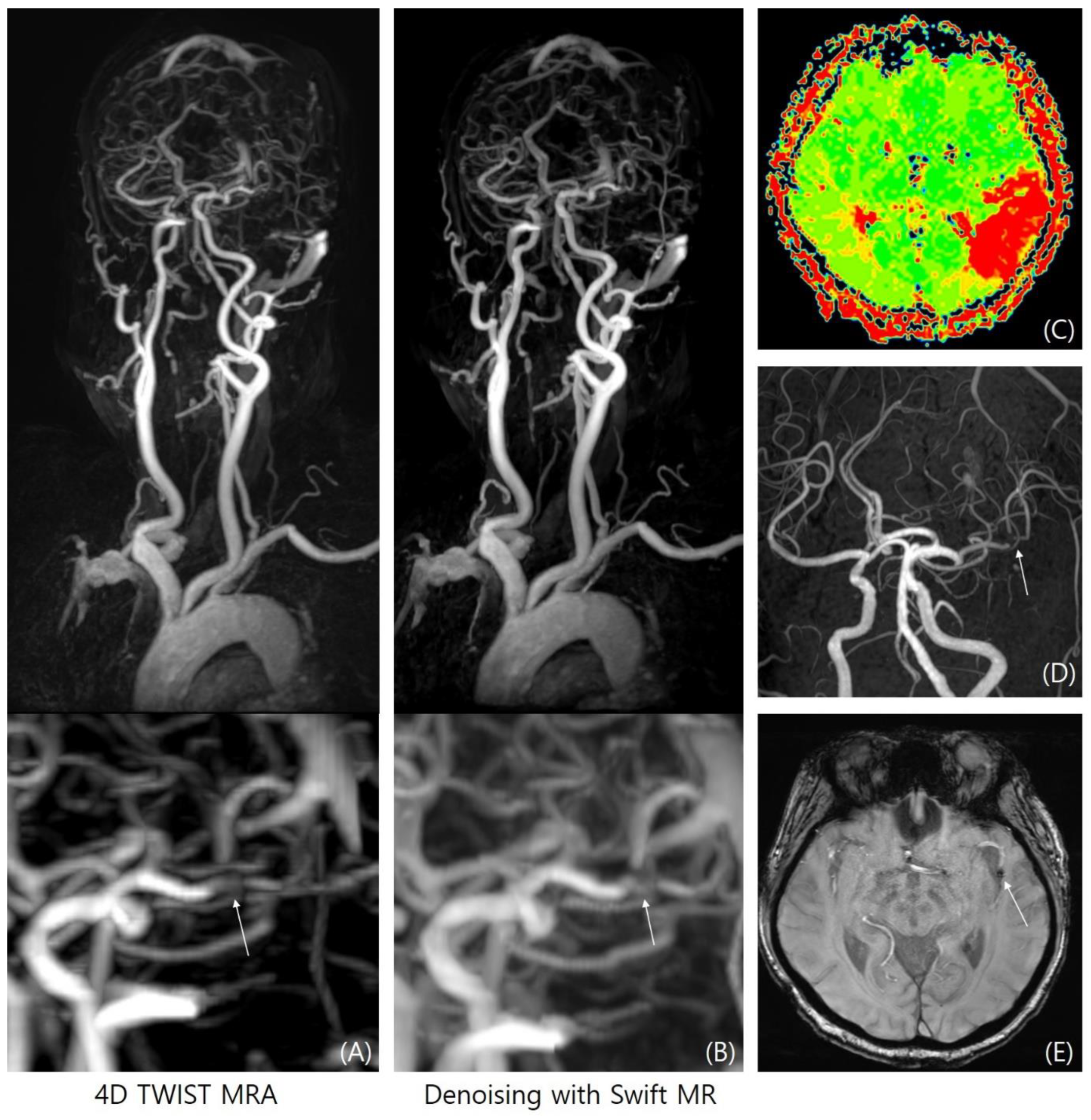
3.3. LVO Evaluation
4. Discussion
Author Contributions
Funding
Institutional Review Board Statement
Informed Consent Statement
Data Availability Statement
Conflicts of Interest
References
- Powers, W.J.; Rabinstein, A.A.; Ackerson, T.; Adeoye, O.M.; Bambakidis, N.C.; Becker, K.; Biller, J.; Brown, M.; Demaerschalk, B.M.; Hoh, B.; et al. Guidelines for the Early Management of Patients with Acute Ischemic Stroke: 2019 Update to the 2018 Guidelines for the Early Management of Acute Ischemic Stroke: A Guideline for Healthcare Professionals From the American Heart Association/American Stroke. Stroke 2019, 50, e344–e418. [Google Scholar] [CrossRef] [PubMed]
- Schellinger, P.D.; Jansen, O.; Fiebach, J.B.; Hacke, W.; Sartor, K. A Standardized MRI Stroke Protocol. Stroke 1999, 30, 765–768. [Google Scholar] [CrossRef] [PubMed]
- Davis, S.M.; Donnan, G.A.; Parsons, M.W.; Levi, C.; Butcher, K.S.; Peeters, A.; Barber, P.A.; Bladin, C.; De Silva, D.A.; Byrnes, G.; et al. Effects of alteplase beyond 3 h after stroke in the Echoplanar Imaging Thrombolytic Evaluation Trial (EPITHET): A placebo-controlled randomised trial. Lancet Neurol. 2008, 7, 299–309. [Google Scholar] [CrossRef] [PubMed]
- Albers, G.W.; Thijs, V.N.; Wechsler, L.; Kemp, S.; Schlaug, G.; Skalabrin, E.; Bammer, R.; Kakuda, W.; Lansberg, M.G.; Shuaib, A.; et al. Magnetic resonance imaging profiles predict clinical response to early reperfusion: The diffusion and perfusion imaging evaluation for understanding stroke evolution (DEFUSE) study. Ann. Neurol. 2006, 60, 508–517. [Google Scholar] [CrossRef] [PubMed]
- Boujan, T.; Neuberger, U.; Pfaff, J.; Nagel, S.; Herweh, C.; Bendszus, M.; Möhlenbruch, M.A. Value of Contrast-Enhanced MRA versus Time-of-Flight MRA in Acute Ischemic Stroke MRI. Am. J. Neuroradiol. 2018, 39, 1710–1716. [Google Scholar] [CrossRef] [PubMed]
- Hernández-Pérez, M.; Puig, J.; Blasco, G.; Pérez de la Ossa, N.; Dorado, L.; Dávalos, A.; Munuera, J. Dynamic magnetic resonance angiography provides collateral circulation and hemodynamic information in acute ischemic stroke. Stroke 2016, 47, 531–534. [Google Scholar] [CrossRef] [PubMed]
- Le Bras, A.; Raoult, H.; Ferré, J.C.; Ronzière, T.; Gauvrit, J.Y. Optimal MRI Sequence for Identifying Occlusion Location in Acute Stroke: Which Value of Time-Resolved Contrast-Enhanced MRA? Am. J. Neuroradiol. 2015, 36, 1081–1088. [Google Scholar] [CrossRef]
- Dhundass, S.; Savatovsky, J.; Duron, L.; Fahed, R.; Escalard, S.; Obadia, M.; Zuber, K.; Metten, M.A.; Mejdoubi, M.; Blanc, R.; et al. Improved detection and characterization of arterial occlusion in acute ischemic stroke using contrast enhanced MRA. J. Neuroradiol. 2020, 47, 278–283. [Google Scholar] [CrossRef]
- Wetzl, J.; Forman, C.; Wintersperger, B.J.; D’Errico, L.; Schmidt, M.; Mailhe, B.; Maier, A.; Stalder, A.F. High-resolution dynamic CE-MRA of the thorax enabled by iterative TWIST reconstruction. Magn. Reson. Med. 2017, 77, 833–840. [Google Scholar] [CrossRef]
- Grossberg, J.A.; Howard, B.M.; Saindane, A.M. The use of contrast-enhanced, time-resolved magnetic resonance angiography in cerebrovascular pathology. Neurosurg. Focus 2019, 47, E3. [Google Scholar] [CrossRef]
- Villablanca, J.P.; Nael, K.; Habibi, R.; Nael, A.; Laub, G.; Finn, J.P. 3 T contrast-enhanced magnetic resonance angiography for evaluation of the intracranial arteries: Comparison with time-of-flight magnetic resonance angiography and multislice computed tomography angiography. Investig. Radiol. 2006, 41, 799–805. [Google Scholar] [CrossRef] [PubMed]
- Saver, J.L.; Chapot, R.; Agid, R.; Hassan, A.E.; Jadhav, A.P.; Liebeskind, D.S.; Lobotesis, K.; Meila, D.; Meyer, L.; Raphaeli, G.; et al. Thrombectomy for Distal, Medium Vessel Occlusions. Stroke 2020, 51, 2872–2884. [Google Scholar] [CrossRef] [PubMed]
- Liang, Y.; Wang, J.; Li, B. Coexistence of internal carotid artery stenosis with intracranial aneurysm. Int. J. Stroke 2014, 9, 306–307. [Google Scholar] [CrossRef]
- Wicaksono, K.P.; Fujimoto, K.; Fushimi, Y.; Sakata, A.; Okuchi, S.; Hinoda, T.; Nakajima, S.; Yamao, Y.; Yoshida, K.; Miyake, K.K.; et al. Super-resolution application of generative adversarial network on brain time-of-flight MR angiography: Image quality and diagnostic utility evaluation. Eur. Radiol. 2022, 33, 936–946. [Google Scholar] [CrossRef] [PubMed]
- Yasaka, K.; Akai, H.; Sugawara, H.; Tajima, T.; Akahane, M.; Yoshioka, N.; Kabasawa, H.; Miyo, R.; Ohtomo, K.; Abe, O.; et al. Impact of deep learning reconstruction on intracranial 1.5 T magnetic resonance angiography. Jpn. J. Radiol. 2022, 40, 476–483. [Google Scholar] [CrossRef] [PubMed]
- Montalt-Tordera, J.; Quail, M.; Steeden, J.A.; Muthurangu, V. Reducing contrast agent dose in cardiovascular MR angiography with deep learning. J. Magn. Reson. Imaging 2021, 54, 795–805. [Google Scholar] [CrossRef] [PubMed]
- Jun, Y.; Eo, T.; Shin, H.; Kim, T.; Lee, H.J.; Hwang, D. Parallel imaging in time-of-flight magnetic resonance angiography using deep multistream convolutional neural networks. Magn. Reson. Med. 2019, 81, 3840–3853. [Google Scholar] [CrossRef] [PubMed]
- Koktzoglou, I.; Huang, R.; Ankenbrandt, W.J.; Walker, M.T.; Edelman, R.R. Super-resolution head and neck MRA using deep machine learning. Magn. Reson. Med. 2021, 86, 335–345. [Google Scholar] [CrossRef]
- Ronneberger, O.; Fischer, P.; Brox, T. (Eds.) U-net: Convolutional networks for biomedical image segmentation. In Medical Image Computing and Computer-Assisted Intervention–MICCAI 2015, Proceedings of the 18th International Conference, Munich, Germany, 5–9 October 2015, Proceedings, Part III 18; Springer: Berlin/Heidelberg, Germany, 2015. [Google Scholar]
- Kingma, D.P.; Ba, J. Adam: A method for stochastic optimization. arXiv 2014, arXiv:14126980. [Google Scholar]
- Jeong, G.; Kim, H.; Yang, J.; Jang, K.; Kim, J. All-in-One Deep Learning Framework for MR Image Reconstruction. arXiv 2024, arXiv:240503684. [Google Scholar]
- Dissaux, B.; Eugène, F.; Ognard, J.; Gauvrit, J.-Y.; Gentric, J.-C.; Ferré, J.-C. Assessment of 4D MR angiography at 3T compared with DSA for the follow-up of embolized brain dural arteriovenous fistula: A dual-center study. Am. J. Neuroradiol. 2021, 42, 340–346. [Google Scholar] [CrossRef]
- Machet, A.; Portefaix, C.; Kadziolka, K.; Robin, G.; Lanoix, O.; Pierot, L. Brain arteriovenous malformation diagnosis: Value of time-resolved contrast-enhanced MR angiography at 3.0 T compared to DSA. Neuroradiology 2012, 54, 1099–1108. [Google Scholar] [CrossRef] [PubMed]
- Krishnamurthy, R.; Bahouth, S.M.; Muthupillai, R. 4D contrast-enhanced MR angiography with the keyhole technique in children: Technique and clinical applications. Radiographics 2016, 36, 523–537. [Google Scholar] [CrossRef] [PubMed]
- Sakata, A.; Sakamoto, R.; Fushimi, Y.; Nakajima, S.; Hinoda, T.; Oshima, S.; Wetzl, J.; Schmidt, M.; Okawa, M.; Yoshida, K.; et al. Low-dose contrast-enhanced time-resolved angiography with stochastic trajectories with iterative reconstruction (IT-TWIST-MRA) in brain arteriovenous shunt. Eur. Radiol. 2022, 32, 5392–5401. [Google Scholar] [CrossRef] [PubMed]
- Huf, V.I.; Fellner, C.; Wohlgemuth, W.A.; Stroszczynski, C.; Schmidt, M.; Forman, C.; Wetzl, J.; Uller, W. Fast TWIST with iterative reconstruction improves diagnostic accuracy of AVM of the hand. Sci. Rep. 2020, 10, 16355. [Google Scholar] [CrossRef] [PubMed]
- Kidoh, M.; Shinoda, K.; Kitajima, M.; Isogawa, K.; Nambu, M.; Uetani, H.; Morita, K.; Nakaura, T.; Tateishi, M.; Yamashita, Y.; et al. Deep Learning Based Noise Reduction for Brain MR Imaging: Tests on Phantoms and Healthy Volunteers. Magn. Reson. Med. Sci. 2020, 19, 195–206. [Google Scholar] [CrossRef]
- You, S.H.; Cho, Y.; Kim, B.; Yang, K.S.; Kim, I.; Kim, B.K.; Pak, A.; Park, S.E. Deep Learning–Based Synthetic TOF-MRA Generation Using Time-Resolved MRA in Fast Stroke Imaging. Am. J. Neuroradiol. 2023, 44, 1391–1398. [Google Scholar] [CrossRef]


| TOF-MRA (n = 397) | 4D-TWIST-MRA (n = 520) | 4D-DNR (n = 520) | p-Value | TOF vs. 4D-TWIST-MRA | TOF vs. 4D-DNR | 4D-TWIST-MRA vs. 4D-DNR | |
|---|---|---|---|---|---|---|---|
| No. | 395 | 395 | 395 | N/A | N/A | N/A | N/A |
| Overall image quality | 4.45 ± 0.52 | 2.55 ± 0.52 | 3.25 ± 0.42 | 0.001 | 0.001 | 0.010 | 0.040 |
| Noise | 4.46 ± 0.52 | 2.68 ± 0.62 | 3.40 ± 0.52 | 0.001 | 0.001 | 0.001 | 0.016 |
| Sharpness | 4.87 ± 0.34 | 2.12 ± 0.33 | 3.18 ± 0.51 | 0.001 | 0.001 | 0.001 | 0.035 |
| Vascular conspicuity | 3.52 ± 0.58 | 2.80 ± 0.50 | 3.27 ± 0.52 | 0.001 | 0.045 | 0.356 | 0.037 |
| Venous contamination | 5.00 ± 0.00 | 3.33 ± 0.80 | 3.18 ± 0.55 | 0.001 | 0.001 | 0.001 | 0.679 |
| SI (M1) | 558.9 ± 104.0 | 552.3 ± 350.2 | 561.7 ± 370.3 | 0.523 | 1.000 | 1.000 | 1.000 |
| SI (M2) | 420.3 ± 88.2 | 511.1 ± 373.6 | 575.3 ± 415.1 | 0.023 | 0.105 | 0.001 | 0.235 |
| SI (M3) | 325.4 ± 70.3 | 352.4 ± 231.2 | 434.6 ± 285.1 | 0.001 | 0.877 | 0.001 | 0.001 |
| SI (BA) | 659.4 ± 103.8 | 431.6 ± 271.0 | 570.9 ± 265.6 | 0.001 | 0.001 | 0.042 | 0.001 |
| Background Noise | 11.3 ± 5.6 | 19.9 ± 14.6 | 9.37 ± 6.7 | 0.001 | 0.001 | 0.813 | 0.001 |
| SNR (M1) | 62.4 ± 33.0 | 21.4 ± 7.0 | 30.5 ± 12.4 | 0.001 | 0.001 | 0.001 | 0.001 |
| SNR (M2) | 46.2 ± 23.3 | 14.1 ± 12.8 | 28.3 ± 12.9 | 0.001 | 0.001 | 0.001 | 0.001 |
| SNR (M3) | 34.0 ± 18.8 | 13.3 ± 11.0 | 18.0 ± 9.2 | 0.001 | 0.001 | 0.001 | 0.171 |
| SNR (M4) | 73.1 ± 35.0 | 20.5 ± 8.7 | 31.4 ± 9.5 | 0.001 | 0.001 | 0.001 | 0.021 |
| TOF-MRA | 4D-TWIST-MRA | 4D-DNR | p-Value | TOF vs. 4D-TWIST-MRA | TOF vs. 4D-DNR | 4D-TWIST-MRA vs. 4D-DNR | |
|---|---|---|---|---|---|---|---|
| Aneurysm detection | 54 (100%) | 42 (77.8%) | 44 (81.5%) | 0.001 | <0.001 | 0.001 | 0.814 |
| Aneurysm size | 2.66 ± 0.51 | 1.75 ± 0.62 | 2.10 ± 0.41 | 0.033 | 0.029 | 0.327 | 0.251 |
| Reader 1 | Reader 2 | |||||
|---|---|---|---|---|---|---|
| 4D-TWIST-MRA | 4D-DNR | p-Value | 4D-TWIST-MRA | 4D-DNR | p-Value | |
| No. | 123 | 123 | 123 | 123 | ||
| Confidence level of LVO diagnosis | 3.92 ± 0.70 | 4.41 ± 0.58 | 0.007 | 3.82 ± 0.56 | 4.51 ± 0.61 | 0.003 |
| Decision time (s) | 33.76 ± 11.0 | 30.42 ± 9.6 | 0.056 | 31.61 ± 13.4 | 27.15 ± 12.3 | 0.042 |
Disclaimer/Publisher’s Note: The statements, opinions and data contained in all publications are solely those of the individual author(s) and contributor(s) and not of MDPI and/or the editor(s). MDPI and/or the editor(s) disclaim responsibility for any injury to people or property resulting from any ideas, methods, instructions or products referred to in the content. |
© 2024 by the authors. Licensee MDPI, Basel, Switzerland. This article is an open access article distributed under the terms and conditions of the Creative Commons Attribution (CC BY) license (https://creativecommons.org/licenses/by/4.0/).
Share and Cite
Kim, B.K.; You, S.-H.; Kim, B.; Shin, J.H. Deep Learning-Based High-Resolution Magnetic Resonance Angiography (MRA) Generation Model for 4D Time-Resolved Angiography with Interleaved Stochastic Trajectories (TWIST) MRA in Fast Stroke Imaging. Diagnostics 2024, 14, 1199. https://doi.org/10.3390/diagnostics14111199
Kim BK, You S-H, Kim B, Shin JH. Deep Learning-Based High-Resolution Magnetic Resonance Angiography (MRA) Generation Model for 4D Time-Resolved Angiography with Interleaved Stochastic Trajectories (TWIST) MRA in Fast Stroke Imaging. Diagnostics. 2024; 14(11):1199. https://doi.org/10.3390/diagnostics14111199
Chicago/Turabian StyleKim, Bo Kyu, Sung-Hye You, Byungjun Kim, and Jae Ho Shin. 2024. "Deep Learning-Based High-Resolution Magnetic Resonance Angiography (MRA) Generation Model for 4D Time-Resolved Angiography with Interleaved Stochastic Trajectories (TWIST) MRA in Fast Stroke Imaging" Diagnostics 14, no. 11: 1199. https://doi.org/10.3390/diagnostics14111199
APA StyleKim, B. K., You, S.-H., Kim, B., & Shin, J. H. (2024). Deep Learning-Based High-Resolution Magnetic Resonance Angiography (MRA) Generation Model for 4D Time-Resolved Angiography with Interleaved Stochastic Trajectories (TWIST) MRA in Fast Stroke Imaging. Diagnostics, 14(11), 1199. https://doi.org/10.3390/diagnostics14111199


