Article

Classical Orbital Floor Post-Traumatic Reconstruction vs. Customized Reconstruction with the Support of “In-House” 3D-Printed Models: A Retrospective Study with an Analysis of Volumetric Measurement

by
Elvis Kallaverja
1,
Ida Barca
2,
Francesco Ferragina
1 and
Maria Giulia Cristofaro
1,*
1
Department of Experimental and Clinical Medicine, Maxillofacial Surgery Unit, Renato Dulbecco Hospital, Magna Graecia University of Catanzaro, 88100 Catanzaro, Italy
2
Department of Experimental and Clinical Medicine, Maxillofacial Surgery Unit, Magna Graecia University, 88100 Catanzaro, Italy
*
Author to whom correspondence should be addressed.
Diagnostics 2024, 14(12), 1248; https://doi.org/10.3390/diagnostics14121248
Submission received: 23 May 2024 / Revised: 7 June 2024 / Accepted: 9 June 2024 / Published: 13 June 2024

Abstract

:
Background: Orbital floor fractures (OFFs) represent an interesting chapter in maxillofacial surgery, and one of the main challenges in orbit reconstruction is shaping and cutting the precise contour of the implants due to its complex anatomy. Objective: The aim of the retrospective study was to demonstrate, through pre- and postoperative volumetric measurements of the orbit, how the use of a preformed titanium mesh based on the stereolithographic model produced with 3D printers (“In-House” reconstruction) provides a better reconstruction volumetric compared to the intraoperatively shaped titanium mesh. Materials and Methods: The patients with OFF enrolled in this study were divided into two groups according to the inclusion criteria. In Group 1 (G1), patients surgically treated for OFF were divided into two subgroups: G1a, patients undergoing orbital floor reconstruction with an intraoperatively shaped mesh, and G1b, patients undergoing orbital floor reconstruction with a preoperative mesh shaped on a 3D-printed stereolithographic model. Group 2 (G2) consisted of patients treated for other traumatic pathologies (mandible fractures and middle face fractures not involving orbit). Pre- and postoperative orbital volumetric measurements were performed on both G1 and G2. The patients of both groups were subjected to the measurement of orbital volume using Osirix software (Pixmeo SARL, CH-1233 Bernex, Switzerland) on the new CT examination. Both descriptive (using central tendency indices such as mean and range) and regressive (using the Bravais–Pearson index, calculated using the GraphPad program) statistical analyses were performed on the recorded data. Results: From 1 January 2017 to 31 December 2021, of the 176 patients treated for OFF at the “Magna Graecia” University Hospital of Catanzaro 10 fulfilled the study’s inclusion criteria: 5 were assigned to G1a and 5 to G1b, with a total of 30 volumetric measurements. In G2, we included 10 patients, with a total of 20 volumetric measurements. From the volumetric measurements and statistical analysis carried out, it emerged that the average of the volumetric differences of the healthy orbits was ±0.6351 cm3, the standard deviation of the volumetric differences was ±0.3383, and the relationship between the treated orbit and the healthy orbit was linear; therefore, the treated orbital volumes tend to approach the healthy ones after surgical treatment. Conclusion: This study demonstrates that if the volume is restored within the range of the standardized mean, the diplopia is completely recovered already after surgery or after one month. For orbital volumes that do not fall within this range, functional recovery could occur within 6 months or be lacking. The restoration of the orbital volume using pre-modeled networks on the patient’s anatomical model, printed internally in 3D, allows for more accurate reconstructions of the orbital floor in less time, with clinical advantages also in terms of surgical timing.

1. Introduction

Orbital floor fractures (OFFs) represent an interesting chapter of maxillofacial traumatology that is much debated in the scientific community regarding its epidemiological, clinical, and demographic characteristics [1,2]. Their incidence is increasing, probably due to the increase in road accidents, sporting accidents, and physical assaults. The literature contains conflicting indications and recommendations regarding orbital floor reconstruction, in particular on the operating times, the surgical approach, and the materials to be implanted, because of the great variety of options available today. All authors, however, agree in establishing that a detailed clinical examination associated with a computed tomography (CT) study is fundamental for the definition of an indication and an adequate treatment. Traumatic alterations of the orbital structures can cause entrapment of the periorbital tissue with symptoms such as diplopia, dystopia, impaired ocular motility, and enophthalmos [3]. The indication for surgery should be assessed individually for each patient after careful clinical and radiographic evaluation [4,5,6]. The primary goal of surgery is to restore orbital volume by preserving both function and aesthetics [7]. The material used to restore orbital volume should be similar to the orbital bone, easy to handle, and provide a stable result throughout life [8]. Nowadays, intraoperatively conformed titanium mesh is frequently used. This involves modeling the plates and adapting them to the shape and size of the OFF [9], until reaching an ideal conformation. For this practical reason, combined with the complex three-dimensional anatomy of the orbit and limited surgical access (which limits vision during surgery), orbital reconstructions remain a significant challenge for surgeons [10,11]. Three-dimensional technology has certainly helped modern medicine and has offered the possibility to create customized anatomical models through 3D printing. In this way, it was possible to evaluate traumatic pathologies not only on a virtual screen but also on stereolithographic models. Titanium plates shaped before surgery on the stereolithographic model (produced with 3D printers) are much more precise and require less surgical time and less damage to the tissues of the patient. The future projects us toward specific patient implants, but currently, not all centers can afford such a model, especially concerning costs. In the beginning, 3D printing had high costs and long production times [10,12]; often, 3D printing was carried out at other facilities, even distant ones, and consequently delivery times were long [13]. All this led to a limited use of such prototyping in the medical field. Nowadays, the introduction of in-house 3D printers for producing orbital models is gaining increasing popularity in clinical and surgical practice [14,15,16], achieving high precision at a low cost. Through volumetric measurements of isolated and unilateral orbital floor fractures, comparing the postoperative volume with the healthy contralateral, the authors wanted to demonstrate how the use of a preformed titanium mesh based on the stereolithographic model produced with 3D printers (“In-House” reconstruction) provides a better volumetric reconstruction compared to intraoperatively conformed titanium mesh.

2. Materials and Methods

This retrospective study included patients surgically treated for OFF at the Maxillofacial Traumatology Center of the “Magna Grecia” University Hospital of Catanzaro, from 1 January 2017 to 31 January 2021. The study was carried out according to the guidelines set out in the Helsinki Declaration and was approved by the Ethics Committee of the “Magna Grecia” University of Catanzaro, Italy. The aim of this study was to compare two titanium mesh modeling techniques using a volumetric analysis of the orbits in patients subjected to surgery for OFF.
The inclusion criteria were as follows:
-
A diagnosis of unilateral isolated OFF in male patients aged >18 years;
-
Pre- and postoperative ophthalmological examination with preoperative diplopia confirmed by an Orthoptic examination with Hess Lancaster screens;
-
Pre- and postoperative CT in our Radiology Center with slice of 0.5 mm;
-
An absence of muscle deficits and nerve or vascular damage;
-
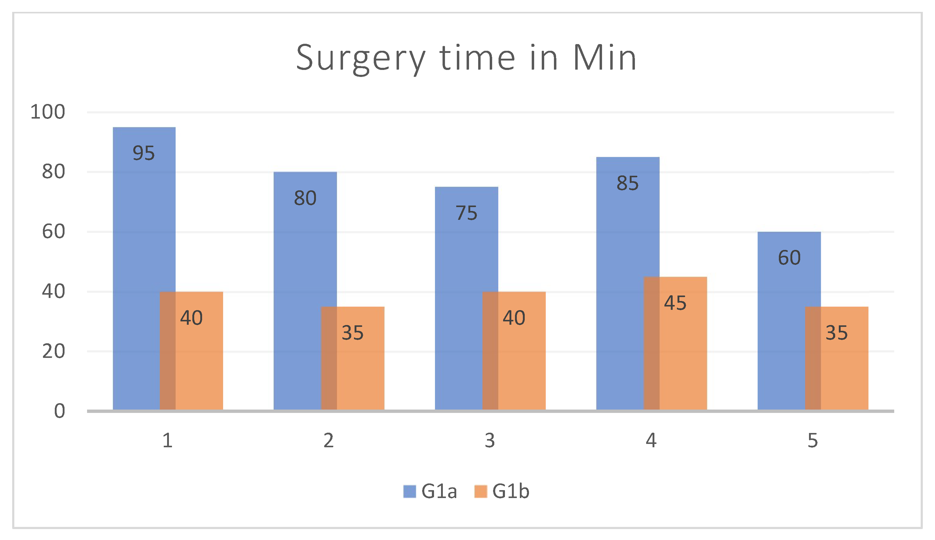
Surgery performed within 2 weeks from trauma;
-
Three-dimensional stereolithographic printed model, volumetric measurements, and reconstruction with titanium mesh (both modeled intraoperatively and preoperatively on a 3D model) performed by the same surgeon.
The exclusion criteria were as follows:
-
Patients with multiple maxilla-facial fractures;
-
Prior surgery for orbital trauma or ophthalmic surgery;
-
Patients who were not cooperating;
-
Patients with incomplete clinical-radiological documentation;
-
A reconstruction of the orbital floor with other materials.

2.1. Procedure

All patients arrived, hemodynamically stable, from the Emergency Departments of other local hospitals with radiological exams and urgent specialist visits. After clinical and radiological consultation, patients were hospitalized in case of surgical indication. The study design and treatment protocol are shown in Table 1.
Each patient underwent a general and loco-regional objective examination (including weight, height, and vital signs), instrumental examinations such as thin layer CT (0.5 mm) with axial, coronal, and sagittal acquisitions and 3D reconstructions, ophthalmological examination pre- and postoperatively, and orthoptic examination with Hess-Lancaster screens. We also classified the blow-out fractures according to Harris’ classification. Supported by the literature, indications for surgical reconstruction were fractures with defects ≥ 3 cm2, enophthalmos ≥ 2 mm, dislocated fractures, and soft tissue herniation [5,6].
Patients enrolled in the study were divided into two groups:
  • Group 1 (G1): patients surgically treated for isolated OFF, divided into 2 subgroups: G1a (patients undergoing orbital floor reconstruction with an intraoperatively shaped mesh) and G1b (patients undergoing orbital floor reconstruction with a preoperative mesh shaped on a 3D-printed stereolithographic model, obtained from the patient’s CT scan and using the Ultimaker S5 3D printer—Manufat Engineering Srl, 160 23900 Lecco (LC), Italy).
  • Group 2 (G2): patients treated for other traumatic pathologies such as mandibular or middle third fracture not involving orbit (zygmatic arch, Le Fort I, etc.).
Pre- and postoperative orbital volumetric measurements were performed on both G1 and G2.
Patients of both groups were subjected to the following:
-
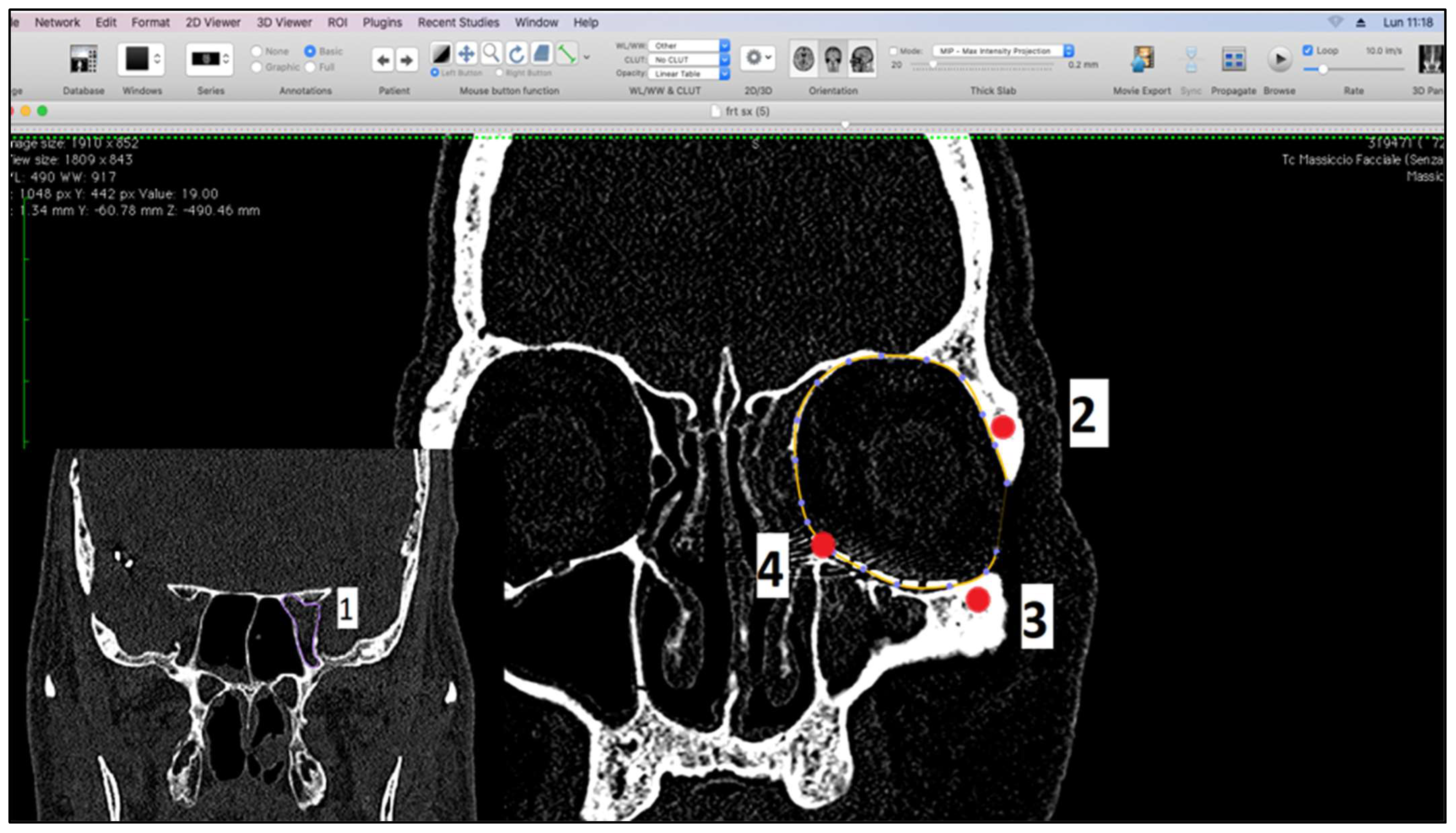
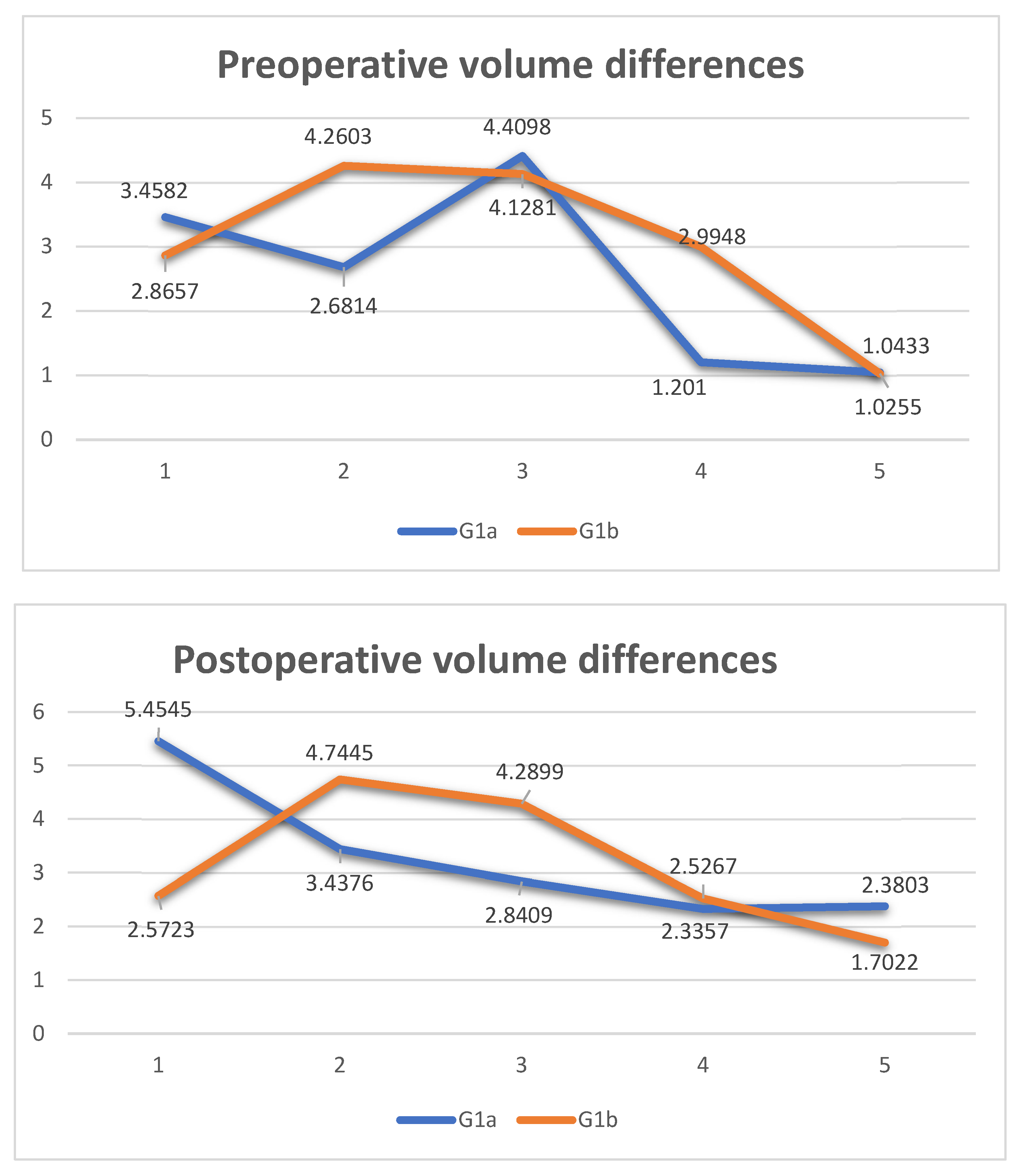
Measurement of orbital volume using Osirix software (Version 12.0) (Pixmeo SARL, CH-1233 Bernex, Switzerland) on the new CT examination. This was carried out both semi-automatically and manually. For each patient, the orbital volume on the preoperative CT was calculated for both healthy and fractured orbits. On the postoperative CT, however, the orbital volume reconstructed with titanium mesh was calculated. We then calculated the difference in volumes between healthy and traumatized orbit, both before and after surgery. Finally, we evaluated the recovery difference, which is made up of the difference between the two volumes, pre-op and post-op, of the traumatized orbit.
-
Post-surgery thin layer CT (0.5 mm) with axial, coronal, and sagittal acquisitions and 3D reconstructions.
On this printed model we modeled, before surgery, the titanium mesh. All preoperative stages (study and printing), surgery, and postoperative studies were performed by the same surgeon, to reduce bias.

2.2. Measurement of Orbital Volumes

We performed the measurements using Osirix software in semi-automatic and manual mode on CT scans [17,18], on both preoperative and postoperative CT examinations, with slices of 0.5 mm and with the head positioned in its “neutral position”. Preoperative CT was carried out within 24 h of the trauma. We corrected the minimal movement discrepancies using the MPR function of the software with anatomical references in the planes: the Frankfurt horizontal plane, a vertical plane passing between the supraorbital foramen and the maxilla-malar suture of the lower orbital frame, and the tangential horizontal plane to the lower orbital frames. Volumetric analysis after CT orientation was performed on coronal scans in bone cuts. Since the orbital cavity reflects the geographical shape of a cone, with the base at the level of the orbital frames, several reference points have been identified to standardize the measurement procedure which are (1) the optic foramen; (2) the frontal zygomatic suture; (3) the maxillo-malar suture at the level of the lower orbital frame; and (4) the lacrimal canal. The plane passing through points 2, 3, and 4 was considered the base of the cone (Figure 1).
Using the option ROI (region of interest) and selecting the tool “closed polygon”, present on the software Osirix, we could manually identify and delineate the regions of interest for each section of the cut. This was done using the reference points in the posterior to the anterior direction (from the apex to the base of the orbit). Figure 2A shows the preoperative orbital volume measurement including herniated soft tissues. Instead, Figure 2B shows the measurement of the postoperative orbital volume; we performed the measurements following the neo-floor (following the titanium mesh).
After contouring each slice of the CT scan, we applied the volume calculation function of the selected ROI (Figure 3). We reported all calculated data in an Excel (Version Microsoft 365) table on which the statistical analysis was carried out.

2.3. Production of the 3D Model

After acquiring the preoperative CT (performed on 0.5 mm bone cuts), we exported the DICOM file using the Radiant software and created the 3D model with the acquisition of the post-processing stereolithographic file. So, we created an stl. file with the bone part of interest (Figure 4A). The stereolithographic file was opened with another open-source software called Meshmixer (Version 3.5) (Figure 4B). In this software, the processing and cleaning of the model were practiced creating a solid file that could be printed on 3D printers. Finally, the stereolithographic file was linked to the CURA software (version 4.2) specific to the Ultimaker (Zaltbommel, The Netherlands) 3D printer used for this study (Figure 4C). The stereolithographic model generated by 3D printing was inserted and modeled on the fractured orbit of the patient (Figure 4D).
The modeling criteria were based on basic principles, such as fixed points of the residual orbital floor where the mesh can be placed to have a support base; restoring the anatomy of the floor as faithfully as possible by comparing it with the contralateral floor; and retaining important anatomical structures that may be damaged during placement, such as the lower orbital fissure, the lacrimal sac, and the infraorbital nerve.

2.4. Statistical Analysis

We performed both descriptive and regressive statistical analyses on the recorded data.
We performed descriptive statistical analysis using central tendency indices (such as mean and range) and absolute and relative frequencies for categorical data. A descriptive analysis was performed for an overview of the data. The averages of the orbital volumes give us an idea of the central value of the data, while the standard deviations give us a measure of how dispersed the data were around the mean.
We performed regressive statistical analysis using the Bravais–Pearson index, calculated using the GraphPad program (Version 10.2.3) (GraphPad Company, San Diego, CA, USA). This was used to evaluate the relationship between the healthy orbital volume and the surgically treated orbital volume in both groups.

3. Results

In the timeframe analyzed from 1 January 2017 to 31 December 2021, we evaluated 176 patients treated for OFF and 10 fulfilled the study’s inclusion criteria. The sample examined included patients with an average age of 39.5 years and a range of 16–88 years. The most frequent cause of trauma was road accidents (n. 4, 40%), followed by domestic accidents (n. 3, 30%), sports injuries (n. 2, 20%), and interpersonal violence (n. 1, 10%).
The 10 patients of G1 were divided into two groups, with 5 assigned to G1a and 5 to G1b, and a total of 30 volumetric measurements were performed, 20 preoperatively and 10 postoperatively.
In G2, we included 10 patients treated for other traumatic pathologies (face lower third fractures), and a total of 20 measurements were performed.
The average volume based on the literature is about 24.5 ± 3.08 cm3 [19]. Compared to these data, ours remains an effective and reliable measurement method with a volumetric mean of healthy orbits of 24.511 cm3, with a standard deviation of 2.30. Instead, the mean volume of fractured orbits was 27.318 cm3, with a standard deviation of 2.87. After treatment, the orbits reconstructed with the mesh had a volumetric mean of 24,089 cm3, with a standard deviation of 2.674. The maximum herniation was approximately 4.4098 cm3, and the minimum herniation was approximately 1.0433 cm3.
Table 2 and Table 3 present the data of patients treated with a conventional intra-op-shaped titanium mesh and with a titanium mesh pre-op-shaped based on the 3D printed model, respectively.
We saw that in four out of five patients the volume of the traumatized orbit, after surgery, was greater than the healthy contralateral; the recovery difference had a negative value. In one case, it was smaller, and the recovery difference had a positive value. The range of recovery difference was from −1.9963 to +1.5689 cm3. These patients had a resolution of diplopia within 2–6 months, as evaluated with the Hess Lancaster test. The average surgical timing was 79 min. In two cases, the patient suffered from postoperative exophthalmos due to hypercorrection. No patient had dystopia.
The descriptive analysis of G2 in Table 4, demonstrated that the mean of the volumetric differences was ±0.6351 cm3 and the standard deviation of the volumetric differences was ±0.3383. These data allowed us to evaluate and compare how much the mean and standard deviation of the volumetric differences approached or moved away from G1 patients compared to G2.
We saw that in three out of five patients, the volume of the traumatized orbit, after surgery, was greater than the healthy contralateral. In two cases, it was smaller. The range of recovery difference was from −0.6767 to +0.4681 cm3. These patients had a resolution of diplopia in less than 1 month, as evaluated with the Hess Lancaster test. The average surgical timing was 39 min. No patient had dystopia, exophthalmos, or enophthalmos.
Figure 5 shows two graphs comparing the differences in the volumes of patients in groups G1a and G1b, preoperatively and postoperatively. A very important result, despite the limited number of patients, is the trend of the two orange and blue lines, respectively, of groups G1a and G1b. In both charts, the blue line has a much more uniform pattern than the orange one. Therefore, we can say that in G1b, the volume recovery from the preoperative to the postoperative period was more linear than in G1a.
Figure 6 shows the recovery volume in the two groups. This difference should ideally be 0. The closer the result is to 0, the more likely it is that the surgically treated orbit will have an optimal recovery. This is well highlighted in group G1b.
As a final analysis, we calculated the Bravais–Pearson index. It emerged that the relationship between healthy orbital volumes and treated orbital volumes was linear (Figure 7), and both variables were distributed according to a normal one. Significance was confirmed by a value p < 0.0001, with an r value = 0.9350. The 95% confidence interval was between 0.7425 and 0.9849.
From the analysis of the surgical times used, it appears that G1b had an average surgical timing of about 40 min less than G1a (Figure 8).

4. Discussion

At present, there is no doubt that the presence of diplopia represents a surgical indication in patients with OFF, but even today, the debate remains open in the literature regarding the best way to perform the operation and the type of material to be implanted. The advantages and disadvantages of the most used materials for reconstruction as well as the methods of their use have been widely described in the literature [20,21,22]. Titanium is an excellent material for implants due to its properties such as high biocompatibility, less-than-one-millimetre thickness, and excellent malleability and stability. The increased precision of titanium implants improves the fit of the implant and minimizes the number of insertion tests, causing minor damage to the surrounding soft tissues [23,24,25]. Titanium plates modeled before surgery on the stereolithographic model (produced with 3D printers) are much more precise and require less surgical time and less damage to the patient’s tissues. In the beginning, 3D printing had high costs and long production times [10,26]; often, 3D printing was carried out at other facilities, even distant ones, and consequently delivery times were long [27]. All this led to a limited use of such prototyping in the medical field. In our study, the models were made using an in-house 3D printer, and this was certainly less expensive and took less time than the external printing of standard stereolithographic models [25]. The rigorous inclusion criteria adopted allowed us to minimize selection bias and to make the sample even more homogeneous, as we considered only male patients. Similarly, including asymptomatic patients immediately after surgical treatment did not influence the functional results. This allowed us to choose patients with great precision for about 5 years. From the analyses carried out and the results obtained, we can say that the measurements are quite reliable. From these results, we can deduce that a post-treatment volumetric recovery of the fractured orbit will be closer to a healthy orbit with less probability of having distant diplopia. Observing the differences in the standard deviation between G1a and G1b, it was seen that in the five patients belonging to G1a, the standard deviation moved away from that measured in group G1b (±0.338), with an excess or deficit volumetric recovery. In four of these patients, the residual diplopia resolved within 6 months. In one patient (standard deviation difference of −1.9963), diplopia persisted one year later. Therefore, although the volume restored by surgery is not exactly the pre-trauma volume, it is included within a standardized range of orbital volume. All this translates into better aesthetic and functional results and therefore less likelihood of diplopia persisting after surgery. The study by Holmes and Schlittler [28] showed a statistically significant improvement through the use of patient-specific implants, especially in larger and more complex fractures. Zimmerer et al. [29] showed no significant differences in postoperative diplopia and ocular motility between the two groups examined. Instead, Kozakiewicz et al. [30] showed that ophthalmic results were better in the long term for individual orbital systems. Kim et al. [31] showed a higher rate of postoperative resolution or the persistence of only mild symptoms in the group with the patient-specific pre-shaped implants, while the conventional group also experienced moderate to severe postoperative symptoms. [29,32,33,34]. According to previous studies, the reduction in surgical timing in G1b showed a significant statistically significant difference, with an average recovery of 40 min, less intraoperative stress, and a faster postoperative recovery [35,36]. To improve the result and comparison in the study, we preferred to perform measurements of orbital volume. The literature does not report a gold standard method for measuring orbital volume. We chose to use medical software like Osirix MD that allows measurements to be performed on CT scans. The models were made using an in-house 3D printer, and this made it less expensive and time-consuming than externally printed standard stereolithographic models [25]. The virtual design, 3D printing, and preoperative bending of the titanium mesh took 50 to 120 min, without causing delays in surgical timing. The accuracy of low-cost 3D printed models was found to be satisfactory for daily maxillofacial procedures, such as teaching purposes, preoperative surgical planning, and plate bending [14,15,32,36].

5. Conclusions

“In-house” pre-modelling on stereolithographic models, compared to that practiced intraoperatively, has a significantly higher accuracy in restoring the fractured floor as faithfully as possible and thus also restoring orbital volume. This study demonstrated that in patients with a pre-modeled mesh, the orbital volume fell within the target range of the standard deviation, with diplopia disappearing immediately postoperatively or after 1 month. This result demonstrates that this modeling method allows for greater precision in the contours and a less invasive insertion of the implant, avoiding phenomena of multiple attempts at adaptation or malpositioning. Our results in the orbital volumetric analysis suggest the use of this method which involves better volumetric recovery, greater precision, better aesthetic results, earlier functional results, less surgical time, and better post-op recovery. Therefore, ours remains a valid and reproducible type of surgical treatment.

Author Contributions

Conceptualization, E.K. and M.G.C.; Methodology, E.K. and I.B.; Software, E.K. and F.F.; Validation, E.K. and M.G.C.; Formal analysis, F.F.; Resources, F.F.; Data curation, E.K., I.B. and F.F.; Writing—original draft, E.K., I.B. and F.F.; Writing—review & editing, M.G.C.; Visualization, M.G.C.; Supervision, M.G.C.; Funding acquisition, E.K. and I.B. All authors have read and agreed to the published version of the manuscript.

Funding

Open access funding was provided by “Magna Graecia” University of Catanzaro, Italy, within the CRUI-CARE Agreement.

Institutional Review Board Statement

The study was conducted following the Declaration of Helsinki; the Magna Graecia University Ethics Committee approved the study (protocol number 003589_15, approval date 02.11.2023).

Informed Consent Statement

Informed consent was obtained from all subjects involved in the study.

Data Availability Statement

The data presented in this study are available on request from the corresponding author.

Conflicts of Interest

The authors declare no conflicts of interest.

References

  1. Megafu, M.N.; Megafu, E.C.; Nguyen, J.T.; Mian, H.S.; Singhal, S.S.; Parisien, R.L. The Statistical Fragility of Orbital Fractures: A Systematic Review of Randomized Controlled Trials. J. Oral Maxillofac. Surg. 2023, 81, 752–758. [Google Scholar] [CrossRef] [PubMed]
  2. Colangeli, W.; Ferragina, F.; Kallaverja, E.; Celano, C.; Cristofaro, M.G. Orbital fractures treated in a university hospital of southern Italy: Epidemiology, outcomes and prognostic factors resulting from 538 retrospectively analyzed cases. Oral Maxillofac. Surg. 2024, 1–7. [Google Scholar] [CrossRef] [PubMed]
  3. Shin, J.W.; Lim, J.S.; Yoo, G.; Byeon, J.H. An Analysis of Pure Blowout Fractures and Associated Ocular Symptoms. J. Craniofacial Surg. 2013, 24, 703–707. [Google Scholar] [CrossRef] [PubMed]
  4. Manson, P.N.; Iiff, N. Management of blow-out fractures of the orbital floor II. Early repair for selected injuries. Surv. Ophthalmol. 1991, 35, 280–292. [Google Scholar] [CrossRef] [PubMed]
  5. Kunz, C.; Sigron, G.R.; Jaquiéry, C. Functional outcome after non-surgical management of orbital fractures—The bias of decisionmaking according to size of defect: Critical review of 48 patients. Br. J. Oral Maxillofac. Surg. 2013, 51, 486–492. [Google Scholar] [CrossRef] [PubMed]
  6. Burnstine, M.A. Clinical recommendations for repair of isolated orbital floor fractures: An evidence-based analysis. Ophthalmology 2002, 109, 1207–1210. [Google Scholar] [CrossRef] [PubMed]
  7. Hammer, B.; Prein, J. Correction of post-traumatic orbital deformities: Operative techniques and review of 26 patients. J. Cranio-Maxillofac. Surg. 1995, 23, 81–90. [Google Scholar] [CrossRef] [PubMed]
  8. Potter, J.K.; Ellis, E. Biomaterials for reconstruction of the internal orbit. J. Oral Maxillofac. Surg. 2004, 62, 1280–1297. [Google Scholar] [CrossRef] [PubMed]
  9. Sugar, A.W.; Kurlakose, M.; Walshaw, N.D. Titanium mesh in orbital wall reconstruction. Int. J. Oral Maxillofac. Surg. 1992, 21, 140–144. [Google Scholar] [CrossRef] [PubMed]
  10. Metzger, M.C.; Schön, R.; Weyer, N.; Rafii, A.; Gellrich, N.-C.; Schmelzeisen, R.; Strong, B.E. Anatomical 3-dimensional Pre-bent Titanium Implant for Orbital Floor Fractures. Ophthalmology 2006, 113, 1863–1868. [Google Scholar] [CrossRef]
  11. Kärkkäinen, M.; Wilkman, T.; Mesimäki, K.; Snäll, J. Primary reconstruction of orbital fractures using patient-specific titanium milled implants: The Helsinki protocol. Br. J. Oral Maxillofac. Surg. 2018, 56, 791–796. [Google Scholar] [CrossRef] [PubMed]
  12. Strong, E.B.; Fuller, S.C.; Wiley, D.F.; Zumbansen, J.; Wilson, M.D.; Metzger, M.C. Preformed vs Intraoperative Bending of Titanium Mesh for Orbital Reconstruction. Otolaryngol. Head Neck Surg. 2013, 149, 60–66. [Google Scholar] [CrossRef] [PubMed]
  13. Kozakiewicz, M.; Elgalal, M.; Loba, P.; Komuński, P.; Arkuszewski, P.; Broniarczyk-Loba, A.; Stefańczyk, L. Clinical application of 3D pre-bent titanium implants for orbital floor fractures. J. Cranio-Maxillofac. Surg. 2009, 37, 229–234. [Google Scholar] [CrossRef] [PubMed]
  14. Msallem, B.; Sharma, N.; Cao, S.; Halbeisen, F.S.; Zeilhofer, H.-F.; Thieringer, F.M. Evaluation of the Dimensional Accuracy of 3D-Printed Anatomical Mandibular Models Using FFF, SLA, SLS, MJ, and BJ Printing Technology. J. Clin. Med. 2020, 9, 817. [Google Scholar] [CrossRef] [PubMed]
  15. Hatz, C.R.; Msallem, B.; Aghlmandi, S.; Brantner, P.; Thieringer, F.M. Can an entry-level 3D printer create high-quality anatomical models? Accuracy assessment of mandibular models printed by a desktop 3D printer and a professional device. Int. J. Oral Maxillofac. Surg. 2020, 49, 143–148. [Google Scholar] [CrossRef] [PubMed]
  16. Maglitto, F.; Orabona, G.D.; Committeri, U.; Salzano, G.; De Fazio, G.R.; Vaira, L.A.; Abbate, V.; Bonavolontà, P.; Piombino, P.; Califano, L. Virtual Surgical Planning and the “In-House” Rapid Prototyping Technique in Maxillofacial Surgery: The Current Situation and Future Perspectives. Appl. Sci. 2021, 11, 1009. [Google Scholar] [CrossRef]
  17. Kwon, J.; Barrera, J.E.; Jung, T.Y.; Most, S.P. Measurements of orbital volume change using computed tomography in isolated orbital blowout fractures. Arch. Facial Plast. Surg. 2009, 11, 395–398. [Google Scholar] [CrossRef] [PubMed]
  18. Koppel, D.A.; Foy, R.H.; McCaul, J.A.; Logan, J.; Hadley, D.M.; Ayoub, A. The reliability of “Analyze” software in measuring orbital volume utilizing CT-derived data. J. Craniomaxillofac. Surg. 2003, 31, 88–91. [Google Scholar] [CrossRef] [PubMed]
  19. Andrades, P.; Cuevas, P.; Hernández, R.; Danilla, S.; Villalobos, R. Characterization of the orbital volume in normal population. J. Craniomaxillofac. Surg. 2018, 46, 594–599. [Google Scholar] [CrossRef] [PubMed]
  20. Ellis, E.; Tan, Y. Assessment of internal orbital reconstructions for pure blowout fractures: Cranial bone grafts versus titanium mesh. J. Oral Maxillofac. Surg. 2003, 61, 442–453. [Google Scholar] [CrossRef]
  21. Ellis, E.; Messo, E. Use of nonresorbable alloplastic implants for internal orbital reconstruction. J. Oral Maxillofac. Surg. 2004, 62, 873–881. [Google Scholar] [CrossRef] [PubMed]
  22. Dubois, L.; Steenen, S.A.; Gooris, P.J.J.; Bos, R.R.M.; Becking, A.G. Controversies in orbital reconstruction—III. Biomaterials for orbital reconstruction: A review with clinical recommendations. Int. J. Oral Maxillofac. Surg. 2016, 45, 41–50. [Google Scholar] [CrossRef] [PubMed]
  23. Fan, B.; Chen, H.; Sun, Y.-J.; Wang, B.-F.; Che, L.; Liu, S.-Y.; Li, G.-Y. Clinical effects of 3-D printing-assisted personalized reconstructive surgery for blowout orbital fractures. Graefe’s Arch. Clin. Exp. Ophthalmol. 2017, 255, 2051–2057. [Google Scholar] [CrossRef] [PubMed]
  24. Schön, R.; Metzger, M.C.; Zizelmann, C.; Weyer, N.; Schmelzeisen, R. Individually preformed titanium mesh implants for a true-to-original repair of orbital fractures. Int. J. Oral Maxillofac. Surg. 2006, 35, 990–995. [Google Scholar] [CrossRef] [PubMed]
  25. Lim, C.G.T.; Campbell, D.I.; Clucas, D.M. Rapid Prototyping Technology in Orbital Floor Reconstruction: Application in Three Patients. Craniomaxillofac. Trauma Reconstr. 2014, 7, 143–146. [Google Scholar] [CrossRef] [PubMed]
  26. Callahan, A.B.; Campbell, A.A.; Petris, C.; Kazim, M. Low-Cost 3D Printing Orbital Implant Templates in Secondary Orbital Reconstructions. Ophthalmic Plast. Reconstr. Surg. 2017, 33, 376–380. [Google Scholar] [CrossRef] [PubMed]
  27. Kwon, M.-S.; Shin, H.J. Comparison of Orbital Reconstructive Effect between Customized Orbital Implants Using Three-Dimensional Printed Templates and Conventional Manual-Bending Implants in Blowout Fracture Surgery. Appl. Sci. 2023, 13, 9012. [Google Scholar] [CrossRef]
  28. Holmes, S.; Schlittler, F.L. Going beyond the limitations of the non-patient-specific implant in titanium reconstruction of the orbit. Br. J. Oral Maxillofac. Surg. 2021, 59, 1074–1078. [Google Scholar] [CrossRef]
  29. Zimmerer, R.M.; Ellis, E.; Aniceto, G.S.; Schramm, A.; Wagner, M.E.H.; Grant, M.P.; Cornelius, C.-P.; Strong, E.B.; Rana, M.; Chye, L.T. A prospective multicenter study to compare the precision of posttraumatic internal orbital reconstruction with standard preformed and individualized orbital implants. J. Cranio-Maxillofac. Surg. 2016, 44, 1485–1497. [Google Scholar] [CrossRef]
  30. Kozakiewicz, M.; Elgalal, M.; Piotr, L.; Broniarczyk-Loba, A.; Stefanczyk, L. Treatment with individual orbital wall implants in humans—1-Year ophthalmologic evaluation. J. Cranio-Maxillofac. Surg. 2011, 39, 30–36. [Google Scholar] [CrossRef]
  31. Kim, Y.C.; Jeong, W.S.; Park, T.-K.; Choi, J.W.; Koh, K.S.; Oh, T.S. The accuracy of patient specific implant prebented with 3D-printed rapid prototype model for orbital wall reconstruction. J. Cranio-Maxillofac. Surg. 2017, 45, 928–936. [Google Scholar] [CrossRef]
  32. Sigron, G.R.; Rüedi, N.; Chammartin, F.; Meyer, S.; Msallem, B.; Kunz, C.; Thieringer, F.M. Three-Dimensional Analysis of Isolated Orbital Floor Fractures Pre- and Post-Reconstruction with Standard Titanium Meshes and “Hybrid” Patient-Specific Implants. J. Clin. Med. 2020, 9, 1579. [Google Scholar] [CrossRef]
  33. Murray-Douglass, A.; Snoswell, C.; Winter, C.; Harris, R. Three-dimensional (3D) printing for post-traumatic orbital reconstruction, a systematic review and meta-analysis. Br. J. Oral Maxillofac. Surg. 2022, 60, 1176–1183. [Google Scholar] [CrossRef]
  34. Zieli’nski, R.; Mali’nska, M.; Kozakiewicz, M. Classical versus custom orbital wall reconstruction: Selected factors regarding surgery and hospitalization. J. Cranio-Maxillofac. Surg. 2017, 45, 710–715. [Google Scholar] [CrossRef]
  35. Msallem, D.M.B.; Beiglboeck, F.; Honigmann, P.; Jaquiéry, D.M.C.; Thieringer, F. Craniofacial Reconstruction by a Cost-Efficient Template-Based Process Using 3D Printing. Plast. Reconstr. Surg. Glob. Open 2017, 5, e1582. [Google Scholar] [CrossRef] [PubMed]
  36. Legocki, A.T.; Duffy-Peter, A.; Scott, A.R. Benefits and Limitations of Entry-Level 3-Dimensional Printing of Maxillofacial Skeletal Models. JAMA Otolaryngol. Head Neck Surg. 2017, 143, 389–394. [Google Scholar] [CrossRef] [PubMed]
Figure 1. Measurement landmarks.
Diagnostics 14 01248 g001
Figure 2. (A) Preoperative orbital volume measurement including herniated soft tissues. (B) Postoperative orbital volume measurement.
Diagnostics 14 01248 g002
Figure 3. Volume calculation.
Diagnostics 14 01248 g003
Figure 4. (A) DICOM file of a 3D reconstruction, using Radiant. (B) Meshmixer solid stereolithographic file. (C) Ultimaker CURA 3D printing file. (D) Titanium mesh shaped on the 3D printed patient anatomical model.
Diagnostics 14 01248 g004
Figure 5. Comparison of the differences in preoperative and postoperative volumes.
Diagnostics 14 01248 g005
Figure 6. Comparison of the difference in recovery volumes.
Diagnostics 14 01248 g006
Figure 7. Graph showing the linear relationship between healthy orbital volumes and treated orbital volumes.
Diagnostics 14 01248 g007
Figure 8. Comparison of surgical timing between G1a an G1b.
Diagnostics 14 01248 g008
Table 1. Treatment protocol.
10 patients isolated blow out fracture
CT scans and Hess Lancaster screen
G1a
(intraoperative shaping mesh)
G1b
(preoperative shaping mesh on 3D printed stereolithographic model)
Import Dicom blow out fracture Into Osirix MD software
Blow Out fracture identification, first volume measurements, export the Dicom file in the stl file
3D Printing of the stereolithographic model
Preoperative shaping of the mesh
Sterilization of the pre shaped mesh
Titanium mesh insertion without further adjustment
Postoperative CT
Import the new Dicom into Osirix MD software
Second volume measurement
Creating Table 2 and Table 3
Intraoperative shaping, mesh insertion, and evaluation if shape and insertion are correctAnalysis
Table 2. Data of patients in G1a (treated with intra-op-shaped titanium mesh).
AgeSexType of
Incidente
Fracture
Side
Volume cm3 Right OrbitVolume cm3 Left OrbitVolume cm3 Mesh OrbitVol- cm3
Difference
Pre-op
Vol- cm3
Difference
Post-op
Vol- cm3
Recovery
Difference
Surgery Time in Min
133MaleaggressionRight29.035925.577723.58143.45825.4545−1.996395
259MaleaggressionRight 23.974121.292720.53652.68143.4376−0.756280
337Malecar accidentLeft 23.20427.613824.77294.40982.84091.568975
474Maleaccidental fallLeft 25.279726.480724.1451.2012.3357−1.134785
541MaleaggressionLeft 25.981527.024824.64451.04332.3803−1.33760
Table 3. Data of patients in G1b (treated with preop-shaped titanium mesh).
AgeSexType of
Incidente
Fracture SideVolume cm3 Right OrbitVolume cm3 Left OrbitVolume cm3 Mesh OrbitVol- cm3
Difference
Pre-op
Vol- cm3
Difference
Post-op
Vol- cm3
Recovery
Difference
Surgery Time in Min
119Malesports traumaRight 27.695424.829725.12312.86572.57230.293440
272Malecar accidentLeft 24.052128.312423.56794.26034.7445−0.484235
337MaleaggressionRight 21.954626.082721.79284.12814.2899−0.161840
430Malesports traumaLeft 21.790424.785222.25852.99482.52670.468145
519Malesports traumaLeft 31.14932.174530.47231.02551.7022−0.676735
Table 4. Data of patients in G2.
AgeSexVolume cm3 Right OrbitVolume cm3 Left OrbitVolume cm3 Difference
123Male24.383525.51331.1298
255Male24.249524.76790.5184
339Male24.443324.52610.0828
447Male20.265920.57630.0828
553Male23.579222.83340.7458
628Male22.747123.38600.6389
719Male27.940427.20110.7393
838Male23.051721.91581.1359
946Male23.567724.19430.6266
1057Male28.671729.19440.5227
Disclaimer/Publisher’s Note: The statements, opinions and data contained in all publications are solely those of the individual author(s) and contributor(s) and not of MDPI and/or the editor(s). MDPI and/or the editor(s) disclaim responsibility for any injury to people or property resulting from any ideas, methods, instructions or products referred to in the content.

