Next Issue
Volume 14, July-1
Previous Issue
Volume 14, June-1


Diagnostics, Volume 14, Issue 12 (June-2 2024) – 107 articles

Cover Story (view full-size image): 
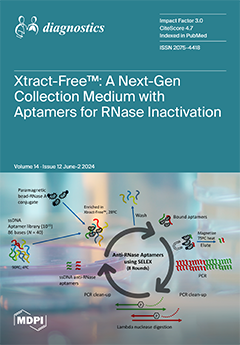
This study presents the first-ever utilization of anti-RNase aptamers, specifically chosen to target and inactivate RNases in Xtract-Free™, a next-generation sample collection and transport medium. The incorporation of aptamers for RNase inactivation in Xtract-Free™ provides the following advantages: (1) it offers a safe and eco-friendly alternative for both molecular and lateral flow diagnostic tests, eliminating the requirement for nucleic acid extraction prior to PCR, (2) it ensures the preservation and stabilization of RNA for PCR and other RNA-related genomic applications such as RNA-Seq and transcriptome analysis; (3) it broadens the range of collection media beyond traditional viral transport media and caustic molecular transport media, catering to the increasing demand for a safe medium suitable for point-of-care, self-collection, return mail, and home collection purposes. View this paper
  • Issues are regarded as officially published after their release is announced to the table of contents alert mailing list.
  • You may sign up for e-mail alerts to receive table of contents of newly released issues.
  • PDF is the official format for papers published in both, html and pdf forms. To view the papers in pdf format, click on the "PDF Full-text" link, and use the free Adobe Reader to open them.
Order results
Result details
Section
Select all
Export citation of selected articles as:
14 pages, 3006 KiB  
Article
Lung Disease Detection Using U-Net Feature Extractor Cascaded by Graph Convolutional Network
by Pshtiwan Qader Rashid and İlker Türker
Diagnostics 2024, 14(12), 1313; https://doi.org/10.3390/diagnostics14121313 - 20 Jun 2024
Viewed by 481
Abstract
Computed tomography (CT) scans have recently emerged as a major technique for the fast diagnosis of lung diseases via image classification techniques. In this study, we propose a method for the diagnosis of COVID-19 disease with improved accuracy by utilizing graph convolutional networks [...] Read more.
Computed tomography (CT) scans have recently emerged as a major technique for the fast diagnosis of lung diseases via image classification techniques. In this study, we propose a method for the diagnosis of COVID-19 disease with improved accuracy by utilizing graph convolutional networks (GCN) at various layer formations and kernel sizes to extract features from CT scan images. We apply a U-Net model to aid in segmentation and feature extraction. In contrast with previous research retrieving deep features from convolutional filters and pooling layers, which fail to fully consider the spatial connectivity of the nodes, we employ GCNs for classification and prediction to capture spatial connectivity patterns, which provides a significant association benefit. We handle the extracted deep features to form an adjacency matrix that contains a graph structure and pass it to a GCN along with the original image graph and the largest kernel graph. We combine these graphs to form one block of the graph input and then pass it through a GCN with an additional dropout layer to avoid overfitting. Our findings show that the suggested framework, called the feature-extracted graph convolutional network (FGCN), performs better in identifying lung diseases compared to recently proposed deep learning architectures that are not based on graph representations. The proposed model also outperforms a variety of transfer learning models commonly used for medical diagnosis tasks, highlighting the abstraction potential of the graph representation over traditional methods. Full article
(This article belongs to the Topic AI in Medical Imaging and Image Processing)
Show Figures

Figure 1

12 pages, 1539 KiB  
Article
Comparison of Small Blood Vessel Diameter with Intravascular Ultrasound and Coronary Angiography for Guidance of Percutaneous Coronary Intervention
by Sergio A. Zuñiga-Mendoza, Emanuel Zayas-Diaz, Victoria R. Armenta-Velazquez, Ana A. Silva-Baeza, Juan J. Beltran-Ochoa, Misael A. Medina-Servin and Maria G. Zavala-Cerna
Diagnostics 2024, 14(12), 1312; https://doi.org/10.3390/diagnostics14121312 - 20 Jun 2024
Viewed by 407
Abstract
Major cardiovascular events (MACEs) are a cause of major mortality worldwide. The narrowing and blockage of coronary arteries with atherosclerotic plaques are diagnosed and treated with percutaneous coronary intervention (PCI). During this procedure, coronary angiography (CAG) remains the most widely used guidance modality [...] Read more.
Major cardiovascular events (MACEs) are a cause of major mortality worldwide. The narrowing and blockage of coronary arteries with atherosclerotic plaques are diagnosed and treated with percutaneous coronary intervention (PCI). During this procedure, coronary angiography (CAG) remains the most widely used guidance modality for the evaluation of the affected blood vessel. The measurement of the blood vessel diameter is an important factor to consider in order to decide if stent colocation is suitable for the intervention. In this regard, a small blood vessel (<2.75 mm) is majorly left without stent colocation; however, small vessel coronary artery disease (SvCAD) is a significant risk factor for the recurrence of MACEs, maybe due to the lack of a standardized treatment related to the diameter of the affected blood vessel; therefore, a more precise measurement is needed. The use of CAG for the measurement of the blood vessel diameter has some important limitations that can be improved with the use of newer techniques such as intravascular ultrasound (IVUS), although at higher costs, which might explain its underuse. To address differences in blood vessel diameter measurements and identify specific cases where IVUS might be of additional benefit for the patient, we conducted a retrospective study in patients who underwent PCI for MACEs with affection for at least one small blood vessel. We compared the measurements of the affected small blood vessels’ diameter obtained by CAG and IVUS to identify cases of reclassification of the affected blood vessel; additionally, we underwent a multivariate analysis to identify risk factors associated with blood vessel reclassification. We included information from 48 patients with a mean ± SD age of 69.1 ± 11.9 years; 70.8% were men and 29.2% were women. The mean diameter with CAG and IVUS was 2.1 mm (95% CI 1.9–2.2), and 2.8 (2.8–3.0), respectively. The estimated difference was of 0.8 mm (95% CI 0.7–0.9). We found a significant positive low correlation in diameter measurements of small blood vessels obtained with CAG and IVUS (r = 0.1242 p = 0.014). In total, 37 (77%) patients had a reclassification of the affected blood vessel with IVUS. In 21 cases, the affected blood vessel changed from a small to a medium size (2.75–3.00 mm), and in 15 cases, the affected vessel changed from a small to a large size (<3.00 mm). The Bland–Altman plot was used to evaluate agreement in measurements with CAG and IVUS. The change in blood vessel classification with IVUs was important for the decision of intervention and stent collocation. The only variable associated with reclassification of blood vessels after adjustment in a multivariate analysis was T2D (type 2 diabetes) (p = 0 0.035). Our findings corroborate that blood vessels might appear smaller with CAG, especially in patients with T2D; therefore, at least in these cases, the use of IVUS is recommended over CAG. Full article
(This article belongs to the Special Issue Advanced Imaging in Acute Coronary Syndrome)
Show Figures

Figure 1

15 pages, 2289 KiB  
Review
Presepsin in Critical Illness: Current Knowledge and Future Perspectives
by Paolo Formenti, Miriam Gotti, Francesca Palmieri, Stefano Pastori, Vincenzo Roccaforte, Alessandro Menozzi, Andrea Galimberti, Michele Umbrello, Giovanni Sabbatini and Angelo Pezzi
Diagnostics 2024, 14(12), 1311; https://doi.org/10.3390/diagnostics14121311 - 20 Jun 2024
Viewed by 339
Abstract
The accurate identification of infections is critical for effective treatment in intensive care units (ICUs), yet current diagnostic methods face limitations in sensitivity and specificity, alongside cost and accessibility issues. Consequently, there is a pressing need for a marker that is economically feasible, [...] Read more.
The accurate identification of infections is critical for effective treatment in intensive care units (ICUs), yet current diagnostic methods face limitations in sensitivity and specificity, alongside cost and accessibility issues. Consequently, there is a pressing need for a marker that is economically feasible, rapid, and reliable. Presepsin (PSP), also known as soluble CD14 subtype (sCD14-ST), has emerged as a promising biomarker for early sepsis diagnosis. PSP, derived from soluble CD14, reflects the activation of monocytes/macrophages in response to bacterial infections. It has shown potential as a marker of cellular immune response activation against pathogens, with plasma concentrations increasing during bacterial infections and decreasing post-antibiotic treatment. Unlike traditional markers such as procalcitonin (PCT) and C-reactive protein (CRP), PSP specifically indicates monocyte/macrophage activation. Limited studies in critical illness have explored PSP’s role in sepsis, and its diagnostic accuracy varies with threshold values, impacting sensitivity and specificity. Recent meta-analyses suggest PSP’s diagnostic potential for sepsis, yet its standalone effectiveness in ICU infection management remains uncertain. This review provides a comprehensive overview of PSP’s utility in ICU settings, including its diagnostic accuracy, prognostic value, therapeutic implications, challenges, and future directions. Full article
Show Figures

Figure 1

10 pages, 246 KiB  
Article
The Association between Echocardiographic Parameters of Heart Failure with Preserved Ejection Fraction and Fluid Status Biomarkers in Hemodialysis Patients
by Mariusz Lupa, Agnieszka Pardała, Anna Bednarek, Jolanta Mrochem-Kwarciak, Regina Deja, Katarzyna Mizia-Stec and Aureliusz Kolonko
Diagnostics 2024, 14(12), 1310; https://doi.org/10.3390/diagnostics14121310 - 20 Jun 2024
Viewed by 403
Abstract
Overhydration and cardiac function abnormalities are common in hemodialysis patients. The association of N-terminal prohormone for brain natriuretic peptide (NT-proBNP) and other fluid status biomarkers with echocardiographic parameters of heart failure with preserved ejection fraction (HFpEF) is scarcely investigated in this population. A [...] Read more.
Overhydration and cardiac function abnormalities are common in hemodialysis patients. The association of N-terminal prohormone for brain natriuretic peptide (NT-proBNP) and other fluid status biomarkers with echocardiographic parameters of heart failure with preserved ejection fraction (HFpEF) is scarcely investigated in this population. A total of 100 separate measurements performed in 50 dialysis patients (29 male, aged 60 ± 17 years) in NYHA class II/II and preserved left ventricle ejection fraction were analyzed. Plasma levels of NT-proBNP, mid-regional prohormone for atrial natriuretic peptide (MR-proANP) and copeptin (CPP) were measured. The E/e’ ratio as an index of HFpEF and other echocardiographic parameters were calculated. An E/e’ ratio >9 was associated with higher median right ventricular systolic pressure (RVSP) and LVMI values. Left atrium volume index (LAVI) as well as NT-proBNP and MR-proANP, but not CPP levels were significantly higher in this group. In a stepwise multivariate analysis, only CPP and IL-6 levels were found to be independently associated with the E/e’ ratio in the study group, whereas NT-proBNP and MR-proANP were associated only with left heart structure parameters and LVEF. Of the analyzed biomarkers, only the CPP level was found to be independently associated with the E/e’ ratio in maintenance hemodialysis patients. Full article
(This article belongs to the Special Issue Advances in Nephrology)
23 pages, 4549 KiB  
Article
Enhancing Lung Cancer Survival Prediction: 3D CNN Analysis of CT Images Using Novel GTV1-SliceNum Feature and PEN-BCE Loss Function
by Muhammed Oguz Tas and Hasan Serhan Yavuz
Diagnostics 2024, 14(12), 1309; https://doi.org/10.3390/diagnostics14121309 - 20 Jun 2024
Viewed by 439
Abstract
Lung cancer is a prevalent malignancy associated with a high mortality rate, with a 5-year relative survival rate of 23%. Traditional survival analysis methods, reliant on clinician judgment, may lack accuracy due to their subjective nature. Consequently, there is growing interest in leveraging [...] Read more.
Lung cancer is a prevalent malignancy associated with a high mortality rate, with a 5-year relative survival rate of 23%. Traditional survival analysis methods, reliant on clinician judgment, may lack accuracy due to their subjective nature. Consequently, there is growing interest in leveraging AI-based systems for survival analysis using clinical data and medical imaging. The purpose of this study is to improve survival classification for lung cancer patients by utilizing a 3D-CNN architecture (ResNet-34) applied to CT images from the NSCLC-Radiomics dataset. Through comprehensive ablation studies, we evaluate the effectiveness of different features and methodologies in classification performance. Key contributions include the introduction of a novel feature (GTV1-SliceNum), the proposal of a novel loss function (PEN-BCE) accounting for false negatives and false positives, and the showcasing of their efficacy in classification. Experimental work demonstrates results surpassing those of the existing literature, achieving a classification accuracy of 0.7434 and an ROC-AUC of 0.7768. The conclusions of this research indicate that the AI-driven approach significantly improves survival prediction for lung cancer patients, highlighting its potential for enhancing personalized treatment strategies and prognostic modeling. Full article
(This article belongs to the Section Machine Learning and Artificial Intelligence in Diagnostics)
Show Figures

Figure 1

44 pages, 3556 KiB  
Article
Enhancing Clinical Validation for Early Cardiovascular Disease Prediction through Simulation, AI, and Web Technology
by Md Abu Sufian, Wahiba Hamzi, Sadia Zaman, Lujain Alsadder, Boumediene Hamzi, Jayasree Varadarajan and Md Abul Kalam Azad
Diagnostics 2024, 14(12), 1308; https://doi.org/10.3390/diagnostics14121308 - 20 Jun 2024
Viewed by 474
Abstract
Cardiovascular diseases (CVDs) remain a major global health challenge and a leading cause of mortality, highlighting the need for improved predictive models. We introduce an innovative agent-based dynamic simulation technique that enhances our AI models’ capacity to predict CVD progression. This method simulates [...] Read more.
Cardiovascular diseases (CVDs) remain a major global health challenge and a leading cause of mortality, highlighting the need for improved predictive models. We introduce an innovative agent-based dynamic simulation technique that enhances our AI models’ capacity to predict CVD progression. This method simulates individual patient responses to various cardiovascular risk factors, improving prediction accuracy and detail. Also, by incorporating an ensemble learning model and interface of web application in the context of CVD prediction, we developed an AI dashboard-based model to enhance the accuracy of disease prediction and provide a user-friendly app. The performance of traditional algorithms was notable, with Ensemble learning and XGBoost achieving accuracies of 91% and 95%, respectively. A significant aspect of our research was the integration of these models into a streamlit-based interface, enhancing user accessibility and experience. The streamlit application achieved a predictive accuracy of 97%, demonstrating the efficacy of combining advanced AI techniques with user-centered web applications in medical prediction scenarios. This 97% confidence level was evaluated by Brier score and calibration curve. The design of the streamlit application facilitates seamless interaction between complex ML models and end-users, including clinicians and patients, supporting its use in real-time clinical settings. While the study offers new insights into AI-driven CVD prediction, we acknowledge limitations such as the dataset size. In our research, we have successfully validated our predictive proposed methodology against an external clinical setting, demonstrating its robustness and accuracy in a real-world fixture. The validation process confirmed the model’s efficacy in the early detection of CVDs, reinforcing its potential for integration into clinical workflows to aid in proactive patient care and management. Future research directions include expanding the dataset, exploring additional algorithms, and conducting clinical trials to validate our findings. This research provides a valuable foundation for future studies, aiming to make significant strides against CVDs. Full article
(This article belongs to the Special Issue Artificial Intelligence in Cardiology Diagnosis )
Show Figures

Figure 1

12 pages, 1156 KiB  
Article
Patients with Severe Trauma Having an Injury Severity Score of 24 and above Develop Nutritional Disorders
by Minori Mizuochi, Junko Yamaguchi, Nobutaka Chiba and Kosaku Kinoshita
Diagnostics 2024, 14(12), 1307; https://doi.org/10.3390/diagnostics14121307 - 20 Jun 2024
Viewed by 436
Abstract
In this single-center, retrospective, observational study, we aimed to assess the severity at which patients with trauma tend to develop metabolic disturbances that worsen their Controlling Nutritional Status (CONUT) scores. Participants were general adult patients with trauma hospitalized for at least one week. [...] Read more.
In this single-center, retrospective, observational study, we aimed to assess the severity at which patients with trauma tend to develop metabolic disturbances that worsen their Controlling Nutritional Status (CONUT) scores. Participants were general adult patients with trauma hospitalized for at least one week. Injury Severity Scores (ISSs) at admission and CONUT scores one week later were calculated, and correlation coefficients were examined. The receiver operating characteristic (ROC) curve was used to calculate the ISS cutoff value for a CONUT score of 5 or more on day 7 of hospitalization. The ISS was assessed using multiple logistic regression analysis to determine whether it predicts worse nutritional status. Forty-nine patients were included. ISSs correlated with CONUT scores on day 7 (r = 0.373, p = 0.008). Using the ROC curve, the cutoff value for the ISS was 23.5. Multiple logistic regression analyses showed that a high ISS (odds ratio [OR], 1.158; 95% confidence interval [CI], 1.034–1.296; p = 0.011) and older age (OR, 1.094; 95% CI, 1.027–1.165; p = 0.005) were associated with a CONUT score 5 or more on day 7 of hospitalization. Patients with trauma with an ISS of 24 or higher have worsening CONUT scores during hospitalization; these patients require careful nutritional management. Full article
(This article belongs to the Special Issue ICU Disease Diagnosis)
Show Figures

Figure 1

9 pages, 1294 KiB  
Article
Macular Ischemia Changes in Patients with Diabetic Macular Edema Treated with Aflibercept and Ranibizumab
by Dimitrios Maris, Anna Dastiridou, Maria Kotoula, Aikaterini Karathanou, Evangelia E. Tsironi, Alexandra Bargiota and Sofia Androudi
Diagnostics 2024, 14(12), 1306; https://doi.org/10.3390/diagnostics14121306 - 20 Jun 2024
Viewed by 414
Abstract
Τhis study aims to assess changes in the fovea avascular zone (FAZ) in treatment naïve patients receiving aflibercept or ranibizumab injections for diabetic macular edema (DME). Best corrected visual acuity (BCVA) testing, OCT, and OCT-angiography imaging were performed at baseline and 1 month [...] Read more.
Τhis study aims to assess changes in the fovea avascular zone (FAZ) in treatment naïve patients receiving aflibercept or ranibizumab injections for diabetic macular edema (DME). Best corrected visual acuity (BCVA) testing, OCT, and OCT-angiography imaging were performed at baseline and 1 month after each injection. Injections of either aflibercept or ranibizumab were administered monthly for 6 consecutive months. FAZ in the superficial (SCP) and the deep capillary plexus (DCP) using OCT angiography was recorded for each visit. Fifty eyes from fifty patients with a mean age of 67.0 ± 10.7 years were included in the study. Twenty-five patients received aflibercept and twenty-five received ranibizumab. BCVA was 40.8 ± 10.0 and increased to 52.1 ± 7.9 ETDRS letters at the last visit (p < 0.001). CRT was 295.6 ± 34.0 at baseline and 247.9 ± 29.7 at the last study visit (p < 0.001). SCP FAZ was 350.6 ± 79.5 μm2 at baseline and 339.0 ± 71.3 μm2 after sox monthly injections (p = 0.132). DCP FAZ was 558.6 ± 199.0 μm2 at baseline and 459.5 ± 156.1 μm2 after six monthly injections (p < 0.001). There was no effect of the choice of ranibizumab or aflibercept on DCP FAZ change (p = 0.277). In conclusion, treatment with 6 monthly injections of ranibizumab and aflibercept led to an increase in BCVA and a decrease in CRT and DCP FAZ area. Both drugs led to an improvement in DCP ischemia. Full article
(This article belongs to the Special Issue Optical Coherence Tomography in Diagnosis of Ophthalmology Disease)
Show Figures

Figure 1

13 pages, 2348 KiB  
Review
Speckle-Tracking Echocardiography in Right Ventricular Function of Clinically Well Patients with Heart Transplantation
by Xiang Ji, Junmin Zhang, Yuji Xie, Wenyuan Wang, Yiwei Zhang, Mingxing Xie and Li Zhang
Diagnostics 2024, 14(12), 1305; https://doi.org/10.3390/diagnostics14121305 - 20 Jun 2024
Viewed by 254
Abstract
Heart transplantation (HT) is the mainstream therapy for end-stage heart disease. However, the cardiac graft function can be affected by several factors. It is important to monitor HT patients for signs of graft dysfunction. Transthoracic echocardiography is a simple, first-line, and non-invasive method [...] Read more.
Heart transplantation (HT) is the mainstream therapy for end-stage heart disease. However, the cardiac graft function can be affected by several factors. It is important to monitor HT patients for signs of graft dysfunction. Transthoracic echocardiography is a simple, first-line, and non-invasive method for the assessment of cardiac function. The emerging speckle-tracking echocardiography (STE) could quickly and easily provide additive information over traditional echocardiography. STE longitudinal deformation parameters are markers of early impairment of ventricular function. Although once called the “forgotten ventricle”, right ventricular (RV) assessment has gained attention in recent years. This review highlights the potentially favorable role of STE in assessing RV systolic function in clinically well HT patients. Full article
(This article belongs to the Section Medical Imaging and Theranostics)
Show Figures

Figure 1

11 pages, 248 KiB  
Article
Exploring Relationships of Heart Rate Variability, Neurological Function, and Clinical Factors with Mortality and Behavioral Functional Outcome in Patients with Ischemic Stroke
by Mei-Jung Wu, Sari R. K. Dewi, Wan-Ting Hsu, Tien-Yu Hsu, Shu-Fen Liao, Lung Chan and Ming-Chin Lin
Diagnostics 2024, 14(12), 1304; https://doi.org/10.3390/diagnostics14121304 - 20 Jun 2024
Viewed by 326
Abstract
Ischemic stroke is a leading cause of mortality and disability. The relationships of heart rate variability (HRV) and stroke-related factors with mortality and functional outcome are complex and not fully understood. Understanding these relationships is crucial for providing better insights regarding ischemic stroke [...] Read more.
Ischemic stroke is a leading cause of mortality and disability. The relationships of heart rate variability (HRV) and stroke-related factors with mortality and functional outcome are complex and not fully understood. Understanding these relationships is crucial for providing better insights regarding ischemic stroke prognosis. The objective of this study is to examine the relationship between HRV, neurological function, and clinical factors with mortality and 3-month behavioral functional outcome in ischemic stroke. We prospectively collected the HRV data and monitored the behavioral functional outcome of patients with ischemic stroke. The behavioral functional outcome was represented by a modified Rankin Scale (mRS) score. This study population consisted of 58 ischemic stroke patients (56.9% male; mean age 70) with favorable (mRS score ≤ 2) and unfavorable (mRS score ≥ 3) outcome. The analysis indicated that the median of the mean RR interval (RR mean) showed no statistical difference between mortality groups. Conversely, the median of the RR mean had significant association with unfavorable outcome (OR = 0.989, p = 0.007). Lower hemoglobin levels had significant association with unfavorable outcome (OR = 0.411, p = 0.010). Higher National Institute of Health Stroke Scale (NIHSS) score at admission had significant association with unfavorable outcome (OR = 1.396, p = 0.002). In contrast, age, stroke history, NIHSS score at admission, and hemoglobin showed no significant association with mortality in ischemic stroke. These results imply that HRV, as indicated by the median of RR mean, alongside specific clinical factors and neurological function at admission (measured by NIHSS score), may serve as potential prognostic indicators for 3-month behavioral functional outcome in ischemic stroke. Full article
(This article belongs to the Section Pathology and Molecular Diagnostics)
35 pages, 2280 KiB  
Review
Endocrine Petrified Ear: Associated Endocrine Conditions in Auricular Calcification/Ossification (A Sample-Focused Analysis)
by Ana Valea, Claudiu Nistor, Mihai-Lucian Ciobica, Oana-Claudia Sima and Mara Carsote
Diagnostics 2024, 14(12), 1303; https://doi.org/10.3390/diagnostics14121303 - 19 Jun 2024
Viewed by 208
Abstract
Petrified ear (PE), an exceptional entity, stands for the calcification ± ossification of auricular cartilage (CAC/OAC); its pathogenic traits are still an open matter. Endocrine panel represents one of the most important; yet, no standard protocol of assessments is available. Our objective was [...] Read more.
Petrified ear (PE), an exceptional entity, stands for the calcification ± ossification of auricular cartilage (CAC/OAC); its pathogenic traits are still an open matter. Endocrine panel represents one of the most important; yet, no standard protocol of assessments is available. Our objective was to highlight most recent PE data and associated endocrine (versus non-endocrine) ailments in terms of presentation, imagery tools, hormonal assessments, biopsy, outcome, pathogenic features. This was a comprehensive review via PubMed search (January 2000–March 2024). A total of 75 PE subjects included: 46 case reports/series (N = 49) and two imagery-based retrospective studies (N = 26) with CAC/OAC prevalence of 7–23% (N = 251) amid routine head/temporal bone CT scans. Endocrine PE (EPE): N = 23, male/female ratio = 10.5; average age = 56.78, ranges: 22–79; non-EPE cohort: N = 26; male/female ratio = 1.88, mean age = 49.44; ranges: 18–75 (+a single pediatric case).The longest post-diagnosis follow-up was of 6–7 years. The diagnosis of PE and endocrine anomalies was synchronous or not (time gap of 10–20 years). A novel case in point (calcified EPE amid autoimmune poly-endocrine syndrome type 2 with a 10-year post-diagnosis documented follow-up) was introduced. We re-analyzed EPE and re-classified another five subjects as such. Hence, the final EPE cohort (N = 50) showed: adrenal insufficiency was the most frequent endocrine condition (36%) followed by hypopituitarism (22%) and hypothyroidism (18%); 39% of the patients with adrenal failure had Addison’s disease; primary type represented 72% of all cases with hypothyroidism; an endocrine autoimmune (any type) component was diagnosed in 18%. We propose the term of “endocrine petrified ear” and a workflow algorithm to assess the potential hormonal/metabolic background in PE. Full article
(This article belongs to the Special Issue Biomarkers: Diagnostic Indicators for Human Ailments)
Show Figures

Figure 1

11 pages, 2482 KiB  
Article
Long-Term Results of Surgical Treatment for Popliteal Artery Entrapment Syndrome
by Wassim Mansour, Francesca Miceli, Alessia Di Girolamo, Ada Dajci, Antonio Marzano, Rocco Cangiano, Marta Ascione and Luca di Marzo
Diagnostics 2024, 14(12), 1302; https://doi.org/10.3390/diagnostics14121302 - 19 Jun 2024
Viewed by 263
Abstract
Introduction: Popliteal artery entrapment syndrome (PAES) is a rare disease of the lower limbs, mainly affecting young patients, due to extrinsic compression of the neurovascular bundle at the popliteal fossa. The aim of this study was to describe our experience during a median [...] Read more.
Introduction: Popliteal artery entrapment syndrome (PAES) is a rare disease of the lower limbs, mainly affecting young patients, due to extrinsic compression of the neurovascular bundle at the popliteal fossa. The aim of this study was to describe our experience during a median 15-year period. Methods: Patients treated for PAES in our institution from 1979 to 2024 were included. Preoperative, intraoperative, and postoperative data were analyzed. Results: A total of 47 patients with a total of 78 limbs were treated. Duplex ultrasound with active maneuvers was performed in all limbs (100%). Angiography was performed in almost all patients (97.4%), computed tomography angiography in 56 (71.8%), and magnetic resonance angiography in 22 (28.2%). Concerning surgical treatment, musculotendinous section was performed in 60 limbs (76.9%), and autologous venous bypass was achieved in 18 limbs (23.1%). The rates for freedom from target lesion revascularization—meaning that no significant stenosis or occlusion during follow-up required revascularization—and 15-year primary patency were 92.4% and 98%, respectively. Conclusion: Long-term results of surgical treatment for PAES seem to be very satisfying. Myotomy with or without arterial reconstruction using venous bypass can lead to good patency at 15 years of follow-up. Full article
(This article belongs to the Special Issue Recent Advances in the Diagnosis and Treatment of Vascular Diseases)
Show Figures

Figure 1

14 pages, 2678 KiB  
Article
MRI-Derived Dural Sac and Lumbar Vertebrae 3D Volumetry Has Potential for Detection of Marfan Syndrome
by Omar Naas, Tobias Norajitra, Christian Lückerath, Matthias A. Fink, Klaus Maier-Hein, Hans-Ulrich Kauczor and Fabian Rengier
Diagnostics 2024, 14(12), 1301; https://doi.org/10.3390/diagnostics14121301 - 19 Jun 2024
Viewed by 225
Abstract
Purpose: To assess the feasibility and diagnostic accuracy of MRI-derived 3D volumetry of lower lumbar vertebrae and dural sac segments using shape-based machine learning for the detection of Marfan syndrome (MFS) compared with dural sac diameter ratios (the current clinical standard). Materials and [...] Read more.
Purpose: To assess the feasibility and diagnostic accuracy of MRI-derived 3D volumetry of lower lumbar vertebrae and dural sac segments using shape-based machine learning for the detection of Marfan syndrome (MFS) compared with dural sac diameter ratios (the current clinical standard). Materials and methods: The final study sample was 144 patients being evaluated for MFS from 01/2012 to 12/2016, of whom 81 were non-MFS patients (46 [67%] female, 36 ± 16 years) and 63 were MFS patients (36 [57%] female, 35 ± 11 years) according to the 2010 Revised Ghent Nosology. All patients underwent 1.5T MRI with isotropic 1 × 1 × 1 mm3 3D T2-weighted acquisition of the lumbosacral spine. Segmentation and quantification of vertebral bodies L3-L5 and dural sac segments L3-S1 were performed using a shape-based machine learning algorithm. For comparison with the current clinical standard, anteroposterior diameters of vertebral bodies and dural sac were measured. Ratios between dural sac volume/diameter at the respective level and vertebral body volume/diameter were calculated. Results: Three-dimensional volumetry revealed larger dural sac volumes (p < 0.001) and volume ratios (p < 0.001) at L3-S1 levels in MFS patients compared with non-MFS patients. For the detection of MFS, 3D volumetry achieved higher AUCs at L3-S1 levels (0.743, 0.752, 0.808, and 0.824) compared with dural sac diameter ratios (0.673, 0.707, 0.791, and 0.848); a significant difference was observed only for L3 (p < 0.001). Conclusion: MRI-derived 3D volumetry of the lumbosacral dural sac and vertebral bodies is a feasible method for quantifying dural ectasia using shape-based machine learning. Non-inferior diagnostic accuracy was observed compared with dural sac diameter ratio (the current clinical standard for MFS detection). Full article
Show Figures

Figure 1

34 pages, 354 KiB  
Review
Beyond Diagnosis and Comorbidities—A Scoping Review of the Best Tools to Measure Complexity for Populations with Mental Illness
by Grace Kapustianyk, Anna Durbin, Ali Shukor and Samuel Law
Diagnostics 2024, 14(12), 1300; https://doi.org/10.3390/diagnostics14121300 - 19 Jun 2024
Viewed by 277
Abstract
Beyond the challenges of diagnosis, complexity measurement in clients with mental illness is an important but under-recognized area. Accurate and appropriate psychiatric diagnoses are essential, and further complexity measurements could contribute to improving patient understanding, referral, and service matching and coordination, outcome evaluation, [...] Read more.
Beyond the challenges of diagnosis, complexity measurement in clients with mental illness is an important but under-recognized area. Accurate and appropriate psychiatric diagnoses are essential, and further complexity measurements could contribute to improving patient understanding, referral, and service matching and coordination, outcome evaluation, and system-level care planning. Myriad conceptualizations, frameworks, and definitions of patient complexity exist, which are operationalized by a variety of complexity measuring tools. A limited number of these tools are developed for people with mental illness, and they differ in the extent to which they capture clinical, psychosocial, economic, and environmental domains. Guided by the PRISMA Extension for Scoping Reviews, this review evaluates the tools best suited for different mental health settings. The search found 5345 articles published until November 2023 and screened 14 qualified papers and corresponding tools. For each of these, detailed data on their use of psychiatric diagnostic categories, definition of complexity, primary aim and purpose, context of use and settings for their validation, best target populations, historical references, extent of biopsychosocial information inclusion, database and input technology required, and performance assessments were extracted, analyzed, and presented for comparisons. Two tools—the INTERMED, a clinician-scored and multiple healthcare data-sourced tool, and the VCAT, a computer-based instrument that utilizes healthcare databases to generate a comprehensive picture of complexity—are exemplary among the tools reviewed. Information on these limited but suitable tools related to their unique characteristics and utilities, and specialized recommendations for their use in mental health settings could contribute to improved patient care. Full article
(This article belongs to the Special Issue Advances in Mental Health Diagnosis and Screening)
11 pages, 276 KiB  
Review
The Liver and Lysosomal Storage Diseases: From Pathophysiology to Clinical Presentation, Diagnostics, and Treatment
by Patryk Lipiński and Anna Tylki-Szymańska
Diagnostics 2024, 14(12), 1299; https://doi.org/10.3390/diagnostics14121299 - 19 Jun 2024
Viewed by 404
Abstract
The liver, given its role as the central metabolic organ, is involved in many inherited metabolic disorders, including lysosomal storage diseases (LSDs). The aim of this manuscript was to provide a comprehensive overview on liver involvement in LSDs, focusing on clinical manifestation and [...] Read more.
The liver, given its role as the central metabolic organ, is involved in many inherited metabolic disorders, including lysosomal storage diseases (LSDs). The aim of this manuscript was to provide a comprehensive overview on liver involvement in LSDs, focusing on clinical manifestation and its pathomechanisms. Gaucher disease, acid sphingomyelinase deficiency, and lysosomal acid lipase deficiency were thoroughly reviewed, with hepatic manifestation being a dominant clinical phenotype. The natural history of liver disease in the above-mentioned lysosomal disorders was delineated. The importance of Niemann–Pick type C disease as a cause of cholestatic jaundice, preceding neurological manifestation, was also highlighted. Diagnostic methods and current therapeutic management of LSDs were also discussed in the context of liver involvement. Full article
11 pages, 1756 KiB  
Review
Giant Myxofibrosarcoma in the Lower Limb: An Overview of Diagnostic and Clinical Management
by Răzvan Ene, Alexandru Lisias Dimitriu, Ileana Peride, Mirela Țigliș, Elisa Georgiana Popescu, Eduard Cătălin Georgescu, Tiberiu Paul Neagu, Ionel Alexandru Checherita and Andrei Niculae
Diagnostics 2024, 14(12), 1298; https://doi.org/10.3390/diagnostics14121298 - 19 Jun 2024
Viewed by 325
Abstract
Myxofibrosarcoma (MFS), an aggressive soft tissue sarcoma, is one of the undifferentiated pleomorphic sarcomas; it has a low incidence, affecting people in the sixth to eighth decades of life. It usually involves the extremities and is painless with a slow-growing pattern. Based on [...] Read more.
Myxofibrosarcoma (MFS), an aggressive soft tissue sarcoma, is one of the undifferentiated pleomorphic sarcomas; it has a low incidence, affecting people in the sixth to eighth decades of life. It usually involves the extremities and is painless with a slow-growing pattern. Based on the case of a 52-year-old female patient who presented with a painful, massive, rapid-growing, ulcerated tumor of the anterior surface of the left thigh, we performed a literature review regarding the current standard of care for patients with MFS. Computed tomography examination, followed by magnetic resonance imaging and surgical biopsy with histopathological examination, confirmed the diagnosis and the presence of lung and inguinal lymph node metastases. Due to the rapid-growing pattern and the local aggressiveness, our tumor board team recommended emergency excisional surgery, with subsequent reconstructive procedures followed by referral to an oncological center. This review emphasizes the importance of proper and rapid diagnosis, followed by multidisciplinary management, for MFS cases with atypical presentation and distal metastases to improve overall outcomes. Full article
(This article belongs to the Special Issue Soft Tissue Sarcoma: From Diagnosis to Prognosis)
Show Figures

Figure 1

26 pages, 3945 KiB  
Article
Revolutionizing Chronic Heart Disease Management: The Role of IoT-Based Ambulatory Blood Pressure Monitoring System
by Ganesh Yenurkar, Sandip Mal, Vincent O. Nyangaresi, Shailesh Kamble, Lalit Damahe and Nandkishor Bankar
Diagnostics 2024, 14(12), 1297; https://doi.org/10.3390/diagnostics14121297 - 19 Jun 2024
Viewed by 428
Abstract
Chronic heart disease (CHD) is a widespread and persistent health challenge that demands immediate attention. Early detection and accurate diagnosis are essential for effective treatment and management of this condition. To overcome this difficulty, we created a state-of-the-art IoT-Based Ambulatory Blood Pressure Monitoring [...] Read more.
Chronic heart disease (CHD) is a widespread and persistent health challenge that demands immediate attention. Early detection and accurate diagnosis are essential for effective treatment and management of this condition. To overcome this difficulty, we created a state-of-the-art IoT-Based Ambulatory Blood Pressure Monitoring System that provides real-time blood pressure readings, systolic, diastolic, and pulse rates at predefined intervals. This unique technology comes with a module that forecasts CHD’s early warning score. Various machine learning algorithms employed comprise Naïve Bayes, K-Nearest Neighbors (K-NN), random forest, decision tree, and Support Vector Machine (SVM). Using Naïve Bayes, the proposed model has achieved an impressive 99.44% accuracy in predicting blood pressure, a vital aspect of real-time intensive care for CHD. This IoT-based ambulatory blood pressure monitoring (IABPM) system will provide some advancement in the field of healthcare. The system overcomes the limitations of earlier BP monitoring devices, significantly reduces healthcare costs, and efficiently detects irregularities in chronic heart diseases. By implementing this system, we can take a significant step forward in improving patient outcomes and reducing the global burden of CHD. The system’s advanced features provide an accurate and reliable diagnosis that is essential for treating and managing CHD. Overall, this IoT-based ambulatory blood pressure monitoring system is an important tool for the early identification and treatment of CHD in the field of healthcare. Full article
Show Figures

Figure 1

23 pages, 3524 KiB  
Article
Predicting Sudden Sensorineural Hearing Loss Recovery with Patient-Personalized Seigel’s Criteria Using Machine Learning
by Sanghyun Shon, Kanghyeon Lim, Minsu Chae, Hwamin Lee and June Choi
Diagnostics 2024, 14(12), 1296; https://doi.org/10.3390/diagnostics14121296 - 19 Jun 2024
Viewed by 371
Abstract
Background: Accurate prognostic prediction is crucial for managing Idiopathic Sudden Sensorineural Hearing Loss (ISSHL). Previous studies developing ISSHL prognosis models often overlooked individual variability in hearing damage by relying on fixed frequency domains. This study aims to develop models predicting ISSHL prognosis one [...] Read more.
Background: Accurate prognostic prediction is crucial for managing Idiopathic Sudden Sensorineural Hearing Loss (ISSHL). Previous studies developing ISSHL prognosis models often overlooked individual variability in hearing damage by relying on fixed frequency domains. This study aims to develop models predicting ISSHL prognosis one month after treatment, focusing on patient-specific hearing impairments. Methods: Patient-Personalized Seigel’s Criteria (PPSC) were developed considering patient-specific hearing impairment related to ISSHL criteria. We performed a statistical test to assess the shift in the recovery assessment when applying PPSC. The utilized dataset of 581 patients comprised demographic information, health records, laboratory testing, onset and treatment, and hearing levels. To reduce the model’s reliance on hearing level features, we used only the averages of hearing levels of the impaired frequencies. Then, model development, evaluation, and interpretation proceeded. Results: The chi-square test (p-value: 0.106) indicated that the shift in recovery assessment is not statistically significant. The soft-voting ensemble model was most effective, achieving an Area Under the Receiver Operating Characteristic Curve (AUROC) of 0.864 (95% CI: 0.801–0.927), with model interpretation based on the SHapley Additive exPlanations value. Conclusions: With PPSC, providing a hearing assessment comparable to traditional Seigel’s criteria, the developed models successfully predicted ISSHL recovery one month post-treatment by considering patient-specific impairments. Full article
(This article belongs to the Special Issue Machine Learning in Precise and Personalized Diagnosis)
Show Figures

Figure 1

9 pages, 12432 KiB  
Article
Involvement of Sclera in Lattice Retinal Degeneration: An Optical Coherence Tomography Study
by Dmitrii S. Maltsev, Alexey N. Kulikov, Maria A. Burnasheva, Alexander S. Vasiliev, Yana A. Kalinicheva and Alina A. Kazak
Diagnostics 2024, 14(12), 1295; https://doi.org/10.3390/diagnostics14121295 - 19 Jun 2024
Viewed by 226
Abstract
The aim of the study was to evaluate the local status of the sclera in lattice retinal degeneration. Patients with lattice degeneration, snail-track degeneration, or horseshoe retinal breaks were included. One lesion of a single eye in each patient was captured with cross-sectional [...] Read more.
The aim of the study was to evaluate the local status of the sclera in lattice retinal degeneration. Patients with lattice degeneration, snail-track degeneration, or horseshoe retinal breaks were included. One lesion of a single eye in each patient was captured with cross-sectional optical coherence tomography (OCT) along and across the greatest lesion dimension. The maximum height of scleral indentation was measured and compared between different lesion types and between lattice lesions with and without retinal breakage or local detachment. The correlation between the maximum height of the scleral indentation of lattice lesions and the age of the patients was calculated. Seventy-five eyes of 75 patients (44.4 ± 14.7 years; 35 males and 30 females) were included. OCT showed variable local scleral indentation in 52 out of 55 (94.5%) lattice lesions, in five out of nine (55.5%) snail-tack lesions, and in three out of eleven (27.3%) horseshoe breaks. The maximum scleral indentation within lattice lesions, snail-tack lesions, and horseshoe breaks was 227.2 ± 111.3, 22.0 ± 49.2, and 88.5 ± 48.4 µm, respectively (p < 0.001 for snail-tack lesions and horseshoe breaks compared to lattice lesions). Lattice lesions with retinal breaks and/or local retinal detachment had statistically significantly lower scleral indentation than those without (p = 0.01). The height of the scleral indentation of lattice lesions was positively correlated with patient age (r = 0.51, p = 0.03). In conclusion, scleral indentation is one of the hallmarks of lattice retinal degeneration and may be associated with a reduced risk of rhegmatogenous retinal detachment. Full article
(This article belongs to the Special Issue Vitreo-Retinal Disorders: Pathophysiology and Diagnostic Imaging)
Show Figures

Figure 1

16 pages, 4162 KiB  
Article
Nasal Airflow Dynamics following LeFort I Advancement in Cleft Nasal Deformities: A Retrospective Preliminary Study
by Daniel Charles, Lucas Harrison, Fatemeh Hassanipour and Rami R. Hallac
Diagnostics 2024, 14(12), 1294; https://doi.org/10.3390/diagnostics14121294 - 19 Jun 2024
Viewed by 308
Abstract
Unilateral cleft lip and palate (UCLP) nasal deformity impacts airflow patterns and pressure distribution, leading to nasal breathing difficulties. This study aims to create an integrated approach using computer-aided design (CAD) and computational fluid dynamics (CFD) to simulate airway function and assess outcomes [...] Read more.
Unilateral cleft lip and palate (UCLP) nasal deformity impacts airflow patterns and pressure distribution, leading to nasal breathing difficulties. This study aims to create an integrated approach using computer-aided design (CAD) and computational fluid dynamics (CFD) to simulate airway function and assess outcomes in nasal deformities associated with unilateral cleft lip and palate (UCLP) after LeFort I osteotomy advancement. Significant alterations were observed in nasal geometry, airflow velocity, pressure dynamics, volumetric flow rate, and nasal resistance postoperatively, indicating improved nasal airflow. The cross-sectional area increased by 26.6%, airflow rate by 6.53%, and nasal resistance decreased by 6.23%. The study offers quantitative insights into the functional impacts of such surgical interventions, contributing to a deeper understanding of UCLP nasal deformity treatment and providing objective metrics for assessing surgical outcome. Full article
Show Figures

Figure 1

11 pages, 2402 KiB  
Article
Microparticles in Human Perspiration as an Inflammatory Response Index
by Zuha Imtiyaz, Veena M. Bhopale, Awadhesh K. Arya, Abid R. Bhat and Stephen R. Thom
Diagnostics 2024, 14(12), 1293; https://doi.org/10.3390/diagnostics14121293 - 19 Jun 2024
Viewed by 297
Abstract
A blood component analysis is an early step for evaluating inflammatory disorders, but it can be unfeasible in some settings. This pilot study assessed whether extracellular vesicle (EV) changes in perspiration are parallel to those occurring in blood as an alternative or complementary [...] Read more.
A blood component analysis is an early step for evaluating inflammatory disorders, but it can be unfeasible in some settings. This pilot study assessed whether extracellular vesicle (EV) changes in perspiration are parallel to those occurring in blood as an alternative or complementary option to diagnose an inflammatory response. In parallel studies, EVs were analyzed in perspiration and blood obtained before and after five self-contained underwater breathing apparatus (SCUBA) divers at the National Aquarium in Baltimore performed a dive to 3.98 m of sea water for 40 min, and five non-divers performed an exercise routine at ambient atmospheric pressure. The results demonstrated that microparticles (MPs) are present in perspiration, their numbers increase in the blood in response to SCUBA diving, and the interleukin (IL)-1β content increases. In contrast, while blood-borne MPs became elevated in response to terrestrial exercise, no statistically significant increases occurred in perspiration, and there were no changes in IL-1β. There were no statistically significant elevations in the exosomes in perspiration or blood in response to SCUBA diving and few changes following terrestrial exercise. These findings suggest that an MP perspiration analysis could be a non-invasive method for detecting inflammatory responses that can occur due to the oxidative stress associated with SCUBA diving. Full article
Show Figures

Figure 1

12 pages, 606 KiB  
Article
A Comparison of the Clinical Characteristics of Short-, Mid-, and Long-Term Mortality in Patients Attended by the Emergency Medical Services: An Observational Study
by Rodrigo Enriquez de Salamanca Gambara, Ancor Sanz-García, Carlos del Pozo Vegas, Raúl López-Izquierdo, Irene Sánchez Soberón, Juan F. Delgado Benito, Raquel Martínez Diaz, Cristina Mazas Pérez-Oleaga, Nohora Milena Martínez López, Irma Domínguez Azpíroz and Francisco Martín-Rodríguez
Diagnostics 2024, 14(12), 1292; https://doi.org/10.3390/diagnostics14121292 - 19 Jun 2024
Viewed by 373
Abstract
Aim: The development of predictive models for patients treated by emergency medical services (EMS) is on the rise in the emergency field. However, how these models evolve over time has not been studied. The objective of the present work is to compare the [...] Read more.
Aim: The development of predictive models for patients treated by emergency medical services (EMS) is on the rise in the emergency field. However, how these models evolve over time has not been studied. The objective of the present work is to compare the characteristics of patients who present mortality in the short, medium and long term, and to derive and validate a predictive model for each mortality time. Methods: A prospective multicenter study was conducted, which included adult patients with unselected acute illness who were treated by EMS. The primary outcome was noncumulative mortality from all causes by time windows including 30-day mortality, 31- to 180-day mortality, and 181- to 365-day mortality. Prehospital predictors included demographic variables, standard vital signs, prehospital laboratory tests, and comorbidities. Results: A total of 4830 patients were enrolled. The noncumulative mortalities at 30, 180, and 365 days were 10.8%, 6.6%, and 3.5%, respectively. The best predictive value was shown for 30-day mortality (AUC = 0.930; 95% CI: 0.919–0.940), followed by 180-day (AUC = 0.852; 95% CI: 0.832–0.871) and 365-day (AUC = 0.806; 95% CI: 0.778–0.833) mortality. Discussion: Rapid characterization of patients at risk of short-, medium-, or long-term mortality could help EMS to improve the treatment of patients suffering from acute illnesses. Full article
(This article belongs to the Special Issue Advances in Emergency Medicine and Point-of-Care Testing)
Show Figures

Figure 1

14 pages, 11777 KiB  
Article
68Ga-PSMA PET/CT in Recurrent Prostate Cancer after Radical Prostatectomy Using PSMA-RADS Version 2.0
by Gabriele Masselli, Saadi Sollaku, Cristina De Angelis, Elisabetta Polettini, Gianfranco Gualdi and Emanuele Casciani
Diagnostics 2024, 14(12), 1291; https://doi.org/10.3390/diagnostics14121291 - 19 Jun 2024
Viewed by 446
Abstract
Background: 68Ga-PSMA PET/CT is superior to standard-of-care imaging for detecting regional and distant metastatic recurrent prostate cancer. The objective of our study was to evaluate the performance of 68Ga-PSMAPET/CT in our patient population, using the new PSMA-RADS version 2.0. Methods: A [...] Read more.
Background: 68Ga-PSMA PET/CT is superior to standard-of-care imaging for detecting regional and distant metastatic recurrent prostate cancer. The objective of our study was to evaluate the performance of 68Ga-PSMAPET/CT in our patient population, using the new PSMA-RADS version 2.0. Methods: A total of 128 patients scanned with 68Ga-PSMA PET/CT for detection of recurrence after RP were analyzed with PSMA-RADS version 2.0. For the analysis of the detection rate, categories PSMA-RADS 3 to 5 were considered as “positive for malignancy” and 1–2 as “negative”. Results: According to PSMA-RADS v2.0, we classified patients as follows: 23 patients without PSMA-RADS because they were negative; PSMA-RADS 1: 10 patients; PSMA-RADS 2: 4 patients; PSMA-RADS 3A: 11 patients; PSMA-RADS 3B: 2 patients; PSMA-RADS 3C: 2 patients; PSMA-RADS 3D: 2 patients; PSMA-RADS 4: 13 patients; PSMA-RADS 5: 61 patients. Conclusions: The overall detection rate of 68Ga-PSMA PET/CT was 71%. By dividing the patients into fourgroups according to PSA level before examination, we obtained the following detection rates: PSA < 0.2 ng/mL 38%; 0.2 ≤ PSA < 0.5 ng/mL 57%; 0.5 ≤ PSA ≤ 1 ng/mL 77%; and PSA > 1 ng/mL 95%. Conclusion: Using PSMA-RADS version 2.0, we obtained detection rate values comparable with recent literature both in absolute terms and in relation to different PSA levels. Full article
(This article belongs to the Special Issue Nuclear Medicine Imaging and Therapy in Prostate Cancer)
Show Figures

Figure 1

21 pages, 2475 KiB  
Article
Application of a Machine Learning-Based Classification Approach for Developing Host Protein Diagnostic Models for Infectious Disease
by Thomas F. Scherr, Christina E. Douglas, Kurt E. Schaecher, Randal J. Schoepp, Keersten M. Ricks and Charles J. Shoemaker
Diagnostics 2024, 14(12), 1290; https://doi.org/10.3390/diagnostics14121290 - 18 Jun 2024
Viewed by 420
Abstract
In recent years, infectious disease diagnosis has increasingly turned to host-centered approaches as a complement to pathogen-directed ones. The former, however, typically requires the interpretation of complex multiple biomarker datasets to arrive at an informative diagnostic outcome. This report describes a machine learning [...] Read more.
In recent years, infectious disease diagnosis has increasingly turned to host-centered approaches as a complement to pathogen-directed ones. The former, however, typically requires the interpretation of complex multiple biomarker datasets to arrive at an informative diagnostic outcome. This report describes a machine learning (ML)-based classification workflow that is intended as a template for researchers seeking to apply ML approaches for developing host-based infectious disease biomarker classifiers. As an example, we built a classification model that could accurately distinguish between three disease etiology classes: bacterial, viral, and normal in human sera using host protein biomarkers of known diagnostic utility. After collecting protein data from known disease samples, we trained a series of increasingly complex Auto-ML models until arriving at an optimized classifier that could differentiate viral, bacterial, and non-disease samples. Even when limited to a relatively small training set size, the model had robust diagnostic characteristics and performed well when faced with a blinded sample set. We present here a flexible approach for applying an Auto-ML-based workflow for the identification of host biomarker classifiers with diagnostic utility for infectious disease, and which can readily be adapted for multiple biomarker classes and disease states. Full article
Show Figures

Figure 1

20 pages, 871 KiB  
Review
Biochemical Markers for Neuroendocrine Tumors: Traditional Circulating Markers and Recent Development—A Comprehensive Review
by Marianna Franchina, Federica Cavalcoli, Olga Falco, Marta La Milia, Alessandra Elvevi and Sara Massironi
Diagnostics 2024, 14(12), 1289; https://doi.org/10.3390/diagnostics14121289 - 18 Jun 2024
Viewed by 551
Abstract
Neuroendocrine neoplasms (NENs) are a heterogeneous group of neoplasms presenting unique challenges in diagnosis and management. Traditional markers such as chromogranin A (CgA), pancreatic polypeptide (PP), and neuron-specific enolase (NSE) have limitations in terms of specificity and sensitivity. Specific circulating markers such as [...] Read more.
Neuroendocrine neoplasms (NENs) are a heterogeneous group of neoplasms presenting unique challenges in diagnosis and management. Traditional markers such as chromogranin A (CgA), pancreatic polypeptide (PP), and neuron-specific enolase (NSE) have limitations in terms of specificity and sensitivity. Specific circulating markers such as serotonin and its metabolite 5-hydroxyindoleacetic acid (5-HIAA) and various gastrointestinal hormones such as gastrin, glucagon, somatostatin, and vasoactive intestinal peptide (VIP) have a role in identifying functional NENs. Recent advances in molecular and biochemical markers, also accounting for novel genomic and proteomic markers, have significantly improved the landscape for the diagnosis and monitoring of NENs. This review discusses these developments, focusing on both traditional markers such as CgA and NSE, as well as specific hormones like gastrin, insulin, somatostatin, glucagon, and VIP. Additionally, it covers emerging genomic and proteomic markers that are shaping current research. The clinical applicability of these markers is highlighted, and their role in improving diagnostic accuracy, predicting surgical outcomes, and monitoring response to treatment is demonstrated. The review also highlights the need for further research, including validation of these markers in larger studies, development of standardized assays, and integration with imaging techniques. The evolving field of biochemical markers holds promise for improving patient outcomes in the treatment of NENs, although challenges in standardization and validation remain. Full article
(This article belongs to the Special Issue Diagnosis and Management of Neuroendocrine Tumors)
Show Figures

Figure 1

7 pages, 212 KiB  
Brief Report
Modified “C” Haptic Intraocular Lens Orientation and Negative Dysphotopsia
by Valeria Cuevas-Lozano, Rosario Gulias-Cañizo and Oscar Guerrero-Berger
Diagnostics 2024, 14(12), 1288; https://doi.org/10.3390/diagnostics14121288 - 18 Jun 2024
Viewed by 258
Abstract
Phacoemulsification is the standard of care in cataract surgery in the developed world, with patients having high expectations regarding visual results. Postoperative dissatisfaction due to negative dysphotopsia (ND) ranges from rare to very frequent; its etiology is unclear, and it affects postoperative satisfaction. [...] Read more.
Phacoemulsification is the standard of care in cataract surgery in the developed world, with patients having high expectations regarding visual results. Postoperative dissatisfaction due to negative dysphotopsia (ND) ranges from rare to very frequent; its etiology is unclear, and it affects postoperative satisfaction. Since one of the most frequently used strategies to avoid ND is related to intraocular lens (IOL) haptic orientation, we conducted a prospective interventional study that enrolled 197 patients who underwent standard phacoemulsification. All patients had a one-piece hydrophobic acrylic IOL implanted; in one group, the haptics were placed in any meridional axis except inferotemporal (IT) meridians, and in the other group, the IOL was implanted with the haptics in an IT position. Our results showed no statistically significant differences between groups when analyzing the correlation between the position of IOL haptics and the presence of ND in week one and month one. Also, pupillary diameter showed no statistically significant differences between patients with or without ND. Despite some studies claiming that haptic orientation prevents ND, we found that haptic orientation does not correlate with ND incidence and that ND decreases from day 1 to month 1. Our results support previous findings on the decrease in ND over time and that haptic orientation should not be considered an intraoperative strategy to avoid this unwanted phenomenon. Full article
24 pages, 6903 KiB  
Review
Role of Clinical and Multimodality Neuroimaging in the Evaluation of Brain Death/Death by Neurologic Criteria and Recent Highlights from 2023 Updated Guidelines
by Pokhraj Prakashchandra Suthar, Miral D. Jhaveri, Avin Kounsal, Lillian D. Pierce and Jagadeesh S. Singh
Diagnostics 2024, 14(12), 1287; https://doi.org/10.3390/diagnostics14121287 - 18 Jun 2024
Viewed by 287
Abstract
Purpose of Review: This review aims to provide a comprehensive overview of the diagnosis of brain death/death by neurologic criteria (BD/DNC) by emphasizing the clinical criteria established by the American Academy of Neurology (AAN) in light of their updated guidelines released in 2023. [...] Read more.
Purpose of Review: This review aims to provide a comprehensive overview of the diagnosis of brain death/death by neurologic criteria (BD/DNC) by emphasizing the clinical criteria established by the American Academy of Neurology (AAN) in light of their updated guidelines released in 2023. In this review, we will focus on the current implementation of ancillary tests including the catheter cerebral angiogram, nuclear scintigraphy, and transcranial Doppler, which provide support in diagnoses when clinical examination and apnea tests are inconclusive. Finally, we will also provide examples to discuss the implementation of certain imaging studies in the context of diagnosing BD/DNC. Recent Findings: Recent developments in the field of neurology have emphasized the importance of clinical criteria for diagnosing BD/DNC, with the AAN providing clear updated guidelines that include coma, apnea, and the absence of brainstem reflexes. Current ancillary tests, including the catheter cerebral angiogram, nuclear scintigraphy, and transcranial Doppler play a crucial role in confirming BD/DNC when the clinical assessment is limited. The role of commonly used imaging studies including computed tomography and magnetic resonance angiographies of the brain as well as CT/MR perfusion studies will also be discussed in the context of these new guidelines. Summary: BD/DNC represents the permanent cessation of brain functions, including the brainstem. This review article provides the historical context, clinical criteria, and pathophysiology that goes into making this diagnosis. Additionally, it explores the various ancillary tests and selected imaging studies that are currently used to diagnose BD/DNC under the newly updated AAN guidelines. Understanding the evolution of how to effectively use these diagnostic tools is crucial for healthcare professionals who encounter these BD/DNC cases in their practice. Full article
(This article belongs to the Section Medical Imaging and Theranostics)
Show Figures

Figure 1

14 pages, 2022 KiB  
Article
Comparative Analysis of Lymphocyte Populations in Post-COVID-19 Condition and COVID-19 Convalescent Individuals
by Luisa Berger, Johannes Wolf, Sven Kalbitz, Nils Kellner, Christoph Lübbert and Stephan Borte
Diagnostics 2024, 14(12), 1286; https://doi.org/10.3390/diagnostics14121286 - 18 Jun 2024
Viewed by 296
Abstract
Reduced lymphocyte counts in peripheral blood are one of the most common observations in acute phases of viral infections. Although many studies have already examined the impact of immune (dys)regulation during SARS-CoV-2 infection, there are still uncertainties about the long-term consequences for lymphocyte [...] Read more.
Reduced lymphocyte counts in peripheral blood are one of the most common observations in acute phases of viral infections. Although many studies have already examined the impact of immune (dys)regulation during SARS-CoV-2 infection, there are still uncertainties about the long-term consequences for lymphocyte homeostasis. Furthermore, as persistent cellular aberrations have been described following other viral infections, patients with “Post-COVID-19 Condition” (PCC) may present similarly. In order to investigate cellular changes in the adaptive immune system, we performed a retrospective analysis of flow cytometric data from lymphocyte subpopulations in 106 patients with confirmed SARS-CoV-2 infection who received medical care at our institution. The patients were divided into three groups according to the follow-up date; laboratory analyses of COVID-19 patients were compared with 28 unexposed healthy controls. Regarding B lymphocyte subsets, levels of IgA + CD27+, IgG + CD27+, IgM + CD27− and switched B cells were significantly reduced at the last follow-up compared to unexposed healthy controls (UHC). Of the 106 COVID-19 patients, 56 were clinically classified as featuring PCC. Significant differences between PCC and COVID-19 convalescents compared to UHC were observed in T helper cells and class-switched B cells. However, we did not detect specific or long-lasting immune cellular changes in PCC compared to the non-post-COVID-19 condition. Full article
Show Figures

Figure 1

16 pages, 7286 KiB  
Article
Glaucoma Detection through a Novel Hyperspectral Imaging Band Selection and Vision Transformer Integration
by Ching-Yu Wang, Hong-Thai Nguyen, Wen-Shuang Fan, Jiann-Hwa Lue, Penchun Saenprasarn, Meei-Maan Chen, Shuan-Yu Huang, Fen-Chi Lin and Hsiang-Chen Wang
Diagnostics 2024, 14(12), 1285; https://doi.org/10.3390/diagnostics14121285 - 18 Jun 2024
Viewed by 332
Abstract
Conventional diagnostic methods for glaucoma primarily rely on non-dynamic fundus images and often analyze features such as the optic cup-to-disc ratio and abnormalities in specific retinal locations like the macula and fovea. However, hyperspectral imaging techniques focus on detecting alterations in oxygen saturation [...] Read more.
Conventional diagnostic methods for glaucoma primarily rely on non-dynamic fundus images and often analyze features such as the optic cup-to-disc ratio and abnormalities in specific retinal locations like the macula and fovea. However, hyperspectral imaging techniques focus on detecting alterations in oxygen saturation within retinal vessels, offering a potentially more comprehensive approach to diagnosis. This study explores the diagnostic potential of hyperspectral imaging for glaucoma by introducing a novel hyperspectral imaging conversion technique. Digital fundus images are transformed into hyperspectral representations, allowing for a detailed analysis of spectral variations. Spectral regions exhibiting differences are identified through spectral analysis, and images are reconstructed from these specific regions. The Vision Transformer (ViT) algorithm is then employed for classification and comparison across selected spectral bands. Fundus images are used to identify differences in lesions, utilizing a dataset of 1291 images. This study evaluates the classification performance of models using various spectral bands, revealing that the 610–780 nm band outperforms others with an accuracy, precision, recall, F1-score, and AUC-ROC all approximately at 0.9007, indicating its superior effectiveness for the task. The RGB model also shows strong performance, while other bands exhibit lower recall and overall metrics. This research highlights the disparities between machine learning algorithms and traditional clinical approaches in fundus image analysis. The findings suggest that hyperspectral imaging, coupled with advanced computational techniques such as the ViT algorithm, could significantly enhance glaucoma diagnosis. This understanding offers insights into the potential transformation of glaucoma diagnostics through the integration of hyperspectral imaging and innovative computational methodologies. Full article
(This article belongs to the Special Issue Artificial Intelligence in Eye Disease—3rd Edition)
Show Figures

Figure 1

16 pages, 4238 KiB  
Article
A Machine Learning Model for the Prediction of COVID-19 Severity Using RNA-Seq, Clinical, and Co-Morbidity Data
by Sahil Sethi, Sushil Shakyawar, Athreya S. Reddy, Jai Chand Patel and Chittibabu Guda
Diagnostics 2024, 14(12), 1284; https://doi.org/10.3390/diagnostics14121284 - 18 Jun 2024
Viewed by 612
Abstract
The premise for this study emanated from the need to understand SARS-CoV-2 infections at the molecular level and to develop predictive tools for managing COVID-19 severity. With the varied clinical outcomes observed among infected individuals, creating a reliable machine learning (ML) model for [...] Read more.
The premise for this study emanated from the need to understand SARS-CoV-2 infections at the molecular level and to develop predictive tools for managing COVID-19 severity. With the varied clinical outcomes observed among infected individuals, creating a reliable machine learning (ML) model for predicting the severity of COVID-19 became paramount. Despite the availability of large-scale genomic and clinical data, previous studies have not effectively utilized multi-modality data for disease severity prediction using data-driven approaches. Our primary goal is to predict COVID-19 severity using a machine-learning model trained on a combination of patients’ gene expression, clinical features, and co-morbidity data. Employing various ML algorithms, including Logistic Regression (LR), XGBoost (XG), Naïve Bayes (NB), and Support Vector Machine (SVM), alongside feature selection methods, we sought to identify the best-performing model for disease severity prediction. The results highlighted XG as the superior classifier, with 95% accuracy and a 0.99 AUC (Area Under the Curve), for distinguishing severity groups. Additionally, the SHAP analysis revealed vital features contributing to prediction, including several genes such as COX14, LAMB2, DOLK, SDCBP2, RHBDL1, and IER3-AS1. Notably, two clinical features, the absolute neutrophil count and Viremia Categories, emerged as top contributors. Integrating multiple data modalities has significantly improved the accuracy of disease severity prediction compared to using any single modality. The identified features could serve as biomarkers for COVID-19 prognosis and patient care, allowing clinicians to optimize treatment strategies and refine clinical decision-making processes for enhanced patient outcomes. Full article
(This article belongs to the Special Issue Artificial Intelligence in Clinical Decision Support)
Show Figures

Figure 1

Previous Issue
Next Issue
Back to TopTop