Article

Comparative Efficacy and Safety of Self-Expandable vs. Balloon-Expandable Stent Grafts in Visceral Artery Aneurysm Management

1
Vascular and Interventional Radiology, Department of Radiology, University of Minnesota, Minneapolis, MN 55455, USA
2
Department of Mechanical and Aerospace Engineering, University of Missouri, Columbia, MO 65211, USA
*
Author to whom correspondence should be addressed.
Diagnostics 2024, 14(15), 1695; https://doi.org/10.3390/diagnostics14151695
Submission received: 5 June 2024 / Revised: 6 July 2024 / Accepted: 30 July 2024 / Published: 5 August 2024
(This article belongs to the Special Issue Diagnosis and Management of Cardiovascular Disorders)

Abstract

:
Purpose: This study assesses the efficacy and safety of self-expandable (SE) versus balloon-expandable (BE) stent grafts for managing visceral artery aneurysms (VAAs), focusing on procedural success and complication rates. Materials and Methods: We conducted a retrospective analysis of VAA patients treated at our institution from April 2006 to September 2021. The study reviewed patient demographics, aneurysm characteristics, treatment details, and outcomes, including endoleaks. Results: Among the 23 patients analyzed, splenic artery aneurysms represented 44% of cases. Fifteen patients were treated with balloon-expandable stent grafts (BE SGs), and eight patients were treated with self-expandable stent grafts (SE SGs). For saccular aneurysms, the average neck size was 10.10 ± 8.70 mm in the BE group versus 18.50 ± 3.40 mm in the SE group (p = 0.23), with an average sac size of 20.10 ± 18.9 mm in the BE group versus 15.60 ± 12.7 mm in the SE group (p = 0.16). The average sac-to-neck ratio was 1.69 ± 2.23 in the BE group versus 1.38 ± 0.33 in the SE group (p = 0.63). The BE group exhibited a significantly higher endoleak rate (60%) compared to the SE group (12.5%; p = 0.03). Conclusions: While further investigation is needed to fully assess the outcomes of stent graft treatment for VAAs, initial data show a significantly higher endoleak rate with BE SGs compared to SE SGs. The SE SGs may offer better outcomes due to their superior ability to conform to tortuous and mobile visceral arteries.

1. Introduction

Visceral artery aneurysms (VAAs), though rare, pose significant clinical risks, with a prevalence estimated between 0.1% to 2% in the general population. These aneurysms primarily affect the splanchnic arteries and may arise from degenerative processes or idiopathic conditions. The advent of advanced imaging technologies has improved the detection of asymptomatic VAAs, but the high risk of rupture—with mortality rates ranging from 8.5% to 100%, depending on the aneurysm’s state and presentation—necessitates timely and effective intervention [1,2,3,4,5].
The criteria for intervention generally include aneurysms larger than 2 cm or those showing signs of growth, due to the significant rupture risk. The selection of treatment modalities is influenced by various factors, including the aneurysm’s size, location, and patient-specific comorbidities, complicating the management approach [2,3,6]. Endovascular techniques have transformed the management landscape of VAAs, offering high technical success with minimal complications, thereby providing a suitable option for high-risk surgical candidates. Among these techniques, stent graft exclusion—which utilizes either self-expandable (SE) or balloon-expandable (BE) stents—remains fundamental. SE stents, typically made from flexible materials like nitinol, are preferred for their ability to conform to the vessel’s natural contours. In contrast, BE stents are chosen for their precise placement capabilities in straighter vascular sections [4,5,6,7,8,9,10,11].
Despite the critical roles of these stents, comparative studies between SE and BE stents in VAA treatment are limited, often anecdotal, or derived from small patient series [6,11,12,13,14,15,16,17,18]. This study seeks to fill this gap by retrospectively analyzing the efficacy, safety, and technical outcomes of SE versus BE stents in the treatment of VAAs at our institution, supported by an extensive review of existing literature and detailed subgroup evaluations. This research aims to provide a detailed comparative analysis, setting a foundation for future studies that might further optimize endovascular strategies for VAA management.

2. Materials and Methods

2.1. Study Design and Patient Demographics

This retrospective, single-center study analyzed patient records from January 2006 to December 2021. The research adhered to the ethical standards set by the institutional review board, and all participants or their legal representatives provided written informed consent. The inclusion criteria targeted patients with visceral artery aneurysms (VAAs) treated with either self-expandable (SE) or balloon-expandable (BE) stent grafts, ensuring a comprehensive overview of the diverse treatment scenarios and outcomes within this group.

2.2. Patient Selection and Preoperative Assessment

Patients were selected based on clinical indications such as imminent risk of rupture identified through imaging, as well as patient-specific factors like pregnancy and portal hypertension [12,13,14,15,16,17,18,19]. The therapeutic approach was guided by aneurysm characteristics, including size (typically over 2 cm), morphology (saccular vs. fusiform), and vascular anatomy (tortuosity, vessel size, proximity to critical branches). This approach balanced vessel-sacrificing and vessel-sparing techniques to optimize outcomes. Visceral artery aneurysms smaller than 2 cm were treated in cases where either the patient’s symptomatic presentation or specific aneurysm characteristics (e.g., rapid growth or saccular morphology) indicated a higher risk of rupture.

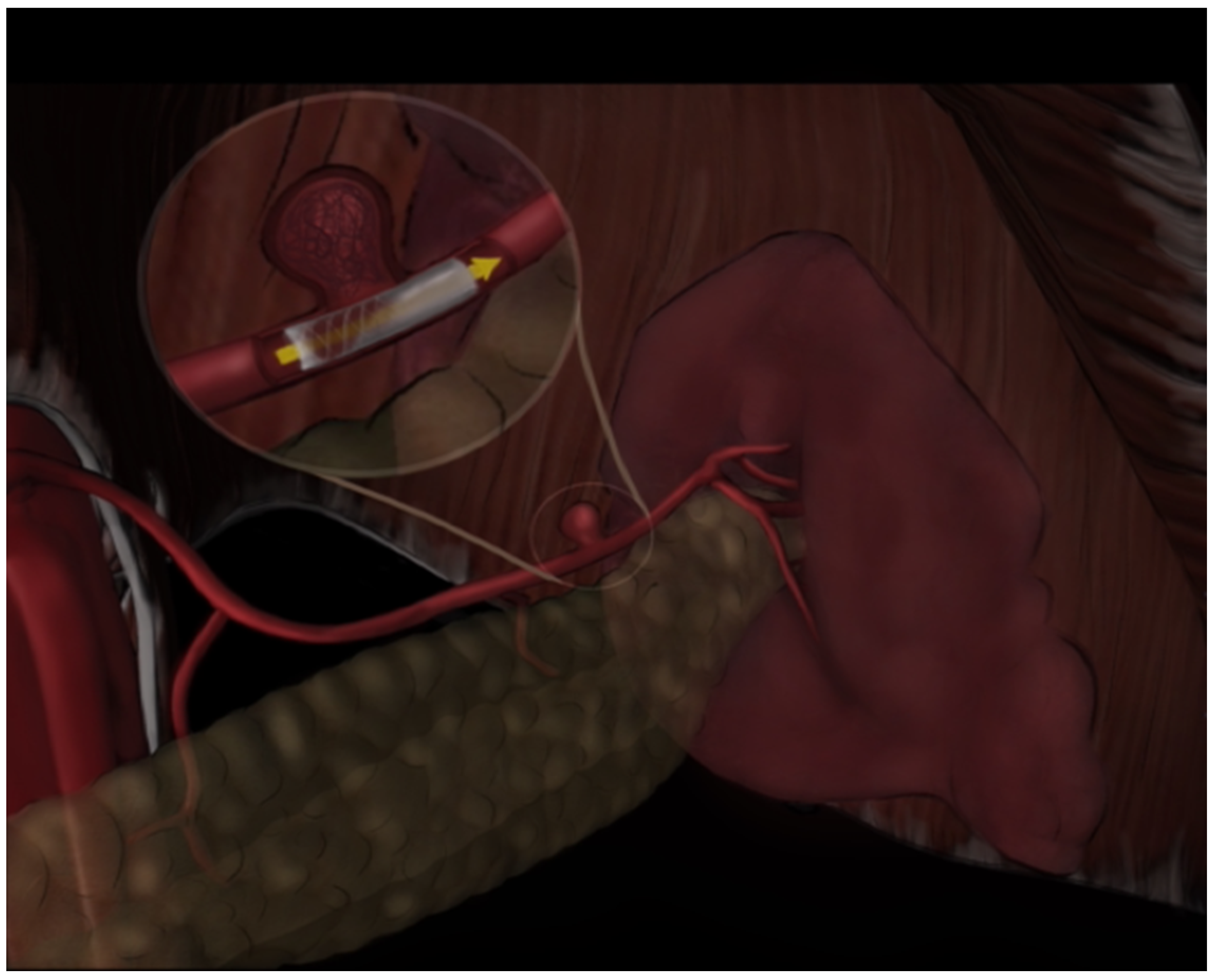
2.3. Interventional Procedure

The choice of endovascular treatment was determined by the operator based on imaging findings, expediency, hemodynamic status, lesion morphology, systemic or local comorbidities, and anatomical details. The choice between SE and BE stents was based not only on vessel tortuosity but also on the operator’s assessment of the need for precision placement versus flexibility, historical performance data of the stent types in similar anatomical settings, and the patient’s previous interventions. Covered stents (CSs) were the first-line therapy, selected based on the probability of reaching the affected artery with the introducer sheath or guiding catheter, the distance between the lesion and the aorta, and the tortuosity of the affected artery. All procedures were performed under moderate sedation in an angiographic suite by an interventional radiology attending, assisted by a fellow physician, using digital subtraction angiography (DSA). Angiograms were conducted via a trans-femoral approach. After cannulating the parent artery and characterizing the VAA angiographically in multiprojectional and magnified views, the operator made the final treatment choice. Typically, self-expandable stent grafts (SE SGs) were considered for more tortuous vessels, while balloon-expandable stent grafts (BE SGs) were preferred when precise deployment was the priority. Wire access was obtained beyond the planned treatment area. For CS placement, a guiding catheter or vascular sheath was advanced into the parent vessel giving rise to the VAA, and the CS was deployed. To ensure proper sealing post-deployment, angioplasty was performed using appropriately sized balloons. In cases with side branches or bifurcations, prophylactic embolization with vascular coils or plugs was performed at the operator’s discretion to prevent endoleaks. Prophylactic embolization was performed in 12 out of the 23 patients to secure side branches at risk of endoleak. Completion angiography was conducted to confirm the appropriate exclusion of the VAA and continued flow in the parent and desired distal vessel. Post-procedure, patients were administered dual antiplatelet therapy comprising aspirin (100 mg daily) and clopidogrel (75 mg daily) for six months to ensure stent patency.

2.4. Imaging Techniques and Data Collection

Extensive pre-interventional imaging was undertaken to measure vessel angles and other critical anatomical features (Figure 1). This data informed procedural planning and stent selection. Comprehensive data collection included aneurysm characteristics, procedural details, post-treatment outcomes such as morbidity, mortality, reintervention rates, complications, and the etiology of the aneurysms (Table 1).

2.5. Outcome Measures and Definitions

Technical success was defined as complete angiographic exclusion of the aneurysm without any detectable endoleak post-procedure. Clinical and imaging success included criteria such as aneurysm shrinkage or stabilization, sustained patency of the stent and the parent vessel, absence of organ ischemia, and resolution of symptoms without subsequent aneurysm-related mortality. Primary patency and aneurysm exclusion were verified through follow-up imaging such as computed tomography angiography (CTA) or DSA, ensuring no residual perfusion within the aneurysm sac.

2.6. Statistical Analysis

Continuous variables were presented as mean ± standard deviation, and categorical variables as percentages. Long-term outcomes, such as stent-graft patency and re-intervention-free survival, were analyzed using Kaplan–Meier methods with the statistical software IBM SPSS Statistics (Version 20, IBM, Armonk, NY, USA). Associations between qualitative variables were evaluated using Fisher’s exact test, and relationships between quantitative variables were examined using Spearman’s correlation test. A p-value of less than 0.05 was considered statistically significant.

3. Results

3.1. Patient Characteristics

Our study included 23 patients with a mean age of 61 ± 12.2 years, consisting of 13 males and 10 females. The majority, approximately 78%, presented with abdominal pain. Aneurysms were predominantly located in the splenic artery (44%), followed by the hepatic artery (26%) and renal artery (17.5%). The celiac trunk was involved in two cases, and the gastroduodenal and superior mesenteric arteries were used in one case each. Balloon-expandable stent grafts (BE SGs) were used in 15 patients, while self-expandable stent grafts (SE SGs) were chosen for 8 patients based on anatomical considerations and clinical needs. Subgroup analysis based on aneurysm location revealed similar procedural success and complication rates (p > 0.05) across all groups, despite anatomical challenges.

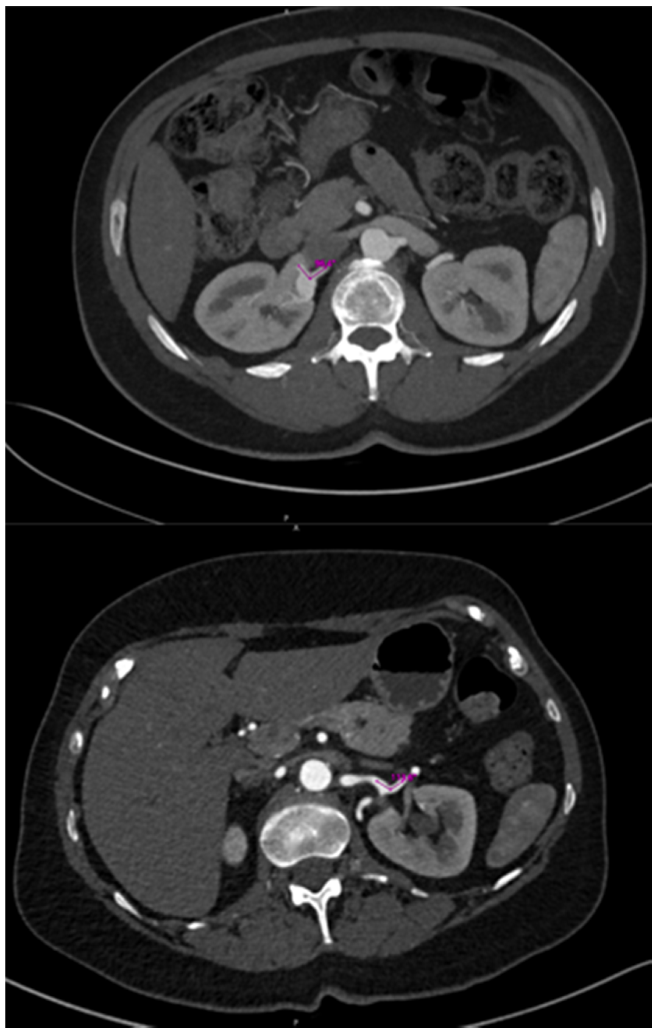
3.2. Imaging Findings

Pre-intervention imaging showed a mean neck size of 10.10 ± 8.70 mm in the BE group versus 18.50 ± 3.40 mm in the SE group, with no statistical significance (p = 0.23). The mean aneurysm sac size was 20.10 ± 18.9 mm for the BE group and 15.60 ± 12.7 mm for the SE group, with the sac-to-neck ratio also showing no significant difference (1.69 ± 2.23 vs. 1.38 ± 0.33, p = 0.63).

3.3. Clinical Outcomes and Follow-Up

During the follow-up period, stent-graft patency and aneurysm exclusion were evaluated in all patients, although the length of follow-up varied due to the availability of imaging data. Imaging during the follow-up was available for 18 patients at 6 months, 12 patients at 12 months, and 7 patients at 36 months, with a mean follow-up time of 28 months (range, 0–111 months).
Out of the 23 patients, 21 underwent follow-up after the procedure. Imaging assessments provided information on aneurysm exclusion and stent-graft patency, with a mean imaging follow-up time of 31 months for these 21 patients. Seven patients required re-intervention due to endoleak or occluded stent-grafts identified on the follow-up imaging. Clinical and imaging success at 30 days post-procedure was achieved in 11 patients.
When evaluating outcomes based on the type of stent used, both self-expandable (SE) and balloon-expandable (BE) stents achieved a 100% technical success rate. However, the BE group exhibited a significantly higher endoleak rate compared to the SE group. Despite this difference, other complications and overall procedural success rates showed no significant differences between the stent types (p > 0.05). Endoleak complications were observed in ten patients, with the BE group experiencing a significantly higher rate of endoleaks at 60%, compared to 12.5% in the SE group (p = 0.03). Specifically, the BE group had five Type IA endoleaks, three Type IB, one Type II, and one Type III, whereas the SE group had only a single Type II endoleak. Initial patency rates were 80% for BE stents and 100% for SE stents after 30 days of the procedure (p = 0.52).

3.4. Long-Term Patency and Survival

A regression survival model with repeated measures (recurrent survival analysis) demonstrated no statistically significant difference in the time until the occurrence of adverse events between patients receiving BE SGs and those receiving SE SGs, at the 95% confidence level (p = 1) (Table 2). Kaplan–Meier curves for stent-graft patency and reintervention-free survival also indicated no significant differences across five separate follow-up periods (Figure 2). The mean efferent-to-afferent vessel angle was 63.29 ± 46.34 degrees in the SE group and 65.00 ± 40.89 degrees in the BE group. A two-sample independent t-test revealed no statistically significant difference between the mean angles in the two groups at the 95% confidence level (p = 0.907) (Figure 3). The Spearman correlation test, used to examine the relationship between vessel angles and the presence of endoleak, resulted in a correlation coefficient of 0.11, indicating no statistically significant relationship between the two variables (p = 0.627) at the 95% confidence level (Table 3, Figure 4, Figure 5, Figure 6 and Figure 7).

4. Discussion

The treatment paradigm for visceral artery aneurysms (VAAs) has shifted from traditional surgical approaches to endovascular techniques, with self-expandable (SE) and balloon-expandable (BE) stent grafts (SGs) being prominent options. Endovascular repair with SE or BE SGs allows for aneurysm exclusion while preserving the parent vessel and distal perfusion, theoretically reducing the risk of downstream organ ischemia [11,18,20,21,22]. This approach significantly improves outcomes by decreasing recovery time, hospital stay [1], perioperative morbidity, and 30-day mortality [17,23]. Currently, there is no consensus on the treatment indications for VAAs [11], and image-based monitoring is often recommended for the initial management of small aneurysms, particularly those less than 25 mm in diameter [24].
While endovascular intervention is effective for both symptomatic and asymptomatic patients, asymptomatic lesions have shown lower 30-day mortality compared to symptomatic lesions [25]. This difference may be due to the more severe presentation in symptomatic patients, often with already ruptured aneurysms, compared to asymptomatic patients with intact or smaller aneurysms. Although coil embolization (CS) has shown effectiveness in the VAA treatment, with technical success rates ranging from 84.2% to 98.3% [11,18,20,22], comparative data between SE and BE SGs are limited. Our comparative analysis of SE and BE SGs in managing VAAs has elucidated critical differences in clinical and technical performance. Notably, the lower incidence of endoleaks with the SE stents (12.5%) compared to the BE stents (60%) is significant (p = 0.03). This outcome aligns with existing literature, emphasizing the superior adaptability of SE stents to complex vascular architectures, largely due to the flexible properties of nitinol, which enable better conformity to vessel walls [11,18,20,21,22,23,24].
Despite achieving a 100% technical success rate for both stent types over the follow-up period, the higher prevalence of early complications, primarily Type I and Type III endoleaks, in the BE group points to potential issues with their rigidity, especially in anatomically challenging scenarios. The rigidity of BE stents, while beneficial for precise deployment, appears less effective in irregular or highly tortuous vessels [24,25]. This suggests that technical success at deployment does not necessarily equate to optimal clinical outcomes, highlighting a critical area for improvement [26,27,28,29,30,31,32,33,34]. Moreover, our findings did not reveal significant differences in the sac-to-neck ratios between groups, indicating that the geometric compatibility of the stents alone does not reliably predict success in preventing endoleaks. Instead, the material and mechanical properties of the stents emerge as pivotal factors influencing clinical outcomes.
Long-term outcomes, assessed through survival analysis and Kaplan–Meier curves, showed no significant differences in stent-graft patency or reintervention rates between the groups. This suggests that both stent types may stabilize within the vascular system once initial complications are managed, implying their comparable efficacy over the long term. Several anatomical considerations are crucial for ensuring technical success and long-term patency. These include neck length, aneurysm location, and the angulation of the aneurysmal tract, particularly for saccular aneurysms, which were most common in this cohort [2,35]. The tortuosity of the involved arteries plays a significant role and may explain the differences in endoleak complications between BE and SE SG procedures [16,36]. SE SGs, due to their shape memory, are more flexible and conform better to the tortuous visceral arteries. In contrast, BE SGs are relatively rigid, forcing the artery to adapt to the stent’s shape, potentially increasing endoleak rates [22]. Therefore, BE SGs should be considered primarily for VAAs located in short, straight arterial segments or proximal vessels near the aorta.
Another risk factor for the endoleak is the length of the landing zone [37,38]. Adequate proximal and distal landing zones are necessary for proper sealing and aneurysm exclusion [39]. During balloon inflation, the BE SGs and vessels are straightened, but post-deflation the vessel’s tortuosity can lead to decreased seal and increased Type I endoleaks [40]. While a landing zone of 5 mm is generally effective in atherosclerotic lesions, BE SGs require longer zones. Stent oversizing, typically 15–20%, can enhance sealing but must be approached with caution to avoid stent graft folding and subsequent endoleaks [28,41,42,43].
While vessel tortuosity significantly impacts stent deployment and occurrence of the endoleaks, vessel angle remains an important consideration in procedural planning and stent selection. Interestingly, our study found no significant correlation between pre-procedural vessel angles and the occurrence of endoleaks, challenging traditional views that prioritize anatomical considerations in the stent selection [5]. This underscores the need for a more nuanced approach that also considers stent characteristics and deployment strategies during procedural planning. The placement of CS in visceral arteries is also technically challenging due to the severe tortuosity and small caliber of these arteries [9,44]. Utilizing stable carrier systems at the artery origin, hydrophilic guidewires for engaging efferent vessels, or stiff guidewires can facilitate SG advancement during the procedure [22]. This nuanced evaluation underscores the complexity of vascular anatomy and guides the optimization of endovascular treatments for achieving successful outcomes in clinical practice.
Considering the evolving landscape of VAA management, it is pertinent to acknowledge emerging techniques such as flow diversion stents, which offer an alternative strategy and are particularly valuable when preserving branch vessel patency is critical. Flow diversion techniques redirect blood flow away from the aneurysm sac while maintaining the perfusion of vital branches, reflecting evolving strategies that aim to balance effective aneurysm exclusion with minimal disruption to vascular anatomy. Furthermore, discussion of the open surgical approaches remains relevant, especially in cases involving complex or hilar aneurysms where endovascular techniques may pose technical limitations. Surgical intervention continues to play a pivotal role in scenarios requiring direct vessel repair or when endovascular options prove insufficient or impractical. Our study has limitations, including its retrospective design and the small sample size, which may affect the generalizability of the findings. Additionally, being confined to a single institution might introduce biases related to specific interventional practices and patient selection. Future research should expand to multicenter studies to enhance the validity of the results across diverse settings. Also, the absence of long-term follow-up for all participants and a non-randomized design limits our ability to definitively assert the superiority of one stent type over another. Prospective, randomized trials are needed to further delineate the relative benefits and risks of SE and BE stents in VAA management.

5. Conclusions

In conclusion, while both SE and BE stents demonstrate high technical success rates, SE stents may offer better clinical outcomes in the management of VAAs, particularly in cases involving complex vascular anatomy. The choice of stent types should be carefully considered, with attention to both the anatomical challenges and the specific properties of the stents. Continued research, especially studies with larger and more diverse populations, is crucial to further elucidate these findings and optimize the treatment strategies for VAA patients.

Author Contributions

Conceptualization, A.M. and R.T.; Methodology, J.L.; Validation, A.S.; Resources, P.S.; Writing—original draft, P.T.; Writing—review & editing, S.M.F., M.R. and J.G. All authors have read and agreed to the published version of the manuscript.

Funding

This research received no external funding.

Institutional Review Board Statement

Ethical approval and IRB approval were obtained. The study was conducted in accordance with the Declaration of Helsinki, and approved by the Institutional Review Board (or Ethics Committee) of University of Minnesota, protocol code STUDY0013971.

Informed Consent Statement

Informed consent was obtained from all subjects involved in the study.

Data Availability Statement

The original contributions presented in the study are included in the article, further inquiries can be directed to the corresponding author.

Conflicts of Interest

The authors declare no conflicts of interest.

References

  1. Barrionuevo, P.; Malas, M.B.; Nejim, B.; Haddad, A.; Morrow, A.; Ponce, O.; Hasan, B.; Seisa, M.; Chaer, R.; Murad, M.H. A systematic review and meta-analysis of the management of visceral artery aneurysms. J. Vasc. Surg. 2020, 72 (Suppl. S1), 40S–45S. [Google Scholar] [CrossRef] [PubMed]
  2. Obara, H.; Kentaro, M.; Inoue, M.; Kitagawa, Y. Current management strategies for visceral artery aneurysms: An overview. Surg. Today 2020, 50, 38–49. [Google Scholar] [CrossRef] [PubMed]
  3. Hosn, M.A.; Xu, J.; Sharafuddin, M.; Corson, J.D. Visceral Artery Aneurysms: Decision Making and Treatment Options in the New Era of Minimally Invasive and Endovascular Surgery. Int. J. Angiol. 2019, 28, 11–16. [Google Scholar] [CrossRef] [PubMed]
  4. Bedford, P.D.; Lodge, B. Aneurysm of the Splenic Artery. Gut 1960, 1, 312–320. [Google Scholar] [CrossRef] [PubMed]
  5. Spiliopoulos, S.; Sabharwal, T.; Karnabatidis, D.; Brountzos, E.; Katsanos, K.; Krokidis, M.; Gkoutzios, P.; Siablis, D.; Adam, A. Endovascular Treatment of Visceral Aneurysms and Pseudoaneurysms: Long-term Outcomes from a Multicenter European Study. Cardiovasc. Intervent. Radiol. 2012, 35, 1315–1325. [Google Scholar] [CrossRef] [PubMed]
  6. Tulsyan, N.; Kashyap, V.S.; Greenberg, R.K.; Sarac, T.P.; Clair, D.G.; Pierce, G.; Ouriel, K. The endovascular management of visceral artery aneurysms and pseudoaneurysms. J. Vasc. Surg. 2007, 45, 276–283. [Google Scholar] [CrossRef] [PubMed]
  7. Sahajwani, S.; Tolaymat, B.; Khalifeh, A.; Hosseini, M.; Santini-Dominguez, R.; Blitzer, D.; Sarkar, R.; Toursavadkohi, S. Presentation and management of rare saccular superior mesenteric artery trunk and branch aneurysms. J. Vasc. Surg. Cases Innov. Tech. 2022, 8, 281–286. [Google Scholar] [CrossRef] [PubMed]
  8. Meyer, A.; Uder, M.; Lang, W.; Croner, R. Visceral artery aneurysms. Zentralbl. Chir. 2010, 135, 416–420. [Google Scholar] [CrossRef] [PubMed]
  9. Wang, L.; Shu, C.; Li, Q.; Jiang, X.; Li, X.; He, H.; Li, M. Experience of managing superior mesenteric artery aneurysm and its midterm follow-up results with 18 cases. Vascular 2021, 29, 516–526. [Google Scholar] [CrossRef]
  10. Kueper, M.A.; Ludescher, B.; Koenigsrainer, I.; Kirschniak, A.; Mueller, K.; Wiskirchen, J.; Koenigsrainer, A. Successful Coil Embolization of a Ruptured Gastroduodenal Artery Aneurysm. Vasc. Endovascular. Surg. 2008, 41, 568–571. [Google Scholar] [CrossRef]
  11. Martinelli, O.; Giglio, A.; Irace, L.; Di Girolamo, A.; Gossetti, B.; Gattuso, R. Single-Center Experience in the Treatment of Visceral Artery Aneurysms. Ann. Vasc. Surg. 2019, 60, 447–454. [Google Scholar] [CrossRef] [PubMed]
  12. Juntermanns, B.; Bernheim, J.; Karaindros, K.; Walensi, M.; Hoffmann, J.N. Visceral artery aneurysms. Gefasschirurgie 2018, 23 (Suppl. S1), 19–22. [Google Scholar] [CrossRef] [PubMed]
  13. Coleman, D.M.; Stanley, J.C. Renal artery aneurysms. J. Vasc. Surg. 2015, 62, 779–785. [Google Scholar] [CrossRef] [PubMed]
  14. Abbas, M.A.; Stone, W.M.; Fowl, R.J.; Gloviczki, P.; Oldenburg, W.A.; Pairolero, P.C.; Hallett, J.W.; Bower, T.C.; Panneton, J.M.; Cherry, K.J. Splenic Artery Aneurysms: Two Decades Experience at Mayo Clinic. Ann. Vasc. Surg. 2002, 16, 442–449. [Google Scholar] [CrossRef] [PubMed]
  15. Chaer, R.A.; Abularrage, C.J.; Coleman, D.M.; Eslami, M.H.; Kashyap, V.S.; Rockman, C.; Murad, M.H. The Society for Vascular Surgery clinical practice guidelines on the management of visceral aneurysms. J. Vasc. Surg. 2020, 72 (Suppl. S1), 3S–39S. [Google Scholar] [CrossRef] [PubMed]
  16. Mauri, G.; Sconfienza, L.M.; Casilli, F.; Massaro, S.; Inglese, L. Cardiatis Multilayer Stent for Endovascular Treatment of Peripheral and Visceral Aneurysms: Where Do We Stand? J. Endovasc. Ther. 2013, 20, 575–577. [Google Scholar] [CrossRef] [PubMed]
  17. Zhang, Y.; Xiang, D.; Lu, Q.; Wu, M.; Cui, J. A systematic review and meta-analysis of the performance of flow-diverting stents in the treatment of peripheral and visceral artery aneurysms. Catheter. Cardiovasc. Interv. 2021, 97, 461–469. [Google Scholar] [CrossRef] [PubMed]
  18. Künzle, S.; Glenck, M.; Puippe, G.; Schadde, E.; Mayer, D.; Pfammatter, T. Stent-Graft Repairs of Visceral and Renal Artery Aneurysms Are Effective and Result in Long-term Patency. J. Vasc. Interv. Radiol. 2013, 24, 989–996. [Google Scholar] [CrossRef] [PubMed]
  19. Lee, P.C.; Rhee, R.Y.; Gordon, R.Y.; Fung, J.J.; Webster, M.W. Management of splenic artery aneurysms: The significance of portal and essential hypertension11No competing interests declared. J. Am. Coll. Surg. 1999, 189, 483–490. [Google Scholar] [CrossRef] [PubMed]
  20. Venturini, M.; Marra, P.; Colombo, M.; Panzeri, M.; Gusmini, S.; Sallemi, C.; Salvioni, M.; Lanza, C.; Agostini, G.; Balzano, G.; et al. Endovascular Repair of 40 Visceral Artery Aneurysms and Pseudoaneurysms with the Viabahn Stent-Graft: Technical Aspects, Clinical Outcome and Mid-Term Patency. Cardiovasc. Interv. Radiol. 2018, 41, 385–397. [Google Scholar] [CrossRef]
  21. You, Y.; Choi, S.H.; Choi, D.W.; Heo, J.S.; Han, I.W.; Han, S.; Shin, S.W.; Park, K.B.; Park, H.S.; Cho, S.K.; et al. Long-term clinical outcomes after endovascular management of ruptured pseudoaneurysm in patients undergoing pancreaticoduodenectomy. Ann. Surg. Treat. Res. 2019, 96, 237–249. [Google Scholar] [CrossRef]
  22. Venturini, M.; Marra, P.; Colombo, M.; Alparone, M.; Agostini, G.; Bertoglio, L.; Sallemi, C.; Salvioni, M.; Gusmini, S.; Balzano, G.; et al. Endovascular Treatment of Visceral Artery Aneurysms and Pseudoaneurysms in 100 Patients: Covered Stenting vs. Transcatheter Embolization. J. Endovasc. Ther. 2017, 24, 709–717. [Google Scholar] [CrossRef] [PubMed]
  23. Song, C.; Dong, J.; Yu, G.; Zhou, J.; Xiang, F.; Pei, Y.; Lu, Q.; Jing, Z. Comparison of open surgery and endovascular procedures as a therapeutic choice for visceral artery aneurysms. Vascular 2018, 26, 387–392. [Google Scholar] [CrossRef] [PubMed]
  24. Corey, M.R.; Ergul, E.A.; Cambria, R.P.; English, S.J.; Patel, V.I.; Lancaster, R.T.; Kwolek, C.J.; Conrad, M.F. The natural history of splanchnic artery aneurysms and outcomes after operative intervention. J. Vasc. Surg. 2016, 63, 949–957. [Google Scholar] [CrossRef] [PubMed]
  25. Guo, B.; Guo, D.; Xu, X.; Chen, B.; Shi, Z.; Luo, J.; Jiang, J.; Fu, W. Early and intermediate results of endovascular treatment of symptomatic and asymptomatic visceral artery aneurysms. J. Vasc. Surg. 2016, 64, 140–148. [Google Scholar] [CrossRef]
  26. Grande, W.; Stavropoulos, S.W. Treatment of Complications Following Endovascular Repair of Abdominal Aortic Aneurysms. Semin. Interv. Radiol. 2006, 23, 156–164. [Google Scholar] [CrossRef]
  27. Chun, J.Y.; Morgan, R. Transcatheter Embolisation of Type 1 Endoleaks after Endovascular Aortic Aneurysm Repair with Onyx: When No Other Treatment Option is Feasible. Eur. J. Vasc. Endovasc. Surg. 2013, 45, 141–144. [Google Scholar] [CrossRef] [PubMed]
  28. Rehman, Z.U. Endoleaks: Current concepts and treatments. J. Pak. Med. Assoc. 2021, 35, 141–144. [Google Scholar] [CrossRef]
  29. Orgera, G.; Tipaldi, M.A.; Laurino, F.; Lucatelli, P.; Rebonato, A.; Paraskevopoulos, I.; Rossi, M.; Krokidis, M. Techniques and future perspectives for the prevention and treatment of endoleaks after endovascular repair of abdominal aortic aneurysms. Insights Imaging 2019, 10, 91. [Google Scholar] [CrossRef]
  30. Sampram, E.S.K.; Karafa, M.T.; Mascha, E.J.; Clair, D.G.; Greenberg, R.K.; Lyden, S.P.; O’Hara, P.J.; Sarac, T.P.; Srivastava, S.D.; Butler, B.; et al. Nature, frequency, and predictors of secondary procedures after endovascular repair of abdominal aortic aneurysm. J. Vasc. Surg. 2003, 37, 930–937. [Google Scholar] [CrossRef]
  31. Jones, J.E.; Atkins, M.D.; Brewster, D.C.; Chung, T.K.; Kwolek, C.J.; LaMuraglia, G.M.; Hodgman, T.M.; Cambria, R.P. Persistent type 2 endoleak after endovascular repair of abdominal aortic aneurysm is associated with adverse late outcomes. J. Vasc. Surg. 2007, 46, 1–8. [Google Scholar] [CrossRef] [PubMed]
  32. Becquemin, J.P.; Kelley, L.; Zubilewicz, T.; Desgranges, P.; Lapeyre, M.; Kobeiter, H. Outcomes of secondary interventions after abdominal aortic aneurysm endovascular repair. J. Vasc. Surg. 2004, 39, 298–305. [Google Scholar] [CrossRef] [PubMed]
  33. Baum, R.A.; Carpenter, J.P.; Golden, M.A.; Velazquez, O.C.; Clark, T.W.; Stavropoulous, S.; Cope, C.; Fairman, R.M. Treatment of type 2 endoleaks after endovascular repair of abdominal aortic aneurysms: Comparison of transarterial and translumbar techniques. J. Vasc. Surg. 2002, 35, 23–29. [Google Scholar] [CrossRef] [PubMed]
  34. Görich, J.; Rilinger, N.; Sokiranski, R.; Krämer, S.C.; Ermis, C.; Schütz, A.; Brambs, H.-J.; Söldner, J.; Kaiser, W.; Sunder-Plassmann, L.; et al. Treatment of Leaks after Endovascular Repair of Aortic Aneurysms1. Radiology 2000, 215, 414–420. [Google Scholar] [CrossRef] [PubMed]
  35. Ibrahim, F.; Dunn, J.; Rundback, J.; Pellerito, J.; Galmer, A. Visceral Artery Aneurysms: Diagnosis, Surveillance, and Treatment. Curr. Treat. Options Cardiovasc. Med. 2018, 20, 97. [Google Scholar] [CrossRef] [PubMed]
  36. Hemp, J.H.; Sabri, S.S. Endovascular Management of Visceral Arterial Aneurysms. Tech. Vasc. Interv. Radiol. 2015, 18, 14–23. [Google Scholar] [CrossRef] [PubMed]
  37. Gaffey, A.C.; Damrauer, S.M. Evolving Concepts, Management, and Treatment of Type 1 Endoleaks after Endovascular Aneurysm Repair. Semin. Interv. Radiol. 2020, 37, 395–404. [Google Scholar] [CrossRef] [PubMed]
  38. Risk Factors for Early and Late Type Ib Endoleak Following Endovascular Abdominal Aortic Aneurysm Repair—ClinicalKey. Available online: https://www-clinicalkey-com.ezp3.lib.umn.edu/#!/content/playContent/1-s2.0-S089050962030830X?returnurl=null&referrer=null (accessed on 22 July 2022).
  39. Fankhauser, G.T.; Stone, W.M.; Naidu, S.G.; Oderich, G.S.; Ricotta, J.J.; Bjarnason, H.; Money, S.R. The minimally invasive management of visceral artery aneurysms and pseudoaneurysms. J. Vasc. Surg. 2011, 53, 966–970. [Google Scholar] [CrossRef] [PubMed]
  40. Tenorio, E.R.; Kärkkäinen, J.M.; Mendes, B.C.; DeMartino, R.R.; Macedo, T.A.; Diderrich, A.; Hofer, J.; Oderich, G.S. Outcomes of directional branches using self-expandable or balloon-expandable stent grafts during endovascular repair of thoracoabdominal aortic aneurysms. J. Vasc. Surg. 2020, 71, 1489–1502.e6. [Google Scholar] [CrossRef]
  41. Gargiulo, M.; Gallitto, E.; Wattez, H.; Verzini, F.; Massoni, C.B.; Loschi, D.; Freyrie, A.; Haulon, S. Outcomes of endovascular aneurysm repair performed in abdominal aortic aneurysms with large infrarenal necks. J. Vasc. Surg. 2017, 66, 1065–1072. [Google Scholar] [CrossRef]
  42. Mohan, I.V.; Laheij, R.J.F.; Harris, P.L. Risk Factors for Endoleak and the Evidence for Stent-graft Oversizing in Patients Undergoing Endovascular Aneurysm Repair. Eur. J. Vasc. Endovasc. Surg. 2001, 21, 344–349. [Google Scholar] [CrossRef] [PubMed]
  43. van Prehn, J.; Schlösser, F.J.V.; Muhs, B.E.; Verhagen, H.J.M.; Moll, F.L.; van Herwaarden, J.A. Oversizing of Aortic Stent Grafts for Abdominal Aneurysm Repair: A Systematic Review of the Benefits and Risks. Eur. J. Vasc. Endovasc. Surg. 2009, 38, 42–53. [Google Scholar] [CrossRef] [PubMed]
  44. Qiu, C.; Liu, Z.; Huang, L.; Guo, L.; Lu, W.; Zhang, H.; He, Y.; Tian, L.; Li, D.; Wang, X.; et al. Covered Stents for Treatment of Visceral Artery Aneurysms: A Multicenter Study. J. Vasc. Interv. Radiol. 2022, 33, 640–647. [Google Scholar] [CrossRef] [PubMed]
Figure 1. The efferent-to-afferent angle was measured in the axial plane on the most recent cross-sectional imaging prior to intervention.
Diagnostics 14 01695 g001
Figure 2. Kaplan–Meier plots comparing the survival rates between the two groups (AE).
Diagnostics 14 01695 g002aDiagnostics 14 01695 g002b
Figure 3. Bar plot comparing the angles between the two groups.
Diagnostics 14 01695 g003
Figure 4. Scatter plot assessing the relationship between angles and endoleak.
Diagnostics 14 01695 g004
Figure 5. A schematic showing an endoleak classification system. Types of endoleaks are classified according to established criteria: Type Ia represents leakage at the proximal stent graft attachment site, Type Ib at the distal attachment site, Type II results from retrograde flow into the aneurysm sac via collateral vessels, Type III is due to stent graft fabric porosity or structural failure, and Type IV is associated with stent graft material degradation.
Diagnostics 14 01695 g005
Figure 6. A schematic showing occurrence of endoleaks, one of the most common complications in endovascular procedures, resulting in increased morbidity, mortality, and the need for re-intervention.
Diagnostics 14 01695 g006
Figure 7. A 57-year-old woman with a significant medical history including gastroparesis, eosinophilic esophagitis with prior dilation in 2020, type 2 diabetes mellitus, multiple sclerosis, anxiety, postoperative nausea and vomiting triggered by narcotics, and multiple medication allergies. She sought evaluation due to an 18 mm right renal aneurysm discovered during a recent CT scan prompted by right flank pain. After consultation, she opted to undergo the recommended surgical intervention for the aneurysm. The computed tomographic angiography of the abdomen and pelvis, both without and with contrast, revealed a saccular aneurysm located at the bifurcation of the right renal artery. The neck size of the aneurysm measures 14 mm, with a maximal diameter of approximately 1.8 cm in the axial plane. After the procedure, follow-up imaging revealed a new stent extending along most of the right renal artery, spanning across an aneurysm located at the renal hilum, which is positioned at a trifurcation. The stent is patent, and the aneurysm remains perfused, measuring approximately 1.7 × 1.5 cm, unchanged from the previous examination (AC).
Diagnostics 14 01695 g007
Table 1. Characteristics of included patients.
Patient NoAgeStent TypeGenderLocationSize mmType of AneurysmDOPStent SizeEndoleakTechnical SuccessClinical SuccessPatent at Latest FU
153BEFRAA17.3S1/6/2021, 3/1/20216 mm × 39 mm Gore VBX and
6 mm × 19 mm Gore VBX
Endoleak Type IBNoNoYes
268BEFRAA28.7S9/8/205 mm × 22 mm Atrium iCastNoYesYesYes
364BEMHAA48.7S2/20/207 mm × 59 mm Gore VBXNoYesYesYes
463BEFRAA14.8S10/18/195 × 22 mm Atrium iCastNoYesYesYes
554BEFHAA19F8/2/2018, 8/15/2018, 11/1/20186 × 39 mm Gore VBXEndoleak Type IANoNoNo
659BEMSAA20S8/9/178 × 59 mm Gore VBXNoYesYesYes
762BEMSAA17S7/7/2017, 1/18/20186 × 39 mm Gore VBX and
8 × 39 mm Gore VBX
Endoleak Type IBNoNoYes
867BEMGDA77.5F12/17/2016, 1/25/2017, 1/26/2017Procedure 1: 36 × 7 Atrium iCast
Procedure 2: 7 mm × 22 Atrium iCast and 7 × 38 Atrium iCast
Procedure 3: 8 mm × 50 Viabahn Stent graft and 9 mm × 38 mm Atrium iCast
Endoleak Type IIINoNoYes
952BEFHAA16S2/6/137 mm × 22 mm Atrium iCast and
6 mm × 22 mm Atrium iCast and
8 mm × 25 mm Viabahn stent graft
Endoleak Type I ANoNoYes
1071BEFRAA19.5S4/13/2011, 5/29/2020Procedure 1: 5 × 22 Atrium iCast Procedure 2: 6 × 19 mm Gore VBXEndoleak Type IANoNoYes
1179BEMCeliac AA33.8S2/19/097 mm × 38 cm Atrium iCast and
8 mm × 38 mm Atrium iCast
NoYesYesYes
1270BEFCeliac AA36F4/26/079 mm × 59 mm Atrium iCastEndoleak Type IBNoNoYes
1364BEMSAA18.6S4/27/167 × 22 Atrium iCast (×2)Endoleak Type IANoNoUnclear (limited follow up; splenic artery subsequetly extensively embolized)
1435BEMSMA26.1S5/5/2017, 5/8/2017Procedure 1: 7 × 19 mm and
6 × 19 mm Gore VBX
Procedure 2: 8 × 50 mm Gore VBX
Endoleak Type IANoNoNo
1559BEMSAA20.7S8/9/178 mm × 59 mm Gore VBX.Repeat (No endoleak)NoNoYes
1630SEFSAA20.4F6/4/186 mm × 5 cm Viabahn stent graftEndoleak Type II (resolved)NoNoYes
1770SEMSAA21.1S5/4/188 × 50 mm Viabahn stent graftNoYesYesYes
1881SEFHAA18.3F10/26/165 mm × 4 cm Viabahn stent graft and
5 mm × 5 cm Viabahn stent graft
NoYesYesYes
1966SEMHAA16.2S1/1/2016, 1/14/20166 × 25 Viabahn stent graft and
6 × 50 Viabahn stent graft
NoNoNoYes
2066SEMSAA24S12/13/137 mm × 5 cm Viabahn stent graftNoYesYesYes
2169SEMSAA18.8F2/14/136 mm × 5 cm Viabahn stent graftNoYesYesYes
2258SEFSAA35.5F6/5/085 mm × 5 cm Viabahn stent graftNoYesYesYes
2347SEMHAA?F9/16/066 × 40 Viabahn stent graftNoYesYesYes
Abbreviations: DOP, date of procedure; HAA, hepatic artery; RAA, renal artery; SAA, splenic artery; SMA, superior mesenteric artery.
Table 2. Comparison of mean angles in SE and BE groups using two-sample independent t-test.
GroupnMeanStd. Deviationp-Value
AnglesBE1565.6640.890.907
SE763.2946.34
In the table, results of two-sample independent t-test showed that the difference between the mean angles in the two groups was not significant (p-value > 0.05).
Table 3. Relationship between angles and endoleak using Spearman test.
Endoleak
Anglesn22
Correlation Coefficient0.110
p-value0.627
The results of correlation coefficient test showed that there was no statistically significant relationship between angles and endoleak at the 95% confidence level (p-value = 0.627).
Disclaimer/Publisher’s Note: The statements, opinions and data contained in all publications are solely those of the individual author(s) and contributor(s) and not of MDPI and/or the editor(s). MDPI and/or the editor(s) disclaim responsibility for any injury to people or property resulting from any ideas, methods, instructions or products referred to in the content.

