Article

Ultrasound Assessment of Sarcopenia in Alcoholic Liver Disease

by
Vlad-Teodor Enciu
1,2,*,
Priscila Madalina Ologeanu
1,2 and
Carmen Fierbinteanu-Braticevici
1,2
1
Internal Medicine II and Gastroenterology Department, Emergency University Hospital Bucharest, Carol Davila University of Medicine and Pharmacy, 050474 Bucharest, Romania
2
Emergency University Hospital, 050098 Bucharest, Romania
*
Author to whom correspondence should be addressed.
Diagnostics 2024, 14(17), 1891; https://doi.org/10.3390/diagnostics14171891
Submission received: 22 July 2024 / Revised: 23 August 2024 / Accepted: 26 August 2024 / Published: 28 August 2024
(This article belongs to the Section Medical Imaging and Theranostics)

Abstract

:
Malnutrition frequently affects patients with alcoholic liver disease (ALD), with important impacts on disease prognosis. Sarcopenia, the clinical phenotype of malnutrition characterized by skeletal muscle loss, is the major component responsible for adverse events in this population. The aim of this study is to assess the use of ultrasound (US) skeletal muscle performance in stratifying ALD disease severity. We recruited 43 patients with ALD and divided them into two groups: alcoholic hepatitis (AH) and alcoholic cirrhosis (AC). We evaluated disease-specific clinical and biological parameters and their relation to US Rectus Femoris muscle (RFM) measurements, including RFM thickness, stiffness (RFMS) and echogenicity (RFE). A thirty-seconds chairs stand test (30sCST) was used as the sarcopenia surrogate test. RMF thickness correlated with platelet count and serum albumin (p < 0.001). Both RFM and RFMS correlated with disease severity (p < 0.001) and 30sCST (p < 0.001, p = 0.002). Patients with AH had more severe US muscle abnormalities compared to AC (RFMS 1.78 m/s vs. 1.35 m/s, p = 0.001) and the highest prevalence of RFE (χ2 = 8.652, p = 0.003). Rectus Femoris US assessment could represent a reliable tool in the diagnosis and severity stratification of ALD-induced sarcopenia.

1. Introduction

Malnutrition is one of the most significant comorbidities in alcoholic liver disease (ALD), with an important impact on both morbidity and mortality [1]. However, malnutrition is a general term defined as a disorder of inadequate intake or uptake of nutrients leading to a decrease in total body cell mass. Sarcopenia, the clinical phenotype of malnutrition with prognostic importance in ALD, characterized by skeletal muscle loss, has been proven to be a major component responsible for adverse outcomes in this population category [2]. Both alcohol intake and impaired function of the liver contribute to the development of sarcopenia [3].
Sarcopenia is mainly assessed clinically using a combination of anthropometric measurements and other dynamic tests [4]. However, these tests do not measure skeletal muscle mass directly and are not applicable in patients with decompensated ALD, since most of the clinical parameters are influenced by oedema or ascites [5]. Moreover, dynamic tests such as grip strength cannot differentiate between sarcopenia and frailty, a condition that is either attributed to age-related physiological decline or to disease-induced impaired muscle contraction and weakness [6]. Cross-sectional imaging using computer tomography (CT) scans is considered the gold standard for the precise quantification of sarcopenia, but is irradiating, time-consuming, and expensive to implement as a suitable screening tool [7,8,9]. Thus, other non-invasive, point-of-care methods are analyzed for an accurate assessment of sarcopenia allowing prompt disease stratification and therapeutic intervention [7].
The ultrasound (US) measurement of muscle abnormalities is a promising, readily available and reproducible tool used in assessing sarcopenia [10]. This method has already been validated in geriatric [11] and intensive care unit patients [12]. During a consensus conference, a standardization of US sarcopenia assessment was proposed [13]. Five muscle parameters were illustrated for the ultrasound characterization of sarcopenia, specifically muscle thickness, muscle cross-section area, pennation angle, fascicle length and echogenicity. Microcirculation damage and impaired nitric oxide synthesis have also been proposed as pathophysiological mechanisms of sarcopenia development through alterations of cellular metabolism, mitochondrial abnormalities and ultimately apoptosis. Glucose homeostasis is tightly linked to microcirculation, thus capillary rarefaction may contribute to insulin resistance through reduced glucose uptake [14,15]. This theory could also explain the increased muscular deposits of adipose tissue and the development of myosteatosis [16]. The advancements in US techniques that utilize microvascular imaging and contrast-enhanced ultrasound (CEUS) make muscle microvasculature quantifiable [17]. Skeletal muscle stiffness is another researched parameter analyzed through share-wave elastography (SWE) that was applied in some pilot studies [18,19,20]. The results are controversial, correlating a decrease in muscle stiffness with muscle weakness [18,19], or on the contrary, an increase in muscle stiffness with frailty and disease activity [20].
Taking into account the lack of non-invasive, point-of-care methods for the assessment of sarcopenia, our study sought to investigate the utility of skeletal muscle ultrasound alteration in the diagnosis and stratification of ALD-induced sarcopenia as a novel application.

2. Materials and Methods

2.1. Study Design and Inclusion Criteria

This is a prospective pilot study on patients diagnosed with ALD between December 2023 and May 2024. We included patients with alcoholic hepatitis (AH) and alcoholic cirrhosis (AC), divided into 2 groups (group 1—AH, group 2—AC) and a control group. The control group consisted of healthy patients with no major comorbidities that could influence either muscle functional tests or ultrasound measurements. AH diagnosis was established following the National Institute of Alcohol Abuse and Alcoholism (NIAAA) criteria for probable AH, which includes: history of heavy chronic alcohol consumption (>50–60 g/day for men and >40 g/day for women), recent onset of jaundice (<8 weeks), total serum bilirubin >3 mg/dL and characteristic de Ritis ratio AST:ALT > 1.5, with AST not exceeding 600 U/L. We excluded other hepatic etiologies through screening for viral hepatitis, hemochromatosis and Wilson’s disease, and a thorough assessment of patient’s prescription for known liver toxic drugs. Also, patients with AH and clinical signs of ethanol intoxication, ethylic tremor or hepatic encephalopathy grade ≥ II were excluded as this would influence the functional tests for sarcopenia.
Patients with alcoholic cirrhosis were previously diagnosed based on clinical, biologic and ultrasound modifications. We excluded CHILD C decompensated cirrhosis with large-volume ascites (clinically detectable) or oedema that could influence ultrasound measurements.
Following the recommendations of EWGSOP2, we chose the 30 s chair-stand test (30sCST) as the clinical surrogate muscle functional test for probable sarcopenia. Each patient was asked to perform stand-ups from a folding chair without arms, with the hands crossed and held against their chest. The number of successful and correct repetitions within a timespan of 30 s was recorded. The 30sCST was compared between all groups.
Patients were clinically examined and blood samples were obtained on admission. The control group was assessed during a routine check-up that included clinical examination, blood samples and muscle ultrasound measurements. Weight and height were measured and body mass index (BMI as kg/m2) was calculated. Patients with BMI > 30 kg/m2 were excluded as this might additionally alter the 30sCST outside of its intended use. Patients with fever were screened for infections using chest X-ray, urinary analysis and blood cultures. We performed ascites neutrophil counts in patients with ascites and abdominal pain for suspected spontaneous bacterial peritonitis (SBP). Patients with symptomatic infections and renal impairment were also excluded from the study.

2.2. Ultrasound Assessment of Rectus Femoris

The skeletal muscle ultrasound assessment was conducted following the latest SARCUS recommendations [13]. Ultrasonographic measurements were obtained with a B-mode linear transducer (5.0–7.5 MHz) using Acuson S2000 (Siemens AG, 91052 Erlangen, Germany). Patients were assessed in supine position, after resting for at least 5 min, with the lower limbs extended and knees relaxed. The right Rectus Femoris muscle thickness (RFM) was measured via a transverse scan at the mid-part of the thigh by applying minimal pressure with the transducer. The antero-posterior diameter of the RF was measured as the distance between the superficial and deep fascias. Rectus Femoris echogenicity (RFE) was evaluated qualitatively using gray-scale analysis (Heckmatt scale) in comparison to the surrounding adipose tissue and bone (Figure 1) [21]. Rectus Femoris muscle stiffness (RFMS) was calculated via longitudinal scanning by Acoustic Radiation Force Impulse (ARFI) with five measurements and the results have been expressed as mean shear wave velocity (Vm m/s) and interquartile range (IQR) (Figure 1) [22,23]. The measurements were performed by a hepatologist with more than 20 years of experience in ultrasonography and elastography techniques. US skeletal muscle findings were compared between groups and with 30sCST.

2.3. Statistical Analysis

Microsoft Excel 2021 MSO (Version 2407 Build 16.0.17830.20166, Microsoft Corporation, Redmond, WA, USA) was used to compose the database and a statistical analysis was conducted using SPSS Statistics version 26 (IBM Corporation, Armonk, NY, USA).
Quantitative variables with normal distribution and equal variances were analyzed using t-test and one-way ANOVA. Qualitative variables and those without a normal distribution were assessed using Mann–Whitney U and Chi-Square through crosstabulation. The results have been expressed as mean and standard deviation (SD) for normally distributed variables and as median and IQR for non-normally distributed variables. Association between variables was evaluated with Pearson or Spearman correlation and a p-value < 0.05 was considered statistically significant.
The study was approved by the Local Ethics Committee (36439/16 June 2022) and a detailed informed consent form was signed by each patient.

3. Results

3.1. General Characteristics of Study Population

Forty-three patients with ALD were included in the study, and six control subjects. Twenty-eight patients were included with AH (n = 8, 28% women, n = 20, 72% men) and fifteen with alcoholic cirrhosis (n = 6 Child-Pugh Class A, n = 9 Child–Pugh Class B). More males were present in both groups (n = 20 and n = 10), with no significant difference in age (p = 0.19). Average BMI was similar in both groups (p = 0.44). Only AH patients had either low-volume ascites (n = 12) or grade I hepatic encephalopathy (n = 7). Hepatic failure and inflammation biomarkers were significantly higher in the AH group, as expected. Most of the AH patients had mild forms that did not require corticosteroid treatment (Median MDF = 28.5). Only two patients died at 30 days from a severe form of AH. Group characteristics are illustrated in Table 1.

3.2. Clinical and Biological Results

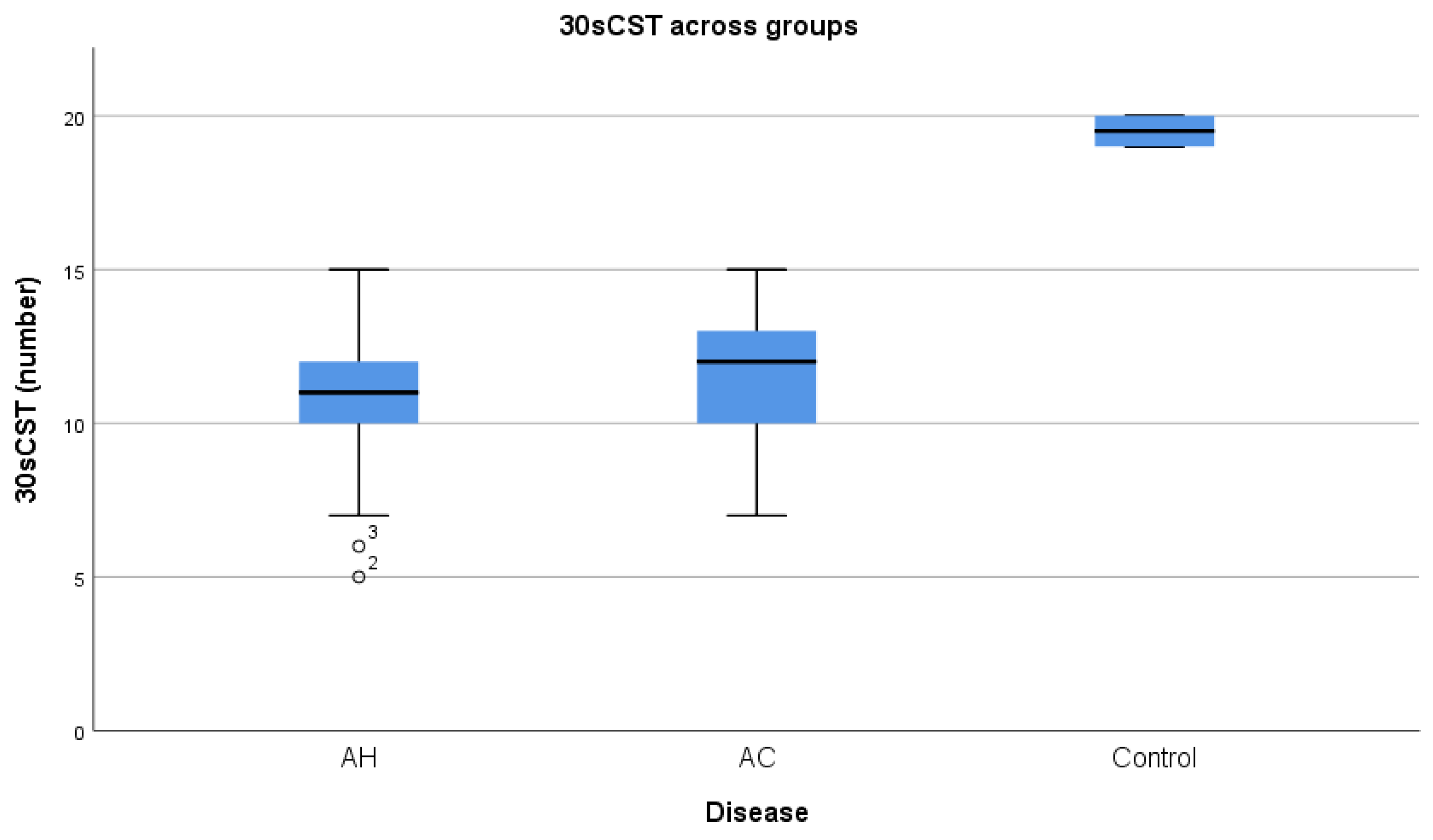
AC performed better than AH at 30sCST (11.47 vs. 10.8) (Figure 2), but without attaining statistical significance (p = 0.38). Additionally, 30sCST was influenced by disease severity (MDF r= −0.616) and parameters of hepatic disfunction (INR r= −0.678, serum albumin r= 0.645). We did not find a correlation between serum creatinine levels and either 30sCST or RF ultrasound measurement.

3.3. Rectus Femoris Ultrasound Measurement Results

Both groups had reduced but similar RFM thicknesses (1.34 cm IQR: 1.12–1.61 vs. 1.24 cm IQR: 1.03–1.65) (Figure 3). Although BMI correlated positively with 30sCST (r = 0.587), we did not find a correlation with RFM (p = 0.09) or RFMS (p = 0.62). Both RFM and RFMS significantly correlated with 30sCST (r = 0.786, r = −0.444), suggesting its utility in the integrated diagnosis of sarcopenia. Compared to control subjects, both AH and AC groups had lower 30sCST (t = −9.51, p < 0.001 and t = −8.58, p < 0.001), with reduced RFM thickness (t = −5.54, p < 0.001 and t = −5.5 p < 0.001). Regarding muscle stiffness, the AC group had lower RFMS (Vm = 1.35 m/s, IQR = 1.22–1.45) than the AH group (Vm = 1.78, IQR = 1.45–2.49), p = 0.001. Interestingly, RFMS was not necessarily corelated with RFM thickness (p = 0.36). Both RMF and RFMS correlated with disease severity in AH (r = −0.483, p = 0.009 and r = 0.558 p = 0.002) (Table 2).
Post-hoc pairwise comparisons using the Bonferroni correction revealed that RFE was the most prevalent in AH (χ2 = 8.652, p = 0.003).

4. Discussion

In this pilot study, we aimed to address the reliability of rectus femoris ultrasound measurements in the diagnosis of ALD-induced sarcopenia. Our results suggest that RF thickness, RFMS and RFE are important non-invasive parameters that correlate with the 30sCST sarcopenia surrogate test and disease severity.
Sarcopenia is a well-known prognostic factor in chronic liver diseases, and recently, novel methods of diagnosis have been developed in order to offer a more feasible diagnostic approach. Although our study did not include a comparison with cross-sectional imaging techniques, ultrasound skeletal muscle measurements have been proven to be a reliable diagnostic alternative to the conventional CT scan gold-standard [5,20,24,25]. These studies validated ultrasound muscle measurements when compared to the CT scan skeletal muscle index (SMI). Moreover, muscle functional tests, specifically the 30sCST, which was used as a sarcopenia surrogate marker in our study, are endorsed by the EWGSOP group as reliable clinical parameters [7]. Our results are similar to those of other studies, wherein decompensated liver states had lower muscle function [26]. As already reported, BMI alone cannot be used as a precise marker of sarcopenia, since the results are influenced by oedema and ascites, hence the need for a more integrated approach. Lastly, although creatinine levels are known to be related to malnutrition and sarcopenia, our study did not find a correlation with muscle functional tests or ultrasound measurement, suggesting that low serum creatinine levels are not reliable for the diagnosis of sarcopenia. Our findings regarding ultrasound skeletal muscle alteration in ALD patients are similar to those of other studies [2,20,24,25,27,28]. We showed that patients with ALD have reduced RFM thickness (1.34 cm and 1.24 cm), indicative of sarcopenia when compared to 30sCST, even in patients with normal BMI. In the AH group, a reduced RFM thickness and a high RFMS indicated disease severity. These findings are consistent with the results found in the study of Becchetti et al. regarding decompensated cirrhosis [20]. Thus, RFM thickness and RFMS could be implemented as severity parameters in alcoholic hepatitis. Interestingly, we did not find a correlation between RFMS and RFM thickness, suggesting that normal muscle fiber composition is influenced by specific pathophysiological mechanisms that can precede muscle mass reduction [29].
On this note, myosteatosis and myofibrosis have been recognized in the literature to induce different degrees of muscle impairment through increased deposits of adipose tissue or extracellular matrix [30,31]. This phenomenon is not necessarily related to obesity and can also be found in patients with cirrhosis, in relation to a chronic inflammatory state [32]. These statements are in concordance with our results regarding RFMS and RFE parameters in AH. Out of the three groups, AH had the highest RFMS and the highest prevalence in RFE, suggesting that this disease category is the most predisposed to either myofibrosis or myosteatosis. Skeletal muscle qualitative modifications in ALD are complex and can be attributed to both disease activity and the toxic effect of alcohol. Alcohol consumption leads to muscle autophagy, decreased proteasome activity and insulin-like growth factor, while disease activity induces cellular and metabolic abnormalities, consisting of differentiations of muscle stem cells into adipocytes, insulin resistance, hyperammonemia, mitochondrial dysfunction and apoptosis [33,34]. Studies including muscle biopsies are warranted in order to further understand qualitative muscle alterations, their ultrasound expression and their relation to disease severity.
Taking into consideration that ALD patients undergo periodic ultrasonography surveillance, ultrasound skeletal muscle measurements could represent an easy, low-cost, readily available strategy to monitor incipient sarcopenic or myosteatotic modifications.
Our study has some limitations. First of all, it has a small sample size, and our protocol did not include CT scans for the comparison of RFMS and RFE with cross-sectional imaging parameters. However, 30sCST is an approved clinical surrogate of sarcopenia, and our intention was to use it specifically, as it targets the muscle group of interest included in our study. Secondly, there is no approved standardized ultrasound skeletal muscle measurement protocol, specifically for muscle stiffness or echogenicity, so the results can be heterogenous. However, we followed the latest SARCUS update recommendations, implementing similar and optimal methods in unison with most ultrasound skeletal muscle studies.
To the best of our knowledge, this is the first study to quantify the muscle stiffness of ALD patients through ARFI.

5. Conclusions

In conclusion, Rectus Femoris ultrasound measurements could be used as a non-invasive diagnostic and prognostic tool in ALD, whether it reflects underlying sarcopenia or myosteatosis. Larger studies and technique standardization are required in order to facilitate the US assessment of ALD-induced muscle changes.

Author Contributions

Conceptualization, V.-T.E. and C.F.-B.; methodology, V.-T.E., P.M.O. and C.F.-B.; investigation, V.-T.E. and C.F.-B.; data curation, V.-T.E. and P.M.O.; writing—original draft preparation, V.-T.E.; writing—review and editing, V.-T.E.; supervision, C.F.-B.; All authors have read and agreed to the published version of the manuscript.

Funding

This research received no external funding.

Institutional Review Board Statement

The study was conducted in accordance with the Declaration of Helsinki, and approved by the Institutional Ethics Committee of Emergency University Hospital Bucharest (protocol code 35439/16.06.2022) for studies involving humans.

Informed Consent Statement

Informed consent was signed by all subjects involved in the study.

Data Availability Statement

The data presented in this study are available on request from the corresponding author. The data are not publicly available due to privacy and ethnical restrictions.

Acknowledgments

Publication of this paper was supported by the University of Medicine and Pharmacy Carol Davila, through the institutional program Publish not Perish.

Conflicts of Interest

The authors declare no conflicts of interest.

References

  1. Tadokoro, T.; Morishita, A.; Himoto, T.; Masaki, T. Nutritional Support for Alcoholic Liver Disease. Nutrients 2023, 15, 1360. [Google Scholar] [CrossRef] [PubMed] [PubMed Central]
  2. Tandon, P.; Low, G.; Mourtzakis, M.; Zenith, L.; Myers, R.P.; Abraldes, J.G.; Shaheen, A.A.M.; Qamar, H.; Mansoor, N.; Carbonneau, M.; et al. A Model to Identify Sarcopenia in Patients with Cirrhosis. Clin. Gastroenterol. Hepatol. 2016, 14, 1473–1480.e3, Erratum in Clin. Gastroenterol. Hepatol. 2022, 20, 1423. [Google Scholar] [CrossRef]
  3. Simon, L.; Jolley, S.E.; Molina, P.E. Alcoholic Myopathy: Pathophysiologic Mechanisms and Clinical Implications. Alcohol. Res. 2017, 38, 207–217. [Google Scholar] [PubMed]
  4. Roberts, H.C.; Denison, H.J.; Martin, H.J.; Patel, H.P.; Syddall, H.; Cooper, C.; Sayer, A.A. A review of the measurement of grip strength in clinical and epidemiological studies: Towards a standardised approach. Age Ageing 2011, 40, 423–429. [Google Scholar] [CrossRef] [PubMed]
  5. Woodward, A.J.; Wallen, M.P.; Ryan, J.; Ward, L.C.; Coombes, J.S.; Macdonald, G.A. Evaluation of techniques used to assess skeletal muscle quantity in patients with cirrhosis. Clin. Nutr. ESPEN 2021, 44, 287–296. [Google Scholar] [CrossRef] [PubMed]
  6. Hari, A. Muscular abnormalities in liver cirrhosis. World J. Gastroenterol. 2021, 27, 4862–4878. [Google Scholar] [CrossRef]
  7. Cruz-Jentoft, A.J.; Bahat, G.; Bauer, J.; Boirie, Y.; Bruyère, O.; Cederholm, T.; Cooper, C.; Landi, F.; Rolland, Y.; Sayer, A.A.; et al. Writing Group for the European Working Group on Sarcopenia in Older People 2 (EWGSOP2), and the Extended Group for EWGSOP2. Sarcopenia: Revised European consensus on definition and diagnosis. Age Ageing 2019, 48, 16–31. [Google Scholar] [CrossRef]
  8. Carey, E.J.; Lai, J.C.; Wang, C.W.; Dasarathy, S.; Lobach, I.; Montano-Loza, A.J.; Dunn, M.A. Fitness, Life Enhancement, and Exercise in Liver Transplantation Consortium A multicenter study to define sarcopenia in patients with end-stage liver disease. Liver Transpl. 2017, 23, 625–633. [Google Scholar] [CrossRef]
  9. European Association for the Study of the Liver. Electronic address: [email protected]; European Association for the Study of the Liver. EASL Clinical Practice Guidelines on nutrition in chronic liver disease. J. Hepatol. 2019, 70, 172–193. [Google Scholar] [CrossRef]
  10. Mirón Mombiela, R.; Vucetic, J.; Rossi, F.; Tagliafico, A.S. Ultrasound Biomarkers for Sarcopenia: What Can We Tell So Far? Semin. Musculoskelet. Radiol. 2020, 24, 181–193. [Google Scholar] [CrossRef]
  11. Ticinesi, A.; Meschi, T.; Narici, M.V.; Lauretani, F.; Maggio, M. Muscle ultrasound and sarcopenia in older individuals: A clinical perspective. J. Am. Med. Dir. Assoc. 2017, 18, 290–300. [Google Scholar] [CrossRef] [PubMed]
  12. Toptas, M.; Yalcin, M.; Akkoc, İ.; Demir, E.; Metin, C.; Savas, Y.; Kalyoncuoglu, M.; Can, M.M. The relation between sarcopenia and mortality in patients at intensive care unit. Biomed Res. Int. 2018, 20185263208. [Google Scholar] [CrossRef] [PubMed]
  13. Perkisas, S.; Bastijns, S.; Baudry, S.; Bauer, J.; Beaudart, C.; Beckwée, D.; Cruz-Jentoft, A.; Gasowski, J.; Hobbelen, H.; Jager-Wittenaar, H.; et al. Application of ultrasound for muscle assessment in sarcopenia: 2020 SARCUS update. Eur. Geriatr. Med. 2021, 12, 45–59. [Google Scholar] [CrossRef] [PubMed]
  14. Morley, J.E.; Anker, S.D.; von Haehling, S. Prevalence, incidence, and clinical impact of sarcopenia: Facts, numbers, and epidemiology-update 2014. J. Cachexia Sarcopenia Muscle 2014, 5, 253–259. [Google Scholar] [CrossRef] [PubMed]
  15. Davies, T.; Wythe, S.; O’Beirne, J.; Martin, D.; Gilbert-Kawai, E. Review article: The role of the microcirculation in liver cirrhosis. Aliment. Pharmacol. Ther. 2017, 46, 825–835. [Google Scholar] [CrossRef]
  16. Di Cola, S.; D’amico, G.; Caraceni, P.; Schepis, F.; Loredana, S.; Lampertico, P.; Toniutto, P.; Martini, S.; Maimone, S.; Colecchia, A.; et al. Myosteatosis is closely associated with sarcopenia and significantly worse outcomes in patients with cirrhosis. J. Hepatol. 2024. epub ahead of print. [Google Scholar] [CrossRef] [PubMed]
  17. Mitchell, W.K.; Phillips, B.E.; Williams, J.P.; Rankin, D.; Smith, K.; Lund, J.N.; Atherton, P.J. Development of a new Sonovue contrast-enhanced ultrasound approach reveals temporal and age-related features of muscle microvascular responses to feeding. Physiol Rep. 2013, 1, e00119. [Google Scholar] [CrossRef]
  18. Sendur, H.N.; Cindil, E.; Cerit, M.N.; Kilic, P.; Gultekin, I.I.; Oktar, S.O. Evaluation of effects of aging on skeletal muscle elasticity using shear wave elastography. Eur. J. Radiol. 2020, 128, 109038. [Google Scholar] [CrossRef]
  19. Alfuraih, A.M.; Tan, A.L.; O’Connor, P.; Emery, P.; Wakefield, R.J. The effect of ageing on shear wave elastography muscle stiffness in adults. Aging Clin. Exp. Res. 2019, 31, 1755–1763. [Google Scholar] [CrossRef]
  20. Becchetti, C.; Lange, N.F.; Delgado, M.G.; Brönnimann, M.P.; Maurer, M.H.; Dufour, J.F.; Berzigotti, A. 2D shear wave elastography of the rectus femoris muscle in patients with cirrhosis: Feasibility and clinical findings. A pilot study. Clin. Res. Hepatol. Gastroenterol. 2023, 47, 102080. [Google Scholar] [CrossRef]
  21. Moreta, M.C.; Fleet, A.; Reebye, R.; McKernan, G.; Berger, M.; Farag, J.; Munin, M.C. Reliability and Validity of the Modified Heckmatt Scale in Evaluating Muscle Changes with Ultrasound in Spasticity. Arch. Rehabil. Res. Clin. Transl. 2020, 2, 100071. [Google Scholar] [CrossRef]
  22. Friedrich-Rust, M.; Nierhoff, J.; Lupsor, M.; Sporea, I.; Fierbinteanu-Braticevici, C.; Strobel, D.; Takahashi, H.; Yoneda, M.; Suda, T.; Zeuzem, S.; et al. Performance of Acoustic Radiation Force Impulse imaging for the staging of liver fibrosis: A pooled meta-analysis. J. Viral Hepat. 2011, 19, e212–e219. [Google Scholar] [CrossRef] [PubMed]
  23. Bruno, C.; Minniti, S.; Bucci, A.; Mucelli, R.P. ARFI: From basic principles to clinical applications in diffuse chronic disease—A review. Insights Imaging 2016, 7, 735–746. [Google Scholar] [CrossRef] [PubMed]
  24. Sakai, M.; Kawaguchi, T.; Koya, S.; Hirota, K.; Matsuse, H.; Torimura, T. Subcutaneous Fat Thickness of the Lower Limb is Associated with Trunk Muscle Mass in Patients with Hepatocellular Carcinoma: A Simple Assessment for Sarcopenia Using Conventional Ultrasonography. Kurume Med. J. 2022, 67, 97–105. [Google Scholar] [CrossRef]
  25. Dhariwal, S.M.; Roy, A.M.; Taneja, S.M.; Bansal, A.M.; Gorsi, U.M.; Singh, S.M.; De, A.M.; Verma, N.M.; Premkumar, M.M.; Duseja, A.M.; et al. Assessment of Sarcopenia Using Muscle Ultrasound in Patients with Cirrhosis and Sarcopenic Obesity (AMUSE STUDY). J. Clin. Gastroenterol. 2023, 57, 841–847. [Google Scholar] [CrossRef] [PubMed]
  26. Daphnee, D.K.; John, S.; Vaidya, A.; Khakhar, A.; Bhuvaneshwari, S.; Ramamurthy, A. Hand grip strength: A reliable, reproducible, cost-effective tool to assess the nutritional status and outcomes of cirrhotics awaiting liver transplant. Clin. Nutr. ESPEN 2017, 19, 49–53. [Google Scholar] [CrossRef]
  27. Ciocîrlan, M.; Mănuc, M.; Diculescu, M.; Ciocîrlan, M. Is rectus abdominis thickness associated with survival among patients with liver cirrhosis? A prospective cohort study. Sao Paulo Med. J. 2019, 137, 401–406. [Google Scholar] [CrossRef]
  28. Pita, A.; Ziogas, I.A.; Ye, F.P.; Chen, Y.; Rauf, M.A.; Matsuoka, L.K.M.; Kaur, N.M.; Whang, G.; Zielsdorf, S.M.; Bastas, G.; et al. Feasibility of Serial Ultrasound Measurements of the Rectus Femoris Muscle Area to Assess Muscle Loss in Patients Awaiting Liver Transplantation in the Intensive Care Unit. Transplant. Direct 2020, 6, e618. [Google Scholar] [CrossRef]
  29. van Kan, G.A.; Chumlea, W.C.; Gillette-Guyonet, S.; Houles, M.; Dupuy, C.; Rolland, Y.; Vellas, B. Clinical trials on sarcopenia: Methodological issues regarding phase 3 trials. Clin. Geriatr. Med. 2011, 27, 471–482. [Google Scholar] [CrossRef]
  30. Montano-Loza, A.J.; Angulo, P.; Meza-Junco, J.; Prado, C.M.M.; Sawyer, M.B.; Beaumont, C.; Esfandiari, N.; Ma, M.; Baracos, V.E. Sarcopenic obesity and myosteatosis are associated with higher mortality in patients with cirrhosis. J. Cachex-Sarcopenia Muscle 2016, 7, 126–135. [Google Scholar] [CrossRef]
  31. Kalafateli, M.; Karatzas, A.; Tsiaoussis, G.; Koutroumpakis, E.; Tselekouni, P.; Koukias, N.; Konstantakis, C.; Assimakopoulos, S.; Gogos, C.; Thomopoulos, K.; et al. Muscle fat infiltration assessed by total psoas density on computed tomography predicts mortality in cirrhosis. Ann. Gastroenterol. 2018, 31, 491–498. [Google Scholar] [CrossRef]
  32. Li, C.W.; Yu, K.; Shyh-Chang, N.; Jiang, Z.; Liu, T.; Ma, S.; Luo, L.; Guang, L.; Liang, K.; Ma, W.; et al. Pathogenesis of sarcopenia and the relationship with fat mass: Descriptive review. J. Cachexia Sarcopenia Muscle 2022, 13, 781–794. [Google Scholar] [CrossRef] [PubMed]
  33. Thapaliya, S.; Runkana, A.; McMullen, M.R.; E Nagy, L.; McDonald, C.; Prasad, S.V.N.; Dasarathy, S. Alcohol-induced autophagy contributes to loss in skeletal muscle mass. Autophagy 2014, 10, 677–690. [Google Scholar] [CrossRef] [PubMed]
  34. Ebadi, M.; Tsien, C.; Bhanji, R.A.; Dunichand-Hoedl, A.R.; Rider, E.; Motamedrad, M.; Mazurak, V.C.; Baracos, V.; Montano-Loza, A.J. Myosteatosis in Cirrhosis: A Review of Diagnosis, Pathophysiological Mechanisms and Potential Interventions. Cells 2022, 11, 1216. [Google Scholar] [CrossRef] [PubMed]
Figure 1. Ultrasound mid-thigh assessment of muscle stiffness (A) and muscle echogenicity (B) in a patient with ALD. Increased RFMS calculated by ARFI elastography expressed as mean shear wave velocity (Vs m/s) (A) and longitudinal ultrasound view of Rectus Femoris (RF) echogenicity with increased brightness compared to subcutaneous fat (B). RFMS: Rectus Femoris muscle stiffness. RF: Rectus Femoris. ALD: Alcoholic liver disease.
Diagnostics 14 01891 g001
Figure 2. Median 30 s chair stand test (30sCST) distribution across groups, AH = alcoholic hepatitis, AC = alcoholic cirrhosis.
Diagnostics 14 01891 g002
Figure 3. Median Rectus Femoris muscle thickness (RFM) distribution across groups. AH = alcoholic hepatitis, AC = alcoholic cirrhosis.
Diagnostics 14 01891 g003
Table 1. Study group characteristics and comparison.
VariableAlcoholic HepatitisAlcoholic CirrhosisMethodp-Value
Number2815--
Sex (M/F)20 M/8 F10 M/5 FChi Square0.746
Age (years)53.89 (± 9.2)50.13 (±8.4)ANOVA0.196
Ascites (0/1)12 (43%)0Chi Square0.003 *
Encephalopathy (0/1)7 (25%)0Chi Square0.034 *
Esophageal Varices (0/1)7 (25%)4 (26%)Chi Square0.905
BMI (kg/m2)23.42 (±4.2)22.36 (±3.5)T test0.440
WBC (×103 cells/uL)9.88 (±5)9.86 (±3.3)T test0.988
PLT (×103 cells/uL)127.61 (±83)164.6 (±53)T test0.126
INR1.7 (±0.3)1.53 (±0.3)T test0.006 *
ALT (U/L)49.86 (±29.3)58.33 (±22.7)T test0.336
AST (U/L)134.11 (±71.4)66.6 (±24.9)T test0.001 *
Creatinine (mg/dL)0.59 (0.51–1.09)0.87(0.7–1.1)M-W U0.161
Urea (mg/dL)26 (17–55)45 (41–54)M-W U0.044 *
Albumin (mg/dL)2.75 (±0.77)3.02 (±0.48)T test0.380
Total Bilirubin (mg/dL)6.15 (4.67–12)1.8 (1.4–2.1)M-W U0.001 *
ALP (mg/dL)121 (94–169.5)42 (41–55)M-W U0.001 *
GGT (U/L)378 (122–768.75) 60 (55–110)M-W U0.001 *
CRP (mg/dL)10.45 (3.15–26.5)0.7 (0.6–2.5)M-W U0.001 *
LDH (U/L)203 (170–250)80 (77–89)M-W U0.001 *
Sodium (mEq/L)131.43 (±7.32)135.2 (±2.95)T test0.235
RFM (cm)1.34 (1.12–1.61)1.24 (1.03–1.65)M-W U0.760
RFMS (m/s)1.78 (1.45–2.49)1.35 (1.22–1.45)M-W U0.001 *
RFE (0/1)22 (78.6%)8 (55.3%)Chi Square0.086
30sCST (n)10.8 (±2.1)11.47(±2.23)T test0.380
MELD17.07 (±7.24)11.41 (±3.45)T test0.007 *
MDF28.45 (25.2–58.44)---
Child–Pugh-A—6 (40%)
B—9 (60%)
--
Continuous normally distributed variables are illustrated as mean and SD = standard deviation. Continuous non-normally distributed variable are illustrated as median and IQR = interquartile range. Categorical variables are illustrated as frequency and percentage. BMI = body mass index, WBC = white blood cell count, PLT = platelet count, INR = international normalized ratio, ALT = alanine aminotransferase, AST = aspartate aminotransferase, ALP = alkaline phosphatase, GGT = gamma glutamyl transpeptidase, CRP= C reactive protein, LDH = lactate dehydrogenase, RFM = Rectus Femoris muscle thickness, RFMS = Rectus Femoris muscle stiffness, RFE= Rectus Femoris echogenicity, 30sCST = 30 s chair stand test, MELD = Model for End-Stage Liver Disease, MDF = Maddrey’s discriminant function, ANOVA = one-way analysis of variance, T test = independent samples T test, M-W U = Mann–Whitney U test, * = statistically significant at the 0.05 level.
Table 2. Bivariate correlations between RFM, RFMS and continuous variables.
Variable, r (p-Value)RFMRFMS
Age (years)−0.178 (0.221)0.021 (0.887)
BMI (kg/m2)0.320 (0.090)−0.025 (0.620)
PLT (×103)0.512 (<0.001) *−0.034 (0.817)
INR−0.571 (<0.001) *0.309 (0.031) *
Creatinine (mg/dL)0.217 (0.135)0.135 (0.355)
Albumin (g/dL)0.549 (p < 0.001) *−0.80 (0.585)
RFM-0.067 (0.647)
RFMS0.067 (0.647)-
30sCST0.786 (p < 0.001) *−0.444 (0.002)
MDF−0.514 (p < 0.001) *0.480 (<0.001) *
BMI—body mass index; PLT—platelet count; INR—international normalized ratio; RFM—Rectus Femoris thickness; RFMS—Rectus Femoris stiffness; 30sCST—30 s chair stand test; MDF—Maddrey’s discriminant function, * = statistically significant at the 0.05 level.
Disclaimer/Publisher’s Note: The statements, opinions and data contained in all publications are solely those of the individual author(s) and contributor(s) and not of MDPI and/or the editor(s). MDPI and/or the editor(s) disclaim responsibility for any injury to people or property resulting from any ideas, methods, instructions or products referred to in the content.