Article

Epidemiological Characterization of Respiratory Pathogens Using the Multiplex PCR FilmArray™ Respiratory Panel

1
Department of Biomedical Laboratory Science, College of Health Sciences, Dankook University, Cheonan 31116, Republic of Korea
2
Department of Laboratory Medicine, College of Medicine, Dankook University, Cheonan 31116, Republic of Korea
*
Author to whom correspondence should be addressed.
These authors contributed equally to this work.
Diagnostics 2024, 14(7), 734; https://doi.org/10.3390/diagnostics14070734
Submission received: 26 February 2024 / Revised: 25 March 2024 / Accepted: 28 March 2024 / Published: 29 March 2024
(This article belongs to the Section Diagnostic Microbiology and Infectious Disease)

Abstract

:
Various pathogens can cause upper respiratory tract infections, presenting challenges in accurate diagnosis due to similar symptomatology. Therefore, rapid and precise diagnostic tests are crucial for effective treatment planning. Traditional culture-based methods for diagnosis are limited by their reliance on skilled personnel and lengthy processing times. In contrast, multiplex polymerase chain reaction (PCR) techniques offer enhanced accuracy and speed in identifying respiratory pathogens. In this study, we aimed to assess the efficacy of the FilmArray™ Respiratory Panel (RP), a multiplex PCR test capable of simultaneously screening 20 pathogens. This retrospective analysis was conducted at Dankook University Hospital, South Korea, between January 2018 and December 2022. Samples from patients with upper respiratory tract infections were analyzed. Results revealed adenovirus as the most prevalent pathogen (18.9%), followed by influenza virus A (16.5%), among others. Notably, a 22.5% co-infection rate was observed. The FilmArray™ RP method successfully identified 20 pathogens within 2 h, facilitating prompt treatment decisions and mitigating unnecessary antibiotic prescriptions. This study underscores the utility of multiplex PCR in respiratory pathogen identification, offering valuable insights for epidemiological surveillance and diagnosis.

1. Introduction

Upper respiratory tract infections, commonly associated with symptoms such as sore throat, fever, runny nose, and cough, are prevalent worldwide, necessitating medical intervention for a significant number of affected individuals. The escalating need for healthcare services has led to substantial financial implications due to the high medical costs incurred [1,2]. Additionally, these infections significantly contribute to the global disease burden, with an estimated 3.5 million deaths worldwide in 2008 attributable to upper respiratory tract infections [3,4]. The pervasive nature of these infections underscores the urgency of addressing their impact on public health systems and implementing effective prevention and management strategies. Upper respiratory tract infections, caused by various pathogens such as bacteria and viruses, present a diagnostic challenge owing to their overlapping symptoms. This similarity often leads to indiscriminate antibiotic prescriptions, fostering the emergence of antibiotic-resistant strains [5]. Distinguishing between bacterial and viral infections is crucial for effective treatment. However, relying solely on symptoms for diagnosis may not suffice. Implementing diagnostic tools and guidelines can aid in accurate identification, curbing unnecessary antibiotic use and combating antimicrobial resistance. Research indicates that a significant portion of pediatric patients (approximately 21%) receive antibiotic prescriptions; of these, 44% patients present with upper respiratory tract infections, indicating a pervasive issue of antibiotic over-prescription in healthcare [6,7]. Addressing this concern requires concerted efforts to minimize unnecessary antibiotic use and adopt treatment strategies guided by precise diagnostic evaluations. Implementing accurate diagnostic tests can assist in discerning the need for antibiotics, thus promoting judicious prescribing practices and mitigating the development of antibiotic resistance.
Several diagnostic tests are available for the identification of upper respiratory pathogens, each with their own set of advantages and limitations. Antigen tests, for instance, offer rapid results but are plagued by low sensitivity [8,9]. On the other hand, traditional methods such as conventional virus culture boast higher sensitivity but entail longer processing times, potentially delaying treatment decisions [10]. Multiplex polymerase chain reaction (PCR) tests have emerged as a valuable tool in hospitals for rapidly and accurately identifying respiratory pathogens. Among these, the FilmArray™ Respiratory Panel (RP) stands out. It is an FDA-approved diagnostic tool and is capable of detecting over 20 respiratory pathogens within 2 h. However, this method requires specialized facilities and skilled personnel for testing and result interpretation. Despite its efficacy, limited studies have evaluated its utility in identifying respiratory pathogens [11]. We employed the FilmArray™ RP to epidemiologically characterize upper respiratory tract infections, assessing its suitability for such research endeavors. Our investigation aimed to furnish foundational data crucial for informing healthcare policies, including the identification of infection patterns, formulation of vaccination strategies, and containment of pathogen spread within communities. Spanning from 2018 to 2022, our study period encompassed both pre- and post-outbreak phases of the coronavirus disease 2019 (COVID-19) pandemic precipitated by severe acute respiratory syndrome coronavirus 2 (SARS-CoV-2). As such, our study could offer invaluable insights into the evolving landscape of upper respiratory tract infections amidst the COVID-19 crisis. By shedding light on the impact of the pandemic on respiratory infection patterns, our study could not only enrich our understanding of disease dynamics but also provide a framework for devising proactive measures to mitigate future outbreaks. In essence, our findings could serve as a cornerstone for evidence-based healthcare strategies aimed at safeguarding public health in the face of infectious disease threats.

2. Materials and Methods

2.1. Samples

Nasopharyngeal swab (NPS) samples, totaling 300 μL in volume, were collected for infection testing at Dankook University (Cheonan, Republic of Korea) over a period spanning from 1 January 2018 to 31 December 2022. These samples underwent analysis without centrifugation. Specifically, only refrigerated and frozen samples adhering to the conditions outlined in the manufacturer’s manual were included in this study.

2.2. Pathogen Identification with the FilmArray™ RP

The NPS samples underwent rigorous testing in accordance with the manufacturer’s manual for the FilmArray™ RP (BioFire Diagnostics, Salt Lake City, UT, USA). Ensuring adherence to stringent safety protocols, all tests were meticulously conducted within a biosafety cabinet, with operators equipped with appropriate personal protective equipment. The sample processing commenced with the injection of samples into the kit using a hydration solution and sample buffer, followed by insertion into the designated equipment. The testing system followed a sequential workflow, beginning with nucleic acid extraction facilitated by sample buffer and zirconium beads, followed by reverse transcription, auto-nested multiplex PCR, and concluding with melting curve analysis. Notably, the initial PCR phase involved a highly multiplexed PCR, followed by an individual PCR step incorporating a cyanine dye for enhanced sensitivity and specificity. The FilmArray™ 2.1 software was used for analyzing the DNA melting curve for every well within the PCR2 array. Upon observing the presence of PCR products in a well and confirming the melting profile aligning with PCR products, the software was used to calculate the melting temperature (Tm) of the curve. If the calculated Tm fell within the specified range designated for analysis, the result was deemed “Detected”. Conversely, if the software determined that the curve did not align within the predetermined melting range, the outcome was labeled as “Not Detected”.
The FilmArray™ RP boasts the capability to detect a comprehensive range of pathogens, encompassing four bacteria and 19 viruses. Among the bacterial targets are Bordetella pertussis (detection of ptxP), Bordetella parapertussis, Chlamydia pneumoniae (previously named Chlamydophila pneumoniae), and Mycoplasma pneumoniae. Additionally, the panel includes detection for various viruses such as adenovirus (AdV), coronavirus 229E (CoV-229E), coronavirus HKU1 (CoV-HKU1), coronavirus NL63 (CoV-NL63), coronavirus OC43 (CoV-OC43), human metapneumovirus (hMPV), human rhinovirus/enterovirus (HRV/EV), influenza virus A (FluA), influenza virus A H1 (FluA H1), influenza virus A H1-2009 (FluA H1-2009), influenza virus A H3 (FluA H3), influenza virus B (FluB), parainfluenza virus 1 (PIV1), parainfluenza virus 2 (PIV2), parainfluenza virus 3 (PIV3), parainfluenza virus 4 (PIV4), respiratory syncytial virus (RSV), Middle East respiratory syndrome coronavirus (MERS-CoV), and SARS-CoV-2.
In our testing process, we screened for 16 viruses (AdV, CoV-229E, CoV-HKU1, CoV-NL63, CoV-OC43, hMPV, HRV/EV, FluA, FluB, PIV1, PIV2, PIV3, PIV4, RSV, MERS-CoV, SARS-CoV-2) and four bacteria (Bordetella pertussis, Bordetella parapertussis, Chlamydia pneumoniae, and Mycoplasma pneumoniae) within the NPS samples. Each testing pouch was equipped with two positive controls, ensuring the reliability and accuracy of our results. The first positive control, known as the RNA Process Control, specifically targeted the transcription of RNA from the yeast Schizosaccharomyces pombe. A positive outcome from this control indicated the successful execution of all steps performed within the pouch. Similarly, the second positive control detected a dried DNA target present in the well of the array, along with the corresponding primers. A positive result from this control confirmed the successful amplification of PCR2. To validate the overall test results, both control outcomes were required to be positive. In the event that either control failed, the test was repeated using a new kit to maintain the integrity and accuracy of the testing process.

3. Results

A total of 6367 respiratory samples underwent analysis, revealing 1538 positive cases. Among these positive cases, 1744 pathogens were identified; these included 1351 instances of single infection, 169 double infections, 17 triple infections, and a solitary case of quadruple infection (Table 1). Among the 20 pathogens detectable using the FilmArray™ RP, 15 were identified in this study, exclusively comprising viruses (AdV, CoV-229E, CoV-HKU1, CoV-NL63, CoV-OC43, FluA, FluB, hMPV, HRV/EV, PIV1, PIV2, PIV3, PIV4 RSV, and SARS-CoV-2). The prevalence of pathogens in this study was in the order of AdV (18.9%), FluA (16.5%), PIV3 (12.3%), HRV/EV (10.4%), and hMPV (9.7%); these collectively constituted over half of the total pathogens detected. Especially, data for SARS-CoV-2 was included starting from 1 October 2022. Out of a total of 73 samples tested since the inception of the study, 28 tested positive. Among these positive samples, twenty-two exhibited a single infection, while five samples showed double infection and one sample displayed triple infection (Table 2).
Notably, the 0–10 age group exhibited the highest distribution of pathogens, accounting for 72.3% of the total age group positivity rate. AdV emerged as the most frequently detected pathogen within the 0–10 age group, comprising 17.0% of cases. Furthermore, within the 0–10 age group, AdV, PIV3, HRV/EV, and RSV constituted 90.3%, 83.6%, 95%, and 97.9% of total pathogens identified, respectively, signifying elevated positivity rates among children. Conversely, unlike other pathogens, FluA and FluB exhibited higher positivity rates in the 51–90 age group, representing 56.4% and 53.3% of the distribution, respectively (Figure 1). This variance in age group distribution underscores the differential susceptibility and manifestation of respiratory pathogens across various age demographics, highlighting the need for age-specific preventive and management strategies.
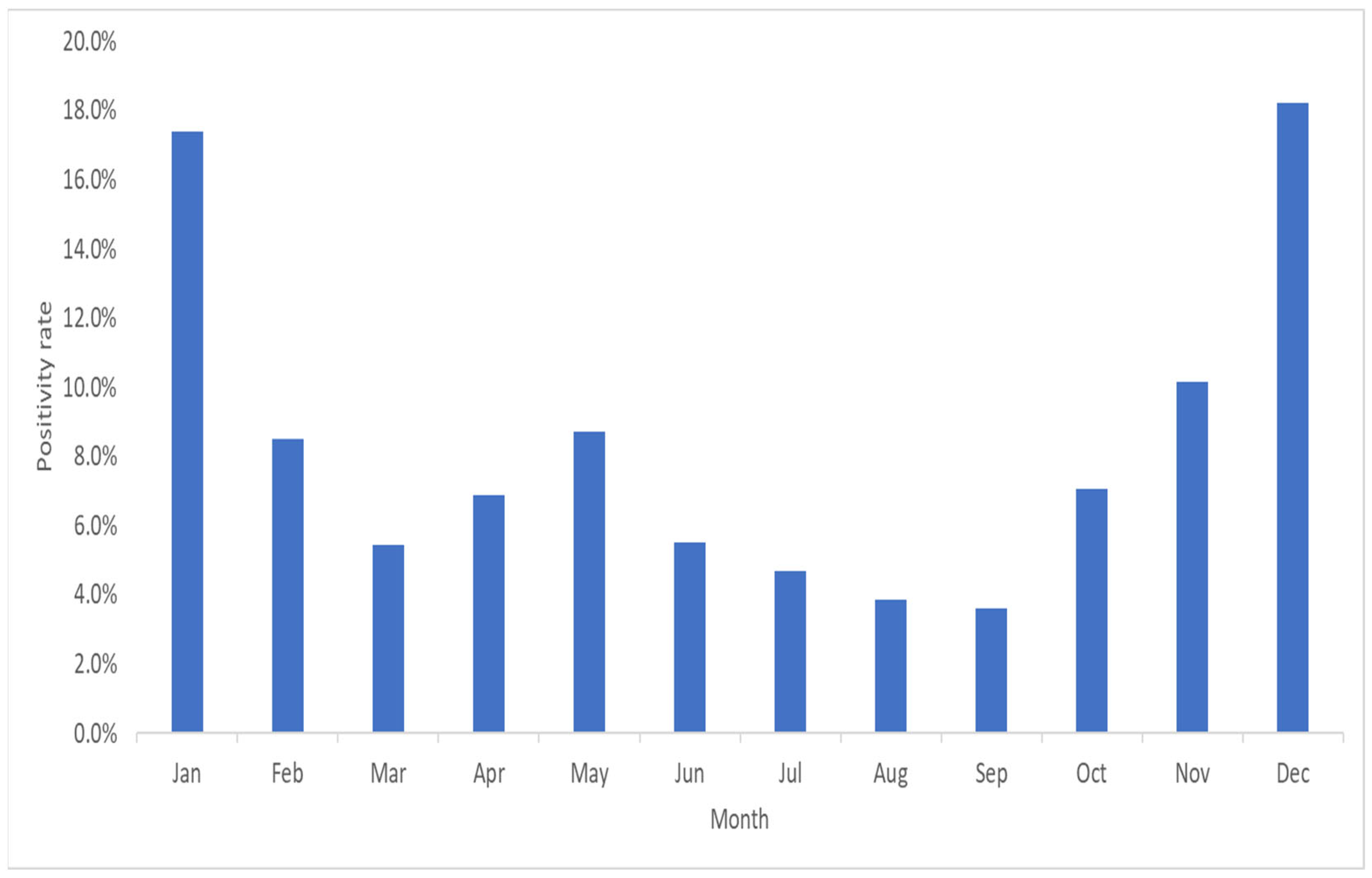
The highest number of pathogens was identified in December, representing 18.2% of all detected pathogens. Notably, a significant portion, accounting for 44.1% of the pathogens, was detected between December and February. During December and February, FluA and FluB exhibited high positivity rates of 87.2% and 71.0%, respectively. Conversely, the positivity rate of PIV3 was notably lower at 51.4% from March to May. Throughout the year, pathogens such as AdV, hMPV, PIV1, PIV3, and HRV/EV showed relatively even distribution patterns (Figure 2).
A total of 187 cases of co-infection were identified, with 393 out of the total 1744 pathogens showing a co-infection rate of 22.5%. Among the pathogens, CoV-HKU1 exhibited the highest co-infection rate at 60% (three out of five), followed by PIV4 at 55.6% (five out of nine), CoV-229E at 40% (sixteen out of forty), and CoV-NL63 at 38.2% (twenty-six out of sixty-eight). Conversely, FluB displayed the lowest co-infection rate at 8.8%, followed by FluA at 13.1% and PIV3 at 13.6%. Regarding double infections by pathogen type, AdV and HRV/EV, as well as HRV/EV and RSV double infections, accounted for the highest proportion at 8.3% (14 out of 169). Co-infections involving AdV, HRV/EV, and PIV3 represented the largest proportion of triple co-infections at 17.6% (three out of seventeen). Additionally, one case of quadruple infection was noted, involving AdV, CoV-NL63, CoV-OC43, and HRV/EV (Table 3).

4. Discussion

Acute respiratory infection is a prevalent disease, accounting for approximately 20–40% of outpatient cases and 12–35% of inpatient cases in general hospitals. Among these, upper respiratory infections, including nasopharyngitis, pharyngitis, and tonsillitis, account for 87.5% of all respiratory infections [12]. Both adults and children show a higher infection rate with viral respiratory pathogens than with bacterial respiratory pathogens; however, children account for more than 80% of all respiratory infection cases [13,14]. Consistent with previous findings, all 1744 (100%) of the FilmArray™ RP-positive pathogens detected in this study were viruses. With respect to the distribution of the pathogens, we found that AdV had the highest positivity rate (18.9%), followed by FluA (16.5%) and PIV3 (12.3%). Previous studies focusing on viral upper respiratory infections have reported the highest positivity rates for HRV/EV and RSV and the lowest positivity rate for FluA [15,16]. However, these results may vary depending on regional and climatic factors, necessitating further research.
In our analysis, we observed 187 instances of co-infection, leading to the detection of 393 pathogens. Among these cases, one hundred and sixty-nine involved double infections, seventeen involved triple infections, and one involved a quadruple infection, reflecting an overall co-infection rate of 22.5%. Comparable co-infection rates have been reported in previous studies utilizing the BioFire FilmArray™ RV and QIAstat-Dx Respiratory Panels, which also documented rates of approximately 20%, mirroring the findings of our investigation [17,18]. However, this rate notably exceeded that reported in a prior study using the FilmArray™ Meningitis/Encephalitis panel [19]. Nevertheless, it remained lower than the rate reported in another study employing the FilmArray™ GI panel [20].
In cases of co-infection, AdV (n = 93) emerged as the most frequently observed pathogen, followed by HRV/EV (n = 57) and FluA (n = 38). Particularly noteworthy were the high co-infection rates of CoV-HKU1 and PIV4, which stood at 60% and 55.6%, respectively, indicating a propensity for co-infection with other pathogens. Comparatively, in previous studies on respiratory viral infections, HRV/EV surfaced as the most common infectious pathogen, trailed by FluB and AdV [21]. Consistent with our findings, another earlier study also reported multiple co-infecting pathogens, primarily HRV/EV, followed by RSV and AdV. Furthermore, elevated co-infection rates were noted for CoV-HKU1 and PIV4 [22].
In the above study, seventeen samples exhibited triple infections, while one sample exhibited quadruple infections, with none exceeding quadruple infections. Samples with triple infections or higher accounted for 1.0% of the total positive samples. In a prior study on a related topic, triple or higher duplicate infections were observed at a rate of approximately 1.3%, which was comparable to the triple double infection rate in this study [23]. Among the eighteen samples with triple infections or higher, AdV was detected in fifteen cases (83.3%), HRV/EVs in ten cases (55.6%), Cov-NL63 in seven cases (38.9%), CoV-OC43 in six cases (33.3%), and PIV-3 in six cases (33.3%), in that order. Notably, one out of five positive samples (20.0%) for Cov-HKU1 and seven out of sixty-eight positive samples (10.3%) for CoV-NL63 exhibited triple or more duplicate infections. Furthermore, SARS-CoV-2 was detected in samples displaying duplicate infections, such as AdV and RSV.
The presence of multiple respiratory Infections carries diverse clinical implications, including prolonged pathogenic infections, extended hospitalization durations, and heightened severity of respiratory-related illnesses [24,25]. However, conflicting findings have also been reported, suggesting that respiratory co-infections may not significantly influence the prevalence or severity of the disease [26,27]. Consequently, the correlation between multiple respiratory pathogen infections and the prevalence and severity of the disease remains unclear, underscoring the necessity for comprehensive investigations into co-infections involving various respiratory pathogens.
In this study, a substantial portion of positive pathogens (1121 cases, constituting 64.3%) were detected in the 1–5 age group, underscoring notably high positivity rates among younger patients. Specifically, within the 1–5 age group, pathogens such as AdV, PIV3, RSV, and HRV/EV displayed positivity rates of 79.3%, 79.4%, 96.9%, and 81.8%, respectively, indicative of robust detection rates in this demographic. Conversely, among older adults (>49 years), FluA and FluB exhibited positivity rates of 58.1% and 53.3%, respectively, suggesting heightened detection rates in this age group. Our findings align with those of previous studies highlighting elevated positivity rates among young patients (aged < 5 years) and robust detection rates for AdV, PIV3, RSV, and HRV/EV [17,28]. However, in contrast to our observations, these studies noted low detection rates for FluA and FluB among older patients but relatively elevated rates among younger individuals.
In this study, 769 pathogens were detected between December and February, corresponding to a detection rate of 44.1%. By contrast, only 245 pathogens were detected between June and August, with a detection rate of 14.0%. In the summer of June-August, 230 samples (16.0%) out of a total of 1443 samples were positive of which AdV was the most common, with 78 (33.9%). This was followed by PIV3 with 69 (30.0%) and HRV/EV with 41 (17.8%). In the winter of December–February, 657 samples (34.5%) out of a total of 1906 samples were positive of which FluA was the most common, with 252 (38.4%). After that, 90 AdV (13.7%) and 76 FluB (11.6%) were different from the summer. These findings indicated a higher detection rate during winter than during summer. Similarly, previous studies reported higher respiratory pathogen detection rates in summer than in winter [29,30]. Research on the age and timing of respiratory pathogen infections can provide fundamental data for implementing various health policies. In particular, such data can help to determine the appropriate age and timing of vaccinations as well as aid in the understanding of the epidemiology of respiratory viruses. Therefore, continuous research and monitoring are necessary to utilize this information as a basis for public health policies.
This present study had several limitations. Firstly, as it was conducted using samples solely obtained from a single university hospital in Cheonan rather than from a range of institutions, the consideration of climatic and regional characteristics may be constrained. Additionally, since the majority of patients visiting the hospital for examinations are residents of the local community, there might be limitations in generalizing the findings to reflect universal trends in respiratory infections. In addition, clinical information related to disease severity, symptoms, and diagnosis was not included, making it challenging to assess treatment outcomes and prognosis. Among the 16 viruses and four bacterial species that could be detected using the FilmArray™ RP, MERS-CoV was not detected. Despite these limitations, in this study, we analyzed 6367 samples according to pathogens, age groups, time periods, and co-infections, allowing us to understand the patterns of respiratory pathogen infections. In particular, analyzing respiratory infections according to age and time can provide valuable clinical data for understanding epidemiological patterns and establishing vaccination strategies. Moreover, the analysis of co-infection rates by specific pathogens can help in the identification of common co-infection pathogens, which can aid in treatment planning and the administration of medications.
Traditionally, microbial culture methods serve as the foundational tests for identifying pathogens responsible for infectious diseases. It is recognized that bacterial and viral culture tests demand specialized facilities and highly trained personnel, more so than molecular biological tests. Bacterial culture typically spans from 24 h to 5 days [31]. The confirmation process for the culture of respiratory viral pathogens, which is the primary focus of this study, can extend over several days to several weeks [32]. In contrast, the FilmArray™ RP offers a rapid and efficient alternative, detecting respiratory pathogens within 2 h. This swift turnaround time facilitates timely decision-making regarding treatment. The expedited results provided by the FilmArray™ RP allow for a shift from empirical antibiotics, such as amoxicillin, clavulanic acid, macrolides, and doxycycline, to more targeted antibiotic prescriptions. This transition enables more precise and effective administration of antibiotics [33]. Furthermore, the ability to test for 20 pathogens in a single sample streamlines the generation and utilization of regional, age-specific, and seasonal data for respiratory pathogen surveillance, vaccination planning, and identification of high-risk populations across different communities. The significance of rapid and accurate diagnostic tests has gained increasing recognition, prompting the adoption of various multiplex PCR diagnostic methods, including the FilmArray™ RP [34]. Epidemiological research on upper respiratory tract infections necessitates the utilization of diverse multiplex PCR methods to comprehensively understand the dynamics of pathogen spread and infection patterns. This underscores the pivotal role of advanced diagnostic technologies in shaping effective public health strategies and interventions.

Author Contributions

Y.J.H.: Conceptualization, Methodology, and Project Administration. Y.J.H. was the lead author and played a central role in developing the research design and methodology. Y.J.H. was responsible for the overall project management and coordination. B.K.J.: Data Collection and Analysis. B.K.J. was responsible for data collection in the field and the subsequent data analysis. Y.J.H. and B.K.J. contributed equally to this study. J.K.K. critically reviewed and edited the manuscript, improving the language, structure, and overall quality of the paper. J.K.K. takes full responsibility for this paper. All authors contributed to the writing, review, and editing of the final manuscript. All authors have read and agreed to the published version of the manuscript.

Funding

This research received no external funding.

Institutional Review Board Statement

The study protocol was approved by the Institutional Review Board of Dankook University (IRB File No. 2023–01–013) on 14 February 2023.

Informed Consent Statement

This study was a retrospective data-only study; therefore, the need for obtaining informed consent from the patients was waived by the Dankook University IRB.

Data Availability Statement

The datasets used and analyzed during this current study are available from the corresponding author upon reasonable request.

Acknowledgments

The authors wish to acknowledge the individuals involved in sample collection and storage.

Conflicts of Interest

The authors declare no conflicts of interest.

References

  1. Fendrick, A.M.; Monto, A.S.; Nightengale, B.; Sarnes, M. The economic burden of non–influenza-related viral respiratory tract infection in the United States. Arch. Intern. Med. 2003, 163, 487–494. [Google Scholar] [CrossRef]
  2. Pillet, S.; Lardeux, M.; Dina, J.; Grattard, F.; Verhoeven, P.; Le Goff, J.; Vabret, A.; Pozzetto, B. Comparative evaluation of six commercialized multiplex PCR kits for the diagnosis of respiratory infections. PLoS ONE 2013, 8, e72174. [Google Scholar] [CrossRef] [PubMed]
  3. Pitrez, P.M.; Pitrez, J.L. Acute upper respiratory tract infections: Outpatient diagnosis and treatment. J. Pediatr. 2003, 79, S77–S86. [Google Scholar] [CrossRef]
  4. Ruopp, M.; Chiswell, K.; Thaden, J.T.; Merchant, K.; Tsalik, E.L. Respiratory tract infection clinical trials from 2007 to 2012. A systematic review of ClinicalTrials.gov. Ann. Am. Thorac. Soc. 2015, 12, 1852–1863. [Google Scholar] [CrossRef] [PubMed]
  5. Kuchar, E.; Miśkiewicz, K.; Nitsch-Osuch, A.; Szenborn, L. Pathophysiology of clinical symptoms in acute viral respiratory tract infections. Adv. Exp. Med. Biol. 2015, 857, 25–38. [Google Scholar]
  6. Hersh, A.L.; Shapiro, D.J.; Pavia, A.T.; Shah, S.S. Antibiotic prescribing in ambulatory pediatrics in the United States. Pediatrics 2011, 128, 1053–1061. [Google Scholar] [CrossRef] [PubMed]
  7. Nyquist, A.-C.; Gonzales, R.; Steiner, J.F.; Sande, M.A. Antibiotic prescribing for children with colds, upper respiratory tract infections, and bronchitis. JAMA 1998, 279, 875–877. [Google Scholar] [CrossRef] [PubMed]
  8. Centers for Disease Control and Prevention. Evaluation of 11 commercially available rapid influenza diagnostic tests—United States, 2011–2012. MMWR Morb. Mortal. Wkly. Rep. 2012, 61, 873–876. [Google Scholar]
  9. Salez, N.; Nougairede, A.; Ninove, L.; Zandotti, C.; De Lamballerie, X.; Charrel, R.N. Xpert Flu for point-of-care diagnosis of human influenza in industrialized countries. Expert Rev. Mol. Diagn. 2014, 14, 411–418. [Google Scholar] [CrossRef] [PubMed]
  10. Hammond, S.P.; Gagne, L.S.; Stock, S.R.; Marty, F.M.; Gelman, R.S.; Marasco, W.A.; Poritz, M.A.; Baden, L.R. Respiratory virus detection in immunocompromised patients with FilmArray Respiratory Panel compared to conventional methods. J. Clin. Microbiol. 2012, 50, 3216–3221. [Google Scholar] [CrossRef] [PubMed]
  11. Kaku, N.; Hashiguchi, K.; Iwanaga, Y.; Akamatsu, N.; Matsuda, J.; Kosai, K.; Uno, N.; Morinaga, Y.; Kitazaki, T.; Hasegawa, H.; et al. Evaluation of FilmArray Respiratory Panel multiplex polymerase chain reaction assay for detection of pathogens in adult outpatients with acute respiratory tract infection. J. Infect. Chemother. 2018, 24, 734–738. [Google Scholar] [CrossRef] [PubMed]
  12. Jain, N.; Lodha, R.; Kabra, S.K. Upper respiratory tract infections. Indian J. Pediatr. 2001, 68, 1135–1138. [Google Scholar] [CrossRef] [PubMed]
  13. Ren, L.; Gonzalez, R.; Wang, Z.; Xiang, Z.; Wang, Y.; Zhou, H.; Li, J.; Xiao, Y.; Yang, Q.; Zhang, J.; et al. Prevalence of human respiratory viruses in adults with acute respiratory tract infections in Beijing, 2005–2007. Clin. Microbiol. Infect. 2009, 15, 1146–1153. [Google Scholar] [CrossRef] [PubMed]
  14. Zhang, G.; Hu, Y.; Wang, H.; Zhang, L.; Bao, Y.; Zhou, X. High incidence of multiple viral infections identified in upper respiratory tract infected children under three years of age in Shanghai, China. PLoS ONE 2012, 7, e44568. [Google Scholar] [CrossRef] [PubMed]
  15. Leber, A.L.; Everhart, K.; Daly, J.A.; Hopper, A.; Harrington, A.; Schreckenberger, P.; McKinley, K.; Jones, M.; Holmberg, K.; Kensinger, B. Multicenter evaluation of BioFire FilmArray Respiratory Panel 2 for detection of viruses and bacteria in nasopharyngeal swab samples. J. Clin. Microbiol. 2018, 56, e01945-17. [Google Scholar] [CrossRef]
  16. Li, J.; Tao, Y.; Tang, M.; Du, B.; Xia, Y.; Mo, X.; Cao, Q. Rapid detection of respiratory organisms with the FilmArray Respiratory Panel in a large Children’s Hospital in China. BMC Infect. Dis. 2018, 18, 510. [Google Scholar] [CrossRef] [PubMed]
  17. Leber, A.L.; Lisby, J.G.; Hansen, G.; Relich, R.F.; Schneider, U.V.; Granato, P.; Young, S.; Pareja, J.; Hannet, I. Multicenter evaluation of the QIAstat-Dx Respiratory Panel for detection of viruses and bacteria in nasopharyngeal swab specimens. J. Clin. Microbiol. 2020, 58, e00155-20. [Google Scholar] [CrossRef] [PubMed]
  18. Loeffelholz, M.J.; Pong, D.L.; Pyles, R.B.; Xiong, Y.; Miller, A.L.; Bufton, K.K.; Chonmaitree, T. Comparison of the FilmArray Respiratory Panel and Prodesse real-time PCR assays for detection of respiratory pathogens. J. Clin. Microbiol. 2011, 49, 4083–4088. [Google Scholar] [CrossRef] [PubMed]
  19. Leber, A.L.; Everhart, K.; Balada-Llasat, J.M.; Cullison, J.; Daly, J.; Holt, S.; Lephart, P.; Salimnia, H.; Schreckenberger, P.C.; DesJarlais, S.; et al. Multicenter evaluation of BioFire FilmArray meningitis/encephalitis panel for detection of bacteria, viruses, and yeast in cerebrospinal fluid specimens. J. Clin. Microbiol. 2016, 54, 2251–2261. [Google Scholar] [CrossRef] [PubMed]
  20. Piralla, A.; Lunghi, G.; Ardissino, G.; Girello, A.; Premoli, M.; Bava, E.; Arghittu, M.; Colombo, M.R.; Cognetto, A.; Bono, P.; et al. FilmArray™ GI panel performance for the diagnosis of acute gastroenteritis or hemorragic diarrhea. BMC Microbiol. 2017, 17, 111. [Google Scholar] [CrossRef] [PubMed]
  21. Busson, L.; Bartiaux, M.; Brahim, S.; Konopnicki, D.; Dauby, N.; Gérard, M.; De Backer, P.; Van Vaerenbergh, K.; Mahadeb, B.; Mekkaoui, L.; et al. Contribution of the FilmArray Respiratory Panel in the management of adult and pediatric patients attending the emergency room during 2015–2016 influenza epidemics: An interventional study. Int. J. Infect. Dis. 2019, 83, 32–39. [Google Scholar] [CrossRef] [PubMed]
  22. Parčina, M.; Schneider, U.V.; Visseaux, B.; Jozić, R.; Hannet, I.; Lisby, J.G. Multicenter evaluation of the QIAstat Respiratory Panel—A new rapid highly multiplexed PCR based assay for diagnosis of acute respiratory tract infections. PLoS ONE 2020, 15, e0230183. [Google Scholar] [CrossRef]
  23. Sanghavi, S.K.; Bullotta, A.; Husain, S.; Rinaldo, C.R. Clinical evaluation of multiplex real-time PCR panels for rapid detection of respiratory viral infections. J. Med. Virol. 2012, 84, 162–169. [Google Scholar] [CrossRef] [PubMed]
  24. Chauhan, J.C.; Slamon, N.B. The impact of multiple viral respiratory infections on outcomes for critically ill children. Pediatr. Crit. Care Med. 2017, 18, e333–e338. [Google Scholar] [CrossRef] [PubMed]
  25. Richard, N.; Komurian-Pradel, F.; Javouhey, E.; Perret, M.; Rajoharison, A.; Bagnaud, A.; Billaud, G.; Vernet, G.; Lina, B.; Floret, D.; et al. The impact of dual viral infection in infants admitted to a pediatric intensive care unit associated with severe bronchiolitis. Pediatr. Infect. Dis. J. 2008, 27, 213–217. [Google Scholar] [CrossRef]
  26. Scotta, M.C.; Chakr, V.C.; de Moura, A.; Becker, R.G.; de Souza, A.P.D.; Jones, M.H.; Pinto, L.A.; Sarria, E.E.; Pitrez, P.M.; Stein, R.T.; et al. Respiratory viral coinfection and disease severity in children: A systematic review and meta-analysis. J. Clin. Virol. 2016, 80, 45–56. [Google Scholar] [CrossRef] [PubMed]
  27. Wishaupt, J.O.; van der Ploeg, T.; de Groot, R.; Versteegh, F.G.; Hartwig, N.G. Single-and multiple viral respiratory infections in children: Disease and management cannot be related to a specific pathogen. BMC Infect. Dis. 2017, 17, 62. [Google Scholar] [CrossRef] [PubMed]
  28. Yang, S.; Li, H.; Tang, Y.; Yu, F.; Ma, C.; Zhang, H.; Pang, L.; Zhao, H.; Wang, L. Multiplex tests for respiratory tract infections: The direct utility of the FilmArray Respiratory Panel in emergency department. Can. Respir. J. 2020, 2020, 6014563. [Google Scholar] [CrossRef]
  29. Lamrani Hanchi, A.; Guennouni, M.; Rachidi, M.; Benhoumich, T.; Bennani, H.; Bourrous, M.; Maoulainine, F.M.R.; Younous, S.; Bouskraoui, M.; Soraa, N. Epidemiology of respiratory pathogens in children with severe acute respiratory infection and impact of the multiplex PCR Film Array Respiratory Panel: A 2-year study. Int. J. Microbiol. 2021, 2021, 2276261. [Google Scholar] [CrossRef] [PubMed]
  30. Visseaux, B.; Burdet, C.; Voiriot, G.; Lescure, F.X.; Chougar, T.; Brugière, O.; Crestani, B.; Casalino, E.; Charpentier, C.; Descamps, D.; et al. Prevalence of respiratory viruses among adults, by season, age, respiratory tract region and type of medical unit in Paris, France, from 2011 to 2016. PLoS ONE 2017, 12, e0180888. [Google Scholar] [CrossRef] [PubMed]
  31. Lagier, J.C.; Edouard, S.; Pagnier, I.; Mediannikov, O.; Drancourt, M.; Raoult, D. Current and Past Strategies for Bacterial Culture in Clinical Microbiology. Clin. Microbiol. Rev. 2015, 28, 208–236. [Google Scholar] [CrossRef] [PubMed]
  32. Talbot, H.K.; Falsey, A.R. The diagnosis of viral respiratory disease in older adults. Clin. Infect. Dis. 2010, 50, 747–751. [Google Scholar] [CrossRef] [PubMed]
  33. Panasiuk, L.; Lukas, W.; Paprzycki, P.; Verheij, T.; Godycki-Ćwirko, M.; Chlabicz, S. Antibiotics in the treatment of upper respiratory tract infections in Poland. Is there any improvement? J. Clin. Pharm. Ther. 2010, 35, 665–669. [Google Scholar] [CrossRef] [PubMed]
  34. Chan, M.; Koo, S.H.; Jiang, B.; Lim, P.Q.; Tan, T.Y. Comparison of the BioFire FilmArray Respiratory Panel, Seegene AnyplexII RV16, and Argene for the detection of respiratory viruses. J. Clin. Virol. 2018, 106, 13–17. [Google Scholar] [CrossRef] [PubMed]
Figure 1. FilmArray™ Respiratory Panel (RP) positive detection of 1744 pathogens by age group collected over five years at Dankook University Hospital in Cheonan, Republic of Korea.
Diagnostics 14 00734 g001
Figure 2. Monthly FilmArray™ Respiratory Panel (RP) positivity rate of 1744 pathogens detected over five years at Dankook University Hospital in Cheonan, Republic of Korea.
Diagnostics 14 00734 g002
Table 1. Positivity rates of FilmArray™ Respiratory Panel pathogens.
ParameterPositivity
Number of Samples% of Total
All samples (N = 6367)
Negative samples4.82975.8%
Positive samples153824.1%
Single detections135121.2%
Co-infections1872.9%
Co-infections (n = 187)
Double infection16990.3%
Triple infection179.1%
Quadruple infection10.6%
Table 2. Detection and co-infection rates for each pathogen.
DetectedDetection Rate (%)Co-InfectionCo-Infection Rate (%)
AdV32918.99328.3
CoV-229E402.31640.0
CoV-HKU150.3360.0
CoV-NL63683.92638.2
CoV-OC431015.83130.7
SARS-CoV-2281.6621.4
hMPV1709.72917.1
HRV/EV18110.45731.5
FluA28916.63813.1
FluB1076.11514.0
PIV1774.41519.5
PIV2291.7413.8
PIV321412.32913.6
PIV490.5555.6
RSV975.62626.8
Total1744100.039322.5
AdV, adenovirus; CoV-229E, coronavirus 229E; CoV-HKU1, coronavirus HKU1; CoV-NL63, coronavirus NL63; CoV-OC43, coronavirus OC43; SARS-CoV-2, severe acute respiratory syndrome coronavirus 2; hMPV, human metapneumovirus; HRV/EV, human rhinovirus/enterovirus; FluA, influenza virus A; FluB, influenza virus B; PIV1, parainfluenza virus 1; PIV2, parainfluenza virus 2; PIV3, parainfluenza virus 3; PIV4, parainfluenza virus 4; RSV, respiratory syncytial virus.
Table 3. Distribution of co-infections of respiratory pathogens.
Single InfectionDouble InfectionTriple Infection
AdV236 (17.5)AdVCoV-229E2 (1.2)AdV & CoV-NL63CoV-229E1 (5.9)
CoV-229E24 (1.8) CoV-OL634 (2.4) CoV-OC431 (5.9)
CoV-HKU12 (0.1) CoV-OC4311 (6.5) PIV31 (5.9)
CoV-NL6342 (3.1) hMPV13 (7.7) HRV/EV1 (5.9)
CoV-OC4370 (5.2) HRV/EV14 (8.3)AdV & HRV/EVPIV11 (5.9)
SARS-CoV-222 (1.6) FluA10 (5.9) RSV2 (11.8)
HRV/EV141 (10.4) FluB2 (1.2) CoV-OC431 (5.9)
RV/EV124 (9.2) PIV12 (1.2) PIV33 (17.6)
FluA251 (18.6) PIV21 (0.6)AdV & CoV-OC43CoV-HKU11 (5.9)
FluB92 (6.8) PIV313 (7.7) PIV31 (5.9)
PIV162 (4.6) PIV41 (0.6)AdV & RSVSARS-CoV-21 (5.9)
PIV225 (1.9) RSV5 (3.0)CoV-NL63 & PIV1PIV31 (5.9)
PIV3185 (13.7)CoV-229ECoV-OL632 (1.2)CoV-NL63 & HRV/EVRSV1 (5.9)
PIV44 (0.3) FluA8 (4.7)CoV-OC43 & hMPVFluA1 (5.9)
RSV71 (5.3) FluB3 (1.8) total17 (100)
Total1351 (100)CoV-HKU1hMPV1 (0.6)
FluA1 (0.6)
CoV-OL63CoV-OC432 (1.2)
hMPV2 (1.2)
HRV/EV1 (0.6)
FluA4 (2.4)
FluB1 (0.6)
PIV11 (0.6)
PIV32 (1.2)
C.OC43HRV/EV1 (0.6)
FluA4 (2.4)
FluB3 (1.8)
PIV33 (1.8)
RSV1 (0.6)
SARShMPV1 (0.6)
HRV/EV2 (1.2)
FluA1 (0.6)
RSV1 (0.6)
MVHRV/EV4 (2.4)
FluB1 (0.6)
PIV12 (1.2)
PIV21 (0.6)
PIV33 (1.8)
HRV/EVFluA1 (0.6)
PIV15 (3.0)
PIV21 (0.6)
PIV34 (2.4)
RSV14 (8.3)
FluAFluB5 (3.0)
PIV11 (0.6)
PIV21 (0.6)
PIV41 (0.6)
PIV1PIV31 (0.6)
RSV1 (0.6)
Total169 (100)
AdV, adenovirus; CoV-229E, coronavirus 229E; CoV-HKU1, coronavirus HKU1; CoV-NL63, coronavirus NL63; CoV-OC43, coronavirus OC43; SARS-CoV-2, severe acute respiratory syndrome coronavirus 2; hMPV, human metapneumovirus; HRV/EV, human rhinovirus/enterovirus; FluA, influenza virus A; FluB, influenza virus B; PIV1, parainfluenza virus 1; PIV2, parainfluenza virus 2; PIV3, parainfluenza virus 3; PIV4, parainfluenza virus 4; RSV, respiratory syncytial virus.
Disclaimer/Publisher’s Note: The statements, opinions and data contained in all publications are solely those of the individual author(s) and contributor(s) and not of MDPI and/or the editor(s). MDPI and/or the editor(s) disclaim responsibility for any injury to people or property resulting from any ideas, methods, instructions or products referred to in the content.