
Occult Breast Cancer Presenting as Sternum Pain

 and
and 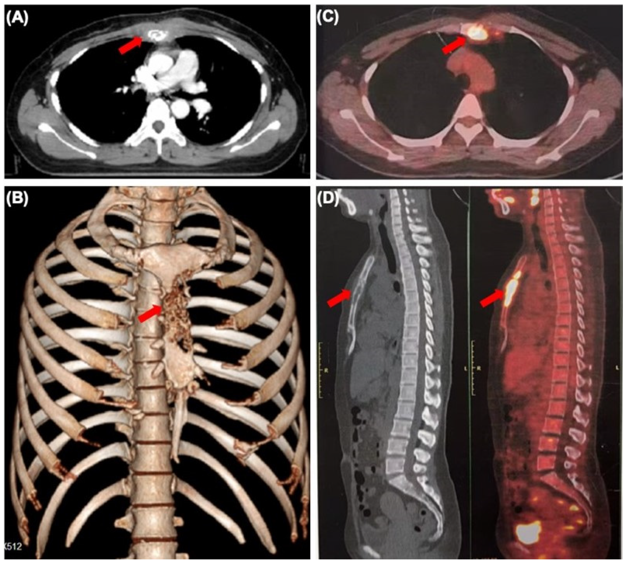
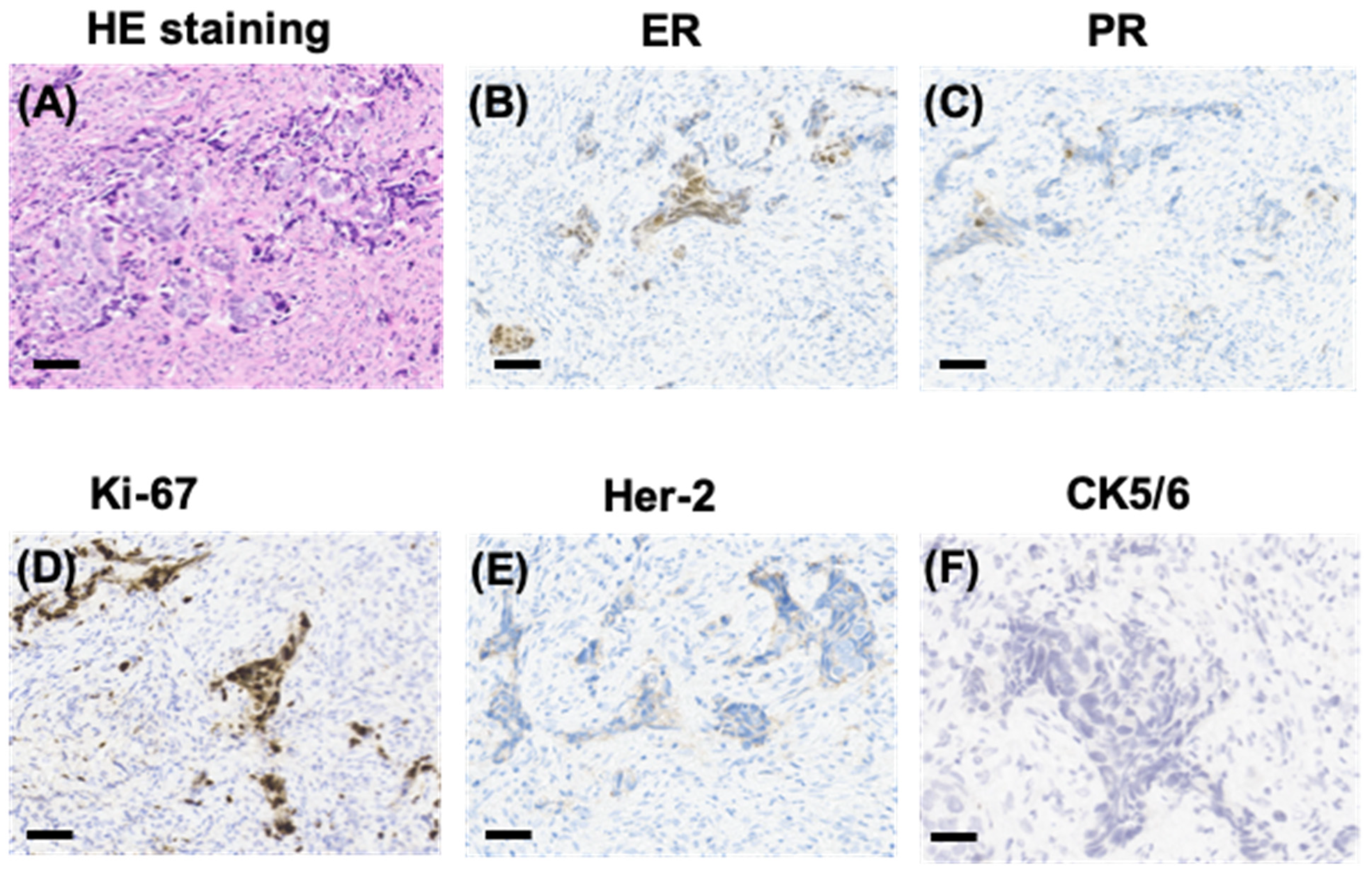
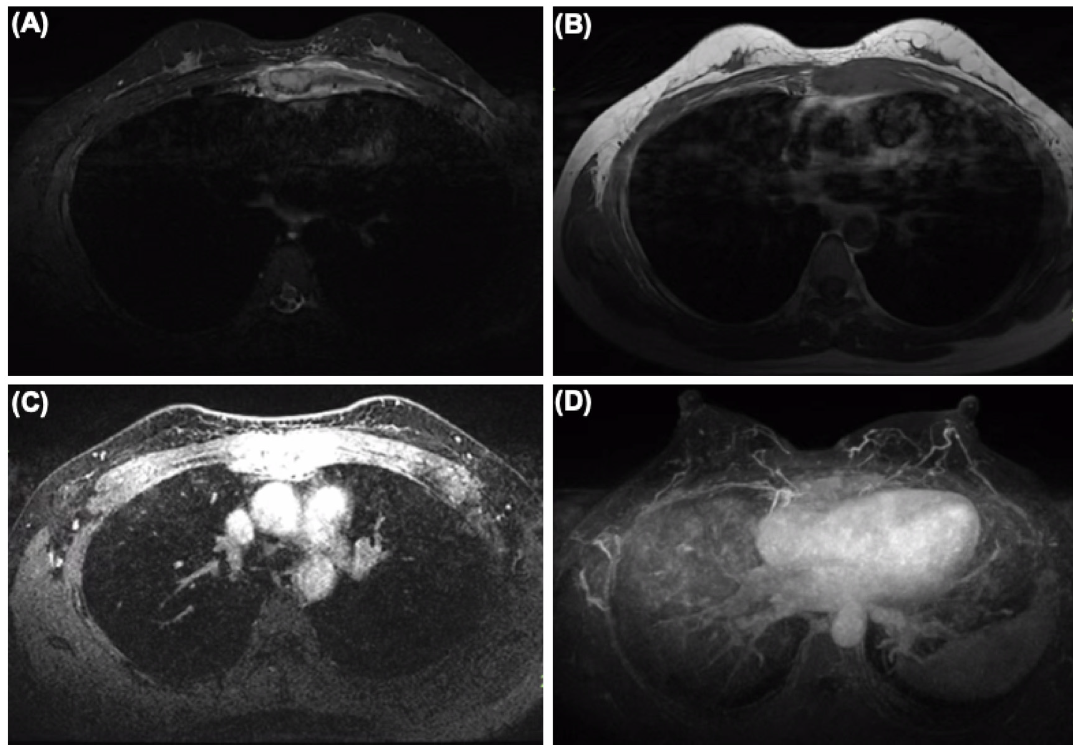
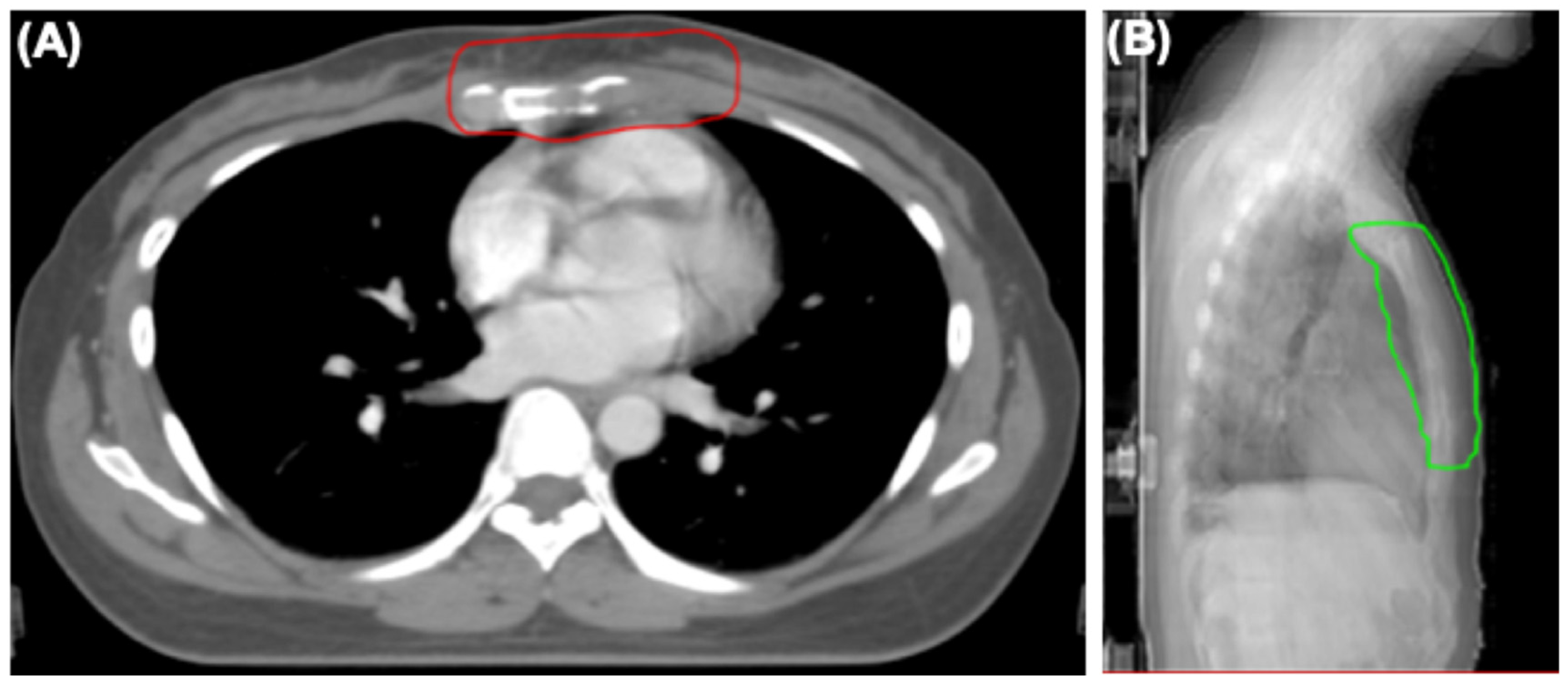
Abstract
:Author Contributions
Funding
Institutional Review Board Statement
Informed Consent Statement
Data Availability Statement
Conflicts of Interest
References
- Halsted, W.S. The Results of Radical Operations for the Cure of Carcinoma of the Breast. Ann. Surg. 1907, 46, 1–19. [Google Scholar] [CrossRef] [PubMed]
- Walker, G.V.; Smith, G.L.; Perkins, G.H.; Oh, J.L.; Woodward, W.; Yu, T.K.; Hunt, K.K.; Hoffman, K.; Strom, E.A.; Buchholz, T.A. Population-based analysis of occult primary breast cancer with axillary lymph node metastasis. Cancer 2010, 116, 4000–4006. [Google Scholar] [CrossRef] [PubMed]
- Toss, A.; Moscetti, L.; Cascinu, S. Occult breast cancer: The uncommon presentation of a common disease. Chin. Clin. Oncol. 2019, 8, S10. [Google Scholar] [CrossRef]
- Buijs, J.T.; van der Pluijm, G. Osteotropic cancers: From primary tumor to bone. Cancer Lett. 2009, 273, 177–193. [Google Scholar] [CrossRef]
- Carsote, M.; Terzea, D.; Vasilescu, F.; Cucu, A.P.; Ciuche, A.; Nistor, C. Sternum Metastases: From Case-Identifying Strategy to Multidisciplinary Management. Diagnostics 2023, 13, 2698. [Google Scholar] [CrossRef] [PubMed]
- Kazemzadeh, F.; Snoek, J.A.A.; Voorham, Q.J.; van Oijen, M.G.H.; Hugen, N.; Nagtegaal, I.D. Association of metastatic pattern in breast cancer with tumor and patient-specific factors: A nationwide autopsy study using artificial intelligence. Breast Cancer 2024, 31, 263–271. [Google Scholar] [CrossRef] [PubMed]
- Bitencourt, A.; Rossi Saccarelli, C.; Morris, E.A.; Flynn, J.; Zhang, Z.; Khan, A.; Gillespie, E.; Cahlon, O.; Mueller, B.; Cuaron, J.J.; et al. Regional Lymph Node Involvement Among Patients With De Novo Metastatic Breast Cancer. JAMA Netw. Open 2020, 3, e2018790. [Google Scholar] [CrossRef] [PubMed]


Disclaimer/Publisher’s Note: The statements, opinions and data contained in all publications are solely those of the individual author(s) and contributor(s) and not of MDPI and/or the editor(s). MDPI and/or the editor(s) disclaim responsibility for any injury to people or property resulting from any ideas, methods, instructions or products referred to in the content. |
© 2024 by the authors. Licensee MDPI, Basel, Switzerland. This article is an open access article distributed under the terms and conditions of the Creative Commons Attribution (CC BY) license (https://creativecommons.org/licenses/by/4.0/).
Share and Cite
Wu, D.; Guo, S.; Zhang, B.; Huang, F.; Qian, W.; Qiu, F.; Wei, Q.; Zhang, T. Occult Breast Cancer Presenting as Sternum Pain. Diagnostics 2024, 14, 765. https://doi.org/10.3390/diagnostics14070765
Wu D, Guo S, Zhang B, Huang F, Qian W, Qiu F, Wei Q, Zhang T. Occult Breast Cancer Presenting as Sternum Pain. Diagnostics. 2024; 14(7):765. https://doi.org/10.3390/diagnostics14070765
Chicago/Turabian StyleWu, Dang, Siyu Guo, Bicheng Zhang, Fengbo Huang, Wei Qian, Fuming Qiu, Qichun Wei, and Ting Zhang. 2024. "Occult Breast Cancer Presenting as Sternum Pain" Diagnostics 14, no. 7: 765. https://doi.org/10.3390/diagnostics14070765
APA StyleWu, D., Guo, S., Zhang, B., Huang, F., Qian, W., Qiu, F., Wei, Q., & Zhang, T. (2024). Occult Breast Cancer Presenting as Sternum Pain. Diagnostics, 14(7), 765. https://doi.org/10.3390/diagnostics14070765