
Mind the Pitfall: Solitary Nodular Fasciitis Mimicking Extra-Nodal Manifestation of Hodgkin Lymphoma on [18F]FDG PET/CT

 ,
,  and
and 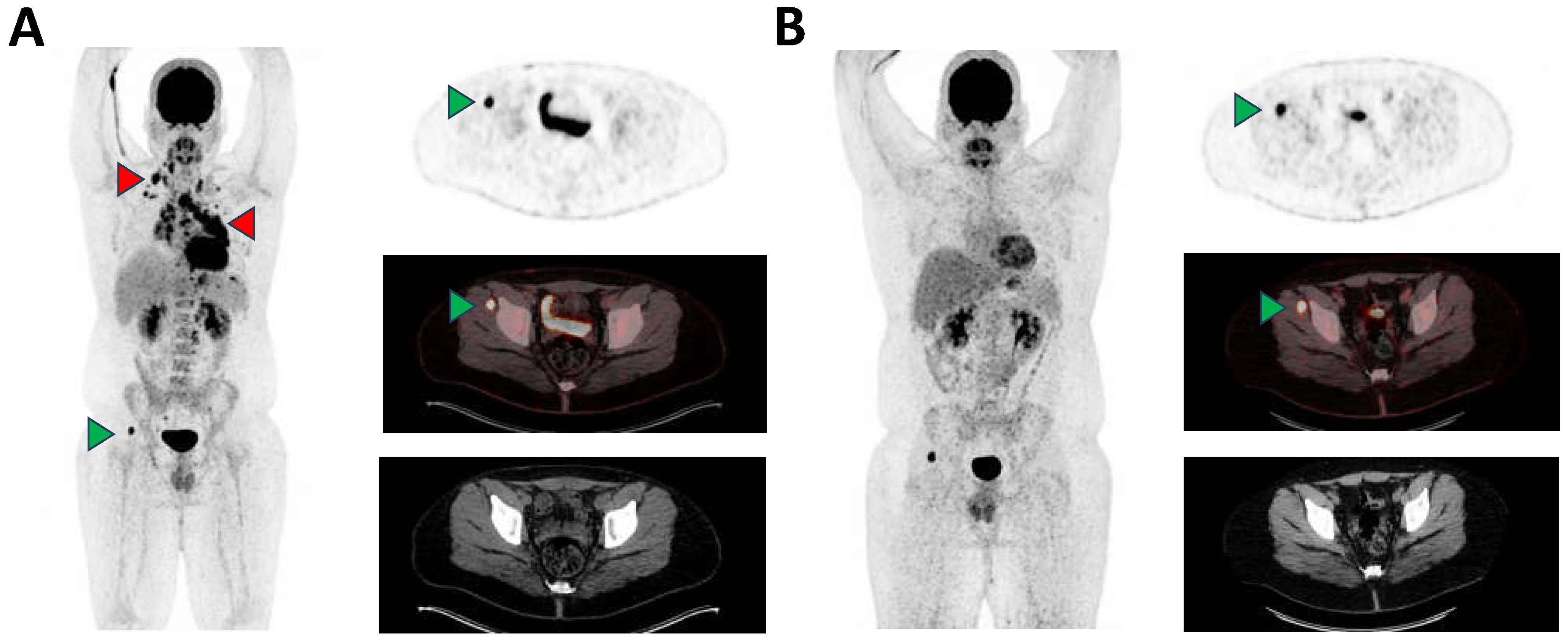
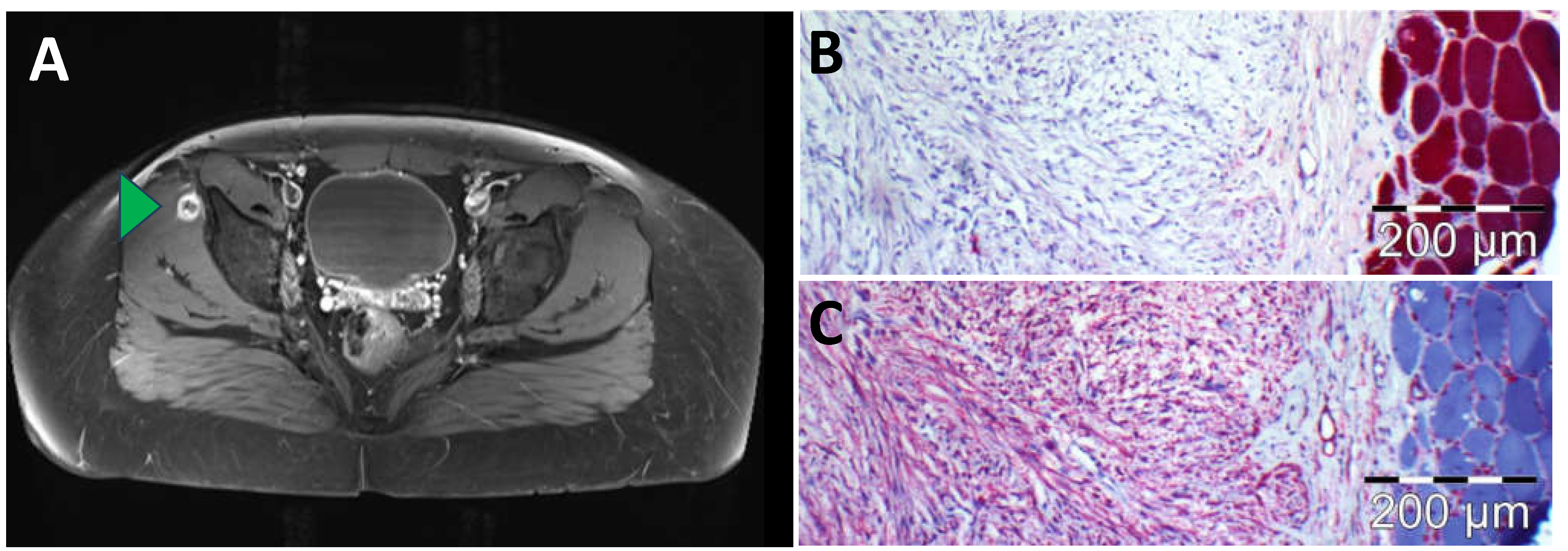
Abstract


Author Contributions
Funding
Institutional Review Board Statement
Informed Consent Statement
Data Availability Statement
Acknowledgments
Conflicts of Interest
References
- Samaratunga, H.; Searle, J.; O’Loughlin, B. Nodular Fasciitis and Related Pseudosarcomatous Lesions of Soft Tissues. Aust. N. Z. J. Surg. 1996, 66, 22–25. [Google Scholar] [CrossRef] [PubMed]
- Berry, A.B.; Jaffee, I.; Greenberg, M.; Eisele, D.W.; Ljung, B.-M. Nodular Fasciitis: Definitive Diagnosis by Fine Needle Aspiration. Acta Cytol. 2016, 60, 19–24. [Google Scholar] [CrossRef] [PubMed]
- Hseu, A.; Watters, K.; Perez-Atayde, A.; Silvera, V.M.; Rahbar, R. Pediatric Nodular Fasciitis in the Head and Neck: Evaluation and Management. JAMA Otolaryngol. Head. Neck Surg. 2015, 141, 54–59. [Google Scholar] [CrossRef] [PubMed]
- Tomita, S.; Thompson, K.; Carver, T.; Vazquez, W.D. Nodular Fasciitis: A Sarcomatous Impersonator. J. Pediatr. Surg. 2009, 44, e17–e19. [Google Scholar] [CrossRef] [PubMed]
- Pandian, T.K.; Zeidan, M.M.; Ibrahim, K.A.; Moir, C.R.; Ishitani, M.B.; Zarroug, A.E. Nodular Fasciitis in the Pediatric Population: A Single Center Experience. J. Pediatr. Surg. 2013, 48, 1486–1489. [Google Scholar] [CrossRef] [PubMed]
- Kim, J.Y.; Park, J.; Choi, Y.Y.; Lee, S.; Paik, S.S. Nodular Fasciitis Mimicking Soft Tissue Metastasis on 18F-FDG PET/CT during Surveillance. Clin. Nucl. Med. 2015, 40, 172–174. [Google Scholar] [CrossRef] [PubMed]
- Barat, M.; Soyer, P.; Audard, V. Nodular Fasciitis: PET/CT and MR Imaging Features. Diagn. Interv. Imaging 2023, 104, 451–452. [Google Scholar] [CrossRef] [PubMed]
- Kessels, L.W.; Simsek, S.; van Hattum, A.H.; Stam, F.; Comans, E.F.I. Nodular Fasciitis: An Unexpected Finding on Computed Tomography and Positron Emission Tomography. Eur. J. Intern. Med. 2004, 15, 183–185. [Google Scholar] [CrossRef] [PubMed]
- Gotthardt, M.; Arens, A.; van der Heijden, E.; de Geus-Oei, L.-F.; Oyen, W.J.G. Nodular Fasciitis on F-18 FDG PET. Clin. Nucl. Med. 2010, 35, 830. [Google Scholar] [CrossRef] [PubMed]
- Zhou, Q.; Ansari, U.; Keshav, N.; Davis, F.; Cundiff, M. Extranodal manifestation of Rosai-Dorfman disease in the breast tissue. Radiol. Case Rep. 2016, 11, 125–128. [Google Scholar] [CrossRef] [PubMed][Green Version]
- Dubreuil, J.; Salles, G.; Bozzetto, J.; Tordo, J.; Djaïleb, L.; Berriolo-Riedinger, A.; Leenhardt, J.; Giammarile, F.; Meignan, M.; Skanjeti, A. Usual and unusual pitfalls of 18F-FDG-PET/CT in lymphoma after treatment: A pictorial review. Nucl. Med. Commun. 2017, 38, 563–576. [Google Scholar] [CrossRef] [PubMed][Green Version]
- Schwerz, S.; Mueller, M.; Lindemann-Docter, K.; Heinzel, A.; Mottaghy, F.M.; Beheshti, M. Hepatic candidiasis mimicking lymphoma on 18F-FDG PET/CT in a patient with T cell lymphoma. Eur. J. Nucl. Med. Mol. Imaging 2020, 47, 2925–2926. [Google Scholar] [CrossRef] [PubMed]
- Akkawi, A.R.; Ezzeddine, L.; Chahinian, R.; Ershaid, F.; Merheb, D.; Mzeihem, M.; El-Cheikh, J.; Haidar, M. Hepatic granuloma mimicking recurrent lymphoma on 18F-FDG PET/CT in a patient with primary mediastinal diffuse large B-cell lymphoma. Asia Ocean. J. Nucl. Med. Biol. 2022, 10, 47–52. [Google Scholar] [CrossRef] [PubMed]
- Bryant, B.S.; Marsh, K.A.; Beuerlein, W.J.; Kalil, D.; Shah, K.K. A Case of Sarcoidosis Mimicking Lymphoma Confounded by Cognitive Decline. Cureus 2021, 13, e13667. [Google Scholar] [CrossRef] [PubMed]
- Henninger, M.; Maurer, T.; Hacker, C.; Eiber, M. 68Ga-PSMA PET/MR Showing Intense PSMA Uptake in Nodular Fasciitis Mimicking Prostate Cancer Metastasis. Clin. Nucl. Med. 2016, 41, e443. [Google Scholar] [CrossRef] [PubMed]
- Plouznikoff, N.; Artigas, C.; Karfis, I.; Flamen, P. Low 68Ga–PSMA PET/CT Uptake in Chronic Intramuscular Nodular Fasciitis. Clin. Nucl. Med. 2020, 45, e41. [Google Scholar] [CrossRef] [PubMed]
- Nagano, H.; Kiyosawa, T.; Aoki, S.; Azuma, R. A Case of Nodular Fasciitis That Was Difficult to Distinguish from Sarcoma. Int. J. Surg. Case Rep. 2019, 65, 27–31. [Google Scholar] [CrossRef] [PubMed]
- Della Volpe, A.; Festa, P.; Varricchio, A.M.; Russo, C.; Covelli, E.M.; Bifano, D.; Piroli, P.; De Lucia, A.; Di Stadio, A.; Ionna, F. Diagnosis and Treatment of Nodular Fasciitis of Ear Region in Children: A Case Report and Review of Literature. Healthcare 2022, 10, 1962. [Google Scholar] [CrossRef] [PubMed]
Disclaimer/Publisher’s Note: The statements, opinions and data contained in all publications are solely those of the individual author(s) and contributor(s) and not of MDPI and/or the editor(s). MDPI and/or the editor(s) disclaim responsibility for any injury to people or property resulting from any ideas, methods, instructions or products referred to in the content. |
© 2024 by the authors. Licensee MDPI, Basel, Switzerland. This article is an open access article distributed under the terms and conditions of the Creative Commons Attribution (CC BY) license (https://creativecommons.org/licenses/by/4.0/).
Share and Cite
Markandu, S.; Blickle, A.; Burgard, C.; Remke, M.; Altmeyer, K.; Wagner, M.; Ezziddin, S.; Rosar, F. Mind the Pitfall: Solitary Nodular Fasciitis Mimicking Extra-Nodal Manifestation of Hodgkin Lymphoma on [18F]FDG PET/CT. Diagnostics 2024, 14, 783. https://doi.org/10.3390/diagnostics14080783
Markandu S, Blickle A, Burgard C, Remke M, Altmeyer K, Wagner M, Ezziddin S, Rosar F. Mind the Pitfall: Solitary Nodular Fasciitis Mimicking Extra-Nodal Manifestation of Hodgkin Lymphoma on [18F]FDG PET/CT. Diagnostics. 2024; 14(8):783. https://doi.org/10.3390/diagnostics14080783
Chicago/Turabian StyleMarkandu, Suginthan, Arne Blickle, Caroline Burgard, Marc Remke, Katrin Altmeyer, Mathias Wagner, Samer Ezziddin, and Florian Rosar. 2024. "Mind the Pitfall: Solitary Nodular Fasciitis Mimicking Extra-Nodal Manifestation of Hodgkin Lymphoma on [18F]FDG PET/CT" Diagnostics 14, no. 8: 783. https://doi.org/10.3390/diagnostics14080783
APA StyleMarkandu, S., Blickle, A., Burgard, C., Remke, M., Altmeyer, K., Wagner, M., Ezziddin, S., & Rosar, F. (2024). Mind the Pitfall: Solitary Nodular Fasciitis Mimicking Extra-Nodal Manifestation of Hodgkin Lymphoma on [18F]FDG PET/CT. Diagnostics, 14(8), 783. https://doi.org/10.3390/diagnostics14080783