Abstract
Here, we describe the case of an asymptomatic 70-year-old male patient who performed a contrast-enhanced computed tomography examination for prostate cancer staging, and an exceptional finding was reported. Specifically, a probable and never before reported minimal V aortic arch remnant with a thin intima–media band that joins together the anterior and posterior aortic walls.
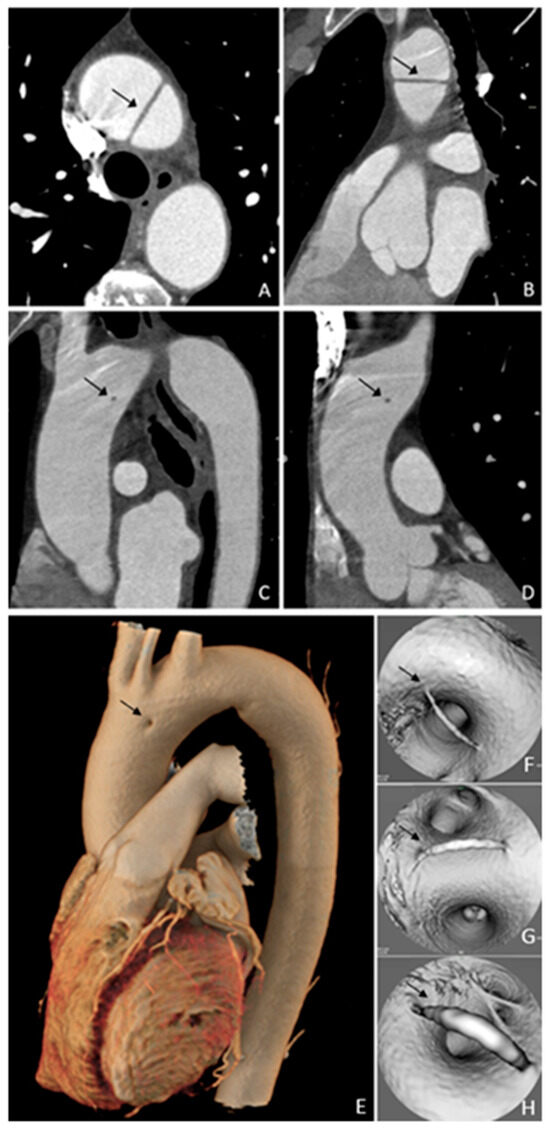
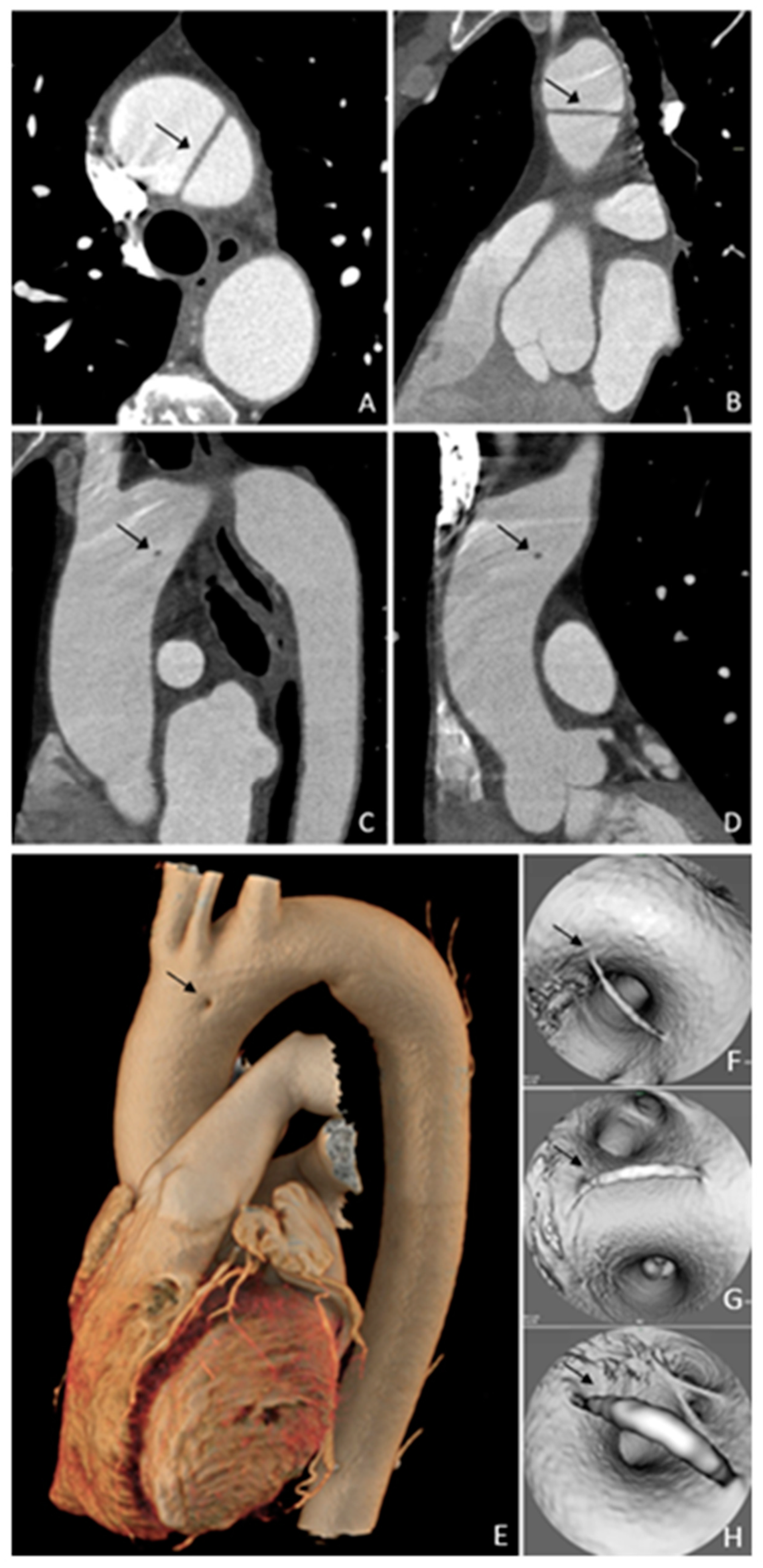
Figure 1.
Axial (A), oblique sagittal (B,C), and oblique coronal (D) computed tomography images in an asymptomatic 70-year-old male patient who performed a computed tomography examination for prostate cancer staging. Images (A,B) could be misinterpreted as type A aortic dissection. However, looking at the oblique images (C,D), is evident that this is not the case—in fact, these images show that a thin line connects the anterior and posterior aortic walls, and it seems to be constituted by intima and media layers. Volume rendering (E) shows a small area with absent contrast enhancement, and the virtual endoluminal images (F,G,H) clearly show the stripe connecting the opposing walls. This seems to be a congenital anomaly due to a minimally imperfect aortic arch formation, and it could be a fifth aortic arch remnant. The patient underwent a cardiac surgery visit, and no follow-up was scheduled. The patient did not show any symptoms related to this condition two years post the computed tomography examination. Previously published articles reported aortic arch anomalies, in particular persistent fifth aortic arch (PFAA), which [1,2,3,4] has been classified into three types by Weinberg. Subsequently, an improved classification divided PFAA into four types based on its anatomical origin and hemodynamic changes. In particular, the imaging findings in our case suggest that it could belong to a previously unreported subtype of type A of the Weinberg classification and type A1 of the improved classification, which are characterized by a double-lumen aortic arch where two parallel arches coexist, with the innominate, left common carotid, and left subclavian arteries arising from the more superior arch, probably as derivatives of the normal embryonic fourth branchial arch [3,5,6]. The persistence of the fifth aortic arch is usually of no hemodynamic significance and it is incidentally diagnosed when it is characterized by a double-lumen aortic arch without other vascular malformations [5]. To the best of our knowledge, no previously published article has reported such an image of the thoracic aorta, so this case can expand the spectrum of aortic arch anomalies.
Author Contributions
Conceptualization, C.T. and G.L.; methodology, C.T., M.F., S.L., C.C., A.Q., A.A.M., D.B., A.B., G.A., I.C., E.D.C. and N.S.; data curation, C.T., M.F., S.L., C.C., D.B. and G.L.; writing—original draft preparation, C.T., M.F., S.L., C.C., A.Q., A.A.M., D.B., A.B., G.A., I.C., E.D.C., N.S. and G.L.; writing—review and editing, C.T., M.F., S.L., C.C., A.Q., A.A.M., D.B., A.B., G.A., I.C., E.D.C., N.S. and G.L.; supervision, G.A., I.C., E.D.C., N.S. and G.L. All authors have read and agreed to the published version of the manuscript.
Funding
This research received no external funding.
Institutional Review Board Statement
Not applicable.
Informed Consent Statement
Written informed consent has been obtained from the patient to publish this paper.
Data Availability Statement
Data are contained within the article.
Conflicts of Interest
The authors declare no conflicts of interest.
References
- Schicchi, N.; Agliata, G.; Giovagnoni, A. CT imaging of a rare case of persistent fifth aortic arch in newborn. BJR Case Rep. 2016, 2, 20150048. [Google Scholar] [CrossRef] [PubMed]
- Liu, Y.; Zhang, H.; Ren, J.; Cao, A.; Guo, J.; Liu, B.; Bao, M.; Zheng, C. Persistent fifth aortic arch: A single-center experience, case series. Transl. Pediatr. 2021, 10, 1566–1572. [Google Scholar] [CrossRef] [PubMed]
- Weinberg, P.M. Aortic arch anomalies. J. Cardiovasc. Magn. Reson. 2006, 8, 633–643. [Google Scholar] [CrossRef] [PubMed]
- Oshitani, T.; Kawasaki, Y.; Murakami, Y.; Fujino, M.; Sasaki, T.; Nakamura, K.; Yoshida, Y.; Suzuki, T.; Ehara, E. A double-barrelled aorta with high aortic Arch. J. Cardiol. Cases 2021, 24, 284–286. [Google Scholar] [CrossRef] [PubMed]
- Shan, H.; Du, X.; Zheng, G.; Ke, T.; Liao, C.; Yang, H. Persistent fifth aortic arch: A comprehensive literature review. Front. Pediatr. 2023, 11, 1183345. [Google Scholar] [CrossRef] [PubMed]
- Naimo, P.S.; del Carmen Vazquez-Alvarez, M.; d’Udekem, Y.; Jones, B.; Konstantinov, I.E. Double-Lumen Aortic Arch: Persistence of the Fifth Aortic Arch. Ann. Thorac. Surg. 2016, 101, e155–e156. [Google Scholar] [CrossRef] [PubMed]
Disclaimer/Publisher’s Note: The statements, opinions and data contained in all publications are solely those of the individual author(s) and contributor(s) and not of MDPI and/or the editor(s). MDPI and/or the editor(s) disclaim responsibility for any injury to people or property resulting from any ideas, methods, instructions or products referred to in the content. |
© 2025 by the authors. Licensee MDPI, Basel, Switzerland. This article is an open access article distributed under the terms and conditions of the Creative Commons Attribution (CC BY) license (https://creativecommons.org/licenses/by/4.0/).