


Abstract
Background: Artificial intelligence (AI) has emerged as a transformative technology across dermatological practice, from automated lesion classification to whole-slide pathology analysis. Despite rapid growth in primary studies, a comprehensive synthesis of diagnostic performance, application breadth, and real-world implementation remains lacking. Methods: We conducted a PRISMA systematic review and meta-analysis of studies published from January 2000 to March 2025. We searched the PubMed, Cochrane, and ScienceDirect databases for studies reporting AI diagnostic performance in dermatology. Results: Of 30 included studies (28 valid after exclusion of two retracted publications), 60% focused on melanoma and related lesions. AI diagnostic performance improved markedly over five identified temporal eras (2003–2025), with a pooled AUROC of 0.92 (95% CI 0.87–0.96), Reitsma sensitivity of 0.88 (0.82–0.93), and Reitsma specificity of 0.85 (0.75–0.91). AI matched or surpassed specialist dermatologists in 71% of direct comparisons. Three randomized controlled trials (RCTs) were identified, with heterogeneous findings across different clinical applications: AI assistance significantly improved non-expert diagnostic accuracy in one trial (53.9% vs. 43.8%; p = 0.019), significantly reduced acne severity via personalized treatment recommendations in a second, and showed non-inferior diagnostic performance, but was not cost-effective in the third. The sole cost-effectiveness analysis found AI-assisted surveillance not cost-effective over a 2-year horizon. Conclusions: AI achieves dermatologist-level diagnostic accuracy in controlled settings; however, real-world evidence, algorithmic equity across skin phototypes, and health economic viability remain critical unresolved challenges. Prospective validation, mandatory demographic subgroup reporting, and cost-effectiveness modeling are essential prerequisites for safe and equitable clinical implementation.
1. Introduction
Skin diseases represent one of the most prevalent categories of human illness worldwide. According to the Global Burden of Disease 2010 study, skin conditions constitute the fourth-leading cause of non-fatal disease burden globally, affecting populations across all 187 countries surveyed [1]. Among skin malignancies, cutaneous melanoma poses the greatest threat to life: GLOBOCAN 2020 data estimate approximately 325,000 new cases and 57,000 deaths annually worldwide, with incidence projected to increase by over 50% by 2040, imposing a substantial and growing burden on healthcare systems in high-incidence regions including Western Europe and Australia [2]. Despite this epidemiological urgency, access to specialist dermatological care remains critically unequal. Geographic maldistribution of the dermatology workforce creates significant disparities in timely diagnosis, particularly in rural and underserved areas [2], where diagnostic delay is directly associated with advanced-stage presentation and poorer survival outcomes. Melanoma itself is not a single clinical or biological entity. Its four principal subtypes (superficial spreading melanoma, nodular melanoma, melanoma in situ, and acral lentiginous melanoma) differ substantially in anatomical distribution, growth pattern, and diagnostic difficulty. Acral lentiginous melanoma disproportionately affects patients with darker skin phototypes and poses distinct dermoscopic and clinical diagnostic challenges [3]. This clinical and technological heterogeneity is directly relevant to the AI equity concerns addressed later in this review, since diagnostic algorithms trained predominantly on non-acral, lighter-skin dermoscopic images may specifically underperform on acral lesions in patients with darker-skin phototypes.
The rapid maturation of AI and deep learning technologies has opened a transformative pathway for addressing these challenges across medicine broadly [2]. Dermatology, a fundamentally image-driven specialty, emerged as one of the earliest and most fertile domains for AI application. The rapid evolution of artificial intelligence has not been limited to diagnostic imaging, but has also extended into adjacent medical disciplines, including forensic medicine, where intelligent materials and AI-assisted technologies have demonstrated promising applications for human identification and personalized forensic investigations, highlighting the broad translational potential of AI across healthcare [4]. In 2017, Esteva et al. demonstrated that a single CNN trained on 129,450 clinical images could classify skin cancer at the level of board-certified dermatologists across 2032 disease categories, a landmark result that galvanized the field [5]. This was subsequently corroborated in a large prospective European reader study by Haenssle et al. in 2018, in which a CNN outperformed 58 international dermatologists in dermoscopic melanoma recognition on both sensitivity and specificity [6].
Since these foundational studies, the scope of AI in dermatology has expanded considerably beyond binary melanoma classification. Contemporary applications encompass teledermatology-integrated decision support, whole-slide-image pathology analysis, total-body photography surveillance, multimodal diagnostic systems, generative AI for patient education, and machine learning-driven treatment recommendation, spanning the full clinical pathway from prevention to precision oncology. Tschandl et al. demonstrated that human–computer collaboration across varying levels of clinical expertise and diverse teledermatology workflows could substantially improve diagnostic accuracy, establishing the “augmented intelligence” paradigm as the operational model for AI deployment in skin disease [7]. Despite this proliferation of published studies, systematic synthesis of this heterogeneous evidence base remains limited. A recent systematic review and meta-analysis by Krakowski et al. conducted in 2024 highlighted that real-world evidence for AI benefit in integrated human–AI clinical workflows remains sparse [8].
The present systematic review and meta-analysis therefore aims to provide a comprehensive, temporally structured synthesis of AI applications across dermatology from 2003 to 2025, evaluating diagnostic performance, clinical utility, technological evolution, and equity considerations, to inform evidence-based deployment of AI in dermatological practice.
2. Materials and Methods
2.1. Study Design
This study was conducted as a meta-analysis in accordance with the Preferred Reporting Items for Systematic Reviews and Meta-Analyses (PRISMA) 2020 guidelines [9]. The protocol was designed a priori to evaluate the development, validation, and clinical application of AI technologies across the spectrum of dermatological practice, with particular emphasis on diagnostic performance, clinical utility, temporal evolution of AI technologies, and equity in AI deployment. The review is registered in PROSPERO—CRD420261416910.
2.2. Eligibility Criteria
Studies were considered eligible if they met all predefined inclusion criteria and none of the exclusion criteria, as outlined in Table 1. Eligible studies were required to be original peer-reviewed research reporting quantitative outcomes for AI or machine learning systems applied to human dermatological conditions. No restriction was placed on AI technology type, disease category, or study design, in order to capture the full breadth of AI application in dermatology from early neural networks to contemporary large language models. Publications in languages other than English were excluded due to translation resource limitations.
Table 1.
Predefined inclusion and exclusion criteria.
2.3. Information Sources and Search Strategy
A systematic literature search was conducted across the PubMed, Cochrane Library, and ScienceDirect databases. The search covered publications from January 2000 to March 2025. No lower date limit was applied because the earliest eligible AI dermatology publications date to the late 1990s/early 2000s and excluding these would have precluded analysis of the historical evolution of AI technology in this field.
Search queries were constructed using medical subject headings (MeSH) [10] terms in PubMed. The core search strategy comprised three conceptual blocks combined with Boolean [11] AND: (1) AI/machine learning technology terms [artificial intelligence OR machine learning OR deep learning OR neural network OR convolutional neural network OR computer vision OR natural language processing OR large language model]; (2) dermatology domain terms [dermatology OR dermatological OR skin OR melanoma OR dermoscopy OR dermatoscopy OR cutaneous OR onychomycosis OR psoriasis OR eczema OR acne OR vitiligo]; and (3) outcome/application terms [diagnosis OR classification OR detection OR screening OR prediction OR treatment OR pathology OR histopathology OR teledermatology].
2.4. Study Selection Process
Following duplicate removal, two independent reviewers screened titles and abstracts against the predefined eligibility criteria. Disagreements at the title/abstract stage were resolved by discussion, and where necessary referral to a third reviewer. Full-text articles were retrieved for all records meeting eligibility criteria at the screening stage, as well as for all records where eligibility could not be determined from abstract information alone.
Full-text eligibility assessment was performed independently by two reviewers. Each article was evaluated against all inclusion and exclusion criteria. Reasons for full-text exclusion were recorded for each excluded article and are reported in the PRISMA flow diagram. Two articles were identified as formally retracted. These articles were retained in the study characteristics table for transparency, but excluded from all quantitative analyses and performance metric syntheses, reducing the analytic cohort to 28 studies. The full search export is in Supplementary Material S1.
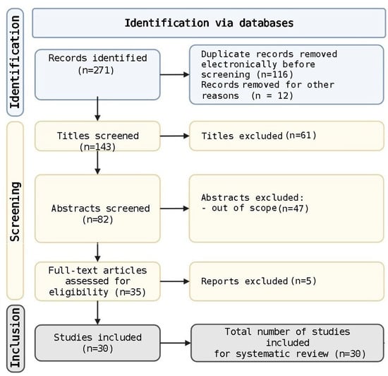
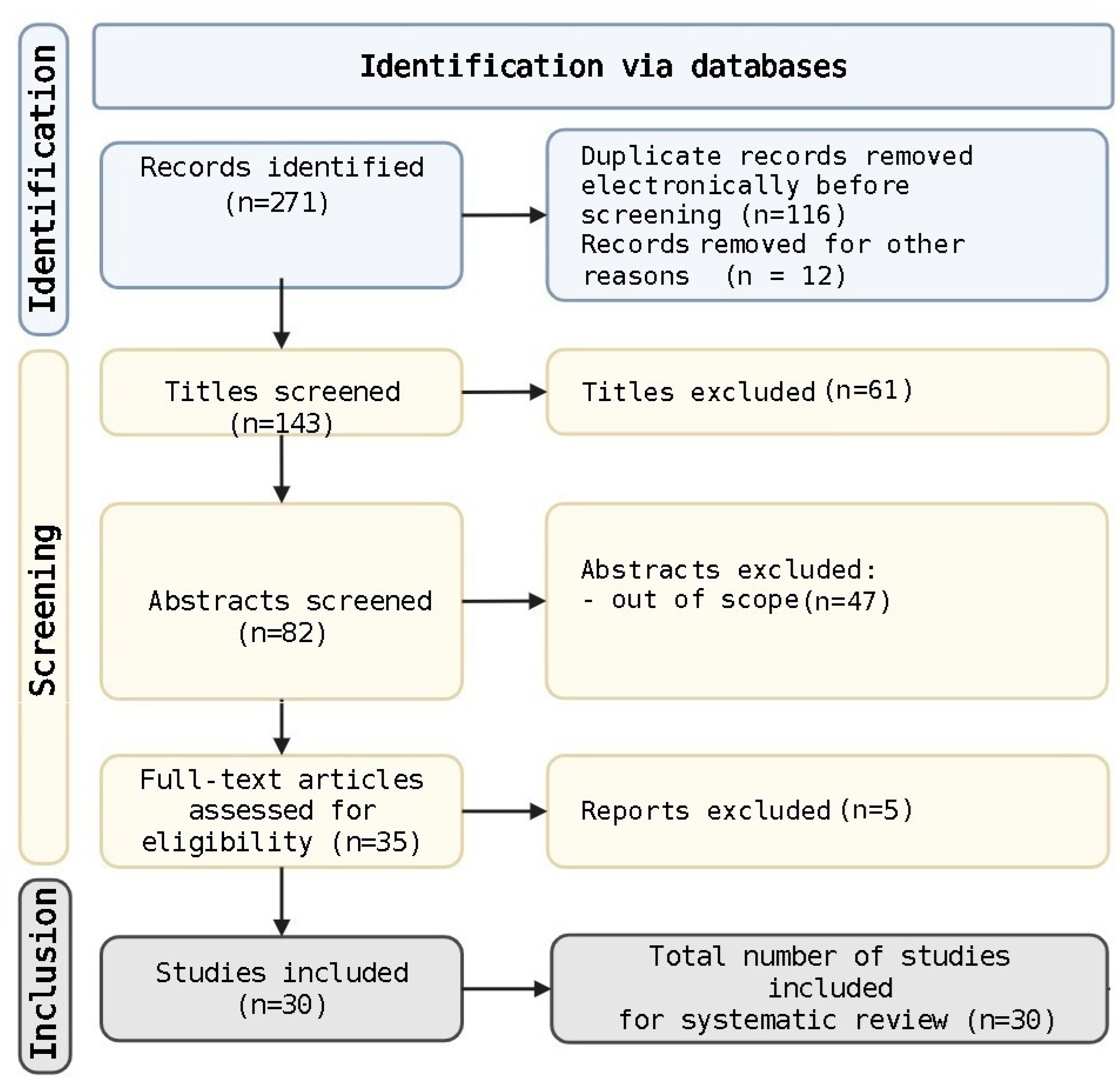
The PRISMA study selection flow is illustrated in the PRISMA 2020 flow diagram (Figure 1).
Figure 1.
PRISMA flow diagram. Created with BioRender. Nelson Twakor, A. (2026). https://BioRender.com/vcmup9b (accessed on 25 August 2026).
2.5. Data Extraction
For studies reporting performance metrics at multiple operating thresholds, the metric corresponding to the primary analysis or the threshold yielding the closest sensitivity–specificity balance was extracted. For studies presenting multiple AI algorithms or model iterations, the estimate corresponding to the algorithm designated by the original authors as their primary or final model was extracted for the main pooled analysis. For studies with multiple cohorts or subgroups, a single prespecified primary effect estimate per study was extracted and used in the main pooled analysis, so that each study contributed exactly one estimate and no clustering adjustment was required. Study-level AUROC values, 2 × 2 counts, and source locators used for Figure 2, Figure 3 and Figure 4 are in Supplementary Material S2.
2.6. Risk-of-Bias Assessment
Risk of bias for included diagnostic accuracy studies was appraised qualitatively by two independent reviewers, informed by the domains of the Quality Assessment of Diagnostic Accuracy Studies-2 (QUADAS-2) tool and by AI-specific methodological concerns raised in the DECIDE-AI and TRIPOD + AI reporting frameworks [12], including provenance and separation of training–testing datasets, reporting of demographic subgroups, and independence of the external validation cohort. Disagreements between the two reviewers were resolved through consensus discussion. A formal domain-by-domain scoring system and a quantitative inter-rater agreement statistic were not applied and are noted as a limitation.
For interventional studies, risk of bias was assessed using the Revised Cochrane Risk of Bias Tool for Randomized Trials (RoB 2), evaluating domains of randomization process, deviations from intended interventions, missing outcome data, measurement of outcomes, and selection of reported results [13]. Risk-of-bias assessments were performed independently by two reviewers. Disagreements were resolved by consensus.
2.7. Statistical Analysis
2.7.1. Software and Primary Meta-Analytic Outcome
Univariate random-effect models and the forest plots in Figure 2, Figure 3 and Figure 4 were generated in IBM SPSS Statistics version 30.0 (IBM Corp., Armonk, NY, USA) [14]. The primary bivariate analysis of sensitivity and specificity used the Reitsma random-effect model, fitted in Python 3 (SciPy) on the logit scale. The primary discrimination outcome was the area under the receiver operating characteristic curve (AUC/AUROC). Study-level AUROC values were transformed to the logit scale, logit(AUC) = ln[AUC/(1 − AUC)]. Standard errors on the logit scale were obtained from published 95% CIs when available, or by the delta method from a Hanley–McNeil standard error when only the AUROC and the numbers of diseased and non-diseased observations were reported. Pooling used a random-effect REML model on the logit scale. Only the pooled estimate and its 95% CI were back-transformed as AUC = 1/(1 + e(−logit)) so that the reported interval was constrained to (0, 1).
2.7.2. Meta-Analytic Model Selection
Given the substantial methodological and clinical heterogeneity anticipated across included studies, a random-effect model was prespecified as the primary meta-analytic model [15]. This model assumes that individual-study effect sizes are drawn from a distribution of true effects and estimate between-study variance (τ2). Fixed-effect modeling was not used as the primary approach, as the assumption of a single common true effect size is implausible given the heterogeneity of AI technologies and clinical contexts represented in this corpus [16].
Sensitivity and specificity were not treated as independent primary outcomes. Studies that provided a reconstructible binary 2 × 2 table at a free operating point (k = 7 independent testing sets) were analyzed with the Reitsma bivariate random-effect model, which jointly estimates logit-sensitivity, logit-specificity, and their between-study correlation. SPSS Figure 3 and Figure 4 display the corresponding univariate logit models for visual inspection. The manuscript primary estimates are the Reitsma summaries. Only one independent testing set per study was included Multiclass top-k accuracy, fixed-sensitivity equity analyses, lesion-detection rates, and duplicate lesion sets were excluded from the quantitative pool and retained in the narrative synthesis. A continuity correction of 0.5 was reserved for any zero cell. None was required in the primary 2 × 2 tables.
2.7.3. Heterogeneity Assessment
Between-study variability in pooled AUC, sensitivity, and specificity estimates was assessed through visual inspection of the forest plots and by comparing effect estimates across six prespecified moderator variables: AI technology category (CNN/deep learning vs. classical neural network vs. mobile app vs. 3D TBP system vs. multimodal DL vs. foundation model), skin condition (melanoma vs. non-melanoma conditions), study design (RCT/prospective trial vs. retrospective/model development study vs. diagnostic accuracy/reader study), clinical setting (specialist secondary/tertiary care vs. primary care/teledermatology vs. digital pathology laboratory), publication era (2000–2009 vs. 2010–2019 vs. 2020–2025), and comparator type (AI vs. specialist dermatologist vs. AI vs. non-specialist clinician vs. standalone AI). Between-study heterogeneity for the SPSS forest plots is reported as Cochran’s Q, I2, and τ2. For the Reitsma model, between-study standard deviations (τ_Se, τ_Sp) and the correlation ρ are reported. Moderator comparisons across AI technology, skin condition, study design, clinical setting, publication era, and comparator type remain descriptive because several subcategories contained too few studies for meta-regression.
3. Results
3.1. Study Selection and Inclusion
The systematic literature search identified a total of 30 studies meeting the predefined inclusion criteria for this review. Following database screening and full-text assessment, all 30 publications were retained for qualitative synthesis. Two articles were subsequently identified as formally retracted by their respective publishers, both as part of a large-scale mass retraction wave involving more than 500 papers across multiple Hindawi-owned open-access journals due to concerns regarding compromised peer review, potential paper mill involvement, and unverifiable data integrity (retraction notices are included in Supplementary Material S3). These two articles were excluded from all quantitative analyses and performance metric syntheses. Their citation information is preserved in the study characteristics table (Table 2) for transparency purposes. The final analytic cohort therefore comprised 28 non-retracted studies. Throughout this manuscript, the two retracted studies are marked with a dagger (†) in every table. They are included only in the descriptive, qualitative-synthesis totals reported as n = 30, while all quantitative pooled analyses, forest plots, and the Reitsma bivariate model are based on the 28 non-retracted studies (n = 28).
Table 2.
Characteristics of included studies.
Included studies span a publication period of 22 years, from 2003 to 2025, reflecting the full trajectory of AI application in dermatology from early computational neural network experiments to contemporary large language model (LLM)- and vision–language model (VLM)-based tools. The distribution of publications was markedly skewed toward the most recent decade, with 25 of 30 studies (83.3%) published between 2020 and 2025, 3 studies (10.0%) published between 2018 and 2019, and 2 studies (6.7%) published before 2010.
3.2. Characteristics of Included Studies
The 30 included studies were published across a diverse range of high-impact peer-reviewed journals, including The Lancet Digital Health, JAMA Network Open, JAMA Dermatology, Annals of Oncology, Nature Communications, British Journal of Dermatology, Journal of the American Academy of Dermatology, European Journal of Cancer, Journal of Investigative Dermatology, Journal of the European Academy of Dermatology and Venereology, PLoS One, and several specialty digital health journals (JMIR Dermatology; Scientific Reports; Journal of Proteomics).
Study designs were heterogeneous (Table 3). The largest category was retrospective cohort and model development studies (n = 9; 30.0%), followed by prospective cohort studies and clinical trials (n = 8; 26.7%) and diagnostic accuracy/reader comparison studies (n = 6; 20.0%). Three studies (10.0%) were RCTs or prespecified analyses derived from RCT data, including the nested cost-effectiveness sub-study of Lindsay et al. [32], providing the highest level of clinical evidence available within the corpus. Cross-sectional and survey-based studies accounted for two publications (6.7%). Notably, only three RCT-level studies exist in the entire 22-year corpus [29,32,35], showing a persistent gap between the availability of AI diagnostic tools and their rigorous evaluation in interventional trial settings.
Table 3.
Distribution of included studies by study design.
Sample sizes varied substantially across study types. Model development and reader studies typically involved hundreds of curated images rather than patient-level cohorts. The largest patient-level cohort was the Melanoma MEGA-study [33] encompassing 1653 patients from an international multicenter population. Other large prospective datasets included Winkler et al. [39] with 236 patients and 1075 clinically relevant melanocytic lesions (CRMLs), and another study with 309 high-risk patients followed over a 2-year RCT [32]. The smallest study was Gantenbein et al. [30], an AI-driven prevention pilot involving 60 healthy young women.
3.3. Geographic Distribution
Geographically, the included studies originated predominantly from European and Asia-Pacific settings (Table 4). European studies accounted for 14 publications (46.7%), with Germany contributing the largest national share (6 studies), followed by the Netherlands, Sweden, Switzerland, France, Spain, and Italy. Asia-Pacific and Oceanian studies comprised five publications (16.7%), including two from Australia, two from South Korea, and one from India. Three studies originated exclusively from China (10.0%), though this includes two retracted publications [40,44]. North American studies numbered three (10.0%), all from the United States. Five additional studies (16.7%) involved international multicenter collaborations spanning multiple continents, typically combining European and North American institutions.
Table 4.
Geographic distribution of included studies.
The geographic concentration of AI dermatology research in Europe and East Asia shows both the presence of large academic dermatology centers with extensive dermoscopy archives and the regulatory environments in these regions that have facilitated CE-marking and clinical validation of AI medical devices. The relative underrepresentation of low- and middle-income country settings, with the exception of India, Marri et al., 2024 [25], is a limitation of the current evidence base, particularly given that access to specialist dermatology care remains severely constrained across large portions of sub-Saharan Africa, South Asia, and Latin America, where AI-enabled teledermatology could have the greatest transformative impact.
3.4. Dermatological Conditions Addressed
The distribution of targeted dermatological conditions was markedly skewed toward melanoma and melanocytic lesions (Table 5). Melanoma constituted the primary disease focus in 18 of 30 studies (60.0%), encompassing applications ranging from dermoscopic diagnosis and whole-body screening to digital pathology, lymph node staging, and precision oncology.
Table 5.
Distribution of included studies by targeted skin condition.
Beyond skin cancer, onychomycosis was the subject of two studies (6.7%): the retrospective EHR cohort of Roustan et al. [19] and the prospective comparative study of Kim et al. [42]. One study each addressed lupus erythematosus [27], acne vulgaris [35], and vitiligo [45]. Two studies evaluated AI across broad arrays of dermatoses, either through wide-spectrum consumer mobile apps (Marri et al. [25]) or through automated histopathological report generation covering multiple malignancy types (Tran et al. [24]).
This concentration of research effort creates a knowledge gap regarding AI performance across the far broader spectrum of inflammatory, infectious, and autoimmune skin conditions that constitute the majority of dermatological workload in clinical practice.
3.5. AI Technology and Temporal Evolution
The AI technologies employed across the included studies reflect the broader evolution of machine learning and computer vision over the last two decades, from early rule-based systems and classical artificial neural networks to contemporary foundation models (Table 6 and Table 7).
Table 6.
Classification of included studies by AI technology category.
Table 7.
Temporal evolution of AI technologies in dermatology.
3.5.1. Early Era: Classical Neural Networks (2003–2009)
The two earliest studies in this corpus [45,46] employed classical artificial neural networks (ANNs) operating on handcrafted feature inputs rather than raw image data. Hoffmann et al. [46] developed the DANAOS (diagnostic and neural analysis of skin cancer) expert system, an ANN trained on 2218 calibrated digital dermoscopic images collected prospectively from 13 dermatology centers in 9 European countries. DANAOS demonstrated performance similar to that of dermatologists as published in the literature, representing a landmark validation of computer-aided dermoscopy analysis and anticipating the concept. Cazzaniga et al. [45] applied discriminant and regression neural networks to clinical and photographic data from 325 vitiligo patches, achieving 66.5% accuracy in classifying excimer laser treatment responders from non-responders, representing the first application of AI to treatment response prediction in a pigmentary skin condition. These early studies were constrained by small datasets, handcrafted feature engineering, and lack of GPU-accelerated training, yet they established the conceptual foundations that subsequent deep learning approaches would operationalize at scale. Despite the proof of concept established by early computational dermoscopy studies, no studies meeting our eligibility criteria were identified in the period 2010 to 2017.
3.5.2. Rise of Deep Learning: Convolutional Neural Networks (2018–2020)
The transition from classical ANNs to deep CNNs fundamentally transformed the field. CNNs eliminated the need for manual feature engineering and enabled end-to-end training on large image corpora. Three pivotal reader comparison studies published between 2018 and 2019 established the paradigm that CNN-based AI could match or surpass specialist dermatologists in melanoma recognition from dermoscopic images.
Haenssle et al. [22] reported that a modified Google Inception V4 CNN achieved an AUC of 0.95 for dermoscopic melanoma classification, outperforming 58 international dermatologists from 17 countries at both experience levels tested (Level I: dermoscopy only, AUC 0.79; Level II: dermoscopy plus clinical information, AUC 0.82). The CNN demonstrated higher sensitivity (95%) and specificity (80%) than both clinician groups, a finding that attracted widespread attention in the dermatological and broader medical AI communities. One year later, Brinker et al. [36] extended this paradigm using a CNN trained exclusively on 12,378 open-source dermoscopic images, demonstrating superior performance over 136 of 157 dermatologists (87%) from 12 German university hospitals across all seniority levels, with CNN sensitivity of 82.3% versus dermatologist sensitivity of 67.2%. Around the same time, Phillips et al. [28] independently validated comparable CNN superiority over board-certified dermatologists, reinforcing the robustness of these findings across different CNN architectures and testing datasets.
In 2020 Kim et al. [42] provided the first prospective evaluation of a DNN for onychomycosis diagnosis, finding that the algorithm (AUC 0.751) was statistically equivalent in performance to both dermoscopic examination (AUC 0.755) and experienced board-certified dermatologists (sensitivity 73.0%), with the additional practical advantage that AI analysis used standard clinical photographs obtainable by non-physicians rather than requiring specialist dermoscopic equipment.
3.5.3. Clinical Validation and Real-World Translation (2021–2023)
Between 2021 and 2023, the field shifted from demonstrating that AI could match experts on curated testing sets toward evaluating clinical utility in real-world settings, validating regulatory-approved devices, and exploring histopathological applications. Höhn et al. [41] in 2021 combined CNN-based whole-slide-image classification with patient demographic data for histopathological melanoma–nevus differentiation, achieving an AUROC of 92.3% for the CNN alone and demonstrating that selective fusion with patient metadata improved balanced accuracy from 83.2% to 86.7% for low-confidence cases. Jansen et al. [31] extended CNN application to melanoma lymph node staging on hematoxylin and eosin-stained sentinel lymph node sections, achieving AUC > 0.96 on two independent test cohorts without requiring immunohistochemistry.
Sangers et al. [34] provided a clinically grounded prospective multicenter validation of a CE-marked AI mHealth application in 785 lesions from 372 patients in the Netherlands, reporting sensitivity of 86.9% and specificity of 70.4%, and identifying a significant performance disparity between iOS (91.0%) and Android (83.0%) platforms. Han et al. [29] published the first RCT demonstrating that AI-assisted diagnosis significantly improved non-expert physician accuracy for skin neoplasms (53.9% vs. 43.8% unaided; p = 0.019), providing evidence for the clinical decision support role of AI. Kommoss et al. [43] demonstrated that a market-approved CNN reduced incorrect management of diagnostically “unclear” face and scalp lesions from 58.8% to 4.1% for malignant cases. Menzies et al. [17] found that a seven-class mobile AI algorithm achieved diagnostic accuracy equivalent to specialist dermatologists (absolute difference +1.2%; 95% CI −6.9 to +9.2), while demonstrating that not all AI algorithms perform equivalently: the ISIC algorithm tested in the same setting was significantly inferior to specialists (Δ = −11.6%).
3.5.4. Foundation Models, Multimodality, and Next-Generation AI (2024–2025)
The most recent stratum of studies reflects a new technological inflection in dermatological AI, characterized by multimodal architectures, foundation models, 3D-imaging integration, and AI applications beyond binary diagnosis. Papachristou et al. [18] demonstrated that a smartphone-based CNN decision support tool used prospectively by primary care physicians achieved AUROC 0.960 for melanoma, with 100% sensitivity for invasive melanoma (AUROC 0.988), establishing a compelling clinical case for AI-enhanced primary care referral pathways. Li Q et al. [27] deployed a multimodal deep learning system integrating clinical images, multicolor immunohistochemistry images, and structured clinical data to classify 13 skin conditions, including lupus erythematosus subtypes, achieving AUC 0.9844 and improving dermatologist diagnostic accuracy from 66.9% to 81.3% (p = 0.0004).
Tran et al. [24] introduced HistoGPT, the first vision–language foundation model for dermatopathology, trained on 15,129 WSIs from 6705 patients. HistoGPT generates free-text dermatopathology reports from gigapixel whole-slide images in a zero-shot fashion, matching human pathologist report quality for common homogeneous malignancies. Aung et al. [23] demonstrated that AI-driven quantification of tumor-infiltrating lymphocytes using the Hover-NeXt deep learning architecture outperformed traditional pathologist scoring in predicting immunotherapy response and survival in advanced melanoma. Guedes et al. [33] integrated proteogenomics, AI-driven digital pathology, and machine learning analytics in 1653 patients through the Melanoma MEGA-Study, identifying novel protein biomarker signatures for immune evasion and therapeutic resistance.
In the domain of prevention, Gantenbein et al. [30] deployed a generative AI-based facial photoaging simulation tool, finding that 91.7% of young women were motivated to reduce UV exposure immediately following the simulation, with 96% reporting persistent behavior modification at a 2-year follow-up.
3.6. Teledermatology and Primary Care AI
Three studies specifically evaluated AI in teledermatology or primary care contexts. Papachristou et al. [18] demonstrated that primary care physicians using an AI smartphone application achieved 100% sensitivity for invasive melanoma in a Swedish primary care setting. Le Lay et al. [38] validated an AI-assisted tele-expertise platform for common skin diseases in a French proof-of-concept study, achieving top-three diagnostic accuracy of 90% with significant improvement between algorithm versions 1 and 2. Marri et al. [25] evaluated the Aysa consumer AI application in an Indian semiurban setting, achieving top-three sensitivity of 86.1% across 12 dermatosis categories, but with notably poor performance for malignant tumors (top sensitivity 10%).
3.7. Treatment, Prevention, and Attitudinal Studies
Beyond diagnosis, three studies evaluated AI for treatment response prediction: Aung et al. [23] for immunotherapy response prediction via TIL quantification, Cazzaniga et al. [45] for excimer laser response prediction in vitiligo, and Guedes et al. [33] for precision oncology stratification in melanoma. Two studies evaluated AI-driven treatment recommendations: Roustan et al. [19] using NLP-based EHR analysis to characterize real-world outcomes in onychomycosis, and Ghazanfar et al. [35] demonstrating that an ML-based skincare recommendation system significantly outperformed self-selected treatment for mild-to-moderate acne vulgaris. Two attitudinal studies [20,26] examined patient and dermatologist preferences for AI integration in skin cancer diagnostics, finding that patients preferred augmented intelligence (AI + dermatologist) over either AI alone or unassisted dermatology, and that 83.4% of patients preferred AI-augmented consultations. Table 8 summarizes the above.
Table 8.
AI application categories across included studies.
3.8. Diagnostic Performance of AI Systems
Reported diagnostic performance metrics varied substantially across studies (Table 9). Among CNN-based dermoscopy studies, reported AUC values ranged from 0.826 (classical ANN, Hoffmann et al. [46]) to 0.960 (smartphone CNN, Papachristou et al. [18]). In the pivotal comparative studies, Haenssle et al. [22] reported CNN AUC of 0.95 versus dermatologist AUC of 0.79–0.82; Brinker et al. [36] found CNN sensitivity of 82.3% and specificity of 77.9% versus dermatologist sensitivity of 67.2% and specificity of 62.2%. Several studies noted that AI performance degrades with image quality, lesion rarity, or mismatched population demographics (as demonstrated by Mitre et al. [21]).
Table 9.
Reported diagnostic performance metrics across included studies.
The single RCT directly evaluating AI-assisted clinical decision support for skin neoplasm diagnosis, Han et al. [29], demonstrated a significant improvement in non-expert physician accuracy with AI assistance (53.9% vs. 43.8%; p = 0.019). The RCT by Ghazanfar et al. [35] extended this to treatment. Their ML-generated personalized skincare recommendations produced significantly greater acne severity reduction compared with self-selected treatment over 8 weeks. Lindsay et al. [32], while demonstrating clinical non-inferiority of 3D TBP-based AI surveillance, found that this approach was not cost-effective at current implementation costs, with 0% probability of cost-effectiveness from a government payer perspective at 2 years.
3.9. Forest Plot Analysis
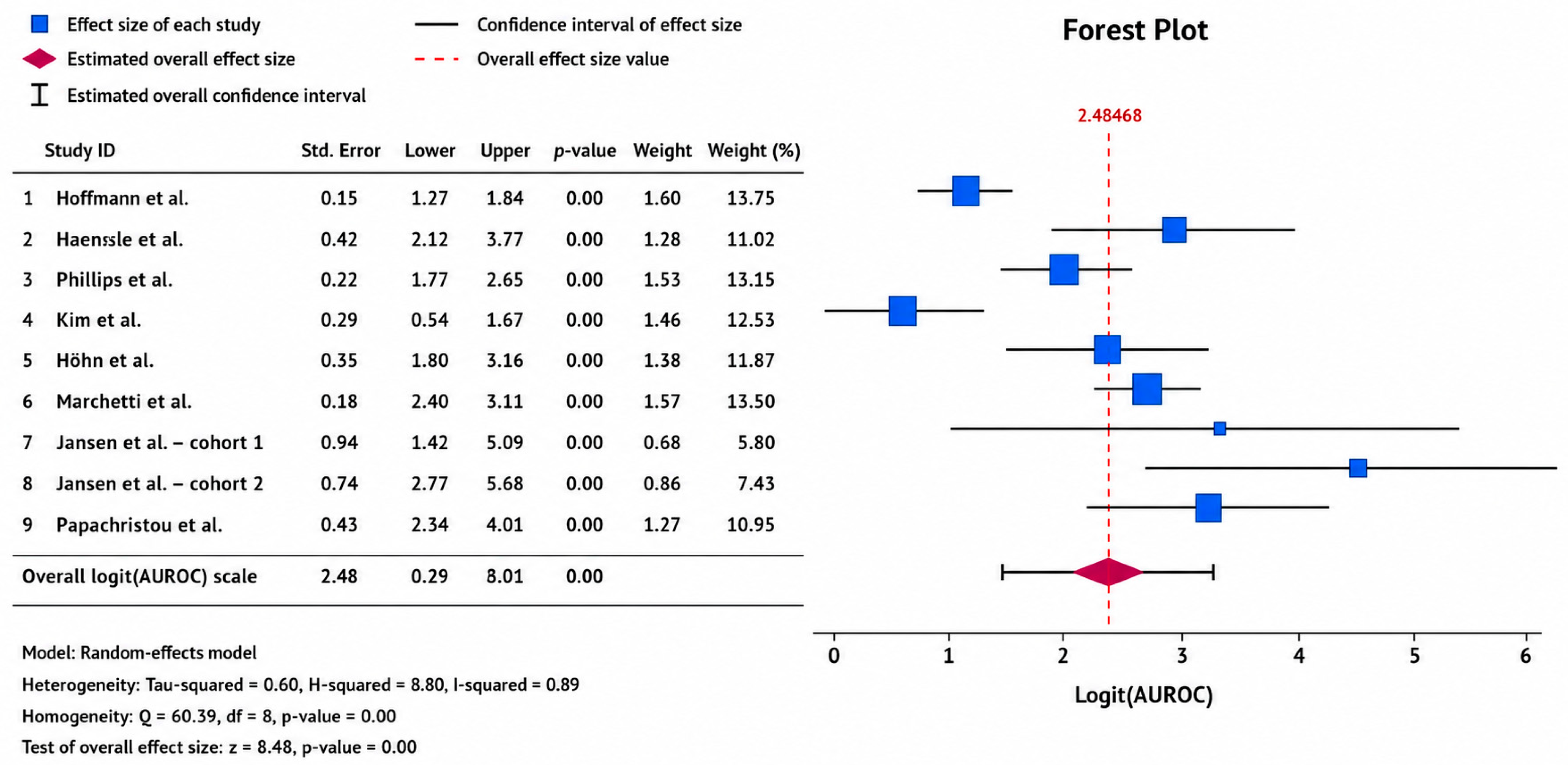
The forest plots summarize diagnostic performance as AUROC. Pooling was done on the logit(AUROC) scale (random-effect REML; k = 9 independent testing sets), so Figure 2 displays logit values. The SPSS summary diamond is 2.48 (95% CI 1.91–3.06; I2 = 89%; τ2 = 0.60). Back-transformation with AUROC = 1/(1 + e(−logit)) gives a pooled AUROC of 0.92 (95% CI 0.87–0.96).
Several melanoma-focused studies reported strong discrimination. Haenssle et al. [22] obtained AUROC 0.95 on the independent test-set-300. Phillips et al. [28] reported an iPhone histology-verified AUROC of 0.901 (0.863–0.940), and Papachristou et al. [18] reported an overall AUROC of 0.960 (0.928–0.980). Hoffmann et al. [46] reported multicenter ROC area of 0.826 ± 0.021, and Jansen et al. [31] reported section-level AUROC values of 0.963 and 0.986 at two independent test centers.
Figure 2.
Forest plot of logit(AUROC) from the random-effect REML model (SPSS vs. 30.0). The pooled logit of 2.48 (1.91–3.06) corresponds to a back-transformed AUROC of 0.92 (0.87–0.96). Studies included Hoffmann et al. [46]; Haenssle et al. [22]; Phillips et al. [28]; Kim et al. [42]; Höhn et al. [41]; Marchetti et al. [37]; Jansen et al. [31]; Papachristou et al. [18].
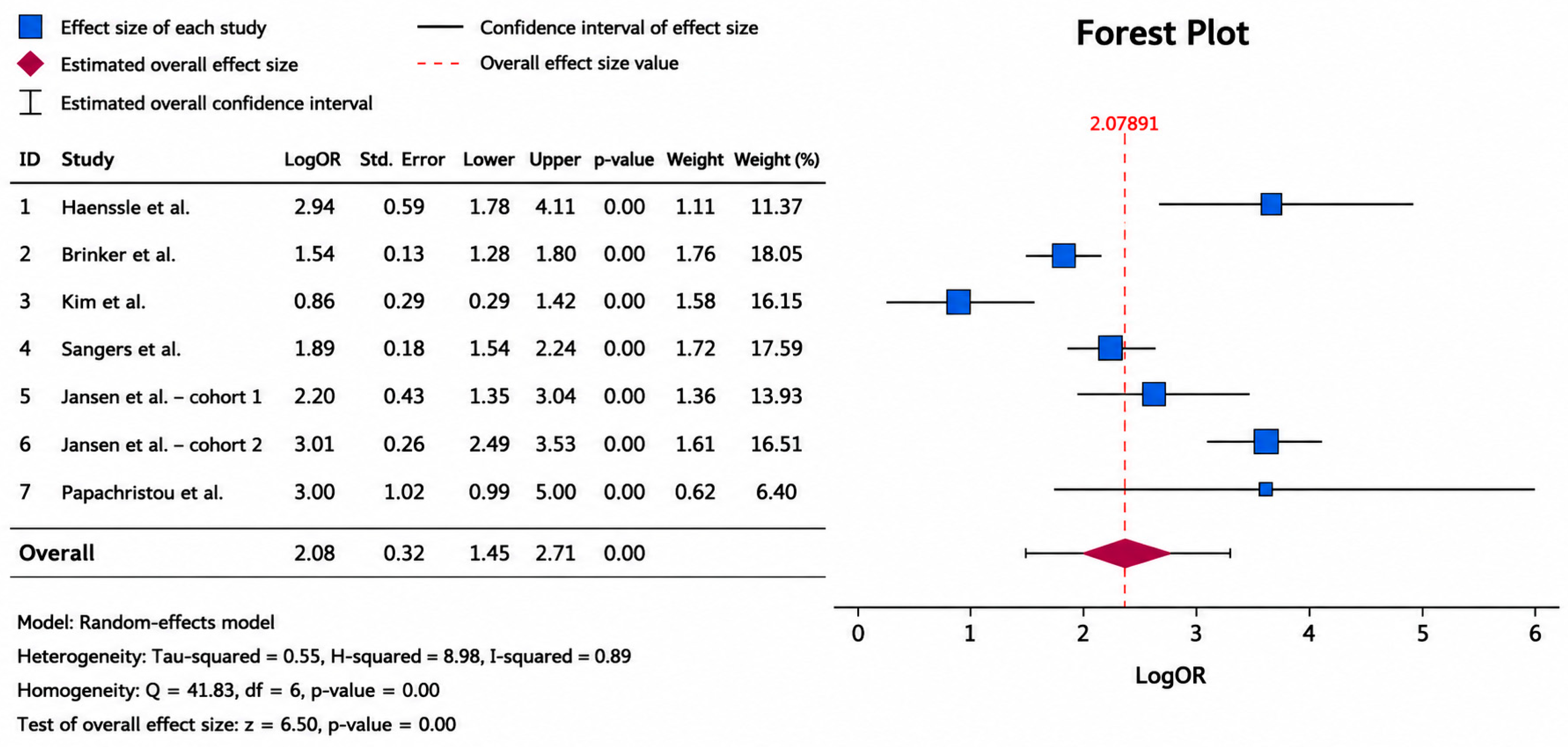
Pooling of sensitivity was done on the logit scale in SPSS (random-effect REML; k = 7 independent 2 × 2 tables), so Figure 3 displays logit values. The SPSS summary diamond is 2.08 (95% CI 1.45–2.71; I2 = 89%; τ2 = 0.55). Back-transformation with sensitivity = 1/(1 + e(−logit)) gives a univariate pooled sensitivity of 0.89 (95% CI 0.81–0.94). The primary manuscript estimate is the Reitsma bivariate model, which jointly analyses sensitivity and specificity: 0.88 (95% CI 0.82–0.93). On the 0–1 scale, study sensitivities ranged from 0.70 (Kim et al. [42]) to 0.95 (Haenssle et al. [22]; Jansen et al. [31] Center 2). Papachristou et al. [18] contributed the overall Youden point (20/21, 0.95), not the invasive-only value of 1.00, which is the same 253 lesions.
Some variability was observed due to differences in study design, populations, imaging modalities, and AI architectures. Technologies ranged from traditional neural networks [46] and CNNs [22,36] to smartphone-based applications [18] and multimodal systems [27] across conditions including melanoma, skin neoplasms, onychomycosis, and lupus erythematosus. Given this substantial heterogeneity (I2 = 89–97% across the three pooled outcomes), the pooled AUROC, sensitivity, and specificity estimates should be interpreted with caution: they summarize a highly diverse set of AI technologies, patient populations, and clinical settings, and should not be read as a single, precise, generalisable performance figure applicable to any individual clinical context.
Figure 3.
Forest plot of logit(sensitivity) from the random-effect REML model. The pooled logit of 2.08 (1.45–2.71) corresponds to a univariate sensitivity of 0.89 (0.81–0.94). The primary pooled estimate is the Reitsma sensitivity of 0.88 (0.82–0.93). Studies included Haenssle et al. [22]; Brinker et al. [36]; Kim et al. [42]; Sangers et al. [34]; Jansen et al. [31]; Papachristou et al. [18].
Pooling of specificity was done on the logit scale in SPSS (random-effect REML; k = 7 independent 2 × 2 tables), so Figure 4 displays logit values. The SPSS summary diamond is 1.74 (95% CI 1.08–2.40; I2 = 97%; τ2 = 0.75). Back-transformation with specificity = 1/(1 + e(−logit)) gives a univariate pooled specificity of 0.85 (95% CI 0.75–0.92). The primary manuscript estimate is the Reitsma bivariate model: 0.85 (95% CI 0.75–0.91).
Figure 4.
Forest plot of logit(specificity) from the random-effect REML model (SPSS vs. 30.0). The pooled logit of 1.74 (1.08–2.40) corresponds to a univariate specificity of 0.85 (0.75–0.92). The primary pooled estimate is the Reitsma specificity of 0.85 (0.75–0.91). Studies included Haenssle et al. [22]; Brinker et al. [36]; Kim et al. [42]; Sangers et al. [34]; Jansen et al. [31]; Papachristou et al. [18].
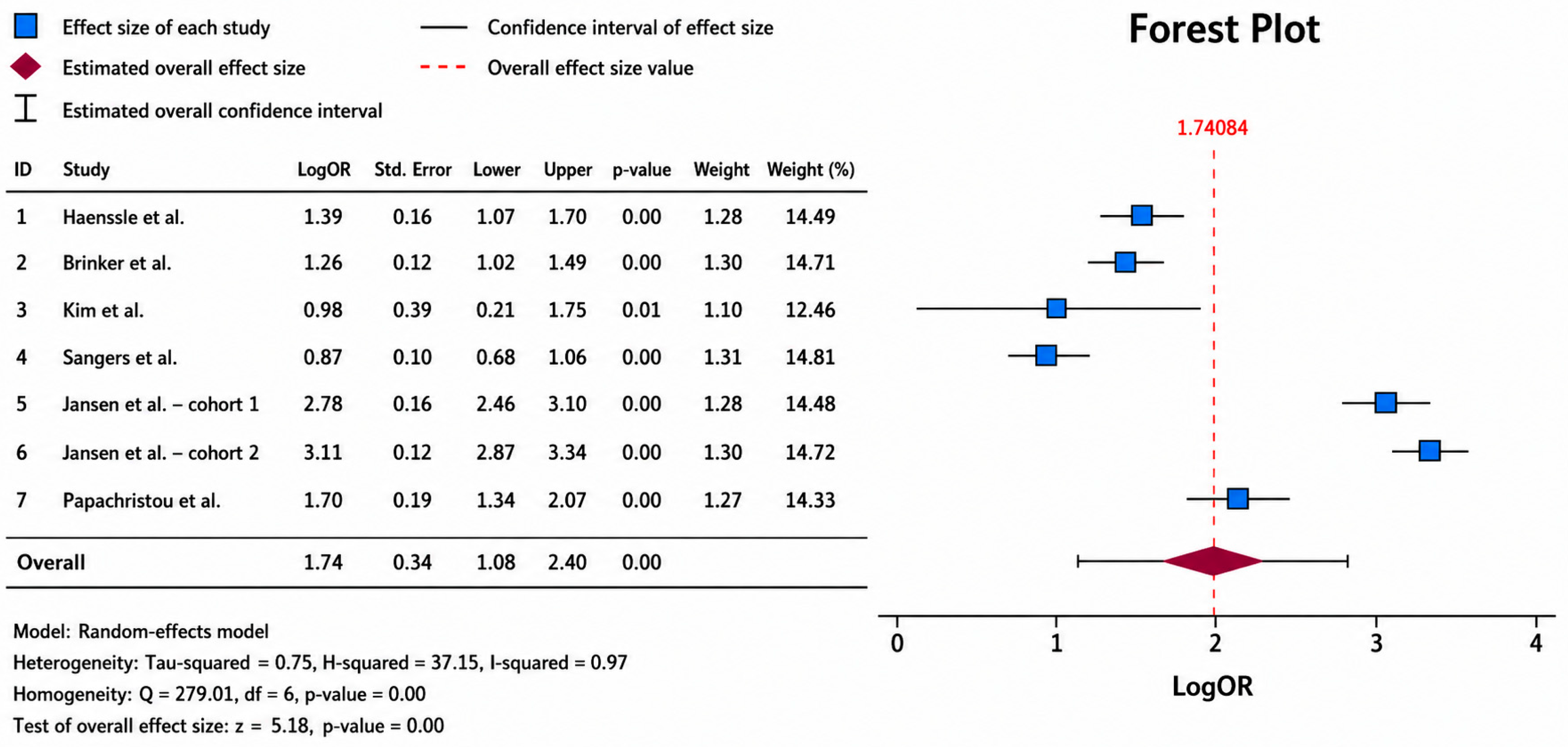
On the 0–1 scale, study specificities ranged from 0.70 (Sangers et al. [34]) to 0.96 (Jansen et al. [31] Center 2). Papachristou et al. [18] contributed the overall Youden specificity of 0.85, not the invasive-only 0.93.
3.10. Data Quality, Risk of Bias, and Retracted Studies
Risk of bias was assessed qualitatively across included studies. First, the majority of dermoscopy-based AI studies used retrospective, curated image datasets with balanced class distributions that are unrepresentative of real-world clinical prevalence. Melanoma prevalence in real-world dermatology practice is typically 2–5% of assessed lesions [47,48,49]. Most CNN evaluation sets used melanoma prevalence of 30–50%, substantially inflating sensitivity estimates and potentially reducing the positive predictive value achievable in clinical deployment.
Second, geographic and demographic homogeneity in training datasets represents a significant source of systematic bias. The ISIC dataset contains a disproportionate representation of Fitzpatrick phototype I–III skin [50,51] from European and Australian populations, with minimal representation of Fitzpatrick phototype V–VI skin. Mitre et al. [21] provide direct empirical evidence of the clinical harm this bias produces, reporting near-zero AI specificity (0–4.3%) for benign acral lesions in Black patients at a standard 95% sensitivity threshold.
Third, comparator standardization was inconsistent across reader studies. Dermatologist comparators ranged from single junior clinicians to panels of 58 international experts. The conditions of clinical information available to human readers varied, and the definition of “gold standard” ranged from histopathological diagnosis to clinical consensus, introducing systematic heterogeneity in relative performance estimates.
The two retracted studies involved claims of AI-based image analysis in dermatological contexts (3D skin CT in pediatric psoriasis [40]; deep learning melanoma detection from histopathological slides [44]) that could not be independently verified following retraction.
4. Discussion
This meta-analysis demonstrates a pooled AUROC of 0.92 (95% CI 0.87–0.96) for AI diagnostic discrimination, with Reitsma sensitivity 0.88 (0.82–0.93) and specificity 0.85 (0.75–0.91), consistently matching or surpassing specialist dermatologist performance in direct comparisons. We found similar evidence in the literature [52,53]. These findings align closely with those of Salinas et al. [54], whose meta-analysis of 30 studies reported a pooled AUC of 0.91 (95% CI 0.89–0.93) for AI versus clinicians in skin cancer diagnosis, with AI performing equivalently to or better than dermatologists in the majority of included studies. The most recent prospective evidence synthesis by Laiouar-Pedari et al. [55] confirmed that AI-assisted decision support reduced melanoma miss rates compared with unaided clinicians.
A recurring theme in our results is the gap between controlled benchmark performance and real-world deployment. Heinlein et al. [56] prospectively demonstrated in a 29-center German study that AI assistance improved melanoma recognition in patient care, though performance gains were smaller than those reported in retrospective reader studies. Thomas et al. [57] similarly documented real-world performance degradation relative to pre-deployment validation in a post-market surveillance analysis. The CLEAR Derm consensus guidelines provide a standardised checklist for evaluating the rigor of image-based AI reports [58]. This broader principle of context-dependent clinical decision-making is also supported by real-world evidence from other medical domains, where patient comorbidities, baseline risk, and treatment characteristics can substantially influence clinical outcomes and therefore need to be considered when translating evidence into individualized care [59].
The near-zero specificity (0–4.3%) observed by Mitre et al. [21] for benign acral lesions in Black patients is the most clinically urgent finding in the present review, and is not an isolated observation. Daneshjou et al. [60] demonstrated significantly worse AI performance across darker Fitzpatrick phototypes on a diverse curated dataset, while Guo et al. [61] documented systematic underrepresentation of non-White skin types in published AI training datasets. Dovigi et al. [62] further reviewed bias, ethics, and regulatory considerations in dermatological AI, concluding that existing regulatory pathways fail to mandate demographic subgroup performance reporting as a condition of market approval.
Our results reveal a pronounced concentration of AI research on melanoma (60% of included studies), with comparatively sparse evidence for inflammatory and infectious dermatoses. This mirrors the evidence gap identified by Cai et al. [63] in a systematic review of AI for inflammatory skin disease severity assessment, which found high heterogeneity and modest performance for conditions such as psoriasis and atopic dermatitis. For acne, CNN-based grading systems have shown promise [64], while machine learning models have demonstrated early utility in predicting atopic dermatitis flares from patient-reported and sensor data [65]. The AI teledermatology literature confirms that algorithms embedded in primary care workflows can improve early detection rates, particularly in geographically underserved populations where specialist access is limited [63].
Beyond image interpretation alone, artificial intelligence is increasingly driving the transition toward personalized medicine through the integration of multimodal clinical, molecular, and microbiome-derived data, enabling more individualized diagnostic, prognostic, and therapeutic strategies [65]. Hyperspectral imaging combined with machine learning is a further emerging modality with reported potential to enhance skin cancer classification beyond conventional RGB dermoscopy. This technology was not represented among the 30 studies included in the present corpus and does not currently map onto the AI technology taxonomy used in Table 6, representing a direction for future updates of this review [66]. The convergence of artificial intelligence with smart biomaterials and connected technologies is also expanding beyond diagnostic support into medical traceability and forensic medicine. Intelligent materials incorporating near-field communication technology have demonstrated feasibility for durable identification [67].
Health economic considerations remain a critical, but underdeveloped domain. Our paper contains only one formal cost-effectiveness analysis [32], which found AI-based total-body photography surveillance not cost-effective over a 2-year horizon. This is consistent with a broader modeling study by Gomez Rossi et al. [68], which found that AI-assisted melanoma detection reached cost-effectiveness thresholds only under specific prevalence and sensitivity assumptions. Regulatory oversight of AI dermatology applications remains fragmented. Wongvibulsin et al. [69] found that the majority of commercial dermatology apps with AI features available in app stores lacked regulatory clearance, a concern also raised by Matin and Dinnes [70] regarding smartphone-based risk assessment tools.
In computational pathology, AI systems for dermatopathology demonstrate consistently high accuracy for melanoma classification on whole slide images, as confirmed by Lalmalani et al. [71]. A Cochrane review by Ferrante di Ruffano et al. [72] identified a high risk of bias in early computer-assisted dermoscopy studies due to methodological deficiencies in patient selection and reference standard definition.
Furthermore, the absence of prospective, registry-level outcome data beyond short-term diagnostic accuracy metrics limits conclusions regarding the impact of AI on patient survival and long-term clinical outcomes. In addition, risk-of-bias appraisal in this review was qualitative rather than based on a formal domain-by-domain QUADAS-2/RoB 2 scoring system with a quantitative inter-rater agreement statistic. A structured, study-level risk-of-bias table is identified as a priority for a future update of this review.
Looking forward, multimodal foundation models such as PanDerm (Yan et al. [73], trained across diverse dermatological image types, represent the next generation of broadly applicable AI in dermatology, while SkinGPT-4 (Zhou et al. [74]) demonstrates that pretrained multimodal large language models can support differential diagnosis and management reasoning from clinical images.
5. Conclusions
This meta-analysis provides the most temporally comprehensive synthesis to date of artificial intelligence applications across dermatology. The evidence base demonstrates a consistent and reproducible trajectory: from early classical neural networks achieving dermatologist-equivalent performance on curated image sets, through the deep learning revolution catalyzed by landmark CNN studies, to the current generation of foundation models, multimodal systems, and AI-integrated clinical workflows spanning diagnosis, pathology, screening, and precision oncology.
Across diagnostic applications, the pooled AUROC was 0.92 (95% CI 0.87–0.96), with Reitsma sensitivity 0.88 (0.82–0.93) and specificity 0.85 (0.75–0.91), frequently matching or surpassing specialist dermatologist performance. Randomized controlled trial evidence confirms that AI decision support significantly improves non-expert diagnostic accuracy, establishing a robust evidentiary basis for AI as a clinical augmentation tool rather than a standalone replacement. In histopathological applications, deep learning systems have demonstrated high accuracy for melanoma detection in whole slide images, lymph node staging, and tumor-infiltrating lymphocyte quantification.
However, critical limitations temper these findings. Algorithmic racial bias represents an urgent patient safety concern requiring immediate attention in dataset curation and regulatory review. Health economic analysis reveals that clinical accuracy alone does not guarantee cost-effectiveness. Furthermore, the absence of prospective, registry-level outcome data beyond short-term diagnostic accuracy metrics limits conclusions regarding the impact of AI on patient survival and long-term clinical outcomes.
In conclusion, AI in dermatology has transitioned from proof of concept to clinically validated reality across multiple application domains. Responsible translation into routine practice requires technology-specific validation, equitable dataset representation, rigorous health economic evaluation, and structured post-market surveillance.
Supplementary Materials
The following supporting information can be downloaded at: https://www.mdpi.com/article/10.3390/diagnostics16172797/s1. Supplementary Material S1: Complete literature search results and study database. Supplementary Material S2: Data underlying the diagnostic accuracy analyses. Supplementary Material S3: PRISMA_checklist.
Author Contributions
Conceptualization, N.I., M.F.P. and A.-O.T.; methodology, N.I., M.F.P. and A.-O.T.; formal analysis, N.I., M.F.P., A.-O.T. and N.C.P.; investigation, N.I., M.F.P., A.-O.T., N.C.P., R.M.F., D.C., A.N.T. and D.V.S.; data curation, N.I., M.F.P., N.C.P., R.M.F. and A.N.T.; writing—original draft preparation, N.I. and M.F.P.; writing—review and editing, A.-O.T., D.C., D.V.S., K.L.C. and R.D.; visualization, N.I., M.F.P. and N.C.P.; supervision, A.-O.T., K.L.C. and R.D.; project administration, A.-O.T.; funding acquisition, none. All authors have read and agreed to the published version of the manuscript.
Funding
We would like to acknowledge Victor Babes University of Medicine and Pharmacy for their support in covering the costs of publication for this research paper. All individuals have consented to the acknowledgement.
Institutional Review Board Statement
Not applicable.
Informed Consent Statement
Not applicable.
Data Availability Statement
No new data were created or analyzed in this study. Data sharing is not applicable to this article.
Acknowledgments
We would like to acknowledge Victor Babes University of Medicine and Pharmacy for their support in covering the costs of publication for this research paper. All individuals have consented to the acknowledgement.
Conflicts of Interest
The authors declare no conflicts of interest.
References
- Hay, R.J.; Johns, N.E.; Williams, H.C.; Bolliger, I.W.; Dellavalle, R.P.; Margolis, D.J.; Marks, R.; Naldi, L.; Weinstock, M.A.; Wulf, S.K.; et al. The global burden of skin disease in 2010: An analysis of the prevalence and impact of skin conditions. J. Investig. Dermatol. 2014, 134, 1527–1534. [Google Scholar] [CrossRef] [Scilit]
- Arnold, M.; Singh, D.; Laversanne, M.; Vignat, J.; Vaccarella, S.; Meheus, F.; Cust, A.E.; de Vries, E.; Whiteman, D.C.; Bray, F. Global burden of cutaneous melanoma in 2020 and projections to 2040. JAMA Dermatol. 2022, 158, 495–503. [Google Scholar] [CrossRef] [Scilit]
- Lu, C.T.; Lin, T.L.; Mukundan, A.; Karmakar, R.; Chandrasekar, A.; Chang, W.Y.; Wang, H.C. Skin Cancer: Epidemiology, Screening and Clinical Features of Acral Lentiginous Melanoma (ALM), Melanoma In Situ (MIS), Nodular Melanoma (NM) and Superficial Spreading Melanoma (SSM). J. Cancer 2025, 16, 3972–3990. [Google Scholar] [CrossRef] [Scilit] [PubMed] [PubMed Central]
- Ştefănescu, C.L.; Neculai-Cândea, L.S.; Popa, M.F.; Zaharia, A.; Murineanu, R.M.; Pricop, Ş.; Sachelarie, L.; Hurjui, L.L.; Danila, V. Intelligent Materials for Labeling Dentures in Forensic Dental Identification—A Pilot Study. Appl. Sci. 2023, 13, 5574. [Google Scholar] [CrossRef] [Scilit]
- Esteva, A.; Kuprel, B.; Novoa, R.A.; Ko, J.; Swetter, S.M.; Blau, H.M.; Thrun, S. Dermatologist-level classification of skin cancer with deep neural networks. Nature 2017, 542, 115–118. [Google Scholar] [CrossRef] [Scilit]
- Hekler, A.; Utikal, J.S.; Enk, A.H.; Solass, W.; Schmitt, M.; Klode, J.; Schadendorf, D.; Sondermann, W.; Franklin, C.; Bestvater, F.; et al. Deep learning outperformed 11 pathologists in the classification of histopathological melanoma images. Eur. J. Cancer 2019, 118, 91–96. [Google Scholar] [CrossRef] [Scilit]
- Tschandl, P.; Rinner, C.; Apalla, Z.; Argenziano, G.; Codella, N.; Halpern, A.; Janda, M.; Lallas, A.; Longo, C.; Malvehy, J.; et al. Human-computer collaboration for skin cancer recognition. Nat. Med. 2020, 26, 1229–1234. [Google Scholar] [CrossRef] [Scilit]
- Krakowski, I.; Kim, J.; Cai, Z.R.; Daneshjou, R.; Lapins, J.; Eriksson, H.; Lykou, A.; Linos, E. Human-AI interaction in skin cancer diagnosis: A systematic review and meta-analysis. npj Digit. Med. 2024, 7, 78. [Google Scholar] [CrossRef] [Scilit]
- Page, M.J.; McKenzie, J.E.; Bossuyt, P.M.; Boutron, I.; Hoffmann, T.C.; Mulrow, C.D.; Shamseer, L.; Tetzlaff, J.M.; Akl, E.A.; Brennan, S.E.; et al. The PRISMA 2020 statement: An updated guideline for reporting systematic reviews. BMJ 2021, 372, n71. [Google Scholar] [CrossRef] [Scilit] [PubMed] [PubMed Central]
- Escobar Liquitay, C.M.; Garegnani, L.; Garrote, V.; Solà, I.; Franco, J.V. Search strategies (filters) to identify systematic reviews in MEDLINE and Embase. Cochrane Database Syst. Rev. 2023, 9, MR000054. [Google Scholar] [CrossRef] [Scilit] [PubMed] [PubMed Central]
- Alharbi, A.; Stevenson, M. Refining Boolean queries to identify relevant studies for systematic review updates. J. Am. Med. Inform. Assoc. 2020, 27, 1658–1666, Erratum in J. Am. Med. Inform. Assoc. 2023, 30, 625. https://doi.org/10.1093/jamia/ocac249. [Google Scholar] [CrossRef] [Scilit] [PubMed] [PubMed Central]
- Guni, A.; Sounderajah, V.; Whiting, P.; Bossuyt, P.; Darzi, A.; Ashrafian, H. Revised Tool for the Quality Assessment of Diagnostic Accuracy Studies Using AI (QUADAS-AI): Protocol for a Qualitative Study. JMIR Res. Protoc. 2024, 13, e58202. [Google Scholar] [CrossRef] [Scilit] [PubMed] [PubMed Central]
- Minozzi, S.; Cinquini, M.; Gianola, S.; Gonzalez-Lorenzo, M.; Banzi, R. The revised Cochrane risk of bias tool for randomized trials (RoB 2) showed low interrater reliability and challenges in its application. J. Clin. Epidemiol. 2020, 126, 37–44. [Google Scholar] [CrossRef] [Scilit] [PubMed]
- Adilovna, S.D. Implementation of Statistical Methods in SPSS Software. Int. J. Pedagog. 2025, 5, 440–443. [Google Scholar] [CrossRef] [Scilit]
- DerSimonian, R.; Laird, N. Meta-analysis in clinical trials revisited. Contemp. Clin. Trials 2015, 45, 139–145. [Google Scholar] [CrossRef] [Scilit] [PubMed] [PubMed Central]
- Higgins, J.P.; Thompson, S.G.; Deeks, J.J.; Altman, D.G. Measuring inconsistency in meta-analyses. BMJ 2003, 327, 557–560. [Google Scholar] [CrossRef] [Scilit] [PubMed] [PubMed Central]
- Menzies, S.W.; Sinz, C.; Menzies, M.; Lo, S.N.; Yolland, W.; Lingohr, J.; Razmara, M.; Tschandl, P.; Guitera, P.; Scolyer, R.A.; et al. Comparison of humans versus mobile phone-powered artificial intelligence for the diagnosis and management of pigmented skin cancer in secondary care: A multicentre, prospective, diagnostic, clinical trial. Lancet Digit. Health 2023, 5, e679–e691. [Google Scholar] [CrossRef] [Scilit]
- Papachristou, P.; Söderholm, M.; Pallon, J.; Taloyan, M.; Polesie, S.; Paoli, J.; Anderson, C.D.; Falk, M. Evaluation of an artificial intelligence-based decision support for the detection of cutaneous melanoma in primary care: A prospective real-life clinical trial. Br. J. Dermatol. 2024, 191, 125–133. [Google Scholar] [CrossRef] [Scilit]
- Roustan, G.; López Estébaranz, J.L.; De La Cueva, P.; Savana Research Group; Pajuelo, F.; Tamarit, M.L.; Valmaseda, A.; Galván, J. Retrospective study of onychomycosis patients treated with ciclopirox 8% HPCH and oral antifungals applying artificial intelligence to electronic health records. Sci. Rep. 2025, 15, 27978. [Google Scholar] [CrossRef] [Scilit]
- Haggenmüller, S.; Maron, R.C.; Hekler, A.; Krieghoff-Henning, E.; Utikal, J.S.; Gaiser, M.; Müller, V.; Fabian, S.; Meier, F.; Hobelsberger, S.; et al. Patients’ and dermatologists’ preferences in artificial intelligence-driven skin cancer diagnostics: A prospective multicentric survey study. J. Am. Acad. Dermatol. 2024, 91, 366–370. [Google Scholar] [CrossRef] [Scilit]
- Mitre, M.; Hosein, S.; Mitri, A.; Kurtansky, N.R.; Mancebo, S.E.; Fonseca, M.; Jacobs, A.K.; Rotemberg, V.; Marchetti, M.A. Dermatoscopic features and potential pitfalls of artificial intelligence-based analysis of benign acral pigmented lesions in Black patients: A multicenter observational study. J. Am. Acad. Dermatol. 2024, 91, 146–148. [Google Scholar] [CrossRef] [Scilit]
- Haenssle, H.A.; Fink, C.; Schneiderbauer, R.; Toberer, F.; Buhl, T.; Blum, A.; Kalloo, A.; Hassen, A.B.H.; Thomas, L.; Enk, A.; et al. Man against machine: Diagnostic performance of a deep learning convolutional neural network for dermoscopic melanoma recognition in comparison to 58 dermatologists. Ann. Oncol. 2018, 29, 1836–1842. [Google Scholar] [CrossRef] [Scilit]
- Aung, T.N.; Liu, M.; Su, D.; Shafi, S.; Boyaci, C.; Steen, S.; Tsiknakis, N.; Vidal, J.M.; Maher, N.; Micevic, G.; et al. Pathologist-read vs AI-driven assessment of tumor-infiltrating lymphocytes in melanoma. JAMA Netw. Open 2025, 8, e2518906. [Google Scholar] [CrossRef] [Scilit]
- Tran, M.; Schmidle, P.; Guo, R.R.; Wagner, S.J.; Koch, V.; Lupperger, V.; Novotny, B.; Murphree, D.H.; Hardway, H.D.; D’Amato, M.; et al. Generating dermatopathology reports from gigapixel whole slide images with HistoGPT. Nat. Commun. 2025, 16, 4886. [Google Scholar] [CrossRef] [Scilit]
- Marri, S.S.; Albadri, W.; Hyder, M.S.; Janagond, A.B.; Inamadar, A.C. Efficacy of an artificial intelligence app (Aysa) in dermatological diagnosis: Cross-sectional analysis. JMIR Dermatol. 2024, 7, e48811. [Google Scholar] [CrossRef] [Scilit]
- Goessinger, E.V.; Niederfeilner, J.C.; Cerminara, S.; Maul, J.T.; Kostner, L.; Kunz, M.; Huber, S.; Koral, E.; Habermacher, L.; Sabato, G.; et al. Patient and dermatologists’ perspectives on augmented intelligence for melanoma screening: A prospective study. J. Eur. Acad. Dermatol. Venereol. 2024, 38, 2240–2249. [Google Scholar] [CrossRef] [Scilit]
- Li, Q.; Yang, Z.; Chen, K.; Zhao, M.; Long, H.; Deng, Y.; Hu, H.; Jia, C.; Wu, M.; Zhao, Z.; et al. Human-multimodal deep learning collaboration in “precise” diagnosis of lupus erythematosus subtypes and similar skin diseases. J. Eur. Acad. Dermatol. Venereol. 2024, 38, 2268–2279. [Google Scholar] [CrossRef] [Scilit]
- Phillips, M.; Marsden, H.; Jaffe, W.; Matin, R.N.; Wali, G.N.; Greenhalgh, J.; McGrath, E.; James, R.; Ladoyanni, E.; Bewley, A.; et al. Assessment of accuracy of an artificial intelligence algorithm to detect melanoma in images of skin lesions. JAMA Netw. Open 2019, 2, e1913436. [Google Scholar] [CrossRef] [Scilit]
- Han, S.S.; Kim, Y.J.; Moon, I.J.; Jung, J.M.; Lee, M.Y.; Lee, W.J.; Won, C.H.; Lee, M.W.; Kim, S.H.; Navarrete-Dechent, C.; et al. Evaluation of artificial intelligence-assisted diagnosis of skin neoplasms: A single-center, paralleled, unmasked, randomized controlled trial. J. Investig. Dermatol. 2022, 142, 2353–2362.e2. [Google Scholar] [CrossRef] [Scilit]
- Gantenbein, L.; Cerminara, S.E.; Maul, J.T.; Navarini, A.A.; Maul, L.V. Artificial intelligence-driven skin aging simulation as a novel skin cancer prevention. Dermatology 2025, 241, 59–71. [Google Scholar] [CrossRef] [Scilit]
- Jansen, P.; Baguer, D.O.; Duschner, N.; Arrastia, J.L.; Schmidt, M.; Landsberg, J.; Wenzel, J.; Schadendorf, D.; Hadaschik, E.; Maass, P.; et al. Deep learning detection of melanoma metastases in lymph nodes. Eur. J. Cancer 2023, 188, 161–170. [Google Scholar] [CrossRef] [Scilit]
- Lindsay, D.; Soyer, H.P.; Janda, M.; Whiteman, D.C.; Osborne, S.; Finnane, A.; Caffery, L.J.; Collins, L.G. Cost-effectiveness analysis of 3D total-body photography for people at high risk of melanoma. JAMA Dermatol. 2025, 161, 482–489. [Google Scholar] [CrossRef] [Scilit]
- Guedes, J.; Szadai, L.; Woldmar, N.; Jánosi, Á.J.; Koroncziová, K.; Lengyel, B.M.; Kelemen, B.; Boltas, E.; Gyulai, R.; Wieslander, E.; et al. The melanoma MEGA-study: Integrating proteogenomics, digital pathology, and AI-analytics for precision oncology. J. Proteom. 2025, 319, 105482. [Google Scholar] [CrossRef] [Scilit]
- Sangers, T.; Reeder, S.; van der Vet, S.; Jhingoer, S.; Mooyaart, A.; Siegel, D.M.; Nijsten, T.; Wakkee, M. Validation of a market-approved artificial intelligence mobile health app for skin cancer screening: A prospective multicenter diagnostic accuracy study. Dermatology 2022, 238, 649–656. [Google Scholar] [CrossRef] [Scilit]
- Ghazanfar, M.N.; Al-Mousawi, A.; Riemer, C.; Björnsson, B.Þ.; Boissard, C.; Lee, I.; Ali, Z.; Thomsen, S.F. Effectiveness of a machine learning-enabled skincare recommendation for mild-to-moderate acne vulgaris: 8-week evaluator-blinded randomized controlled trial. JMIR Dermatol. 2025, 8, e60883. [Google Scholar] [CrossRef] [Scilit]
- Brinker, T.J.; Hekler, A.; Enk, A.H.; Berking, C.; Haferkamp, S.; Hauschild, A.; Weichenthal, M.; Klode, J.; Schadendorf, D.; Holland-Letz, T.; et al. Deep neural networks are superior to dermatologists in melanoma image classification. Eur. J. Cancer 2019, 119, 11–17. [Google Scholar] [CrossRef] [Scilit]
- Marchetti, M.A.; Nazir, Z.H.; Nanda, J.K.; Dusza, S.W.; D’Alessandro, B.M.; DeFazio, J.; Halpern, A.C.; Rotemberg, V.M.; Marghoob, A.A. 3D whole-body skin imaging for automated melanoma detection. J. Eur. Acad. Dermatol. Venereol. 2023, 37, 945–950. [Google Scholar] [CrossRef] [Scilit]
- Le Lay, F.; Barzilay, O.; Cerasuolo, D.; Roger, H.; Abergel, R.; Jouandet, M.; Carvalho-Lallement, P.; Dompmartin, A.; L’Orphelin, J.M. Assessing diagnostic performance for common skin diseases using an AI-assisted tele-expertise platform: A proof of concept. Eur. J. Dermatol. 2024, 34, 595–603. [Google Scholar] [CrossRef] [Scilit]
- Winkler, J.K.; Kommoss, K.S.; Toberer, F.; Enk, A.; Maul, L.V.; Navarini, A.A.; Hudson, J.; Salerni, G.; Rosenberger, A.; Haenssle, H.A. Performance of an automated total body mapping algorithm to detect melanocytic lesions of clinical relevance. Eur. J. Cancer 2024, 202, 114026. [Google Scholar] [CrossRef] [Scilit]
- Li Pomi, F.; Papa, V.; Borgia, F.; Vaccaro, M.; Pioggia, G.; Gangemi, S. Artificial Intelligence: A Snapshot of Its Application in Chronic Inflammatory and Autoimmune Skin Diseases. Life 2024, 14, 516. [Google Scholar] [CrossRef] [Scilit] [PubMed] [PubMed Central]
- Höhn, J.; Krieghoff-Henning, E.; Jutzi, T.B.; von Kalle, C.; Utikal, J.S.; Meier, F.; Gellrich, F.F.; Hobelsberger, S.; Hauschild, A.; Schlager, J.G.; et al. Combining CNN-based histologic whole slide image analysis and patient data to improve skin cancer classification. Eur. J. Cancer 2021, 149, 94–101. [Google Scholar] [CrossRef] [Scilit]
- Kim, Y.J.; Han, S.S.; Yang, H.J.; Chang, S.E. Prospective, comparative evaluation of a deep neural network and dermoscopy in the diagnosis of onychomycosis. PLoS ONE 2020, 15, e0234334. [Google Scholar] [CrossRef] [Scilit]
- Kommoss, K.S.; Winkler, J.K.; Mueller-Christmann, C.; Bardehle, F.; Toberer, F.; Stolz, W.; Kraenke, T.; Hofmann-Wellenhof, R.; Blum, A.; Enk, A.; et al. Observational study investigating the level of support from a convolutional neural network in face and scalp lesions deemed diagnostically “unclear” by dermatologists. Eur. J. Cancer 2023, 185, 53–60. [Google Scholar] [CrossRef] [Scilit]
- Kincaid, E. Wiley and Hindawi to Retract 1200 More Papers for Compromised Peer Review. Retraction Watch. Available online: https://retractionwatch.com/2023/04/05/wiley-and-hindawi-to-retract-1200-more-papers-for-compromised-peer-review (accessed on 29 July 2025).
- Cazzaniga, S.; Sassi, F.; Mercuri, S.R.; Naldi, L. Prediction of clinical response to excimer laser treatment in vitiligo by using neural network models. Dermatology 2009, 219, 133–137. [Google Scholar] [CrossRef] [Scilit]
- Hoffmann, K.; Gambichler, T.; Rick, A.; Kreutz, M.; Anschuetz, M.; Grünendick, T.; Orlikov, A.; Gehlen, S.; Perotti, R.; Andreassi, L.; et al. Diagnostic and neural analysis of skin cancer (DANAOS): A multicentre study for collection and computer-aided analysis of data from pigmented skin lesions using digital dermoscopy. Br. J. Dermatol. 2003, 149, 801–809. [Google Scholar] [CrossRef] [Scilit]
- Musthafa, M.M.; Mahesh, T.R.; Kumar, V.V.; Guluwadi, S. Enhanced skin cancer diagnosis using optimized CNN architecture and checkpoints for automated dermatological lesion classification. BMC Med. Imaging 2024, 24, 201. [Google Scholar] [CrossRef] [Scilit] [PubMed] [PubMed Central]
- Attallah, O. Skin-CAD: Explainable deep learning classification of skin cancer from dermoscopic images by feature selection of dual high-level CNNs features and transfer learning. Comput. Biol. Med. 2024, 178, 108798. [Google Scholar] [CrossRef] [Scilit] [PubMed]
- Kanchana, K.; Kavitha, S.; Anoop, K.J.; Chinthamani, B. Enhancing Skin Cancer Classification using Efficient Net B0-B7 through Convolutional Neural Networks and Transfer Learning with Patient-Specific Data. Asian Pac. J. Cancer Prev. 2024, 25, 1795–1802. [Google Scholar] [CrossRef] [Scilit] [PubMed] [PubMed Central]
- Olsen, D.; Nguyen, C.; Hall, M.; Carletti, M.; Weis, S.E. The Efficacy of the Fitzpatrick Scale in Clinical Practice. HCA Healthc. J. Med. 2026, 7, 5–11. [Google Scholar] [CrossRef] [Scilit] [PubMed] [PubMed Central]
- Sharma, A.N.; Patel, B.C. Laser Fitzpatrick Skin Type Recommendations. In StatPearls [Internet]; StatPearls Publishing: Treasure Island, FL, USA, 2026. [Google Scholar]
- Pham, T.C.; Luong, C.M.; Hoang, V.D.; Doucet, A. AI outperformed every dermatologist in dermoscopic melanoma diagnosis, using an optimized deep-CNN architecture with custom mini-batch logic and loss function. Sci. Rep. 2021, 11, 17485. [Google Scholar] [CrossRef] [Scilit] [PubMed] [PubMed Central]
- Rammal, R.; Mohy U Din, A.; Alam, T. Revolutionizing dermatopathology using AI in skin diagnostics: Scoping review. Front. Med. 2026, 13, 1614681. [Google Scholar] [CrossRef] [Scilit]
- Salinas, M.P.; Sepúlveda, J.; Hidalgo, L.; Peirano, D.; Morel, M.; Uribe, P.; Rotemberg, V.; Briones, J.; Mery, D.; Navarrete-Dechent, C. A systematic review and meta-analysis of artificial intelligence versus clinicians for skin cancer diagnosis. npj Digit. Med. 2024, 7, 125. [Google Scholar] [CrossRef] [Scilit]
- Laiouar-Pedari, S.; Kühn, A.; Wies, C.; Garcia, C.N.; Winterstein, J.T.; Heinlein, L.; Hoffsommer, A.; Chanda, T.; Haggenmüller, S.; Brinker, T.J. Prospective evidence on artificial intelligence-assisted melanoma diagnostics: A systematic review and meta-analysis. JAMA Dermatol. 2026, 162, 478–487. [Google Scholar] [CrossRef] [Scilit]
- Heinlein, L.; Maron, R.C.; Hekler, A.; Haggenmüller, S.; Wies, C.; Utikal, J.S.; Meier, F.; Hobelsberger, S.; Gellrich, F.F.; Sergon, M.; et al. Prospective multicenter study using artificial intelligence to improve dermoscopic melanoma diagnosis in patient care. Commun. Med. 2024, 4, 177. [Google Scholar] [CrossRef] [Scilit]
- Thomas, L.; Hyde, C.; Mullarkey, D.; Greenhalgh, J.; Kalsi, D.; Ko, J. Real-world post-deployment performance of a novel machine learning-based digital health technology for skin lesion assessment and suggestions for post-market surveillance. Front. Med. 2023, 10, 1264846. [Google Scholar] [CrossRef] [Scilit]
- Daneshjou, R.; Barata, C.; Betz-Stablein, B.; Celebi, M.E.; Codella, N.; Combalia, M.; Guitera, P.; Gutman, D.; Halpern, A.; Helba, B.; et al. Checklist for evaluation of image-based artificial intelligence reports in dermatology: CLEAR Derm consensus guidelines from the International Skin Imaging Collaboration Artificial Intelligence Working Group. JAMA Dermatol. 2022, 158, 90–96. [Google Scholar] [CrossRef] [Scilit]
- Suliman, I.L.; Tuta, L.-A.; Panculescu, F.G.; Alexandru, A.; Fasie, D.; Cimpineanu, B.; Cozaru, G.C.; Popescu, S.; Enache, F.-D.; Manac, I.; et al. Comparative outcomes of apixaban and acenocoumarol in advanced chronic kidney disease and atrial fibrillation: A retrospective observational study. J. Clin. Med. 2025, 14, 8860. [Google Scholar] [CrossRef] [Scilit]
- Daneshjou, R.; Vodrahalli, K.; Novoa, R.A.; Jenkins, M.; Liang, W.; Rotemberg, V.; Ko, J.; Swetter, S.M.; Bailey, E.E.; Gevaert, O.; et al. Disparities in dermatology AI performance on a diverse, curated clinical image set. Sci. Adv. 2022, 8, eabq6147. [Google Scholar] [CrossRef] [Scilit]
- Guo, L.N.; Lee, M.S.; Kassamali, B.; Mita, C.; Nambudiri, V.E. Bias in, bias out: Underreporting and underrepresentation of diverse skin types in machine learning research for skin cancer detection—A scoping review. J. Am. Acad. Dermatol. 2022, 87, 157–159. [Google Scholar] [CrossRef] [Scilit]
- Dovigi, E.; Wongvibulsin, S.; Lee, I.; Novoa, R.; Cai, Z.; Rotemberg, V.; Daneshjou, R. Augmented intelligence and dermatology—Part II: Bias, benchmarks, guidelines, ethics, regulation, and future directions. J. Am. Acad. Dermatol. 2026, 94, 11–19. [Google Scholar] [CrossRef] [Scilit]
- Cai, Z.R.; Kim, J.; Rezaei, S.J.; Chen, M.L.; Touma, F.; Zhu, C.; Onyeka, S.; Fonjungo, F.; Omiye, J.A.; Krakowski, I.; et al. Assessing the performance of artificial intelligence models in evaluating inflammatory skin disease severity: A systematic review and meta-analysis. Br. J. Dermatol. 2025, 193, 847–855. [Google Scholar] [CrossRef] [Scilit]
- Lim, Z.V.; Akram, F.; Ngo, C.P.; Winarto, A.A.; Lee, W.Q.; Liang, K.; Oon, H.H.; Thng, S.T.G.; Lee, H.K. Automated grading of acne vulgaris by deep learning with convolutional neural networks. Skin Res. Technol. 2020, 26, 187–192. [Google Scholar] [CrossRef] [Scilit]
- Nielsen, M.L.; Nymand, L.K.; Pena, A.D.; Du Jardin, K.G.; Kasujee, I.; Thomsen, S.F.; Thein, D. Predictors of flares and disease severity in patients with atopic dermatitis using machine learning. JAMA Dermatol. 2025, 161, 950–956. [Google Scholar] [CrossRef] [Scilit]
- Lin, T.L.; Mukundan, A.; Karmakar, R.; Avala, P.; Chang, W.Y.; Wang, H.C. Hyperspectral Imaging for Enhanced Skin Cancer Classification Using Machine Learning. Bioengineering 2025, 12, 755. [Google Scholar] [CrossRef] [Scilit] [PubMed] [PubMed Central]
- Neculai-Cândea, L.; Ștefănescu, C.; Popa, M. A Thermal Resistance Study of Dental Prostheses Marked with Intelligent Materials (NFC, Aerogel)—Importance in Medico-Legal Odontological Identification. Rom. J. Leg. Med. 2021, 29, 91–95. [Google Scholar] [CrossRef] [Scilit]
- Gomez Rossi, J.; Rojas-Perilla, N.; Krois, J.; Schwendicke, F. Cost-effectiveness of artificial intelligence as a decision-support system applied to the detection and grading of melanoma, dental caries, and diabetic retinopathy. JAMA Netw. Open. 2022, 5, e220269. [Google Scholar] [CrossRef] [Scilit]
- Wongvibulsin, S.; Yan, M.J.; Pahalyants, V.; Murphy, W.; Daneshjou, R.; Rotemberg, V. Current state of dermatology mobile applications with artificial intelligence features. JAMA Dermatol. 2024, 160, 646–650. [Google Scholar] [CrossRef] [Scilit]
- Matin, R.N.; Dinnes, J. AI-based smartphone apps for risk assessment of skin cancer need more evaluation and better regulation. Br. J. Cancer 2021, 124, 1749–1750. [Google Scholar] [CrossRef] [Scilit]
- Lalmalani, R.M.; Lim, C.X.Y.; Oh, C.C. Artificial intelligence in dermatopathology: A systematic review. Clin. Exp. Dermatol. 2025, 50, 251–259. [Google Scholar] [CrossRef] [Scilit]
- Ferrante di Ruffano, L.; Takwoingi, Y.; Dinnes, J.; Chuchu, N.; Bayliss, S.E.; Davenport, C.; Matin, R.N.; Godfrey, K.; O’Sullivan, C.; Gulati, A.; et al. Computer-assisted diagnosis techniques (dermoscopy and spectroscopy-based) for diagnosing skin cancer in adults. Cochrane Database Syst. Rev. 2018, 12, CD013186. [Google Scholar] [CrossRef] [Scilit]
- Yan, S.; Yu, Z.; Primiero, C.; Vico-Alonso, C.; Wang, Z.; Yang, L.; Tschandl, P.; Hu, M.; Ju, L.; Tan, G.; et al. A multimodal vision foundation model for clinical dermatology. Nat. Med. 2025, 31, 2691–2702. [Google Scholar] [CrossRef] [Scilit]
- Zhou, J.; He, X.; Sun, L.; Xu, J.; Chen, X.; Chu, Y.; Zhou, L.; Liao, X.; Zhang, B.; Afvari, S.; et al. Pre-trained multimodal large language model enhances dermatological diagnosis using SkinGPT-4. Nat. Commun. 2024, 15, 5649. [Google Scholar] [CrossRef] [Scilit]

Disclaimer/Publisher’s Note: The statements, opinions and data contained in all publications are solely those of the individual author(s) and contributor(s) and not of MDPI and/or the editor(s). MDPI and/or the editor(s) disclaim responsibility for any injury to people or property resulting from any ideas, methods, instructions or products referred to in the content. |
© 2026 by the authors. Licensee MDPI, Basel, Switzerland. This article is an open access article distributed under the terms and conditions of the Creative Commons Attribution (CC BY) license.