Article

Elevated Plasmodium Sporozoite Infection Rates in Primary and Secondary Malaria Vectors in Anopheles stephensi-Infested Areas of Ethiopia

1
School of Public Health, Hawassa University, Hawassa P.O. Box 05, Ethiopia
2
School of Public Health, Wolaita Sodo University, Sodo P.O. Box 138, Ethiopia
3
Department of Public Health, Arbaminch Health Science College, Arba Minch P.O. Box 55, Ethiopia
4
Program in Public Health, University of California at Irvine, Irvine, CA 92697, USA
5
West Valley Mosquito and Vector Control District, Ontario, CA 91761, USA
6
Tropical and Infectious Diseases Research Centre (TIDRC), Jimma University, Jimma P.O. Box 378, Ethiopia
*
Author to whom correspondence should be addressed.
Insects 2025, 16(5), 462; https://doi.org/10.3390/insects16050462 (registering DOI)
Submission received: 4 March 2025 / Revised: 10 April 2025 / Accepted: 26 April 2025 / Published: 27 April 2025
(This article belongs to the Special Issue Insecticide Resistance in Mosquitoes)

Simple Summary

This study investigated malaria transmission by analyzing sporozoite infection rates and bloodmeal sources in adult mosquitoes. Multiple efficient malaria vectors were identified, including the invasive Anopheles stephensi. High Plasmodium infection rates were found in both primary and secondary mosquito vectors, emphasizing the need to include secondary vectors in surveillance and control efforts. Although An. stephensi tested negative for malaria sporozoite infection, its presence complicates malaria control. The findings stress the importance of ongoing monitoring to better understand the roles of various mosquito species in transmission and to strengthen vector control strategies.

Abstract

Assessing bloodmeal sources and sporozoite infection rates in mosquito vectors is essential for understanding their role in malaria transmission. This study investigated these factors in Hawassa, Southern Ethiopia—an area recently invaded by Anopheles stephensi—through adult mosquito collections conducted between January and April 2023 using BG-Pro traps, CDC Light Traps, and Prokopack Aspirators. A total of 738 female Anopheles mosquitoes were collected, including An. arabiensis (72.9%), An. pharoensis (13.4%), An. stephensi (7.5%), and An. coustani (6.2%). Human Blood Index (HBI) values were 23.3% for An. arabiensis, 43.8% for An. pharoensis, 8.3% for An. stephensi, and 25.0% for An. coustani. Plasmodium infection was detected in 8% of An. arabiensis and 4.7% of An. pharoensis, while An. stephensi and An. coustani were uninfected. The findings highlight the presence of multiple competent malaria vectors, with significant infection rates observed in both primary and secondary species, underscoring the need to include secondary vectors in control and surveillance programs. The detection of An. stephensi alongside native vectors adds complexity to malaria control efforts, reinforcing the importance of continuous monitoring to understand the changing dynamics of vector populations and their roles in transmission.

1. Introduction

Understanding vector dynamics in disease transmission is crucial for designing effective vector control interventions against malaria. In Ethiopia, about 47 Anopheles species have been documented [1,2]. The primary vector of malaria is the Anopheles arabiensis, while An. pharoensis, An. funestus, An. coustani complex, and An. nili play secondary roles [2]. Recently, the Asian malaria vector Anopheles stephensi, a competent vector of both P. falciparum and P. vivax, invaded the East African region [3,4,5], complicating the existing malaria control program.
Different malaria vector species exhibit varying bloodmeal preferences, influenced by host availability. Some are anthropophilic (preferring humans), while others are zoophilic (preferring animals) or opportunistic (preferring both) [6,7,8,9,10,11]. These feeding preferences play a critical role in malaria transmission dynamics. Anthropophilic vectors present the highest risk because of their tendency to frequently feed on humans. Both An. arabiensis and An. pharoensis are considered an opportunistic feeder in Ethiopia [12,13,14,15]. Anopheles stephensi, on the other hand, is predominantly documented to have zoophilic behavior [16,17].
In Africa, malaria transmission is facilitated by multiple Anopheles species, each exhibiting different vectorial capacities and behaviors [6]. In recent years, changes in vector dynamics have been documented in South and East Africa. For instance, Mustapha et al. (2021) [18] indicated that secondary vectors may contribute substantially to malaria transmission because of their high densities and Plasmodium-positivity rate in Kenya [18]. This demonstrates that in settings with multiple malaria vectors, continuous monitoring of their vectorial role is essential to understand the role of diverse vector species in malaria transmission and design control measures accordingly.
Hawassa City, the largest city in Southern Ethiopia, is endemic to malaria with both P. falciparum and P. vivax prevalent [19,20]. The city has recently experienced an increase in malaria cases and incidence to an epidemic level. Multiple malaria vectors have been reported in the area, including An. stephensi, An. arabiensis, An. pharoensis, and An. coustani [19]. However, the role of diverse vector species in local malaria transmission has not yet been defined.
This study aims to identify the bloodmeal sources and sporozoite infection rates of Anopheles mosquitoes in Hawassa City, Ethiopia. Better understanding of vectorial characteristics helps guide the development of comprehensive control strategies and prevent recurrent outbreaks.

2. Methods

2.1. Study Design and Period

A targeted entomological survey was conducted from January 2023 to April 2023 in Hawassa City, 275 km from the Capital, Addis Ababa, in Southern Ethiopia (Figure 1). The Study setting has been described elsewhere [19]. For vector surveillance, malarious villages (kebeles) were selected based on the report by the Hawassa City’s Health Department [20].

2.2. Mosquito Survey

Three mosquito surveillance tools were employed to collect adult mosquitoes: CDC Light Traps (Model: John W. Hock CDC Light Trap 512, Gainesville, FL, USA); Bioagent (BG-pro) Traps with attractant lure (Biogents AG, Regensburg, Germany); and Prokopack Aspirator (John W. Hock 1418, Gainesville, FL, USA).
Indoor mosquito collections with CDC Light Trap and BG-Pro were conducted overnight from 18:00 to 06:00, with traps suspended 1.5 m above the ground near sleeping areas. For outdoor sampling, traps were set at least 5 m away from houses. The following morning, the trap collection boxes were gathered and transported to the Malaria Research Laboratory at Hawassa University for the retrieval of mosquitoes and specimen processing. A total of 180 and 120 trap-nights were set with BG-Pro and CDC Light traps, respectively. In addition, Prokopack Aspirator was used to sample indoor resting mosquitoes in the morning between 6:30 and 8:00 in 60 randomly selected houses.

2.3. Mosquito Specimen Processing

Collected mosquito specimens were brought to the Malaria Research Laboratory at Hawassa University for species identification and abdominal status check. Live mosquitoes were euthanized using chloroform. The mosquitoes were then emptied into a Petri dish and sorted into culicines and anophelines. Culicines were counted, recorded, and discarded, while all Anopheles mosquitoes were further sorted into species using a morphological key [21]. The abdominal status of each specimen was also assessed using a stereomicroscope and categorized as unfed, fed, half-gravid, or gravid. Each female Anopheles mosquito was individually kept in labeled Eppendorf tubes for further molecular analysis. Each tube was labeled with the collection date, abdominal status, trapping method, trapping location, morphological identification number, and species.

2.4. Mosquito DNA Extraction

The head-thoracic part of each female adult mosquito was dissected for DNA extraction. DNA extraction was performed following the established automated DNA extraction protocol outlined by Zhong et al. [22]. Briefly, DNA extraction utilized the QIAamp 96 DNA QIAcube HT kit with a QIAcube HT 96 automated nucleic acid purification robot (Qiagen, Valencia, CA, USA), following the manufacturer’s protocol with minor modifications. Specifically, each sample was ground in a ZR Bashing Bead Lysis tube (2.0 mm beads, Zymo Research Corporation, Irvine, CA, USA) and homogenized in 200 μL of lysis solution containing 20 µL of proteinase K (20 mg/mL) using the TissueLyser II system (Qiagen, Hilden, Germany) for 10 min at 30 Hz. The genomic DNA was eluted in a final volume of 100 µL for the head-thoracic portion and 200 µL for the abdominal portion. The DNA extracted from the head–thoracic portion was used for detecting Plasmodium sporozoites, while the DNA from the abdominal portion was used for species and bloodmeal identification [22].

2.5. Molecular Genotyping of Species, Bloodmeal, and Parasite Infections

For confirmation, An. stephensi and An. arabiensis was screened by PCR using a previously established protocol [19,23]. PCR reactions were carried out in a total volume of 17 μL, containing 1 μL of DNA template, 5 pmol of each primer, and 8.5 μL of DreamTaq Green PCR Master Mix (2×) (Thermo Fisher Scientific, Waltham, MA, USA). The thermocycling protocol involved an initial activation step of 3 min at 95 °C, followed by 35 amplification cycles of 30 s at 94 °C, 30 s at 55 °C, and 45 s at 72 °C, with a final extension step of 6 min at 72 °C. Other Anopheles species were identified through DNA sequencing of the ITS2 region of nuclear ribosomal DNA, following the methods described by Zhong et al. [22].
Mosquito bloodmeal sources (humans, cows, pigs, goats, and dogs) were examined using qPCR using previously established protocols [23,24,25]. Plasmodium infections (Plasmodium falciparum, Plasmodium vivax, Plasmodium malariae, and Plasmodium ovale) in mosquitoes were examined by qPCR detection using established protocols [26,27,28]. Multiplexed qPCR was performed on a QuantStudio 3 Real-Time PCR System (Thermo Fisher Scientific, Carlsbad, CA, USA) in a final volume of 12 µL, including 2 µL of sample DNA, 6 µL of PerfeCTa qPCR ToughMix, Low ROX Master Mix (2×) (Quantabio, Beverly, MA, USA), 0.5 µL of each probe (2 µM), and 0.4 µL of each forward primer (10 µM) and reverse primer (10 µM). Positive and negative controls were used in all qPCR runs. Positive controls were DNA from P. falciparum 3D7 (MRA-102G), P. vivax Sal I (MRA-46G), P. malariae 18S (MRA-179), and P. ovale 18S (MRA-180) (all from BEI Resources). Molecular-grade water and uninfected sample DNA served as negative controls. To minimize issues with PCR inhibition, we used the PerfeCTa qPCR ToughMix, noted for its inhibitor resistance, and visually screened amplification curves for signs of inhibition like abnormal shapes or low plateaus. The temperature profile involved a hold stage at 50 °C for 2 min and 95 °C for 2 min, followed by 45 cycles of PCR amplification at 95 °C for 3 s and 60 °C for 30 s.

2.6. Data Analysis

Mosquito density was calculated as the mean number of mosquitoes caught per trap—night for each trap type. Anopheles species density difference across traps was compared using independent Samples Kruskal-Wallis Test. Plasmodium sporozoite infection rate was calculated as the proportion of mosquitoes infected with Plasmodium against those tested. The Entomological Inoculation Rate (EIR) was estimated by multiplying the sporozoite rate by the man-biting rate. The man-biting rate can be determined directly if mosquito collection is conducted through human landing catches (HLC). However, since mosquito collection in this study was done using traps, the man-biting rate was estimated by dividing the mosquito density obtained from trap catches by a conversion factor of 1.605 [29]. The human blood index (HBI) and bovine blood index (BBI) were calculated as the proportion of fed mosquitoes that fed on human and bovine bloodmeals, respectively [8,30,31]. Mixed bloodmeals (human + bovine) were included in the counts for both HBI and BBI. All data was analysed using Microsoft Excel (Version 2016, Microsoft Corp, Redmond, WA, USA) and IBM SPSS version 25.0 (SPSS Inc., Chicago, IL, USA).

2.7. Ethical Considerations

The study protocol was approved by the Institutional Review Board (IRB) of the College of Medicine and Health Sciences, Hawassa University (Ref. No: IRB/069/16). Mosquito collections from each household were conducted after obtaining verbal consent from the household head.

3. Results

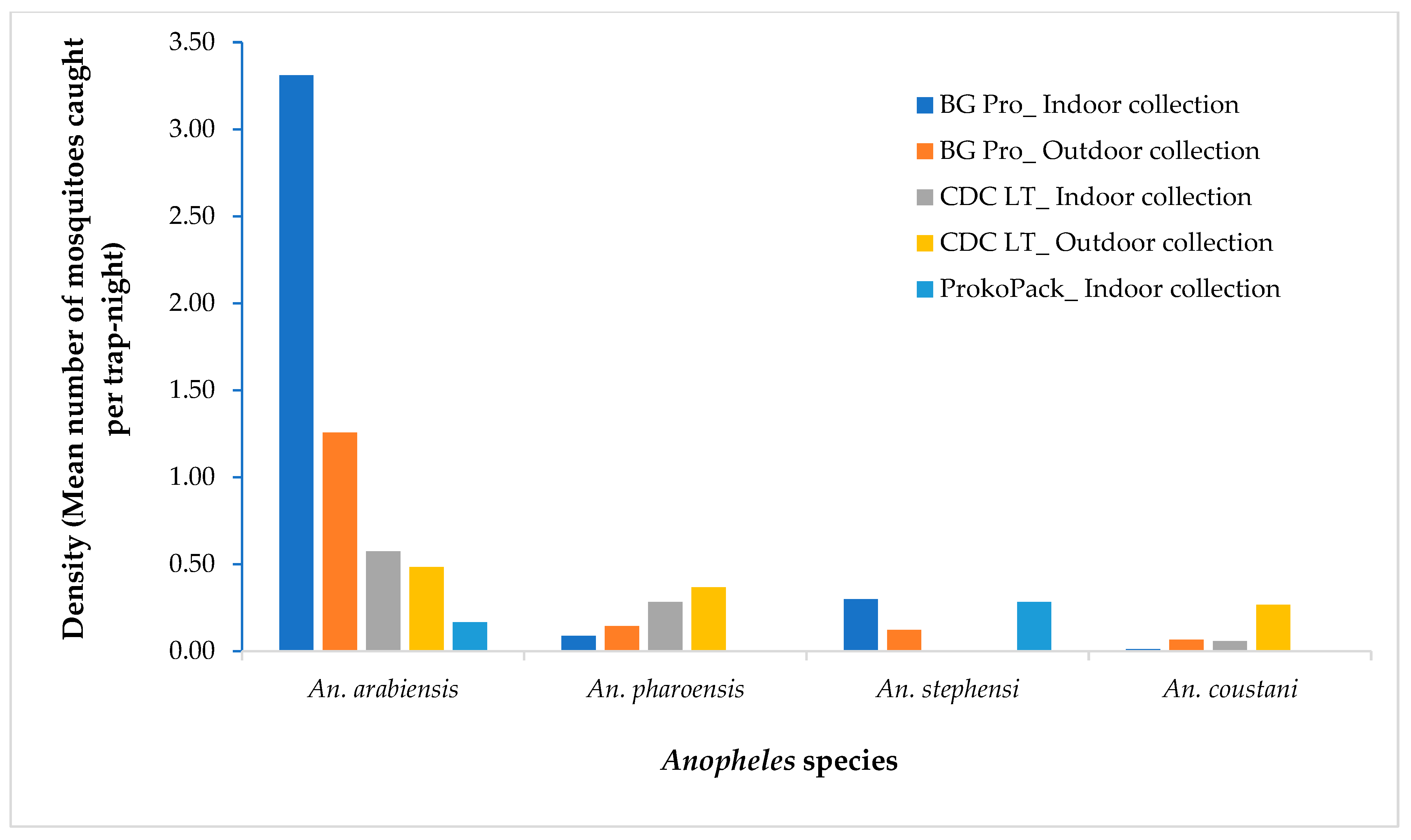
3.1. Anopheles Species and Density

A total of 738 female Anopheles mosquitoes were collected during the study period. Anopheles arabiensis was the predominant species, accounting for 72.9% (n = 538) of the collections, followed by An. pharoensis, 13.4% (n = 99), An. stephensi, 7.5% (n = 55), and An. coustani 6.2% (n = 46). A higher density of An. arabiensis was recorded in the BG-Pro Trap collection compared to other trapping techniques (df = 2; p = 0.004), while a higher density of An. pharoensis was observed in CDC Light Trap collection (p = 0.006). Anopheles stephensi was exclusively captured by the BG Pro Trap and the ProkoPack Aspirator in a comparable density (p = 0.254). In both the BG Pro and CDC LT collections, more An. arabiensis was collected indoors than outdoors. In contrast, for An. pharoensis, outdoor density exceeded indoor density by 1.5 times in the BG Pro collection and by 1.3 times in the CDC collection (Figure 2).

3.2. Bloodmeal Source

A total of 456 Anopheles mosquitoes were tested for bloodmeal sources. The bloodmeal sources identified included human, cow, goat, and dog (Table 1). Some (37.5%) of the samples were unidentified.
Anopheles arabiensis and An. stephensi had lower Human Blood Index (HBI) values, 23.3% and 8.3%, respectively. These species, however, demonstrated a higher Bovine Blood Index (BBI), 61.3% and 75.0%, respectively. An. pharoensis showed similar HBI (43.8%) and BBI (41.7%) values (Figure 3).

3.3. Sporozoite Infection and Entomological Inoculation Rates

The PCR results showed 4.0% of Plasmodium falciparum and 4.0% of Plasmodium vivax sporozoite infection in An. arabiensis. Likewise, 3.5% of P. vivax and 1.2% of P. falciparum was detected in An. pharoensis, while no infection was observed in both An. stephensi and An. coustani (Table 2).
The EIR estimated for the study period indicated that An. arabiensis contributed 4.50 P. falciparum-infective bites and 4.50 P. vivax-infective bites per night in the study area. Anopheles pharoensis accounted for 0.25 P. falciparum-infective bites and 0.72 P. vivax-infective bites per night.

4. Discussion

The present study highlighted an elevated plasmodium sporozoite infection rate in primary and secondary malaria vectors of Ethiopia compared to previous reports. The Plasmodium sporozoite infection rate of An. arabiensis reported was about seven times higher than in previous studies in Central and Southern Ethiopia [8,32,33,34,35,36]. Similarly, the observed sporozoite rate in An. pharoensis was about six times higher than previously reported [32,33,36]. A recent study also documented the increase in malaria incidence in the study area [19], which might be attributed to the high sporozoite infection rate observed in this study in both primary and secondary vectors. This is mainly because the area is prone to malaria epidemics due to the presence of mosquito-breeding lake shorelines of Lake Hawassa coupled with swampy areas surrounding the city. The present findings suggest the need for inclusive interventions considering both primary and secondary vectors for successful control of malaria in the area. Current control measures predominantly target the primary vectors. However, it is noted that the secondary vectors, such as An. pharoensis and An. coustani, could contribute to residual transmission [6]. This suggests that in regions with multiple efficient malaria vectors, interventions targeting a primary vector alone may not effectively reduce malaria transmission, as other vectors could keep augmenting the transmission. Additionally, potential vector shift may also occur if targeted intervention suppresses the primary vector population, as noted by Msugupakulya et al. (2023) in East and Southern Africa [37].
Malaria transmission in Ethiopia is a complex and dynamic issue, with a substantial gap in understanding the bionomics and role of secondary vectors in disease transmission. Anopheles pharoensis was previously incriminated as the second most abundant malaria vector and exhibited the second-highest Plasmodium infection rate after An. arabiensis in this study, suggesting its significant role in local malaria transmission. Several studies also noted that An. pharoensis has consistently ranked second in abundance and sporozoite infection rate elsewhere in Ethiopia [32,33,35]. In another laboratory study, An. pharoensis demonstrated similar susceptibility with An. arabiensis to P. falciparum infection [38]. Unfortunately, less attention has been given to the secondary vectors both in research and vector control interventions in Ethiopia. The study emphasizes the need for a comprehensive understanding of secondary vector biology, ecology, behavior, vector competence, and response to existing interventions. As the country embraces malaria elimination, accurate information on the role of multiple vector species on malaria transmission is critical.
The presence of An. stephensi could further pose a significant threat to malaria control efforts in Ethiopia [3]. As the species alarmingly spread to several areas in Ethiopia [4,19,39], control measures are still not well incorporated in routine vector control strategies. Few studies from Eastern Ethiopia have linked the recent regional malaria outbreak to An. stephensi [40,41]. In this study, this species occurred in sympatry with native vectors with low density in adult collections in Southern Ethiopia. A recent study in the same study area reported high larval density of An. stephensi [19]. This indicates the species is well established in Southern Ethiopia. Although no An. stephensi specimens tested positive for Plasmodium sporozoites in this study—likely due to the small sample size as a result of a lack of optimal vector surveillance tools for adult An. stephensi collection—this species poses a potential risk for malaria transmission [4,39]. Therefore, it is advisable to intensify the development of efficient surveillance tools to collect a good sample size of adult An. stephensi specimens for testing in order better define its role in malaria transmission in the region. The lower HBI and Plasmodium infection rates observed in wild An. stephensi populations, both in this study and earlier works from Ethiopia [4,39], might indicate that the mysorensis form of this species, which has zoophilic behavior, is spreading in Ethiopia (unpublished data). Future research should test these hypotheses in the recently introduced regions of Ethiopia.
This study has several limitations that should be taken into account when interpreting the findings. First, this study was conducted during the dry season. Seasonal variation plays a critical role in influencing vector species composition, density, blood-feeding behavior, and infection status. During the dry season, mosquito densities are typically lower, with resilient species persisting in limited habitats. The relatively small mosquito sample size limits the generalizability of the results of vectors bionomics and their role in malaria transmission. In contrast, the rainy season offers abundant breeding sites, resulting in increased density vectors. Sporozoite infection rates also tend to increase in the rainy season, driven by higher mosquito densities. Second, the choice of mosquito trapping methods can substantially affect estimates of vector density, host preference, and infection status. Mosquito trap type, placement, and maintenance can significantly affect surveillance results by introducing species or behavioral biases and impacting data accuracy. Low Anopheles stephensi counts in CDC light traps are likely due to the species’ weak attraction to light, as it is not strongly phototropic compared to other mosquitoes. Additionally, An. stephensi prefers urban, indoor, and shaded environments, which may be missed by traps placed outdoors or without human-associated cues like CO2 or odor. Its peak biting times may not align with trap effectiveness, and more phototactic species may outcompete it in trap captures. Addressing this limitation through standardized or complementary trapping approaches could improve the accuracy of vector surveillance and malaria control strategies. Third, a considerable proportion of mosquito bloodmeal sources remained unidentified. This is likely due, in part, to the exclusion of some common domestic animals—such as chickens and equines—from the bloodmeal analysis, which may have contributed to the high proportion of unidentified hosts.
In addition, the choice of trapping method can influence species composition in mosquito collections, potentially introducing bias in interpreting species abundance and infection rates. In our study, we employed three complementary trapping tools—BG-Pro traps, CDC Light Traps, and Prokopack Aspirators—to increase species diversity in collections and minimize bias introduced by any single method. However, we acknowledge that An. stephensi may be underrepresented due to its lower attraction to light-based traps and preference for indoor resting, behaviors not optimally captured by all trap types used. To address the concern regarding infection rate comparisons, we clarify that our analysis of Plasmodium infection was not intended to directly compare infection prevalence between species but rather to provide a species-specific snapshot of infection status based on the mosquitoes collected. Nevertheless, we agree that unequal sample sizes can affect interpretation. To improve future analyses, we recommend standardizing infection screening across equal numbers of each species or adjusting for trapping efficiency using known capture biases.

5. Conclusions

In summary, this study confirmed the presence of multiple efficient malaria vectors, with An. arabiensis and An. pharoensis exhibiting high Plasmodium infection rates, underscoring the significant role of secondary vectors in malaria transmission and the need to include them in control strategies. The detection of An. stephensi alongside these primary and secondary vectors further complicates malaria control efforts, emphasizing the importance of ongoing surveillance to better understand vector bionomics and their evolving roles. Additionally, future longitudinal studies should aim to collect larger mosquito samples to more accurately assess the relative contribution of each vector species to malaria transmission in the region.

Author Contributions

D.H.and G.Y. contributed to the conception and design of the study. D.H., T.A., A.A., A.E., T.M., T.Y. and C.A. organized and led the collection of specimens. D.H. analyzed the data and drafted the manuscript. M.-C.L. and G.Z. were involved in data analysis. D.Z., C.W. and J.C. performed the laboratory analysis. G.Y., D.Y. and S.K. critically reviewed the manuscript for intellectual content. All authors have read and agreed to the published version of the manuscript.

Funding

This study was supported by the National Institutes of Health (Grants U19 AI129326 and D43 TW001505). The funders did not participate in the study design, data collection and analysis, publication decisions, or manuscript preparation.

Data Availability Statement

The original contributions presented in this study are included in the article. Further inquiries can be directed to the corresponding author.

Acknowledgments

The authors appreciate Hawassa University, Sidama Regional State Health Office, Hawassa City Administration Health Department, and Sidama Region Public Health Institute for facilitating this study. The Hawassa University’s Malaria Research Laboratory coordinated this study.

Conflicts of Interest

The authors declare no conflicts of interest.

References

  1. Sinka, M.E. Global Distribution of the Dominant Vector Species of Malaria, Anopheles mosquitoes. New Insights into Malar Vectors; IntechOpen: Rijeka, Croatia, 2013. [Google Scholar]
  2. Adugna, F.; Wale, M.; Nibret, E. Review of Anopheles Mosquito Species, Abundance, and Distribution in Ethiopia. J. Trop. Med. 2021, 2021, 6726622. [Google Scholar] [CrossRef] [PubMed]
  3. Tadesse, F.G.; Ashine, T.; Teka, H.; Esayas, E.; Messenger, L.A.; Chali, W.; Meerstein-Kessel, L.; Walker, T.; Behaksra, S.W.; Lanke, K.; et al. Anopheles stephensi Mosquitoes as Vectors of Plasmodium vivax and falciparum, Horn of Africa, 2019. Emerg. Infect. Dis. 2021, 27, 603–607. [Google Scholar] [CrossRef] [PubMed]
  4. Balkew, M.; Mumba, P.; Yohannes, G.; Abiy, E.; Getachew, D.; Yared, S.; Worku, A.; Gebresilassie, A.; Tadesse, F.G.; Gadisa, E.; et al. An update on the distribution, bionomics, and insecticide susceptibility of Anopheles stephensi in Ethiopia, 2018–2020. Malar. J. 2021, 20, 263. [Google Scholar] [CrossRef]
  5. Carter, T.E.; Yared, S.; Gebresilassie, A.; Bonnell, V.; Damodaran, L.; Lopez, K.; Ibrahim, M.; Mohammed, S.; Janies, D. First detection of Anopheles stephensi Liston, 1901 (Diptera: Culicidae) in Ethiopia using molecular and morphological approaches. Acta Trop. 2018, 188, 180–186. [Google Scholar] [CrossRef]
  6. Afrane, Y.A.; Bonizzoni, M.; Yan, G. Secondary Malaria Vectors of Sub-Saharan Africa: Threat to Malaria Elimination on the Continent? Current Topics in Malaria; InTech: London, UK, 2016. [Google Scholar] [CrossRef]
  7. Gueye, A.; Ngom, E.H.M.; Diagne, A.; Ndoye, B.B.; Dione, M.L.; Sambe, B.S.; Sokhna, C.; Diallo, M.; Niang, M.; Dia, I. Host feeding preferences of malaria vectors in an area of low malaria transmission. Sci. Rep. 2023, 13, 16410. [Google Scholar] [CrossRef]
  8. Animut, A.; Balkew, M.; Gebre-michael, T.; Lindtjørn, B. Blood meal sources and entomological inoculation rates of anophelines along a highland altitudinal transect in south-central Ethiopia. Malar. J. 2013, 12, 76. [Google Scholar] [CrossRef]
  9. Katusi, G.C.; Hermy, M.R.G.; Makayula, S.M.; Ignell, R.; Govella, N.J.; Hill, S.R.; Mnyone, L.L. Seasonal variation in abundance and blood meal sources of primary and secondary malaria vectors within Kilombero Valley, Southern Tanzania. Parasites Vectors 2022, 15, 479. [Google Scholar] [CrossRef] [PubMed]
  10. Mlacha, Y.P.; Chaki, P.P.; Muhili, A.; Massue, D.J.; Tanner, M.; Majambere, S.; Killen, G.F.; Govella, N.J. Reduced human—Biting preferences of the African malaria vectors Anopheles arabiensis and Anopheles gambiae in an urban context: Controlled, competitive host—Preference experiments in Tanzania. Malar. J. 2020, 19, 418. [Google Scholar] [CrossRef]
  11. Yewhalaw, D.; Kelel, M.; Getu, E.; Temam, S.; Wessel, G. Blood Meal Sources and Sporozoite Rates of Anophelines in Gilgel-Gibe Dam Area, Southwestern . Afr. J. Vector Biol. 2014. Available online: https://www.researchgate.net/publication/292615632_Blood_meal_sources_and_sporozoite_rates_of_Anophelines_in_Gilgel-Gibe_dam_area_Southwestern_Ethiopia (accessed on 25 April 2025).
  12. Asale, A.; Duchateau, L.; Devleesschauwer, B.; Huisman, G.; Yewhalaw, D. Zooprophylaxis as a control strategy for malaria caused by the vector Anopheles arabiensis (Diptera: Culicidae): A systematic review. Infect. Dis. Poverty 2017, 7, 131. [Google Scholar] [CrossRef]
  13. Fornadel, C.; Norris, L.; Glass, G.; Norris, D. Analysis of Anopheles arabiensis blood feeding behavior in southern zambia during the two years after introduction of insecticide-treated bed nets. Am. J. Trop. Med. Hyg. 2010, 83, 848–853. [Google Scholar] [CrossRef]
  14. Mahande, A.; Mosha, F.; Mahande, J.; Kweka, E. Feeding and resting behaviour of malaria vector, Anopheles arabiensis with reference to zooprophylaxis. Malar. J. 2007, 6, 100. [Google Scholar] [CrossRef] [PubMed]
  15. Garrett-Jones, C.; Boreham, P.; Pant, C. Feeding habits of anophelines (Diptera: Culicidae) in 1971–1978, with reference to the human blood index: A review. Bull. Entomol. Res. 1980, 70, 165–185. [Google Scholar] [CrossRef]
  16. Sinka, M.E.; Bangs, M.J.; Manguin, S.; Rubio-Palis, Y.; Chareonviriyaphap, T.; Coetzee, M.; Mbogo, C.M.; Hemingway, J.; Patil, A.P.; Temperley, W.H.; et al. A global map of dominant malaria vectors. Parasites Vectors 2012, 5, 69. [Google Scholar] [CrossRef] [PubMed]
  17. Thomas, S.; Ravishankaran, S.; Justin, N.A.J.A.; Asokan, A.; Mathai, M.T.; Valecha, N.; Montgomery, J.; Thomas, M.B.; Eapen, A. Resting and feeding preferences of Anopheles stephensi in an urban setting, perennial for malaria. Malar. J. 2017, 16, 111. [Google Scholar] [CrossRef]
  18. Mustapha, A.; Musembi, S.; Nyamache, A. Secondary malaria vectors in western Kenya include novel species with unexpectedly high densities and parasite infection rates. Parasites Vectors 2021, 14, 252. [Google Scholar] [CrossRef]
  19. Hawaria, D.; Kibret, S.; Zhong, D.; Lee, M.-C.; Lelisa, K.; Bekele, B.; Birhanu, M.; Mengesha, M.; Solomon, H.; Yewhalaw, D.; et al. First report of Anopheles stephensi from southern Ethiopia. Malar. J. 2023, 22, 373. [Google Scholar] [CrossRef] [PubMed]
  20. Hawassa City Administration Health Department. Annual Malaria Morbidity Report; Hawassa City Administration Health Department: Hawassa, Ethiopia, 2022. [Google Scholar]
  21. Coetzee, M. Key to the females of Afrotropical Anopheles mosquitoes (Diptera: Culicidae). Malar. J. 2020, 19, 70. [Google Scholar] [CrossRef]
  22. Zhong, D.; Hemming-Schroeder, E.; Wang, X.; Kibret, S.; Zhou, G.; Atieli, H.; Lee, M.-C.; Afrane, Y.A.; Githeko, A.K.; Yan, G. Extensive new Anopheles cryptic species involved in human malaria transmission in western Kenya. Sci. Rep. 2020, 10, 16139. [Google Scholar] [CrossRef]
  23. Scott, J.; Brogdon, W.; Collins, F. Identification of single specimens of the Anopheles gambiae complex by the polymerase chain reaction. Am. J. Trop. Med. Hyg. 1993, 49, 520–529. [Google Scholar] [CrossRef]
  24. Mbewe, R.B.; Keven, J.B.; Mzilahowa, T.; Mathanga, D.; Wilson, M.; Cohee, L.; Laufer, M.K.; Walker, E.D. Blood-feeding patterns of Anopheles vectors of human malaria in Malawi: Implications for malaria transmission and effectiveness of LLIN interventions. Malar. J. 2022, 21, 67. [Google Scholar] [CrossRef]
  25. Keven, J.; Artzberger, G.; Gillies, M.; Mbewe, R.; Walker, E. Probe-based multiplex qPCR identifies blood-meal hosts in Anopheles mosquitoes from Papua New Guinea. Parasites Vectors 2020, 13, 111. [Google Scholar] [CrossRef] [PubMed]
  26. Shokoples, S.E.; Ndao, M.; Kowalewska-Grochowska, K.; Yanow, S.K. Multiplexed real-time PCR assay for discrimination of Plasmodium species with improved sensitivity for mixed infections. J. Clin. Microbiol. 2009, 47, 975–980. [Google Scholar] [CrossRef] [PubMed]
  27. Hofmann, N.; Mwingira, F.; Shekalaghe, S.; Robinson, L.J.; Mueller, I.; Felger, I. Ultra-Sensitive Detection of Plasmodium falciparum by Amplification of Multi-Copy Subtelomeric Targets. PLoS Med. 2015, 12, e1001788. [Google Scholar] [CrossRef]
  28. Veron, V.; Simon, S.; Carme, B. Multiplex real-time PCR detection of P. falciparum, P. vivax and P. malariae in human blood samples. Exp. Parasitol. 2009, 121, 346–351. [Google Scholar] [CrossRef] [PubMed]
  29. Drakeley, C.; Schellenberg, D.; Kihonda, J.; Sousa, C.A.; Arez, A.P.; Lopes, D.; Lines, J.; Mshinda, H.; Lengeler, C.; Schellenberg, J.A.; et al. An estimation of the entomological inoculation rate for Ifakara: A semi-urban area in a region of intense malaria transmission in Tanzania. Trop. Med. Int. Health 2003, 8, 767–774. [Google Scholar] [CrossRef]
  30. Ogola, E.; Villinger, J.; Mabuka, D.; Omondi, D.; Orindi, B.; Mutunga, J.; Owino, V.; Masiga, D.K. Composition of Anopheles mosquitoes, their blood—Meal hosts, and Plasmodium falciparum infection rates in three islands with disparate bed net coverage in Lake Victoria, Kenya. Malar. J. 2017, 16, 360. [Google Scholar] [CrossRef]
  31. Adugna, T.; Yewhelew, D.; Getu, E. Bloodmeal sources and feeding behavior of anopheline mosquitoes in Bure district, northwestern Ethiopia. Parasites Vectors 2021, 14, 166. [Google Scholar] [CrossRef]
  32. Aschale, Y.; Getachew, A.; Yewhalaw, D.; Cristofaro ADe Sciarretta, A. Systematic review of sporozoite infection rate of Anopheles mosquitoes in Ethiopia, 2001–2021. Parasites Vectors 2023, 16, 437. [Google Scholar] [CrossRef]
  33. Kibret, S.; Lautze, J.; Boelee, E.; Mccartney, M. How does an Ethiopian dam increase malaria ? Entomological determinants around the Koka reservoir. Trop. Med. Int. Health 2012, 17, 1320–1328. [Google Scholar] [CrossRef]
  34. Yewhalaw, D.; Legesse, W.; Van Bortel, W.; Gebre-Selassie, S.; Kloos, H.; Duchateau, L.; Speybroeck, N. Malaria and water resource development: The case of Gilgel-Gibe hydroelectric dam in Ethiopia. Malar. J. 2009, 8, 21. [Google Scholar] [CrossRef]
  35. Bedasso, A.H.; Gutto, A.A.; Waldetensai, A.; Eukubay, A.; Bokore, G.E.; Kinde, S.; Gemechu, F.; Debebe, Y.; Aklilu, M.; Tasew, G.; et al. Malaria vector feeding, peak biting time and resting place preference behaviors in line with Indoor based intervention tools and its implication: Scenario from selected sentinel sites of Ethiopia. Hellyon 2022, 8, e12178. [Google Scholar] [CrossRef] [PubMed]
  36. Kibret, S.; Alemu, Y.; Boelee, E.; Tekie, H.; Alemu, D.; Petros, B. The impact of a small-scale irrigation scheme on malaria transmission in Ziway area, Central Ethiopia. Trop. Med. Int. Health 2010, 15, 41–50. [Google Scholar] [CrossRef] [PubMed]
  37. Msugupakulya, B.; Urio, N.; Jumanne, M.; Ngowo, H.; Selvaraj, P. Changes in contributions of different Anopheles vector species to malaria transmission in east and southern Africa from 2000 to 2022. Parasites Vectors 2023, 16, 408. [Google Scholar] [CrossRef] [PubMed]
  38. Abduselam, N.; Zeynudin, A.; Berens-Riha, N.; Seyoum, D.; Pritsch, M.; Tibebu, H.; Eba, K.; Hoelscher, M.; Wieser, A.; Yewhalaw, D. Similar trends of susceptibility in Anopheles arabiensis and Anopheles pharoensis to Plasmodium vivax infection in Ethiopia. Parasites Vectors 2016, 9, 552. [Google Scholar] [CrossRef]
  39. Ashine, T.; Eyasu, A.; Asmamaw, Y.; Simma, E.; Zemene, E.; Epstein, A.; Brown, R.; Negash, N.; Kochora, A.; Reynolds, A.M.; et al. Spatiotemporal distribution and bionomics of Anopheles stephensi in different eco—Epidemiological settings in Ethiopia. Parasites Vectors 2024, 17, 166. [Google Scholar] [CrossRef]
  40. Emiru, T.; Getachew, D.; Murphy, M.; Sedda, L.; Ejigu, L.A.; Bulto, M.G.; Byrne, I.; Demisse, M.; Abdo, M.; Chali, W.; et al. Evidence for a role of Anopheles stephensi in the spread of drug- and diagnosis-resistant malaria in Africa. Nat. Med. 2023, 29, 3203–3211. [Google Scholar] [CrossRef]
  41. de Santi, V.P.; Khaireh, B.A.; Chiniard, T.; Pradines, B.; Taudon, N.; Larréché, S.; Mohamed, A.B.; de Laval, F.; Berger, F.; Gala, F.; et al. Role of Anopheles stephensi Mosquitoes in Malaria Outbreak, Djibouti, 2019. Emerg. Infect. Dis. 2021, 27, 1697–1700. [Google Scholar] [CrossRef]
Figure 1. Map of the study setting, Hawassa City, Ethiopia.
Insects 16 00462 g001
Figure 2. Density of Anopheles mosquitoes in various trapping devices in Hawassa, southern Ethiopia, 2023.
Insects 16 00462 g002
Figure 3. Anopheles species bloodmeal index in Hawassa, Southern Ethiopia, 2023.
Insects 16 00462 g003
Table 1. Bloodmeal sources of Anopheles species in Hawassa, Southern Ethiopia, 2023.
Host An. arabiensisn (%)An. pharoensis
n (%)
An. stephensi
n (%)
An. coustani
n (%)
Human 35 (10.6)14 (16.5)1 (3.3)1 (10.0)
Human + cow 18 (5.4)3 (3.5)1 (3.3)1 (10.0)
Human + cow + goat1 (0.3)2 (2.3)--
Human + goat5 (1.5)2 (2.4)--
Cow 116 (35.0)12 (14.1)16 (53.3)3 (30.0)
Cow + goat19 (5.7)3 (3.5)1 (3.3)-
Cow + goat + dog1 (0.3)---
Dog 1 (0.3)---
Goat 11 (3.3)13 (15.3)3 (10.0)2 (20.0)
Not detected124 (37.5)36 (42.4)8 (26.7)3 (30.0)
Total331853010
Table 2. Plasmodium sporozoite infection rates of Anopheles species in Hawassa, Ethiopia, 2023.
Anopheles SpeciesNumber TestedSporozoite Positive n (%)Parasite Species
Indoor CollectionOutdoor CollectionPlasmodium falciparumPlasmodium vivax
IndoorsOutdoorsIndoorsOutdoors
An. arabiensis33113 (4.0)13 (4.0)7667
An. pharoensis853 (3.5)1(1.2)1021
An. stephensi30000000
An. coustani11000000
Disclaimer/Publisher’s Note: The statements, opinions and data contained in all publications are solely those of the individual author(s) and contributor(s) and not of MDPI and/or the editor(s). MDPI and/or the editor(s) disclaim responsibility for any injury to people or property resulting from any ideas, methods, instructions or products referred to in the content.

