Article

Microstructure and Magnetism of Heavily Helium-Ion Irradiated Epitaxial Iron Films

1
Faculty of Science and Engineering, Iwate University, Morioka 020-8551, Japan
2
Research Institute Applied Mechanics, Kyushu University, Kasuga 816-8580, Japan
*
Author to whom correspondence should be addressed.
Metals 2023, 13(11), 1905; https://doi.org/10.3390/met13111905
Submission received: 16 October 2023 / Revised: 8 November 2023 / Accepted: 9 November 2023 / Published: 18 November 2023

Abstract

:
This study reports on the microstructure and magnetism of pure iron irradiated with high doses of helium ions. Iron alloys are important structural materials used as components in fusion reactors, and a comprehensive database of their various properties has been developed. But little has been investigated on magnetic properties, in particular, the effects of high doses and helium cavities are lacking. Single-crystal iron films, with a thickness of 200 nm, were prepared using the ultra-high vacuum evaporation method. These films were then irradiated with 30 keV He+ ions at room temperature up to a dose of 18 dpa. X-ray diffraction measurements and cross-sectional transmission electron microscope observations revealed significant microstructural changes, including a large lattice expansion perpendicular to the film plane and the formation of high-density cavities after irradiation. However, the saturation magnetization and the shape of the magnetization curve showed almost no change, indicating the robustness of the magnetic properties of iron.

1. Introduction

Iron alloys are important structural materials in advanced nuclear fission and fusion reactors. As a key component of fusion reactors, there is a blanket module, and iron-chromium steel is considered a promising material [1]. Several types of steel have been developed and material databases that tabulate their physical properties are now available [2,3]. For the structural assessment of a blanket module, it is important to consider electromagnetic (EM) loads during normal operation as well as in the case of plasma disruptions. Several numerical analyses using finite element code have been reported [4,5,6], and the magnetization data of ferromagnetic steels are used as input parameters in the calculation. The blankets are subjected to high-energy and high-dose neutron irradiation (a few tens of displacements per atom; dpa) [1,7], which may lead to changes in the magnetic properties of the steels during operation. However, all the above calculations of EM loads have been performed using magnetization data from unirradiated steels due to the lack of information in the corresponding database.
Numerous studies have been carried out to examine the effects of high doses of neutron irradiation on the microstructure and mechanical properties of iron alloys. However, there has been a lack of systematic investigation regarding the magnetic aspects. This is primarily attributed to the difficulties associated with conducting neutron irradiation and post irradiation experiments. For example, neutron irradiation necessitates the use of a nuclear reactor, which is expensive and restricts the flexibility of experimental conditions. Moreover, the handling of radioactive specimens requires a hot laboratory. To address these issues, ion irradiation is commonly used as an alternative experimental approach [8]. It has the advantage that irradiation conditions can be easily changed and the characteristics of nonradioactive specimens can be evaluated in a general laboratory. On the other hand, due to the short penetration depth of ions, the damage is limited to the surface region of the specimen. This poses another problem because it is challenging to distinguish the magnetism of damaged regions from nondamaged regions in irradiated bulk specimens. One approach to solving this problem is by conducting ion irradiation experiments on thin film specimens. This technique involves damaging the entire specimen, which facilitates the assessment of magnetic properties. So far, there have been several studies on the magnetism of ion-irradiated films for pure iron [9,10,11], iron-chromium alloys [12,13] and iron-nickel alloys [14,15]. One of the interesting phenomena observed in these reports is the behavior of magnetism in iron at high doses. The magnetic properties are nearly unaffected after irradiation with a few dpa [9]. However, a significant increase of up to 32% in saturation magnetization was reported after heavy Fe+ irradiation at 72 dpa [10,11].
Research on the magnetism of iron alloys has been ongoing for a long time and continues to be of interest due to the potential for developing materials with high saturation magnetization [16]. The saturation magnetization of a ferromagnetic material is determined by the magnitude of the magnetic moment of its constituent atoms. In 3d transition metals (TMs) like iron, the magnetic moment is related to the spin of unpaired outer shell electrons. The magnetic moment of body-centered cubic (BCC) iron is 2.2 μB in its normal bulk state. However, as the interatomic distance increases, the electron density of states at the Fermi energy also increases, which makes magnetic polarization easier. Consequently, the magnetic moment of iron may potentially increase by up to 3 μB. This phenomenon is commonly referred to as the magneto-volume effect. The mechanism behind the significant increase in saturation magnetization reported in the iron films heavily irradiated with Fe+ ions has not been well understood experimentally. It is suggested that this increase may be attributed to a local magneto-volume effect that occurs as a result of the formation of vacancy clusters [10,11].
Investigating the saturation magnetization behavior of irradiated alloys is necessary for the safe operation of nuclear fusion reactors and is also of academic interest. When studying the magnetism of ion-irradiated films of iron and model alloys as a research method, it is important to consider the differences in microstructure between thin film materials and actual structural materials. The practical iron–chromium alloys of reduced activation ferritic/martensitic steel used for blankets have a tempered martensite structure that includes numerous grain boundaries, dislocations and precipitates [1]. Considering the magnetic properties of iron-based alloys, there are structure-sensitive characteristics such as permeability, coercive force and remanence, as well as structure-insensitive characteristics such as saturation magnetization [17]. Grain boundaries, dislocations and precipitates have a significant effect on the former but have little effect on the latter. This study primarily focuses on the latter characteristics. In this sense, the research approach that utilizes ion-irradiated film is considered beneficial for gaining insights into the magnetism of neutron-irradiated structural materials.
In addition to the formation of irradiation defects such as dislocation loops and vacancy clusters due to high-dose irradiation, there is also interest in studying the effects of helium cavity formation caused by nuclear transmutation in materials used in fusion reactors [7,18]. However, there have been no reports on the effect of this on magnetism. Based on these considerations, we fabricated a high-quality single-crystal iron thin film, irradiated it with helium ions up to a high dose of 18 dpa and conducted a comprehensive investigation of its microstructure and magnetism.

2. Experimental Procedures

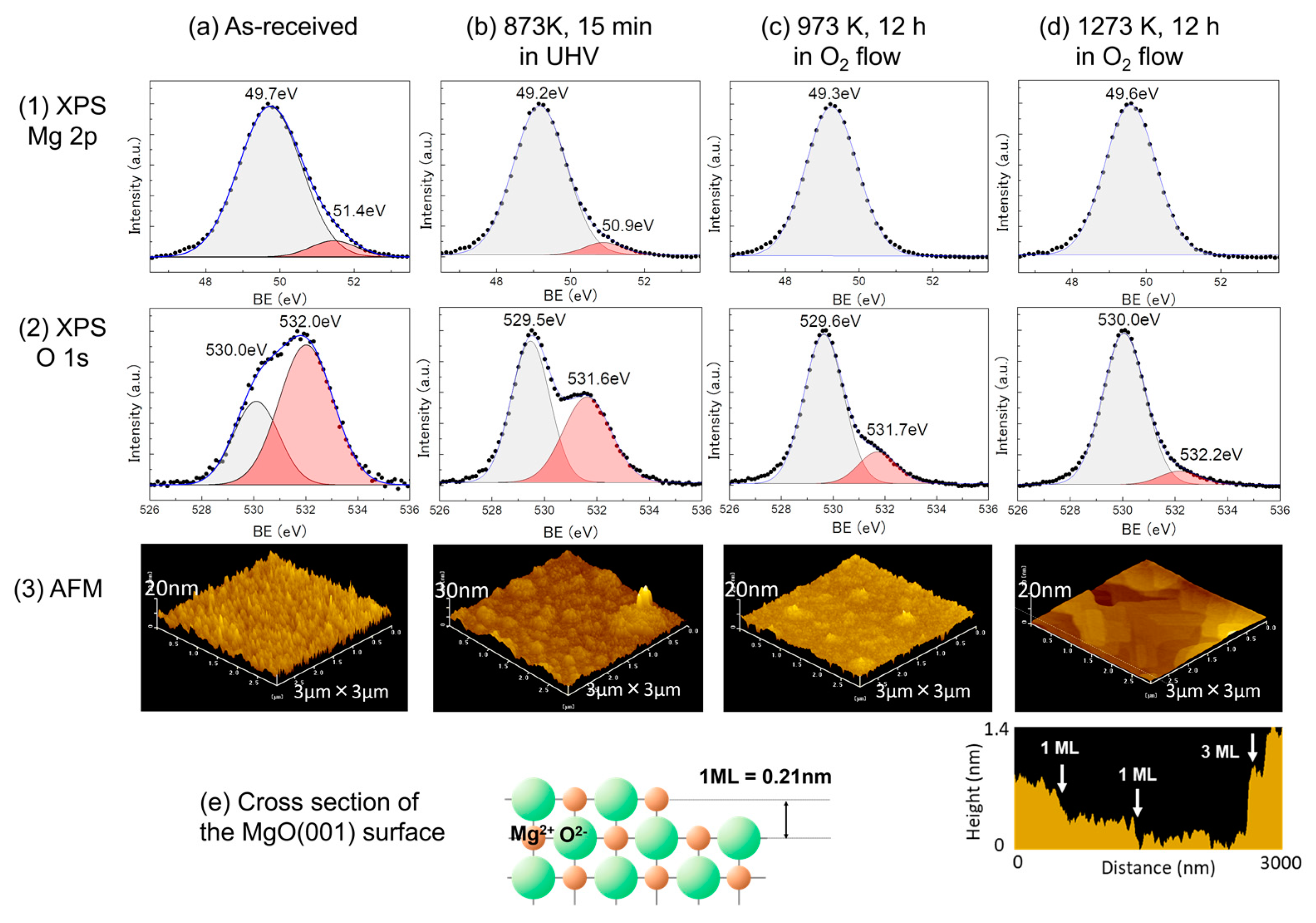
2.1. Specimen Preparation

In order to fabricate high-quality iron thin films, we used commercially available MgO (001) plates that had been mirror-polished as the substrate. Since MgO is deliquescent, it is necessary to remove the altered layer on the surface before depositing the film [19,20]. Figure 1 displays the profile of the X-ray photoelectron spectroscopy (XPS, PHI 5000 Versa Probe, ULVAC-PHI Inc., Chigasaki, Japan) and the surface images captured by the atomic force microscope (AFM, SPA400, SII Inc., Chiba, Japan) of MgO plates. Figure 1a shows the as-received plate stored in a vacuum desiccator for a period of time after purchase. Figure 1b–d include the results obtained after processing the plates under three different conditions. Two peaks are observed in both the Mg 2p and O 1s profiles of the as-received plate (Figure 1a). The binding energies of Mg 2p are reported to be 50.4, 50.8 and 51.6 eV for MgO, hydroxide and carbonate, respectively. The corresponding energies of O 1s are 530.4, 531.5 and 533.8 eV [19]. Therefore, the profiles suggest that a surface-altered layer of hydroxide formed in the as-received plate. Heat treatment at 873 K for 15 min in an ultra-high vacuum (UHV) reduced the peak intensity of the altered layer but not enough (Figure 1b). However, heat treatment at 1273 K for 12 h in an oxygen flow nearly eliminated the hydroxide (Figure 1d). We also obtained a flat surface with a single monolayer (ML) step. Using this substrate, epitaxial iron films with a thickness of 200 nm were fabricated at 323 K through electron beam deposition in a UHV chamber at a base pressure of 2 × 10⁻⁷ Pa. During deposition, the film growth was monitored using in-situ reflection high-energy electron diffraction (RHEED). After deposition, the film was immediately annealed in the chamber at 873 K for 15 min to ensure the production of a high-quality film.
These fabricated specimens were irradiated with 30 keV He+ at room temperature (RT) using a light-element accelerator system at the Research Institute of Applied Mechanics. The ion flux was 1.5 × 10¹³ ions/(cm2s) and the total dose was 3.0 × 10¹⁷ ions/cm2. Figure 2 shows the damage and residual helium profiles of bulk iron, which were calculated using the Stopping and Range of Ions in Matter (SRIM) code with a threshold energy of 40 eV [21]. The damage peak is at a depth of 95 nm and the residual helium peak is at 135 nm, both within the film. As depicted in Figure 2, the level of irradiation-induced damage can be estimated to be around 18 dpa.

2.2. Characterization of Microstructure and Magnetism

The crystal structure and orientation relationship of the thin film were investigated using electron backscatter diffraction (EBSD, HKL Channel5, Oxford Instruments, UK) both before and after irradiation. The diffraction profile perpendicular to the film plane was measured using a conventional X-ray diffraction (XRD) θ-2θ measurement system (Rigaku Ultima IV with CuKa radiation). The profile within the film plane was measured using grazing incidence XRD (GIXRD) measurement (SmartLab, Rigaku Corp., Akishima, Japan). Furthermore, thin sections were cut from both the unirradiated and irradiated specimens using the microsampling method of the focused ion beam of 30 keV Ga+ (FIB, MI-4050, Hitachi High-Tech Corp., Tokyo, Japan). The cross-sectional structures were then observed using a transmission electron microscope (TEM, JEM-2100 and ARM200FC, JEOL Ltd., Tokyo, Japan).
Magnetic hysteresis curves were obtained using a vibrating-sample magnetometer (VSM, VSM-5-15, Toei Industry Co., Ltd., Tokyo, Japan) at RT. The maximum magnetic field was approximately 5 kOe.

3. Results

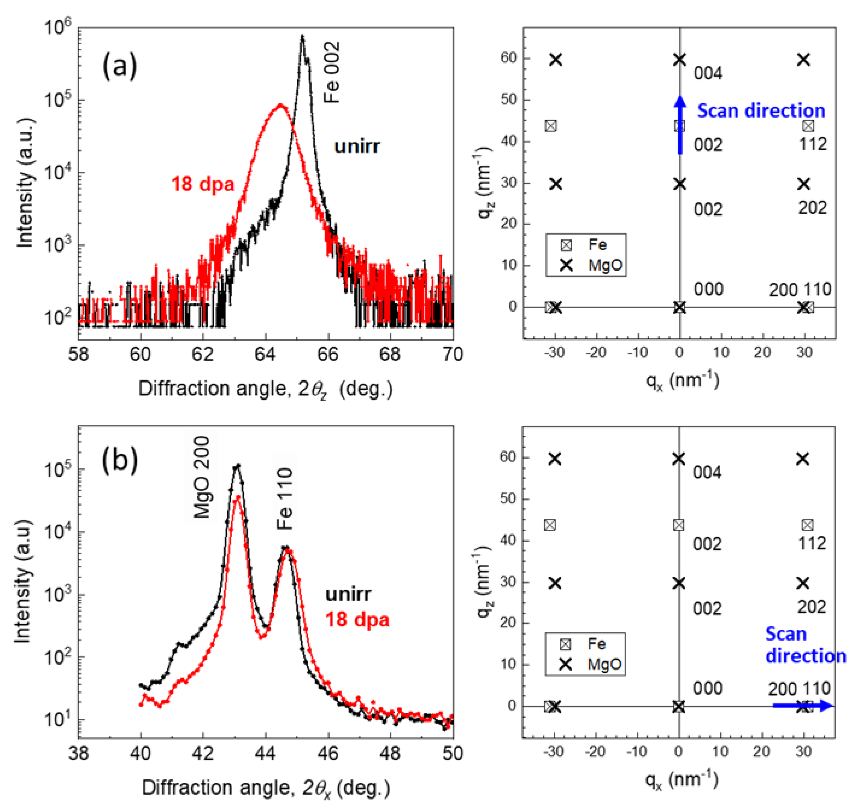
3.1. Epitaxial Structure and Lattice Expansion

The RHEED patterns of the fabricated film were streaky, indicating a smooth and flat surface growth. The analysis revealed that the crystal structure of the film is BCC and the orientation relationship between the film and substrate is presented below.
BCC Fe (001) [110]//MgO (001) [100]
Figure 3a,b show the results obtained using EBSD. The figures display the phase maps, inverse pole figure (IPF) maps and {100}BCC pole figures of the unirradiated and irradiated films, respectively. Figure 3c depicts the schematic drawing of the epitaxial relationship. These results are consistent with RHEED observations, and the irradiation did not affect both the crystal structure and orientation of the film.
Figure 4a,b show XRD profiles around the Fe 002 and Fe 110 reflections, obtained through out-of-plane XRD and in-plane GIXRD measurements, respectively. The accompanying diagrams on the right side illustrate the diffraction maps in reciprocal space and the scan directions. In the out-of-plane scan profile, which was measured by varying the diffraction angle 2θ from 40 degrees to 80 degrees, only the Fe 002 peak was observed, except for the MgO 002 peak. After irradiation, the intensity of the Fe 002 peak decreased, the peak width broadened and the peak position shifted to a lower angle (Figure 4a). These indicate that the lattice spacing perpendicular to the film plane is stretched with a broad distribution. On the other hand, the intensity, width and position of the Fe 110 peak remain almost unchanged (Figure 4b). These observations suggest that while the out-of-plane lattice spacing of the iron expanded due to helium irradiation, the in-plane spacing remained unaffected due to the constraints imposed by the substrate.
Table 1 summarizes the lattice spacings determined from the XRD diffraction profiles of the nonirradiated and irradiated specimens. After irradiation, the lattice spacing of Fe increased by 1.04% in the out-of-plane direction and decreased by 0.22% in the in-plane direction. This indicates that the volume increased by an average of 0.59% due to irradiation.

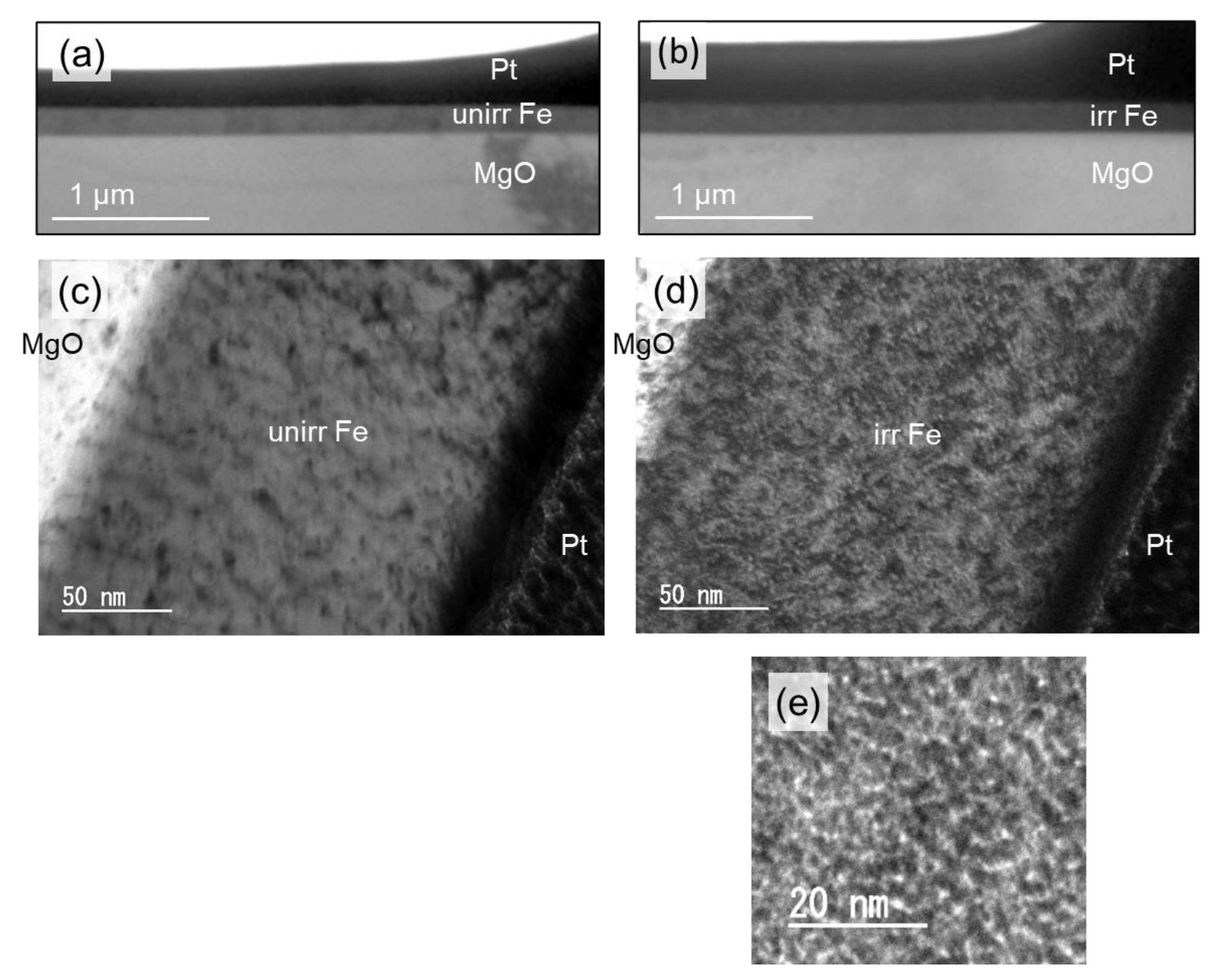
3.2. Formation of Cavities

Figure 5 shows cross-sectional TEM images of the unirradiated specimen (Figure 5a,c) and the specimen irradiated at 18 dpa (Figure 5b,d,e). The low-magnification images (Figure 5a,b) show that the iron film has a band-shaped structure, indicating that it grew flat and smooth on the substrate and remained unchanged after irradiation. A high-magnification image (Figure 5c) of the unirradiated specimen shows dark contrasts, which may indicate defects introduced during FIB sampling. A high-magnification image of the sample after irradiation (Figure 5d) shows similar dark contrasts, but small new bright contrasts can also be seen. The magnified and under-focused image (Figure 5e) revealed numerous small, bright contrasts. When the observations were changing from under-focused to over-focused, the contrast was reversed. This indicates that the small contrasts are helium cavities [22]. The number density of cavities, calculated assuming a TEM specimen thickness of 100 nm, was estimated to be 1.2 × 1024/m3.

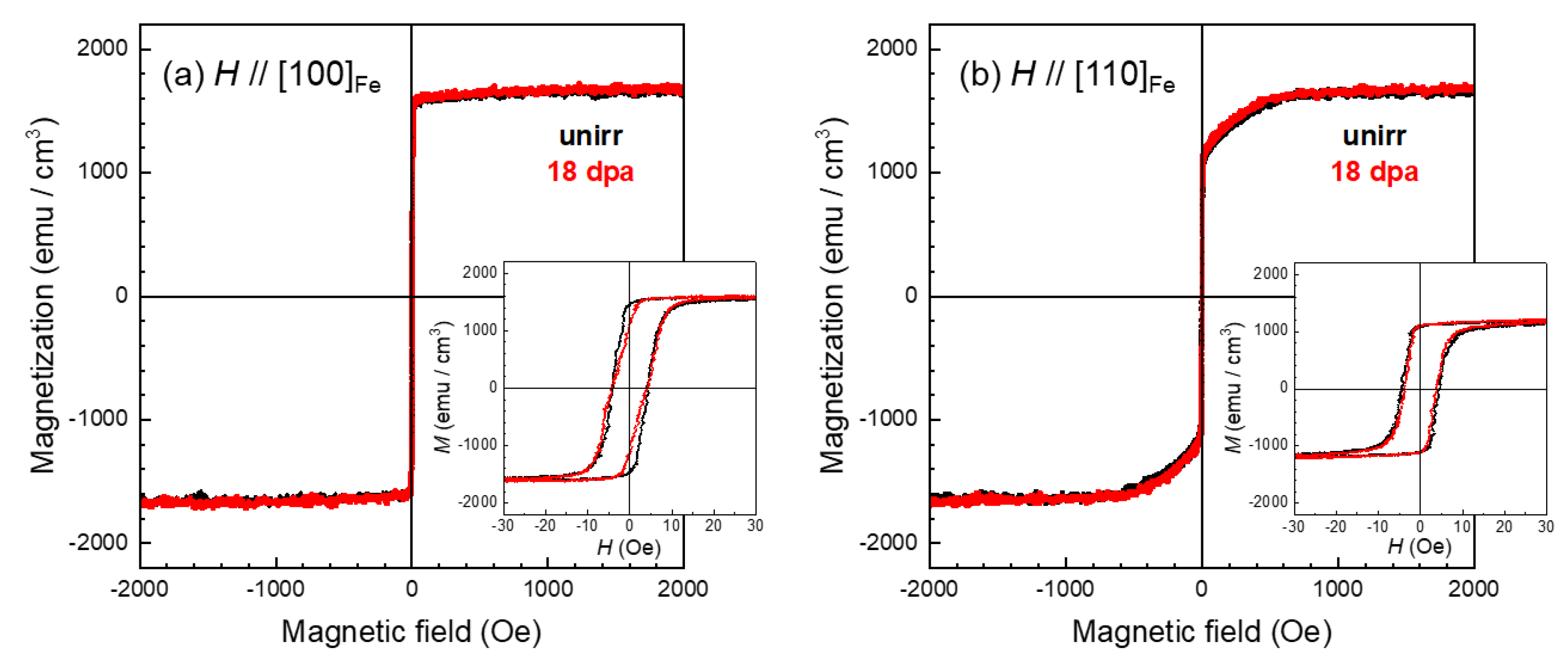
3.3. Irradiation Effects on Magnetization Curves

Figure 6 shows the magnetization curves of the specimens before and after irradiation, which were calculated using a film thickness of 200 nm. In the case of Figure 6a, the magnetic field is applied in the [100]Fe direction of easy magnetization in the film plane, while in the case of Figure 6b it is applied in the [110]Fe direction of hard magnetization. The insets in Figure 6a,b show enlarged views of the area around the origin. Despite the lattice expansion and the formation of dense cavities, the shape of the magnetization curves remains almost unchanged after irradiation. The saturation magnetization value of the unirradiated iron film is 1633 emu/cm3, which is nearly identical to the bulk value of 1707 emu/cm3 [23]. No clear change in saturation magnetization is observed after irradiation, considering the experimental error. The magnetization curve in the hard magnetization direction saturates at around 600 Oe, indicating that the present iron film exhibits the same magneto-crystalline anisotropy as bulk iron [23], and the anisotropy is nearly unaffected by irradiation. Furthermore, the magnified views of the magnetization curves after irradiation reveal that the width of the hysteresis remains nearly unchanged, suggesting that the coercive force is unaffected.
In a previous experiment on low-dose neutron irradiation, conducted on well-annealed bulk iron with a low defect density, it was observed that the coercive force increases monotonically [24]. This phenomenon can be interpreted as the formation and growth of dislocation loops caused by irradiation. The stress field around the dislocations hinders the movement of magnetic domain walls due to magnetoelastic interaction. On the contrary, in cold-rolled bulk iron with a high defect density, the coercive force decreases [25]. This decrease is attributed to the recovery of the dislocation microstructures. Since the coercive force of the current film specimen seems higher than that of well-annealed bulk iron, it is possible that the film may contain defects, such as misfit dislocations, that are formed during the fabrication process before irradiation. It is plausible that the coercive force did not change after irradiation because the recovery of the initial defects and the formation of irradiation defects were balanced.
However, despite the fact that the current specimen contains a large number of cavities, no significant changes were observed in the saturation magnetization and the shape of the magnetization curve. The cavities can be considered as very small, nonmagnetic inclusions. There has been a study on an analytical model that considers the deviation in the direction of magnetic moments around nonmagnetic inclusions and their effects on magnetic properties [26]. The results of this study have led to interesting findings. In nanometer-scale nonmagnetic inclusions, the deviation of surrounding magnetic moments is small and does not significantly affect the magnetism of iron.
In this study, the expansion of the lattice was confirmed after helium irradiation. As mentioned in the introduction, the electron band theory of 3d metals predicts an increase in saturation magnetization due to the magneto-volume effect. However, this study did not observe such behavior. This will be discussed in the next section, Section 4.

4. Discussion

Systematic studies based on first-principles calculations have been reported on the relationship between the magnetic moment and volume expansion of iron [27,28,29]. All of these calculations assume uniform volume expansion. These calculation models do not adequately reproduce the microstructures of the He+ irradiated film, as they do not consider the lateral lattice constraint imposed by the substrate and the nonuniform volume expansion caused by the formation of cavities. However, they are still useful for understanding the average behavior of the magnetic moment. One of the previous studies showed that a 0.59% increase in volume corresponds to less than a 1% increase in magnetic moment [27]. This increase in magnetization is too small to be distinguished using a VSM, which is consistent with our experimental findings.
The calculations described above are all for single-phase pure iron and do not include the effect of helium adjacent to the iron. Therefore, we constructed a simple model that takes into account the presence of helium atoms and investigated the relationship between volume expansion and magnetic moment. The first-principles calculations based on density functional theory were performed using the Vienna ab initio simulation package (VASP) with the generalized gradient approximation and projector augmented wave method [30,31]. Figure 7 shows a schematic view of the model and the corresponding calculation results. We consider a supercell in which one vacancy is introduced into 128 iron atoms (4 × 4 × 4 BCC lattice). Then, a helium atom was inserted into the vacancy one by one [32]. The initial atomic position of the helium atom was set to the center of the vacancy and then the system was relaxed. After finding the stable state, we obtained the volume expansion of the supercell as well as the average magnetic moment of 127 iron atoms. The results are shown in Figure 7b. As the number of helium atoms increases, the volume of the supercell expands and the average magnetic moment increases steadily. The expansion of 0.59% leads to a 0.95% increase in magnetic moment, which is consistent with the current experimental results.
Previous research on iron films heavily irradiated by Fe+ has reported significant changes in their magnetic properties, including a large increase in saturation magnetization and a large reduction in coercive force [10,11]. These trends could not be confirmed using the current iron film specimens after He+ irradiation with a high dose of 18 dpa. The cause of this is unclear; however, it may be attributed to differences in the irradiation microstructures resulting from the different types of ion irradiations. Further investigation is required to examine the effects of irradiation conditions, including the type of ion.
The present research focused on pure iron, which consists of only one type of 3d TM element. However, practical iron alloys are composed of multiple elements, so it is necessary to consider the effects of these elements. In fact, it has been confirmed that adding other 3d TM elements to iron significantly alters the irradiation effect on magnetism. In the case of iron–nickel alloys, the irradiation effect is closely related to the stability of BCC-FCC structures. For example, Cu2+ ion irradiation leads to a significant increase in saturation magnetization of iron–nickel films with a phase boundary composition [15]. In our group, ongoing research on He+ irradiated iron-chromium films has shown that the saturation magnetization is not significantly affected, but there is a noticeable change in the shape of the magnetization curve, indicating a tendency for the films to become magnetically hardened. Further studies on the effects of the second and third elements, using ion-irradiated iron alloy films, are expected to provide a comprehensive understanding of the magnetic properties of neutron-irradiated iron-based structural materials.

5. Conclusions

In this study, we investigated the effects of high-dose irradiation and cavity formation on the microstructure and magnetic properties of iron. Epitaxial iron films of high quality were prepared and subsequently exposed to helium-ion irradiation. Results revealed a significant lattice expansion in the out-of-plane direction and the formation of high-density small cavities after irradiation. However, the magnetization curves remained unchanged and the saturation magnetization was almost the same as that of unirradiated film within experimental accuracy. This result is consistent with the first-principles calculations of a simple model that considers the volume expansion caused by the formation of cavities. This research provides evidence that the magnetic properties of iron remain robust and unaltered during the irradiation experiment conducted in this study.

Author Contributions

Conceptualization, Y.K. and H.W.; investigation (specimen preparation, EBSD, XRD, TEM and VSM measurements), D.U., T.O., T.M., K.S., S.F., N.Y. and H.W.; (first-principles calculation), K.O.; supervision, Y.K.; project administration Y.K., funding acquisition, Y.K. All authors have read and agreed to the published version of the manuscript.

Funding

This study was supported in part by the Japan Society for the Promotion of Science (JSPS) KAKENHI Grant JP23H01890 and was conducted as part of the Collaborative Research Program of the Research Institute for Applied Mechanics, Kyushu University.

Data Availability Statement

The data is included in this article.

Acknowledgments

The authors would like to thank H. Sekimoto for his support in GIXRD measurement, K. Sasaki for his assistance in FIB-TEM specimen preparation at Iwate University and T. Mutaguchi for his contribution to the irradiation experiment at Kyushu University.

Conflicts of Interest

The authors declare no conflict of interest.

References

  1. Cabet, C.; Dalle, F.; Gaganidze, E.; Henry, J.; Tanigawa, H. Ferritic-martensitic steels for fission and fusion applications. J. Nucl. Mater. 2019, 523, 510–537. [Google Scholar]
  2. Hirose, T.; Nozawa, T.; Stoller, R.E.; Hamaguchi, D.; Sakasegawa, H.; Tanigawa, H.; Tanigawa, H.; Enoeda, M.; Katoh, Y.; Snead, L.L. Physical properties of F82H for fusion blanket design. Fusion Eng. Des. 2014, 89, 1595–1599. [Google Scholar] [CrossRef]
  3. Gaganidze, E.; Gillemot, F.; Szenthe, I.; Gorley, M.; Rieth, M.; Diegele, E. Development of EUROFER97 database and material property handbook. Fusion Eng. Des. 2018, 135, 9–14. [Google Scholar]
  4. Roccella, R.; Boccaccini, L.V.; Meyder, R.; Raff, S.; Roccella, M. Assessment of EM loads on the EU HCPB TBM during plasma disruption and normal operating scenario including the ferromagnetic effect. Fusion Eng. Des. 2008, 83, 1212–1216. [Google Scholar]
  5. Maione, I.A.; Roccella, M.; Hernández, F.A.; Lucca, F. Update of electromagnetic loads on HCPB breeding blanked for DEMO 2017 configuration. Fusion Eng. Des. 2020, 156, 111604. [Google Scholar] [CrossRef]
  6. Guan, W.; Gwon, H.; Tanigawa, H.; Hirose, T.; Kawamura, Y. Electromagnetic analysis of cylindrical WCCB TBM. Fusion Eng. Des. 2020, 155, 111719. [Google Scholar]
  7. Knaster, J.; Moeslang, A.; Muroga, T. Materials research for fusion. Nat. Phys. 2016, 12, 424–434. [Google Scholar]
  8. Abromeit, C. Aspects of simulation of neutron damage by ion irradiation. J. Nucl. Mater. 1994, 216, 78–96. [Google Scholar]
  9. Kamada, Y.; Watanabe, H.; Mitani, S.; Echigoya, J.; Mohapatra, J.N.; Kikuchi, H.; Kobayashi, S.; Takanashi, K. Magnetic properties of on irradiated epitaxial Fe films. J. Phys. Conf. Ser. 2011, 266, 012035. [Google Scholar]
  10. Papamihail, K.; Mergia, K.; Ott, F.; Serruys, Y.; Speliotis, T.; Apostolopoulos, G.; Messoloras, S. Magnetic effects induced by self-ion irradiation of Fe films. Phys. Rev. B 2016, 93, 100404(R). [Google Scholar]
  11. Papamihail, K.; Mergia, K.; Ott, F.; Serruys, Y.; Speliotis, T.; Apostolopoulos, G.; Messoloras, S. Fe+ ion irradiation induced changes in structural and magnetic properties of iron films. Nucl. Mater. Energy 2016, 9, 459–464. [Google Scholar] [CrossRef]
  12. Mergia, K.; Tsompopoulou, E.O.; Dellis, S.; Marrows, C.H.; Michelakaki, I.; Kinane, C.; Caruana, A.; Langridge, S.; Douvalis, A.P.; Cabet, C.; et al. Phase stability of Fe-5at%Cr and Fe-10at%Cr films under Fe+ ion irradiation. J. Phys. Condens. Matter. 2020, 32, 185702. [Google Scholar] [CrossRef] [PubMed]
  13. Pantousa, S.; Mergia, K.; Ionescu, A.; Manios, E.; Dellis, S.; Kinane, C.; Langridge, S.; Caruana, A.; Kentsch, U.; Messoloras, S. Fe+ ion irradiation effects in Fe-10at%Cr films irradiated at 300 °C. Nucl. Mater. Energy 2022, 30, 101147. [Google Scholar]
  14. Chimi, Y.; Ishikawa, N.; Iwase, A.; Ono, F. Modification of magnetic properties in Fe-Ni alloy thin films induced by energetic ion irradiation. Nucl. Instrum. Methods Phys. Res. 2007, 257, 388–391. [Google Scholar] [CrossRef]
  15. Kamada, Y.; Oyake, T.; Murakami, T.; Kobayashi, S.; Shimizu, K.; Watanabe, H. Ion Irradiation Effects on the Crystal Structure and Magnetic Properties of Iron-Nickel Binary Alloys. IEEE Trans. Mag. 2023, 59, 2001505. [Google Scholar] [CrossRef]
  16. Scheunert, G.; Heinonen, O.; Hardeman, R.; Lapicki, A.; Gubbins, M.; Bowman, R.M. A review of high magnetic moment thin films for microscale and nanotechnology applications. Appl. Phys. Rev. 2016, 3, 011301. [Google Scholar]
  17. Chen, C.-W. Magnetism and Metallurgy of Soft Magnetic Materials; Dober Publications, Inc.: Mineola, NY, USA, 1986; p. 98. [Google Scholar]
  18. Yamamoto, T.; Wu, Y.; Odette, G.R.; Yabuuchi, K.; Kondo, S.; Kimura, A. A dual ion irradiation study of helium-dpa interactions on cavity evolution in tempered martensitic steels and nanostructured ferritic alloys. J. Nucl. Mater. 2014, 449, 190–199. [Google Scholar]
  19. Aswal, D.K.; Muthe, K.P.; Tawde, S.; Chodhury, S.; Bagkar, N.; Singh, A.; Gupta, S.K.; Yakhami, J.V. XPS and AFM investigations of annealing induced modifications of MgO single crystals. J. Cryst. Growth 2002, 236, 661–666. [Google Scholar]
  20. Zama, H.; Ishii, Y.; Yamamoto, H.; Morishita, T. Atomically Flat MgO Single-Crystal Surface Prepared by Oxygen Thermal Annealing. Jpn. J. Appl. Phys. 2001, 40, L465–L467. [Google Scholar]
  21. Ziegler, J.F.; Ziegler, M.D.; Biersack, J.P. SRIM—The stopping and range of ions in matter (2010). Nucl. Instrum. Methods Phys. Res. B 2010, 268, 1818–1823. [Google Scholar] [CrossRef]
  22. Miyamoto, M.; Nishijima, D.; Baldwin, M.J.; Doerner, R.P.; Ueda, Y.; Yasunaga, K.; Yoshida, N.; Ono, K. Microscopic damage of tungsten exposed to deuterium-helium mixture plasma in PISCES and its impacts on retention property. J. Nucl. Mater. 2011, 415, S567–S660. [Google Scholar] [CrossRef]
  23. O’Handley, R.C. Modern Magnetic Materials Principles and Applications; John Wiley & Sons, Inc.: New York, NY, USA, 2000; pp. 99, 180. [Google Scholar]
  24. Takahashi, S.; Kikuchi, H.; Ara, K.; Ebine, N.; Kamada, Y.; Kobayashi, S.; Suzuki, M. In situ magnetic measurements under neutron radiation in Fe metal and low carbon steel. J. Appl. Phys. 2006, 100, 023902. [Google Scholar]
  25. Kamada, Y.; Mohapatra, J.N.; Kikuchi, H.; Kobayashi, S.; Murakami, T.; Watanabe, H. Neutron Irradiation Effects on Mechanical and Magnetic Properties of Pre-deformed Iron-based Model Alloys. J. Magn. Soc. Jpn. 2013, 37, 147–150. [Google Scholar] [CrossRef]
  26. Schlömann, E. Properties of Magnetic Materials with a Nonuniform Saturation Magnetization. I. General Theory and Calculation of the Static Magnetization. J. Appl. Phys. 1967, 38, 5027–5034. [Google Scholar]
  27. Moruzzi, V.L.; Marcus, P.M.; Schwarz, K.; Mohn, P. Ferromagnetic phases of bcc and fcc Fe, Co, and Ni. Phys. Rev. B 1986, 34, 1784–1791. [Google Scholar] [CrossRef]
  28. Herper, H.C.; Hoffmann, E.; Entel, P. Ab initio full-potential study of the structural and magnetic phase stability of iron. Phys. Rev. B 1999, 60, 3839–3848. [Google Scholar]
  29. Lee, D.; Hong, S. Correlation between Structures and Magnetism in Iron: Ferromagnetism and Antiferromagnetism. J. Magn. 2007, 12, 68–71. [Google Scholar] [CrossRef]
  30. Kresse, G.; Hafner, J. Ab initio molecular dynamics for open-shell transition metals. Phys. Rev. B 1993, 48, 13115–13118. [Google Scholar] [CrossRef]
  31. Kresse, G.; Furthmüller, J. Efficient iterative schemes for ab initio total-energy calculations using a plane-wave basis set. Phys. Rev. B 1996, 54, 11169–11186. [Google Scholar]
  32. Ohsawa, K.; Eguchi, K.; Watanabe, H.; Yamaguchi, M.; Yagi, M. Configuration and binding energy of multiple hydrogen atoms trapped in monovacancy in bcc transition metals. Phys. Rev. B 2012, 85, 094102. [Google Scholar]
Figure 1. XPS profiles and morphology of the surface of MgO substrates. (1) Mg 2p; (2) O 1s profiles; and (3) AFM morphology. (a) The as-received plate stored in a desiccator; (b) annealed at 873 K for 15 min in UHV; (c) at 973 K for 12 h in oxygen flow; and (d) at 1273 K for 12 h in oxygen flow; (e) schematic drawing of the cross-section near the surface of MgO.
Metals 13 01905 g001
Figure 2. Range of damage and helium distribution in iron calculated using the SRIM code.
Metals 13 01905 g002
Figure 3. Phase maps, IPF maps and {100} pole figure maps, and the iron films obtained using EBSD. (a) Unirradiated; (b) irradiated films; and (c) schematic drawing of epitaxial relationship.
Metals 13 01905 g003
Figure 4. XRD profiles of the Fe films before and after irradiation and schematic drawing of their scan directions in reciprocal space. (a) Out-of-plane XRD and (b) in-plane GIXRD measurements. The diagrams on the right side illustrate the diffraction maps in reciprocal space and the scan directions. The scattering vector, denoted as q, is defined as q = (4π/λ) sinθ, where λ represents the wavelength of CuKa.
Metals 13 01905 g004
Figure 5. Cross-sectional TEM observation of Fe film before and after irradiation. (a,b) Low magnification, (ce) high magnification images. (a,c) Unirradiated, (b,d,e) irradiated iron film. The top layer of platinum is a protective coating that was deposited during FIB processing.
Metals 13 01905 g005
Figure 6. Magnetization curves of the Fe film before and after irradiation. (a) A magnetic field H was applied along [100]Fe, and (b) [110]Fe. The insets show enlarged magnetization curves in the vicinity of the origin.
Metals 13 01905 g006
Figure 7. First-principles calculation of the magnetic moment of iron with a helium cavity. (a) Schematic view of the most stable single and multiple helium configurations in an iron vacancy. In the cases of He4 and He6, helium atoms are located in the vicinity of octahedral interstitial sites. (b) Relationship between the average magnetic moment of a Fe atom and the volume expansion ratio of the supercell.
Metals 13 01905 g007
Table 1. Lattice spacing of the film obtained from XRD measurements.
Out-of-PlaneIn-PlaneAtomic Volume
d002
(nm)
Δd/dunirr
×100 (%)
d110
(nm)
Δd/dunirr
×100 (%)
V
(nm3)
ΔV/Vunirr
×100 (%)
unirr0.14303−−−0.20308−−−0.011798−−−
18 dpa0.144511.040.20263−0.220.0118670.59
Disclaimer/Publisher’s Note: The statements, opinions and data contained in all publications are solely those of the individual author(s) and contributor(s) and not of MDPI and/or the editor(s). MDPI and/or the editor(s) disclaim responsibility for any injury to people or property resulting from any ideas, methods, instructions or products referred to in the content.

