Abstract
Background: Rhodococcus equi infection is commonly known in equine medicine to cause frequently fatal rhodococcosis. Infections in other species and people are also reported. Clinical manifestation in goats is relatively similar to horses and humans, but data regarding bacterium prevalence are scarce. Thus, the study aimed to estimate the occurrence of R. equi in goats. Methods: During post mortem examination, submandibular, mediastinal, and mesenteric lymph nodes were collected. Standard methods were used for bacteria isolation and identification. Results: A total of 134 goats were examined, and 272 lymph node samples were collected. R. equi was isolated from four animals. All four isolates carried the choE gene, and one also had traA and pVAPN plasmid genes. Conclusions: To the authors’ best knowledge, this is the first report of R. equi occurrence and genetic diversity in goats. The results may help create a model for treating rhodococcosis in other animal species and assessing the role of meat contamination as a potential source of human infection. This research should be considered a pilot study for further application of the goat as a model of R. equi infection in horses and humans.
1. Introduction
Rhodococcus equi is a ubiquitous bacterium. The genus Rhodococcus is closely related to the Mycobacterium and Corynebacterium genera. Bacteria of this genus are described as aerobic, Gram-positive coccobacillus-invading macrophages. Rhodococcus equi is widely known as a causative agent of purulent bronchopneumonia in foals–rhodococcosis. The disease primarily affects foals in the first three months of life. Due to its high morbidity and mortality and the costs associated with treatment, it has a significant financial impact on the horse industry worldwide. However, our understanding of the disease is limited. Rhodococcosis in foals still presents a clinical and scientific challenge, and many aspects remain unclear [1,2,3,4,5,6].
The combination of a macrolide and rifampicin has been the mainstay of rhodococcosis therapy in foals for decades, and now, increasing antimicrobial resistance is a growing threat, extending beyond equine medicine. R. equi affects other animals and is a human zoonotic pathogen, and these drugs are widely used in humans, for example, to treat tuberculosis [1,2,3,4,5,6,7,8].
Even though R. equi is not considered an essential threat to species other than horses, a growing number of reports of R. equi infection in farm and wild animals, including cattle, goats, sheep, lamas, camels, buffaloes, roe deer, deer, pigs, wild boars, and wild birds have been published in recent years [9,10,11,12,13,14,15,16,17,18,19,20].
The first reports of R. equi infection in goats come from 1974 from India and the USA [21], and only a dozen articles have been published on this topic [15,17,18,22,23,24,25,26,27,28,29]. However, recent reports of infections in companion animals appear to be more thrilling in the possible transmission and even faster increase of antimicrobial resistance. Thus, this tendency is likely to change [9,11,30,31,32,33,34,35,36].
Even though the literature on R. equi infection in animals—especially foals—is extensive, the amount of data regarding disease importance in human medicine is somewhat limited. However, in the last few decades, a growing number of cases has been reported, and R. equi has recently gained attention as an opportunistic pathogen in human beings. The first case dates to 1967: a 29-year-old man with autoimmune hepatitis in chronic steroid therapy—R. equi was isolated from the lung and subcutaneous abscess [37]. However, R. equi became more widely recognized with the onset of the human immunodeficiency virus (HIV)/AIDS epidemic. In 1986, R. equi was first detected in a patient with AIDS [38]. Later, due to the increasing number of HIV infections, the development of transplantation, and diagnostic methods, the number of diagnosed cases of R. equi surged in apparently immunocompetent individuals. Nowadays, the amount of infection in immunocompromised people is growing, which is highly alarming due to the emerging antibiotic resistance of R. equi [8,33,38,39,40,41,42,43,44,45,46].
To date, the source of R. equi infection for humans remains unknown. However, it became apparent that contact with the horses (or their environment) is not the only factor in human disease. Genetic studies have shown more frequent isolation of pig- and cattle-specific strains than equine or environmental strains. Therefore, human infection via meat consumption is the most probable. pVAPB strains specific to pigs and wild boars were isolated from humans who usually did not directly come in contact with those animals and their environment [16,36,47]. The bovine-specific pVAPN strains were detected in the lymph nodes of cattle and goats and the lungs of HIV patients [23,46,48]. However, clinical cases resulting from contact with damaged skin/mucous membranes and contaminated soil, human-to-human transmission, and hospital infections are also described [8].
R. equi infection in foals mainly causes pyogranulomatous bronchopneumonia, but extrapulmonary disorders (EPDs) have also been observed [1,2,3,4,5,6]. In cattle, pigs, and wild boars, R. equi infection is mainly associated with tuberculous-like lesions in lymph nodes [12,16,36,47,48,49,50,51,52]. Disseminated organ abscessation with frequent involvement of the liver and lungs, as well as concurrent lymphadenitis and osteomyelitis, are most often reported in infected goats [15,17,18,21,22,23,24,25,26,27,28,29]. The cutaneous and pulmonary form of the disease is reported in cats and dogs [9,11,30,31]. Pneumonia is the most common manifestation in humans, but EPDs, including pericarditis, mastitis, empyema, pericarditis, mediastinal and intra-abdominal lymphadenopathy, brain and psoas abscesses, osteomyelitis, and spondylodiscitis are also observed. There are also cases of sepsis in preterm infants who had respiratory distress [8,43,44,45].
Given the similarities mentioned above to the disease course in foals, we decided to estimate the occurrence of R. equi in the lymph nodes of goats.
2. Results
A total of 134 goats were examined. Computed tomography showed multiple lesions in the thorax and abdomen in one animal (0.76%) (Figure 1). During necropsy, disseminated abscesses in the lungs (Figure 2), kidneys (Figure 3), spleen (Figure 4), liver (Figure 5), and mesentery were found. R. equi was isolated from all investigated lesions, mostly in pure culture, but in some lesions, the co-infection with Corynebacterium pseudotuberculosis and Trueperella pyogenes was confirmed.
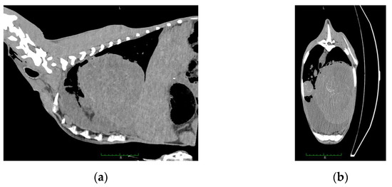
Figure 1.
Computed tomography image of the goat: (a) sagittal section, abscess (19 cm length, 20 cm high) located in the thorax, (b) frontal section, abscess (17 cm) located in the thorax. Both confirmed by R. equi isolation.
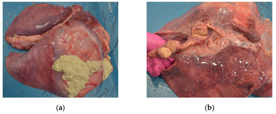
Figure 2.
Necropsy lesions of the goat caused by R. equi: (a) lungs abscessation, (b) abscesses in the mediastinal lymph nodes.
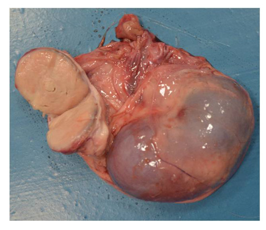
Figure 3.
Necropsy lesions of the goat: kidney abscessation caused by R. equi.
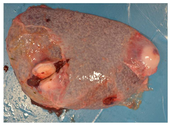
Figure 4.
Necropsy lesions of the goat: spleen abscessation caused by R. equi.
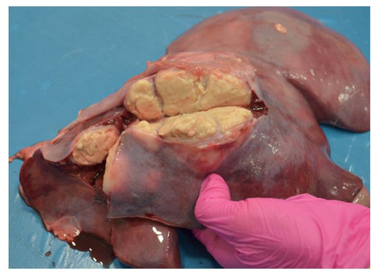
Figure 5.
Necropsy lesions of the goat: liver abscessation caused by R. equi.
In two animals, purulent lesions were present in collected lymph nodes (both in the mediastinal lymph nodes). In addition, extra-lymphatic abscesses were observed in some of the animals, and from most of the lesions, Corynebacterium pseudotuberculosis and Trueperella pyogenes were isolated.
From all 134 investigated animals, 272 lymph node samples were collected: 122 submandibular lymph nodes, 61 mediastinal lymph nodes, and 89 mesenteric lymph nodes.
R. equi was isolated from four animals: in three cases, from lymph node samples (two mesenteric and one submandibular) without any lesions; and the fourth isolate came from a goat suffering multiple organ abscessation. All four isolates were Gram-positive coccobacilli, and their growth characteristics were typical for the R. equi (mucoid, salmon-pink on blood agar, and greyish on CAZ-NB medium, irregular). They were catalase-positive, oxidase-negative, and identified to the genus level as Rhodococcus by API Coryne. In the CAMP test, enhancement of hemolysis was observed in the presence of both indicatory bacterial strains for these two isolates. In addition, all four isolates carried the choE gene specific to the species.
Unfortunately, two of four R. equi isolates, one from the lymph node and an isolate from the goat with multiple organ abscessation because of technical issues, were not available for further investigation.
One of the two remaining isolates carried traA and vapN genes. None of the plasmid-associated virulence genes were found in the second caprine R. equi isolate.
Corynebacterium pseudotuberculosis and Trueperella pyogenes were detected in two and one lymph node samples, respectively. In addition, Staphylococcus spp. was detected in the lymph nodes of nine animals (Table 1).

Table 1.
Result of the microbiological examination of the lesion-free lymph nodes collected from the goats.
PCR product obtained for the vapN gene was sequenced (outsourced to the Genomed, Poland) and analyzed with the Basic Local Alignment Search Tool (BLAST) carried out on the National Center for Biotechnology Information (NCBI) website (http://blast.ncbi.nlm.nih.gov accessed on 8 January 2020). As a result, the vapN gene sequence was deposited in the GenBank database under the accession number MN913373.1.
3. Discussion
This study continues the research on R. equi occurrence in farm animals led by the authors. However, in this case, the outcome may be used not only as epidemiological data, but may also impact further studies on R. equi pathogenicity.
According to the authors’ knowledge, this kind of study, designed to investigate the occurrence of R. equi in goats, was performed for the first time. So far, clinical cases were published [15,17,18,21,22,23,24,25,26,27,28,29], but only one study on the occurrence of R. equi in goats was performed. The serological screening in one breeding farm shoved seroprevalence close to 30% and confirmed exposure of clinically healthy goats for vapN-harboring R. equi [23]. Thus, it is not possible to compare the results with other goat studies.
Generally, ruminants are considered relatively resistant to R. equi infection. Thus, detection of the single clinical case between 134 investigated goats is not surprising. Additionally, detection of R. equi in 1.1% of investigated goats’ lymph nodes is close to other studies on ruminants. Prevalence of R. equi in slaughtered cattle used to be considered very low (0.008%) [53], but more recently, R. equi was isolated from 1.3% of lesion-free lymph nodes of cattle carcasses approved for human consumption [16]. Furthermore, the low prevalence of avirulent, environmental strains of R. equi in red deer (0.7%) and roe deer (0.9%) and lack of tissue lesions indicate an accidental carriage of the pathogen [36]. In addition, isolation of the ruminant-specific (pVAPN-carrying) R. equi aligns with previous studies. It confirms that the goat’s environment or goat meat might be a potential source of infection for humans [23,46,54].
Reported R. equi detection in cattle primarily concerns animals with purulent lesions or those suspected of Mycobacterium spp. infection similarly to the American bison with paratuberculosis [50,51,52]. On the other hand, microbiological examination of purulent lesions and caseous lymphadenitis found in slaughtered sheep during meat inspection detected some bacteria species, but was negative for R. equi [55].
This study has some limitations. Because the material was collected during another project carried out by the same research group, it was impossible to obtain all needed samples from each animal. In some cases, other pathological lesions precluded the collection of the lymph nodes for this survey. Furthermore, the collected material was stored at −20 °C until testing, which could reduce the effectiveness of R. equi isolation, so the accurate scale of the problem may be considerably more significant. However, the same methodology was fully effective in previous studies on the epidemiology of R. equi infection in wild and slaughter animals [16,36] and detection of Corynebacterium pseudotuberculosis, Trueperella pyogenes, and Staphylococcus spp., the most often isolated pathogens from purulent lesions in goats, confirms the usage of appropriate methods [56,57,58,59].
Moreover, samples were collected only from goats from one herd, and the investigated animals were not healthy. Furthermore, individuals were not randomly selected, but eliminated from the herd because of advanced caprine arthritis-encephalitis (CAE) clinical findings and weak conditions. This issue may pose a problem, as this comorbidity may affect the immunological status of the animal and therefore be unrepresentative of the rest of the population. However, CAE infection is widespread in Poland (over 80% of goat herds), and seroprevalence in the herds can approach 100% of adult animals [60,61,62]. Therefore, animals should not be excluded due to CAE infection, as they may represent a considerable part of the goat population.
It is believed that factors predisposing goats to R. equi infection are all immune-suppressing factors, such as stress (transport), co-existing diseases (parasites), and poor environmental conditions that have been published [15,17,18]. The direct impact of the small ruminant lentivirus (SRLV) infection on goats’ immunological status is not known. However, animals with advanced CAE clinical findings are under permanent stress deriving from the illness, for example, pain, the inability to move, and limited access to food and water. In addition, the social behavior of the herd might also have an impact because ill individuals are in the lowest position in the herd hierarchy. All those factors may lead to a weaker immunological response.
Thus, goats’ risk factors and clinical outcomes are similar to those observed in humans, implying that findings of goats suffering from severe CAE might reflect on immunocompromised humans. Therefore, undertaking research using goats as a large animal model may further develop a better understanding of the disease.
4. Materials and Methods
The study was performed on Polish White Improved and Polish Fawn Improved goats. Animals enrolled in this study were eliminated from a large dairy herd due to severe caprine arthritis-encephalitis (CAE) clinical findings, mainly due to emaciation, low milk yield, or progressive arthritis.
The culled animals were used for several research projects. Among others, computed tomography (CT) and necropsy were performed [63,64]. During post mortem examination, material for various laboratory procedures, including swabs from abscesses, was obtained. Moreover, submandibular, mediastinal, and mesenteric lymph nodes were collected and stored at −20 °C until testing.
Standard methods were used for bacteria isolation and identification. Briefly, lymph nodes were initially crushed with sterile scissors, and then 1 g of tissue was homogenized in 3 mL of 0.9% saline solution using a PRO200 homogenizer Multi-Gen 7 (PRO Scientific Inc., Oxford, CT, USA). Next, 100 µL of homogenate was collected and cultured on plates with a differentiation medium of Columbia Agar supplemented with 5% sheep blood (Graso Biotech, Starogard Gdanski, Poland), and selective CAZ-NB medium (Mueller-Hinton agar base supplemented with ceftazidime (0 µg/mL) and novobiocin (25 µg/mL) modified by the addition of 0.026% cycloheximide and 0.005% potassium tellurite. Plates were incubated for 48 h at 37 °C in aerobic conditions [16,36,49].
Colonies were identified based on their morphological, cultural, and biochemical characteristics. Gram staining was used to determine the cell morphology of isolates. Additionally, CAMP with Staphylococcus aureus ATCC 25923 with the R. equi ATCC 33701 reference strain as the control was performed on Columbia Agar with a 5% addition of sheep blood. The test results were evaluated after 24 h of incubation at 37 °C under aerobic conditions. The biochemical properties of isolates were tested using the API Coryne test (bioMérieux, Marcy l’Etoile, France) according to the manufacturer’s instructions.
DNA extracted from cultures of R. equi isolates that were 24 hours old was used as a template for PCR. Each isolate colony was suspended in 500 µL of distilled water and incubated at 99 °C for 10 min. Then, samples were cooled on ice and centrifuged, and stored at −20 °C. The presence of five R. equi genes, choE, traA, vapA, vapB, and vapN, was determined by PCR using specific primers (Table 2). The reaction mixture with a final volume of 25 µL contained 9.5 µL of nuclease-free water (Thermo Scientific, Waltham, MT, USA), 12.5 µL of DreamTaq Green PCR Master Mix (Thermo Scientific, Waltham, MT, USA), 10 pmol of each primer (Genomed, Warsaw, Poland), and 1 µL of DNA template. The thermal cycling conditions for the choE, traA, vapA, vapB, and vapN genes were conducted as previously described [9,16,37,65]. The amplified PCR products were separated by electrophoresis through 1% agarose gel in TAE buffer stained with Midori Green DNA Stain (Nippon, Düren, Germany), visualized, and analyzed using a VersaDoc Model 1000 Imaging System and Quantity One software (version 4.4.0) (Bio-Rad, Hercules, CA, USA).

Table 2.
PCR primers used to detect the selected genes of R. equi.
5. Conclusions
Despite its limitations, the study results show that R. equi is present in the goat population. Therefore, goat meat consumption is a possible source of human infection. It might also be speculated that the goat might be reconsidered as an inexpensive and applicable large animal model for R. equi infection.
Further research on the occurrence and pathogenicity of R. equi in goats might create a possibility for better understanding of the disease and development of the model, enabling the introduction of novel diagnostics and treatment techniques.
Author Contributions
Conceptualization, M.Ż., L.W., M.C., and J.K.; methodology, L.W., and M.R.; formal analysis, M.Ż., L.W., M.R. and M.C.; investigation L.W., A.K., M.R., E.K., I.S., M.C., O.S.-J., M.M., A.M., J.B. and J.K.; resources M.C. and J.K.; writing—original draft preparation M.Ż., M.R., and L.W.; writing—M.Ż., L.W., M.R., M.C., J.K.; supervision J.K.; project administration J.K.; funding acquisition L.W. and J.K. All authors have read and agreed to the published version of the manuscript.
Funding
This research was partially funded by the grant from the Ministry of Science and Higher Education of the Republic of Poland, decision no. 9506/E-385/R/2018.
Institutional Review Board Statement
Ethical review and approval were waived for this study due to the Directive 2010/63/EU and the Act of Polish Parliament of 15 January 2015 on the protection of animals used for scientific purposes (Journal of Laws 2015, item 266).
Informed Consent Statement
Not applicable.
Data Availability Statement
GenBank database under the accession number MN913373.1.
Conflicts of Interest
The authors declare no conflict of interest.
References
- Giguere, S.; Cohen, N.D.; Chaffin, M.K.; Hines, S.A.; Hondalus, M.K.; Prescott, J.F.; Slovis, N.M. Rhodococcus equi: Clinical Manifestations, Virulence, and Immunity. J. Vet. Intern. Med. 2011, 25, 1221–1230. [Google Scholar] [CrossRef]
- Witkowski, L. Treatment and prevention of Rhodococcus equi in foals. Vet. Rec. 2019, 185, 16–18. [Google Scholar] [CrossRef]
- Rakowska, A.; Cywinska, A.; Witkowski, L. Current Trends in Understanding and Managing Equine Rhodococcosis. Animals 2020, 10, 1910. [Google Scholar] [CrossRef]
- Cohen, N.D. Rhodococcus equi foal pneumonia. Vet. Clin. N. Am. Equine Pract. 2014, 30, 609–622. [Google Scholar] [CrossRef]
- Muscatello, G. Rhodococcus equi pneumonia in the foal—Part 2: Diagnostics, treatment and disease management. Vet. J. 2012, 192, 27–33. [Google Scholar] [CrossRef] [PubMed]
- Witkowski, L.; Rzewuska, M.; Takai, S.; Chrobak-Chmiel, D.; Kizerwetter-Swida, M.; Feret, M.; Gawrys, M.; Witkowski, M.; Kita, J. Molecular characterization of Rhodococcus equi isolates from horses in Poland: pVapA characteristics and plasmid new variant, 85-kb type V. BMC Vet. Res. 2017, 13, 35. [Google Scholar] [CrossRef]
- Vazquez-Boland, J.A.; Giguere, S.; Hapeshi, A.; MacArthur, I.; Anastasi, E.; Valero-Rello, A. Rhodococcus equi: The many facets of a pathogenic actinomycete. Vet. Microbiol. 2013, 167, 9–33. [Google Scholar] [CrossRef] [PubMed]
- Yamshchikov, A.V.; Schuetz, A.; Lyon, G.M. Rhodococcus equi infection. Lancet Infect. Dis. 2010, 10, 350–359. [Google Scholar] [CrossRef]
- Bryan, L.K.; Clark, S.D.; Diaz-Delgado, J.; Lawhon, S.D.; Edwards, J.F. Rhodococcus equi Infections in Dogs. Vet. Pathol. 2017, 54, 159–163. [Google Scholar] [CrossRef] [PubMed]
- Kinne, J.; Madarame, H.; Takai, S.; Jose, S.; Wernery, U. Disseminated Rhodococcus equi infection in dromedary camels (Camelus dromedarius). Vet. Microbiol. 2011, 149, 269–272. [Google Scholar] [CrossRef] [PubMed]
- Lechinski de Paula, C.; Silveira Silva, R.O.; Tavanelli Hernandes, R.; de Nardi Junior, G.; Babboni, S.D.; Trevizan Guerra, S.; Paganini Listoni, F.J.; Giuffrida, R.; Takai, S.; Sasaki, Y.; et al. First Microbiological and Molecular Identification of Rhodococcus equi in Feces of Nondiarrheic Cats. BioMed Res. Int. 2019, 2019, 4278598. [Google Scholar] [CrossRef]
- Lohr, C.V.; O’Neill, T.W.; Daw, D.N.; Pitel, M.O.; Schlipf, J.W. Pyogranulomatous enteritis and mesenteric lymphadenitis in an adult llama caused by Rhodococcus equi carrying virulence-associated protein A gene. J. Vet. Diagn. Investig. 2019, 31, 747–751. [Google Scholar] [CrossRef] [PubMed]
- Nakagawa, R.; Moki, H.; Hayashi, K.; Ooniwa, K.; Tokuyama, K.; Kakuda, T.; Yoshioka, K.; Takai, S. A case report on disseminated Rhodococcus equi infection in a Japanese black heifer. J. Vet. Med. Sci. 2018, 80, 819–822. [Google Scholar] [CrossRef]
- Selim, S.A.; Mousa, W.M.; Mohamed, K.F.; Moussa, I.M. Synergistic Haemolytic Activity and Its Correlation to Phospholipase D Productivity by Corynebacteruim Pseudotuberculosis Egyptian Isolates from Sheep and Buffaloes. Braz. J. Microbiol. 2012, 43, 552–559. [Google Scholar] [CrossRef][Green Version]
- Stranahan, L.W.; Plumlee, Q.D.; Lawhon, S.D.; Cohen, N.D.; Bryan, L.K. Rhodococcus equi Infections in Goats: Characterization of Virulence Plasmids. Vet. Pathol. 2018, 55, 273–276. [Google Scholar] [CrossRef]
- Witkowski, L.; Rzewuska, M.; Takai, S.; Kizerwetter-Swida, M.; Kita, J. Molecular epidemiology of Rhodococcus equi in slaughtered swine, cattle and horses in Poland. BMC Microbiol. 2016, 16, 98. [Google Scholar] [CrossRef]
- Haanen, G.A.Y.; Lim, C.K.; Baird, A.N.; Sola, M.F.; Lenz, S.D. Disseminated Rhodococcus equi in an Anglo-Nubian goat. Vet. Radiol. Ultrasound 2020, 61, E22–E25. [Google Scholar] [CrossRef]
- Schlemmer, S.N.; Fratzke, A.P.; Gibbons, P.; Porter, B.F.; Mansell, J.; Ploeg, R.J.; Rodrigues Hoffmann, A.; Older, C.E.; Clark, S.D. Histoplasmosis and multicentric lymphoma in a Nubian goat. J. Vet. Diagn. Investig. 2019, 31, 770–773. [Google Scholar] [CrossRef]
- de Morais, A.B.C.; Bolanos, C.A.D.; Alves, A.C.; Ikuta, C.Y.; Lara, G.H.B.; Heinemann, M.B.; Giuffrida, R.; Listoni, F.P.; Mioni, M.D.R.; Motta, R.G.; et al. Identification of Mycobacterium species and Rhodococcus equi in peccary lymph nodes. Trop. Anim. Health Prod. 2018, 50, 1319–1326. [Google Scholar] [CrossRef]
- Saied, A.A.; Bryan, L.K.; Bolin, D.C. Ulcerative, granulomatous glossitis and enteritis caused by Rhodococcus equi in a heifer. J. Vet. Diagn. Investig. 2019, 31, 783–787. [Google Scholar] [CrossRef] [PubMed]
- Carrigan, M.J.; Links, I.J.; Morton, A.G. Rhodococcus equi Infection in Goats. Aust. Vet. J. 1988, 65, 331–332. [Google Scholar] [CrossRef]
- Rodriguez, J.L.; Acosta, B.; Navarro, R.; Gutierrez, C. Rhodococcus equi infection in goat: Apropos of two cases. J. Appl. Anim. Res. 2000, 18, 149–151. [Google Scholar] [CrossRef]
- Suzuki, Y.; Takahashi, K.; Takase, F.; Sawada, N.; Nakao, S.; Toda, A.; Sasaki, Y.; Kakuda, T.; Takai, S. Serological epidemiological surveillance for vapN-harboring Rhodococcus equi infection in goats. Comp. Immunol. Microbiol. Infect. Dis. 2020, 73, 101540. [Google Scholar] [CrossRef]
- Jeckel, S.; Holmes, P.; King, S.; Whatmore, A.M.; Kirkwood, I. Disseminated Rhodococcus equi infection in goats in the UK. Vet. Rec. 2011, 169, 56. [Google Scholar] [CrossRef]
- Kabongo, P.N.; Njiro, S.M.; Van Strijp, M.F.; Putterill, J.F. Caprine vertebral osteomyelitis caused by Rhodococcus equi. J. S. Afr. Vet. Assoc. 2005, 76, 163–164. [Google Scholar] [CrossRef][Green Version]
- Davis, W.P.; Steficek, B.A.; Watson, G.L.; Yamini, B.; Madarame, H.; Takai, S.; Render, J.A. Disseminated Rhodococcus equi infection in two goats. Vet. Pathol. 1999, 36, 336–339. [Google Scholar] [CrossRef] [PubMed]
- Tkachuk-Saad, O.; Lusis, P.; Welsh, R.D.; Prescott, J.F. Rhodococcus equi infections in goats. Vet. Rec. 1998, 143, 311–312. [Google Scholar] [CrossRef] [PubMed]
- Fitzgerald, S.D.; Walker, R.D.; Parlor, K.W. Fatal Rhodococcus equi infection in an Angora goat. J. Vet. Diagn. Investig. 1994, 6, 105–107. [Google Scholar] [CrossRef] [PubMed]
- Ojo, M.O.; Njoku, C.O.; Freitas, J.; Nurse, L.; Romain, H. Isolation of Rhodococcus equi from the liver abscess of a goat in Trinidad. Can. Vet. J. 1993, 34, 504. [Google Scholar]
- Aslam, M.W.; Lau, S.F.; Chin, C.S.L.; Ahmad, N.I.; Rahman, N.A.; Kuppusamy, K.; Omar, S.; Radzi, R. Clinicopathological and radiographic features in 40 cats diagnosed with pulmonary and cutaneous Rhodococcus equi infection (2012–2018). J. Feline Med. Surg. 2019, 22, 774–790. [Google Scholar] [CrossRef]
- Portilho, F.V.R.; Paes, A.C.; Megid, J.; Hataka, A.; Neto, R.T.; Headley, S.A.; Oliveira, T.E.S.; Colhado, B.S.; de Paula, C.L.; Guerra, S.T.; et al. Rhodococcus equi pVAPN type causing pneumonia in a dog coinfected with canine morbillivirus (Distemper virus) and Toxoplasma gondii. Microb. Pathog. 2019, 129, 112–117. [Google Scholar] [CrossRef]
- Alvarez-Narvaez, S.; Giguere, S.; Cohen, N.; Slovis, N.; Vazquez-Boland, J.A. Spread of Multidrug-Resistant Rhodococcus equi, United States. Emerg. Infect. Dis. 2021, 27, 529–537. [Google Scholar] [CrossRef]
- Alvarez-Narvaez, S.; Huber, L.; Giguere, S.; Hart, K.A.; Berghaus, R.D.; Sanchez, S.; Cohen, N.D. Epidemiology and Molecular Basis of Multidrug Resistance in Rhodococcus equi. Microbiol. Mol. Biol. Rev. 2021, 85, e00011-21. [Google Scholar] [CrossRef] [PubMed]
- Cisek, A.A.; Rzewuska, M.; Witkowski, L.; Binek, M. Antimicrobial resistance in Rhodococcus equi. Acta Biochim. Pol. 2014, 61, 633–638. [Google Scholar] [CrossRef]
- Erol, E.; Locke, S.; Saied, A.; Cruz Penn, M.J.; Smith, J.; Fortner, J.; Carter, C. Antimicrobial susceptibility patterns of Rhodococcus equi from necropsied foals with rhodococcosis. Vet. Microbiol. 2020, 242, 108568. [Google Scholar] [CrossRef]
- Witkowski, L.; Rzewuska, M.; Cisek, A.A.; Chrobak-Chmiel, D.; Kizerwetter-Swida, M.; Czopowicz, M.; Welz, M.; Kita, J. Prevalence and genetic diversity of Rhodococcus equi in wild boars (Sus scrofa), roe deer (Capreolus capreolus) and red deer (Cervus elaphus) in Poland. BMC Microbiol. 2015, 15, 110. [Google Scholar] [CrossRef]
- Golub, B.; Falk, G.; Spink, W.W. Lung Abscess Due to Corynebacterium equi—Report of First Human Infection. Ann. Intern. Med. 1967, 66, 1174. [Google Scholar] [CrossRef]
- Gundelly, P.; Suzuki, Y.; Ribes, J.A.; Thornton, A. Differences in Rhodococcus equi Infections Based on Immune Status and Antibiotic Susceptibility of Clinical Isolates in a Case Series of 12 Patients and Cases in the Literature. BioMed Res. Int. 2016, 2016, 2737295. [Google Scholar] [CrossRef]
- Marrie, T.J. Community acquired pneumonia. Praxis (Bern 1994) 2001, 90, 935–940. [Google Scholar]
- Topino, S.; Galati, V.; Grilli, E.; Petrosillo, N. Rhodococcus equi Infection in HIV-Infected Individuals: Case Reports and Review of the Literature. AIDS Patient Care STDs 2010, 24, 211–222. [Google Scholar] [CrossRef]
- Lin, W.V.; Kruse, R.L.; Yang, K.; Musher, D.M. Diagnosis and management of pulmonary infection due to Rhodococcus equi. Clin. Microbiol. Infect. 2019, 25, 310–315. [Google Scholar] [CrossRef]
- Gray, K.J.; French, N.; Lugada, E.; Watera, C.; Gilks, C.F. Rhodococcus equi and HIV-1 infection in Uganda. J. Infect. 2000, 41, 227–231. [Google Scholar] [CrossRef]
- Gundelly, P.; Thornton, A.; Greenberg, R.N.; McCormick, M.; Myint, T. Rhodococcus equi pericarditis in a patient living with HIV/AIDS. J. Int. Assoc. Provid. AIDS Care 2014, 13, 309–312. [Google Scholar] [CrossRef]
- Mikic, D.; Djordjevic, Z.; Sekulovic, L.; Kojic, M.; Tomanovic, B. Disseminated Rhodococcus equi infection in a patient with Hodgkin lymphoma. Vojnosanit. Pregl. 2014, 71, 317–324. [Google Scholar] [CrossRef]
- Nath, S.R.; Mathew, A.P.; Mohan, A.; Anila, K.R. Rhodococcus equi granulomatous mastitis in an immunocompetent patient. J. Med. Microbiol. 2013, 62, 1253–1255. [Google Scholar] [CrossRef]
- Takai, S.; Sawada, N.; Nakayama, Y.; Ishizuka, S.; Nakagawa, R.; Kawashima, G.; Sangkanjanavanich, N.; Sasaki, Y.; Kakuda, T.; Suzuki, Y. Reinvestigation of the virulence of Rhodococcus equi isolates from patients with and without AIDS. Lett. Appl. Microbiol. 2020, 71, 679–683. [Google Scholar] [CrossRef] [PubMed]
- Lara, G.H.; Takai, S.; Sasaki, Y.; Kakuda, T.; Listoni, F.J.; Risseti, R.M.; de Morais, A.B.; Ribeiro, M.G. VapB type 8 plasmids in Rhodococcus equi isolated from the small intestine of pigs and comparison of selective culture media. Lett. Appl. Microbiol. 2015, 61, 306–310. [Google Scholar] [CrossRef]
- Ribeiro, M.G.; Lara, G.H.B.; da Silva, P.; Franco, M.M.J.; de Mattos-Guaraldi, A.L.; de Vargas, A.P.C.; Sakate, R.I.; Pavan, F.R.; Colhado, B.S.; Portilho, F.V.R.; et al. Novel bovine-associated pVAPN plasmid type in Rhodococcus equi identified from lymph nodes of slaughtered cattle and lungs of people living with HIV/AIDS. Transbound Emerg. Dis. 2018, 65, 321–326. [Google Scholar] [CrossRef]
- Rzewuska, M.; Witkowski, L.; Cisek, A.A.; Stefanska, I.; Chrobak, D.; Stefaniuk, E.; Kizerwetter-Swida, M.; Takai, S. Characterization of Rhodococcus equi isolates from submaxillary lymph nodes of wild boars (Sus scrofa), red deer (Cervus elaphus) and roe deer (Capreolus capreolus). Vet. Microbiol. 2014, 172, 272–278. [Google Scholar] [CrossRef] [PubMed]
- Buergelt, C.D.; Layton, A.W.; Ginn, P.E.; Taylor, M.; King, J.M.; Habecker, P.L.; Mauldin, E.; Whitlock, R.; Rossiter, C.; Collins, M.T. The pathology of spontaneous paratuberculosis in the North American bison (Bison bison). Vet. Pathol. 2000, 37, 428–438. [Google Scholar] [CrossRef]
- Dvorska, L.; Parmova, I.; Lavickova, M.; Bartl, J.; Vrbas, V.; Pavlik, I. Isolation of Rhodococcus equi and atypical mycobacteria from lymph nodes of pigs and cattle in herds with the occurrence of tuberculoid gross changes in the Czech Republic over the period of 1996–1998. Vet. Med. 1999, 44, 321–330. [Google Scholar]
- Sahraoui, N.; Muller, B.; Guetarni, D.; Boulahbal, F.; Yala, D.; Ouzrout, R.; Berg, S.; Smith, N.H.; Zinsstag, J. Molecular characterization of Mycobacterium bovis strains isolated from cattle slaughtered at two abattoirs in Algeria. BMC Vet. Res. 2009, 5, 4. [Google Scholar] [CrossRef]
- Salazar-Rodriguez, D.; Aleaga-Santiesteban, Y.; Iglesias, E.; Plascencia-Hernandez, A.; Perez-Gomez, H.R.; Calderon, E.J.; Vazquez-Boland, J.A.; de Armas, Y. Virulence Plasmids of Rhodococcus equi Isolates from Cuban Patients with AIDS. Front. Vet. Sci. 2021, 8, 628239. [Google Scholar] [CrossRef]
- Didkowska, A.; Zmuda, P.; Kwiecien, E.; Rzewuska, M.; Klich, D.; Krajewska-Wedzina, M.; Witkowski, L.; Zychska, M.; Kaczmarkowska, A.; Orlowska, B.; et al. Microbiological assessment of sheep lymph nodes with lymphadenitis found during post-mortem examination of slaughtered sheep: Implications for veterinary-sanitary meat control. Acta Vet. Scand. 2020, 62, 48. [Google Scholar] [CrossRef]
- Flynn, O.; Quigley, F.; Costello, E.; O’Grady, D.; Gogarty, A.; Mc Guirk, J.; Takai, S. Virulence-associated protein characterisation of Rhodococcus equi isolated from bovine lymph nodes. Vet. Microbiol. 2001, 78, 221–228. [Google Scholar] [CrossRef]
- Szalus-Jordanow, O.; Kaba, J.; Czopowicz, M.; Witkowski, L.; Nowicki, M.; Nowicka, D.; Stefanska, I.; Rzewuska, M.; Sobczak-Filipiak, M.; Binek, M.; et al. Epidemiological features of Morel’s disease in goats. Pol. J. Vet. Sci. 2010, 13, 437–445. [Google Scholar] [PubMed]
- Rzewuska, M.; Kwiecien, E.; Chrobak-Chmiel, D.; Kizerwetter-Swida, M.; Stefanska, I.; Gierynska, M. Pathogenicity and Virulence of Trueperella pyogenes: A Review. Int. J. Mol. Sci. 2019, 20, 2737. [Google Scholar] [CrossRef]
- Kaba, J.; Nowicki, M.; Frymus, T.; Nowicka, D.; Witkowski, L.; Szalus-Jordanow, O.; Czopowicz, M.; Thrusfield, M. Evaluation of the risk factors influencing the spread of caseous lymphadenitis in goat herds. Pol. J. Vet. Sci. 2011, 14, 231–237. [Google Scholar] [CrossRef]
- Moroz, A.; Szalus-Jordanow, O.; Czopowicz, M.; Brodzik, K.; Petroniec, V.; Augustynowicz-Kopec, E.; Lutynska, A.; Roszczynko, M.; Golos-Wojcicka, A.; Korzeniowska-Kowal, A.; et al. Nasal carriage of various staphylococcal species in small ruminant lentivirus-infected asymptomatic goats. Pol. J. Vet. Sci. 2020, 23, 203–209. [Google Scholar] [CrossRef]
- Kaba, J.; Czopowicz, M.; Ganter, M.; Nowicki, M.; Witkowski, L.; Nowicka, D.; Szalus-Jordanow, O. Risk factors associated with seropositivity to small ruminant lentiviruses in goat herds. Res. Vet. Sci. 2013, 94, 225–227. [Google Scholar] [CrossRef]
- Czopowicz, M.; Szalus-Jordanow, O.; Mickiewicz, M.; Moroz, A.; Witkowski, L.; Markowska-Daniel, I.; Stefaniak, T.; Bagnicka, E.; Kaba, J. Haptoglobin and serum amyloid A in goats with clinical form of caprine arthritis-encephalitis. Small Rumin. Res. 2017, 156, 73–77. [Google Scholar] [CrossRef]
- Potarniche, A.V.; Cerbu, C.G.; Czopowicz, M.; Szalus-Jordanow, O.; Kaba, J.; Spinu, M. The epidemiological background of small ruminant lentivirus infection in goats from Romania. Vet. World 2020, 13, 1344–1350. [Google Scholar] [CrossRef]
- Szalus-Jordanow, O.; Bonecka, J.; Pankowski, F.; Barszcz, K.; Tarka, S.; Kwiatkowska, M.; Polguj, M.; Mickiewicz, M.; Moroz, A.; Czopowicz, M.; et al. Postmortem imaging in goats using computed tomography with air as a negative contrast agent. PLoS ONE 2019, 14, e0215758. [Google Scholar] [CrossRef]
- Szalus-Jordanow, O.; Czopowicz, M.; Witkowski, L.; Mickiewicz, M.; Moroz, A.; Kaba, J.; Sapierzynski, R.; Bonecka, J.; Jonska, I.; Garncarz, M.; et al. Malignant thymoma—The most common neoplasm in goats. Pol. J. Vet. Sci. 2019, 22, 475–480. [Google Scholar] [CrossRef] [PubMed]
- Takai, S.; Ikeda, T.; Sasaki, Y.; Watanabe, Y.; Ozawa, T.; Tsubaki, S.; Sekizaki, T. Identification of virulent Rhodococcus equi by amplification of gene coding for 15- to 17-kilodalton antigens. J. Clin. Microbiol. 1995, 33, 1624–1627. [Google Scholar] [CrossRef]
- Ladrón, N.; Fernández, M.; Agüero, J.; González Zörn, B.; Vázquez-Boland, J.A.; Navas, J. Rapid identification of Rhodococcus equi by a PCR assay targeting the choE gene. J. Clin. Microbiol. 2003, 41, 3241–3245. [Google Scholar] [CrossRef] [PubMed]
- Ocampo-Sosa, A.A.; Lewis, D.A.; Navas, J.; Quigley, F.; Callejo, R.; Scortti, M.; Leadon, D.P.; Fogarty, U.; Vazquez-Boland, J.A. Molecular epidemiology of Rhodococcus equi based on traA, vapA, and vapB virulence plasmid markers. J. Infect. Dis. 2007, 196, 763–769. [Google Scholar] [CrossRef] [PubMed]
- Oldfield, C.; Bonella, H.; Renwick, L.; Dodson, H.I.; Alderson, G.; Goodfellow, M. Rapid determination of vapA/vapB genotype in Rhodococcus equi using a differential polymerase chain reaction method. Antonie Van Leeuwenhoek 2004, 85, 317–326. [Google Scholar] [CrossRef]
Publisher’s Note: MDPI stays neutral with regard to jurisdictional claims in published maps and institutional affiliations. |
© 2021 by the authors. Licensee MDPI, Basel, Switzerland. This article is an open access article distributed under the terms and conditions of the Creative Commons Attribution (CC BY) license (https://creativecommons.org/licenses/by/4.0/).