Article

High Prevalence of Virulence-Associated Genes and Length Polymorphism in actA and inlB Genes Identified in Listeria monocytogenes Isolates from Meat Products and Meat-Processing Environments in Poland

by
Iwona Kawacka
* and
Agnieszka Olejnik-Schmidt
*
Department of Food Biotechnology and Microbiology, Poznan University of Life Sciences, Wojska Polskiego 48, 60-627 Poznan, Poland
*
Authors to whom correspondence should be addressed.
Pathogens 2024, 13(6), 444; https://doi.org/10.3390/pathogens13060444
Submission received: 27 March 2024 / Revised: 6 May 2024 / Accepted: 22 May 2024 / Published: 23 May 2024

Abstract

:
Listeria monocytogenes is a human pathogen that has the ability to cause listeriosis, a disease with possible fatal outcomes. The typical route of infection is ingestion of the bacteria with contaminated food. In this study, 13 virulence-associated genes were examined with PCR in the genomes of 153 L. monocytogenes isolates collected from meat products and processing environments in Poland. All isolates possessed genes from LIPI-1—hly, actA, plcA, plcB and mpl—as well as four internalins: inlA, inlB, inlC, inlJ. Invasion-associated protein iap, as well as genes prfA and sigB, encoding regulatory proteins, were also detected in all isolates. Gene flaA, encoding flagellin, was detected in 113 (74%) isolates. This was the only gene that was not detected in all isolates, as its presence is serotype-dependent. Gene actA showed polymorphism with longer and shorter variants in PCR amplicons. Two isolates were characterized by truncated inlB genes, lacking 141 bp in their sequence, which was confirmed by gene sequencing. All isolates were positive in hemolysis assays, proving the synthesis of functional PrfA and Hly proteins. Four genotypes of L. monocytogenes based on actA polymorphism and two genotypes based on inlB polymorphism were distinguished within the isolates’ collection.

1. Introduction

Listeria monocytogenes is a Gram-positive, facultatively anaerobic rod adapted to various conditions. These ubiquitous bacteria are found throughout the environment. Many domestic animals, especially ruminants, are carriers of L. monocytogenes, which leads to the contamination of animal breeding areas, with the subsequent possibility of food contamination. The ingestion of L. monocytogenes with contaminated food may result in self-limiting gastroenteritis occurring with fever and diarrhea. Although usually listeriosis is mild, in severe cases symptoms include sepsis, meningitis, encephalitis, and spontaneous abortion. Overall mortality risk is estimated to be approximately 15% or higher, depending on patient status and comorbidities [1,2,3,4,5,6].
L. monocytogenes is an invasive intracellular pathogen. Its virulence depends on many adhesion and invasion factors that facilitate gastrointestinal tract colonization and crossing of the intestinal barrier [7]. Virulence factors also include proteins facilitating dissemination in the host, including brain and placenta colonization, cell-to-cell spread, the adhesion, and invasion of macrophages and escape from L. monocytogenes-containing vacuoles. Proteins enabling survival in the intestines, such as acid and bile tolerance proteins, are also virulence factors. The most important virulence-associated genes of L. monocytogenes are localized on Listeria pathogenicity island-1 (LIPI-1), namely, hly, actA, plcA, plcB and mpl [5,7,8,9]. All virulence-associated genes from LIPI-1 are positively regulated by a pleiotropic transcriptional regulator PrfA (encoded by the prfA gene), which is considered the main positive regulatory factor of virulence genes in L. monocytogenes [9,10,11].
Other groups of key listerial virulence factors, outside of LIPI-1, include internalins. The most important ones are inlA and inlB, but many have been identified (including inlC, inlJ, inlH, inlK, inlL, inlF and inlP) [8,9,12,13]. Other known listerial virulence factors include, for example, invasion-associated protein (encoded by iap), flagellin (encoded by flaA), a general stress-response regulator, called sigma factor B, (encoded by sigB), Listeria-mucin-binding invasin A, bile salt hydrolase, and cell invasion LPXTG protein, ClpP, a heat shock protein that is involved in intracellular growth or fibronectin-binding protein [7,8,10,12], to just name a few. However, not all L. monocytogenes isolates harbor all discovered virulence genes [14,15,16].
A species-specific characteristic of L. monocytogenes, historically considered to be a virulence marker, is beta-hemolytic activity [17]. This trait is often used to confirm the species identification of the isolates [4,18,19]. However, there are also reports about isolates of this species that do not present hemolytic phenotype, mainly due to mutations within either the hly gene, or, more frequently, prfA mutations [20]. Furthermore, it has been suggested that the spontaneous loss of virulence in natural populations of L. monocytogenes, although rare, is possible due to the fact that some of the virulence genes are under purifying selection. This opens an evolutionary path for potential saprophytism for this pathogen [20]. Hence, tracking potential changes in the virulence-associated genes patterns in food isolates of L. monocytogenes is important, as a trend of reducing pathogenicity in this genus may be observed.

2. Aim of This Study

The aim of this study was to assess the diversity of L. monocytogenes isolates collected in recent years in Poland originating from meat products and meat processing facilities. The presence or absence (or polymorphic form) of selected virulence-associated genes was assessed and the hemolytic phenotype of those isolates was determined. An estimation of the virulence potential of the collected isolates based on the obtained results was an additional aim of this study.

3. Materials and Methods

3.1. Bacterial Isolates and Genetic Material

A collection of 153 L. monocytogenes isolates used in this study originated from both raw meat samples and processed meat products manufactured in Poland (n = 108), as well as from meat processing plants in Poland (n = 45), representing food processing and environmental surfaces. The isolates were collected between October 2020 and November 2021. The DNA of those microorganisms used for gene detection was isolated using a Genomic Mini kit (A&A Biotechnology, Gdańsk, Poland) according to the manufacturer’s instructions. Bacterial isolates preserved in brain heart infusion broth (BHI; Oxoid, Warsaw, Poland) glycerol stocks stored at −80 °C were used for hemolysis assays. Isolates included in the study were confirmed as L. monocytogenes species with two separate genetic analyses, using a PCR-RFLP according to Paillard et al. (2003) [21] and multiplex PCR according to Li et al. (2021) [22] protocols. Details regarding the isolates’ collection process, DNA isolation procedure, and the exact methodology for species affiliation were reported previously [23].

3.2. Detection of Virulence-Associated Genes

The presence of thirteen virulence-associated genes in genomes of L. monocytogenes isolates was analyzed in the study, using standard PCR for most of the genes or multiplex PCR for the simultaneous detection of the two genes inlB and inlC. If an amplicon for any gene was not detected in a multiplex reaction, then separate PCRs using only one pair of primers were performed for verification. If discrepancies occurred between multiplex and singleplex, only the singleplex results were taken into account. Additionally, two pairs of primers (here referred to as actA1 and actA2) were used to analyze the presence of the actA gene.
Reaction mixtures contained 0.2U of RUN polymerase (A&A Biotechnology), along with compatible reaction buffer at the recommended concentration, 0.2 mM of each dNTPs (A&A Biotechnology) and primers (concentrations are given in Table 1), which were ordered in Genomed S.A. (Warsaw, Poland). DNA was added in the amount of 10 ng per reaction. Reactions were performed in a final volume of 10 μL. Thermal cycling was performed in the T-Gradient thermocycler (Biometra, Göttingen, Germany) with annealing temperatures, as presented in Table 1. PCR products were separated by electrophoresis in an agarose gel-containing ethidium bromide and visualized using Gel Doc Imaging System (Bio-Rad, Hercules, CA, USA).
At least one randomly chosen sample representing one amplicon size amplified with one primer pair was sequenced. PCR products were purified prior to sequencing directly from the PCR mixture or after separation in an agarose gel, using the A&A Biotechnology kits, with either Clean-Up Concentrator or Gel-Out Concentrator. Sequencing was performed by Genomed S.A. Obtained sequences were determined to be fragments of genes of interest using Blast software (BLASTN 2.14.1+) [35,36].
In the case of inlB gene, in order to obtain a full-length sequence for more reliable reads, sequences achieved with the forward primer were aligned with the sequence achieved using the reverse primer. BLAST [37] was used for this purpose. The obtained aligned sequences (for isolates 50 and 235) were compared with each other as well as with the inlB gene sequence of L. monocytogenes EDG-e from the GenBank database (accession number NC_003210.1), also using BLAST.

3.3. Hemolysis Assay

Hemolysis assays were performed on a commercially available Columbia Agar with 5% Sheep Blood (Becton, Dickinson and Company, Heidelberg, Germany). Bacterial isolates from glycerol stocks were streaked into BHI agar plates and incubated for 18 h at 37 °C. After incubation, bacteria were re-streaked from BHI to the blood agar plate and incubated at 37 °C for 24 h and 48 h, with assessment after both time periods. Listeria ivanovii ATCC 19119 was used as a positive control and Listeria innocua food isolates 135, 145, and 159 were used as negative controls.

4. Results

4.1. Presence of Virulence-Associated Genes

In the case of ten tested virulence-associated genes, namely, iap, sigB, prfA, hlyA, inlC, inlA, inlJ, plcA, plcB, and mpl, all of the L. monocytogenes isolates included in the study were positive in PCR with amplicons of expected length, in accordance with the literature references. The results were further confirmed with sequencing and BLAST analyses. An amplicon of the flaA gene was present in 113 (74%) isolates, all of which belonged to genoserotype IIa (representing serotypes 1/2a, 3a) (results reported previously; see Reference [23]). Furthermore, all isolates representing genoserotype IIa harbored the flaA gene [23].
In the case of actA and inlB genes, polymorphism of amplicon sizes was detected within the isolates’ collection. Summarized virulence detection results are presented in Table 2.
In the case of /actA/ and /inlB/ genes, polymorphism of amplicon sizes was detected within the isolates’ collection. Summarized virulence detection results are presented in Table 2 above.

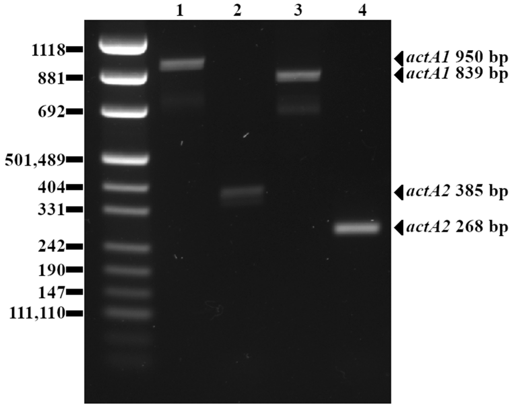
4.2. actA Polymorphism

Typing L. monocytogenes using the two actA primer pairs allowed for four genotypes to be differentiated, which are presented in Table 3.
Interestingly, there is no clear pattern between genoserotype, as determined using the protocol of Doumith et al. protocol [38] (results published previously; see Reference [23]), and the actA amplicon variants achieved in this study, which allowed for the further differentiation of genoserotyped isolates.
Polymorphic variants of the actA gene detected with actA1 and actA2 primer pairs are presented in Figure 1 below.

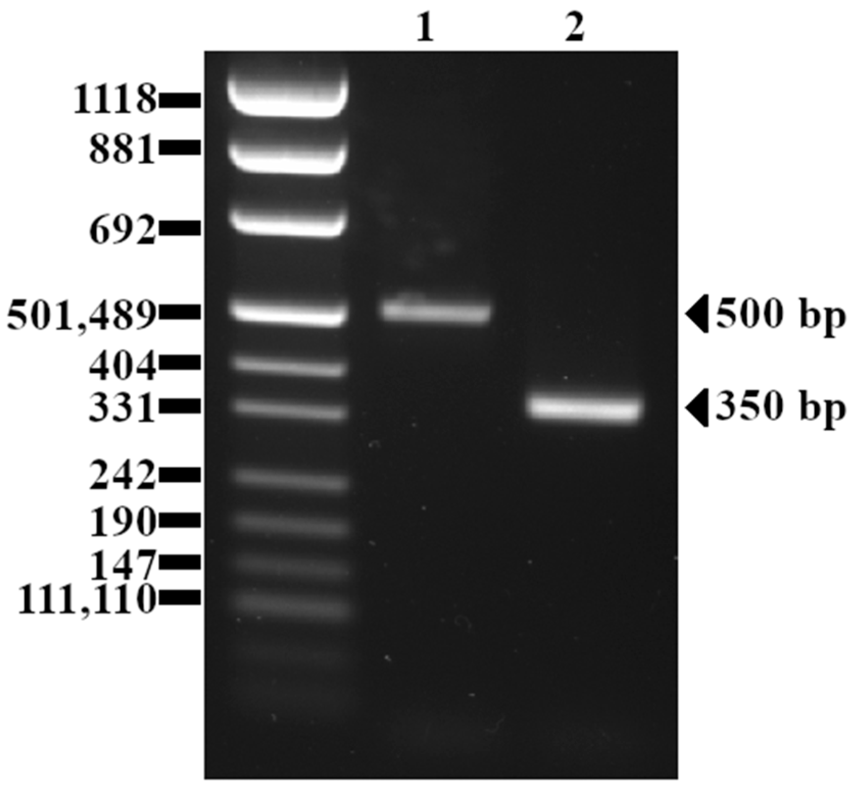
4.3. inlB Polymorphism

Two isolates presented a shorter inlB amplicon than expected. Both of them are characterized by shorter variants of the actA gene, both belonged to the same rare genoserotype IIb (representing serotypes 1/2b, 3b, 7), and both shared the same antibiotic resistance profile, with susceptibility to all 10 tested antibiotics (results published previously; see Reference [39]), although the vast majority (95%) of isolates from this study were characterized by this particular susceptibility profile.
The two isolates with the truncated inlB gene originated from head cheese (processed meat product, also known as brawn) (isolate 50) and the Vienna-type sausage (isolate 235). Sequences of the inlB genes of those two isolates, when aligned with the BLAST, showed 357/366 (99%) identities. When compared to the EGD-e strain, both isolates 50 and 235 lack the 141-bp long fragment (encoding the β-repeat sheet and partially encoding the GW1 domain of the InlB protein [40]) within the inlB gene sequence.
The detected variants of the inlB gene are presented in Figure 2 below.

4.4. Hemolysis

All 153 tested L. monocytogenes isolates presented the hemolytic phenotype. The response was consistent after 24 h and 48 h incubation. Positive results for the hemolysis assay indicate that Hly (listeriolysin O) encoded by the hly gene is functional, and that the PrfA regulator, encoded by prfA, positively regulating hly expression, also remains active.

5. Discussion

5.1. General Prevalence of Virulence-Associated Genes

Recent studies of L. monocytogenes originating from Poland do not always confirm the prevalence of all tested virulence-associated genes in bacterial genomes [14,15]. For example hlyA and prfA were present in 100% of the 27 food isolates and 13 isolates from the food processing environments, whereas inlB and sigB genes were present in 26 (97.5%) and 20 (82.5%) of the samples, respectively [14]. Genes plcB, hlyA, iap, actA, prfA, and sigB were present in all seven isolates originating from fish housed in Poland, whereas inlB was detected in six (85.7%) isolates [15]. However, similarly to our results, internalin family member genes inlA, inlB, inlC, inlE, inlF, and inlJ and the pathogenicity island LIPI-1 were found in 48 (100%) of the tested isolates from different kinds of ready-to-eat (RTE) food of animal origin and from a food processing environment in Poland [40]. Interestingly, the authors also identified one isolate with a deletion of 141 nucleotides in the inlB gene [40].
In recent international studies, some authors report the presence of the tested virulence genes in 100% of the examined isolates (including, e.g., hlyA, prfA, iap, inlA, inlB, mpl, plcA, and plcB [41]), whereas sometimes particular genes were detected only in a subgroup of samples, e.g., only 70% and 80% of the isolates from bovine farms in India harbored plcA and plcB genes, even though hlyA and iap were present in all of them [16].
The only gene not detected in all isolates in our study is flaA, encoding flagellin, which is a protein specific to the 1/2a and 3a serotypes [34]. All isolates positive for the gene encoding flagellin (74%) belong to genoserotype IIa (representing serotypes 1/2a, 3a) [23], and all isolates that were negative belong to other genoserotypes, which is consistent with the theoretical expected results.

5.2. actA Polymorphism

A polymorphism within the actA gene of L. monocytogenes isolates has already been reported in the literature [42,43]. Furthermore, the partial sequencing of this gene has been used as a tool for subtyping L. monoctogenes isolates, enabling the division of the isolates into two [42] or three [43] lineages. In our study, we identified four genotypes based on actA typing using two sets of primers.
Interestingly, the actA gene is located on LIPI-1 between the mpl and plcB genes [44] and, in our study, both flanking genes were identified in all 153 isolates, without detecting any visible polymorphism. This raises questions about the origin of variability within this particular gene, especially considering that the genotypes established based on actA are not correlated with serotypes, which was proved by our study and also reported earlier by other researchers [45].
Although there are studies indicating that the in vitro virulence of L. monocytogenes is not determined by the actA polymorphism [46], some publications found that the actA polymorphism influences the virulence potential of the isolates [42,43,45]. In one study, isolates classified based on the actA polymorphism as lineage II showed significantly lower invasiveness on epithelial Caco-2 cells than lineage I isolates [43]. Similarly, in the paper where L. monocytogenes was subtyped with this method, isolates from lineage I all contained highly invasive isolates, as well as isolates with moderate and low invasiveness, whereas lineage II contained only low-invasive isolates. The invasiveness was established with a cell-invasion assay with the CX-1 human colon cancer cell line [42]. Earlier studies indicated that the deletion of one large unit within a proline-rich region of ActA resulted in a reduction in intracellular bacterial speed, as well as decreased virulence [45]. The primer pairs (actA1 and actA2) used herein include regions translated to proline-rich regions of the protein.

5.3. inlB Polymorphism

Two isolates from the collection examined in this study presented a truncated inlB gene with a 141 bp deletion. Interestingly, Kurpas et al. (2020) also identified an isolate from Poland with 141 bp deletion in the same region of inlB gene [40].
There are also other reports about L. monocytogenes strains harboring mutations within this gene. For example, a food isolate from Mexican-style soft cheese L. monocytogenes F2365 harboring mutations resulting in premature stop codons in inlB was characterized by a reduced invasion efficiency in Caco-2 cells [47]. The same isolate with introduced point mutation resulting in rescued inlB expression showed approximately 9-fold and 1.5-fold higher invasion in HeLa and JEG-3 cells, respectively, when compared to the parental strain [48]. These findings are consistent with a study in which a constructed inlB deletion L. monocytogenes mutant showed significantly decreased invasiveness in a mouse as compared to wild-type isolate [49]. However, there is also a report indicating that the L. monocytogenes A23 strain with inactive internalin B protein remained virulent in a plaque assay on human adenocarcinoma cell line HT-29 [50]. Interestingly, one study demonstrated that InlB domain variants may differ in their ability to support intragastric infection (measured as bacterial loads in livers of intragastrically infected mice), even though, in a cell culture study (measuring the invasion rate of murine colon carcinoma C26 cells), the results were not clearly apparent [51].

5.4. Hemolysis

Hemolysis assay is a phenotypic criterion used for confirmation of the species affiliation of collected isolates [29,52,53,54]. However, there are reports stating that some L. monocytogenes isolates do not present with hemolysis, with a rate of approximately 0.1% according to a study which included 57,820 isolates of food, clinical, veterinary, environmental, and other origins [20]. A lack of hemolytic phenotype can indicate diminished virulence caused by either a nonfunctional Hly protein due to mutations or its hampered expression due to mutations in the prfA gene, encoding the PrfA regulator that positively regulates Hly expression [20].
Hemolysis assays in the case of L. monocytogenes may provide interpretative difficulties. There is no consistency within the literature regarding the most sensitive method for hemolysis assessment or the blood type that would provide optimal test sensitivity [55]. Similarly to many other authors [20,56,57,58,59,60,61,62,63], we decided to apply the blood agar technique. Due to the fact that we observed weaker hemolysis in the case of L. monocytogenes strains than in the positive control, we do not recommend using L. ivanovii ATCC 19119 for that purpose, as this factor could contribute to false-negative reads in the case of L. monocytogenes isolates.

6. Summary and Conclusions

Although all 153 isolates possessed the 12 tested genes (iap, sigB, prfA, hly, actA, inlB, inlC, inlA, inlJ, plcA, plcB, and mpl), a PCR detection of virulence-associated genes allowed us to differentiate L. monocytogenes strains in our collection. The presence of the flaA gene was strictly serotype-dependent, whereas a polymorphism found in the actA gene could further differentiate strains pre-grouped into serogroups. Four major genotypes were identified based on actA typing. Two isolates were also differentiated from the rest of the collection due to a 141 bp deletion within the inlB gene sequence.
In terms of the prevalence of virulence-associated genes, our results are in agreement with the literature. Some authors report an absence of particular genes (e.g., plcA, plcB, sigB, inlB) in a subset of samples, whereas all of them were detected in all isolates included in our study. However, we did not observe that our results broke with any clear trend presented in the literature. The prevalence of particular genes in L. monocytogenes isolates depends on the strain collection itself and minor discrepancies are not only unsurprising but even expected. Due to this fact, the research on this topic is ongoing.
Based on the results and the available literature, we cannot draw conclusions regarding the potentially diminished virulence of some isolates in our isolates collection. However, isolates may differ in terms of their invasiveness due to the presence or absence of other virulence-associated genes that are not included in our study. Furthermore, gene mutations, even those that are undetectable with PCR, may lead to the diminished activity of a synthesized protein or the premature stopping of synthesis. In our study, we detected polymorphic forms in the case of actA and inlB genes, which may influence virulence potential. However, there is no literature consensus about the impact of those two genes on the invasiveness. The hemolytic phenotype observed in all isolates was confirmed to have hemolytic activity, proving that the Hly protein, as well as its regulator PrfA, are functional.
This study provided up-to-date knowledge about the high rate of prevalence of virulence-associated genes in the genomes of L. monocytogenes included in the study. Further research about the importance of detected inlB mutations in terms of the invasion rate should be performed in order to establish its influence on the virulence potential of isolates carrying this mutation.

Author Contributions

Conceptualization, I.K. and A.O.-S.; methodology, I.K. and A.O.-S.; validation, I.K.; formal analysis, I.K.; investigation, I.K.; resources, I.K. and A.O.-S.; data curation, I.K.; writing—original draft preparation, I.K.; writing—review and editing, I.K. and A.O.-S.; supervision, A.O.-S.; project administration, I.K.; funding acquisition, I.K. All authors have read and agreed to the published version of the manuscript.

Funding

This research was funded by the Faculty of Food Science and Nutrition of Poznań University of Life Sciences, Poznań, Poland, Grant for young researchers, Grant number 506.771.03.0 (founding for year 2021 and year 2022).

Institutional Review Board Statement

Not applicable.

Informed Consent Statement

Not applicable.

Data Availability Statement

The data presented in this study are available on request from the corresponding authors.

Acknowledgments

We sincerely thank Marcin Schmidt (Department of Food Biotechnology and Microbiology, Poznan University of Life Sciences) for proofreading the manuscript and providing valuable suggestions.

Conflicts of Interest

The authors declare no conflicts of interest.

References

  1. Murray, E.G.D. A Characterization of Listeriosis in Man and Other Animals. Can. Med. Assoc. J. 1955, 72, 99–103. [Google Scholar] [PubMed]
  2. Osek, J.; Lachtara, B.; Wieczorek, K. Listeria monocytogenes in Foods-From Culture Identification to Whole-Genome Characteristics. Food Sci. Nutr. 2022, 10, 2825–2854. [Google Scholar] [CrossRef] [PubMed]
  3. Reed, R.W.; Gavin, W.F.; Crosby, J.; Dobson, P. Listeriosis in Man. Can. Med. Assoc. J. 1955, 73, 400–402. [Google Scholar] [PubMed]
  4. Rogalla, D.; Bomar, P.A. Listeria monocytogenes. In StatPearls; StatPearls Publishing: Treasure Island, FL, USA, 2020. [Google Scholar]
  5. Wiktorczyk-Kapischke, N.; Skowron, K.; Wałecka-Zacharska, E. Genomic and Pathogenicity Islands of Listeria monocytogenes-Overview of Selected Aspects. Front. Mol. Biosci. 2023, 10, 1161486. [Google Scholar] [CrossRef] [PubMed]
  6. Lungu, B.; Ricke, S.C.; Johnson, M.G. Growth, Survival, Proliferation and Pathogenesis of Listeria monocytogenes under Low Oxygen or Anaerobic Conditions: A Review. Anaerobe 2009, 15, 7–17. [Google Scholar] [CrossRef] [PubMed]
  7. Sibanda, T.; Buys, E.M. Listeria monocytogenes Pathogenesis: The Role of Stress Adaptation. Microorganisms 2022, 10, 1522. [Google Scholar] [CrossRef] [PubMed]
  8. Osek, J.; Wieczorek, K. Listeria monocytogenes-How This Pathogen Uses Its Virulence Mechanisms to Infect the Hosts. Pathogens 2022, 11, 1491. [Google Scholar] [CrossRef] [PubMed]
  9. Quereda, J.J.; Morón-García, A.; Palacios-Gorba, C.; Dessaux, C.; García-del Portillo, F.; Pucciarelli, M.G.; Ortega, A.D. Pathogenicity and Virulence of Listeria monocytogenes: A Trip from Environmental to Medical Microbiology. Virulence 2021, 12, 2509–2545. [Google Scholar] [CrossRef]
  10. Guariglia-Oropeza, V.; Orsi, R.; Wiedmann, M.; Yu, H.; Boor, K.; Guldimann, C. Regulatory Network Features in Listeria monocytogenes—Changing the Way We Talk. Front. Cell. Infect. Microbiol. 2014, 4, 14. [Google Scholar] [CrossRef]
  11. Ollinger, J.; Bowen, B.; Wiedmann, M.; Boor, K.J.; Bergholz, T.M. Listeria monocytogenes sigmaB Modulates PrfA-Mediated Virulence Factor Expression. Infect. Immun. 2009, 77, 2113–2124. [Google Scholar] [CrossRef]
  12. Lopes-Luz, L.; Mendonça, M.; Bernardes Fogaça, M.; Kipnis, A.; Bhunia, A.K.; Bührer-Sékula, S. Listeria monocytogenes: Review of Pathogenesis and Virulence Determinants-Targeted Immunological Assays. Crit. Rev. Microbiol. 2021, 47, 647–666. [Google Scholar] [CrossRef] [PubMed]
  13. Mir, S.A. Structure and Function of the Important Internalins of Listeria monocytogenes. Curr. Protein Pept. Sci. 2021, 22, 620–628. [Google Scholar] [CrossRef] [PubMed]
  14. Wiśniewski, P.; Zakrzewski, A.J.; Zadernowska, A.; Chajęcka-Wierzchowska, W. Antimicrobial Resistance and Virulence Characterization of Listeria monocytogenes Strains Isolated from Food and Food Processing Environments. Pathogens 2022, 11, 1099. [Google Scholar] [CrossRef] [PubMed]
  15. Zakrzewski, A.J.; Chajęcka-Wierzchowska, W.; Zadernowska, A.; Podlasz, P. Virulence Characterization of Listeria monocytogenes, Listeria Innocua, and Listeria Welshimeri Isolated from Fish and Shrimp Using In Vivo Early Zebrafish Larvae Models and Molecular Study. Pathogens 2020, 9, E1028. [Google Scholar] [CrossRef]
  16. Swetha, C.S.; Porteen, K.; Elango, A.; Ronald, B.S.M.; Senthil Kumar, T.M.A.; Milton, A.P.; Sureshkannan, S. Genetic Diversity, Virulence and Distribution of Antimicrobial Resistance among Listeria monocytogenes Isolated from Milk, Beef, and Bovine Farm Environment. Iran. J. Vet. Res. 2021, 22, 1–8. [Google Scholar] [CrossRef]
  17. Fernandez-Garayzabal, J.F.; Delgado, C.; Blanco, M.; Vazquez-Boland, J.A.; Briones, V.; Suarez, G.; Dominguez, L. Role of Potassium Tellurite and Brain Heart Infusion in Expression of the Hemolytic Phenotype of Listeria Spp. on Agar Plates. Appl. Environ. Microbiol. 1992, 58, 434–438. [Google Scholar] [CrossRef] [PubMed]
  18. Fox, E.M.; Bierne, H.; Stessl, B. (Eds.) Listeria monocytogenes: Methods and Protocols, 2nd ed.; Methods in Molecular Biology; Springer: New York, NY, USA, 2021; ISBN 978-1-07-160981-1. [Google Scholar]
  19. Hitchins, A.D.; Jinneman, K.; Chen, Y. Chapter 10: Detection of Listeria monocytogenes in Foods and Environmental Samples, and Enumeration of Listeria monocytogenes in Foods. In FDA Bacteriological Analytical Manual; US Food Drug Administration: Washington, DC, USA, 2017. [Google Scholar]
  20. Maury, M.M.; Chenal-Francisque, V.; Bracq-Dieye, H.; Han, L.; Leclercq, A.; Vales, G.; Moura, A.; Gouin, E.; Scortti, M.; Disson, O.; et al. Spontaneous Loss of Virulence in Natural Populations of Listeria monocytogenes. Infect. Immun. 2017, 85, e00541-17. [Google Scholar] [CrossRef]
  21. Paillard, D.; Dubois, V.; Duran, R.; Nathier, F.; Guittet, C.; Caumette, P.; Quentin, C. Rapid Identification of Listeria Species by Using Restriction Fragment Length Polymorphism of PCR-Amplified 23S rRNA Gene Fragments. AEM 2003, 69, 6386–6392. [Google Scholar] [CrossRef]
  22. Li, F.; Ye, Q.; Chen, M.; Zhou, B.; Xiang, X.; Wang, C.; Shang, Y.; Zhang, J.; Pang, R.; Wang, J.; et al. Mining of Novel Target Genes through Pan-Genome Analysis for Multiplex PCR Differentiation of the Major Listeria monocytogenes Serotypes. Int. J. Food Microbiol. 2021, 339, 109026. [Google Scholar] [CrossRef]
  23. Kawacka, I.; Olejnik-Schmidt, A. Genoserotyping of Listeria monocytogenes Strains Originating from Meat Products and Meat Processing Environments. ŻNTJ 2022, 2, 34–44. [Google Scholar] [CrossRef]
  24. D’Agostino, M.; Wagner, M.; Vazquez-Boland, J.A.; Kuchta, T.; Karpiskova, R.; Hoorfar, J.; Novella, S.; Scortti, M.; Ellison, J.; Murray, A.; et al. A Validated PCR-Based Method to Detect Listeria monocytogenes Using Raw Milk as a Food Model--towards an International Standard. J. Food Prot. 2004, 67, 1646–1655. [Google Scholar] [CrossRef]
  25. Bae, D.; Liu, C.; Zhang, T.; Jones, M.; Peterson, S.N.; Wang, C. Global Gene Expression of Listeria monocytogenes to Salt Stress. J. Food Prot. 2012, 75, 906–912. [Google Scholar] [CrossRef]
  26. Notermans, S.H.; Dufrenne, J.; Leimeister-Wächter, M.; Domann, E.; Chakraborty, T. Phosphatidylinositol-Specific Phospholipase C Activity as a Marker to Distinguish between Pathogenic and Nonpathogenic Listeria Species. Appl. Environ. Microbiol. 1991, 57, 2666–2670. [Google Scholar] [CrossRef]
  27. Nishibori, T.; Cooray, K.; Xiong, H.; Kawamura, I.; Fujita, M.; Mitsuyama, M. Correlation between the Presence of Virulence-Associated Genes as Determined by PCR and Actual Virulence to Mice in Various Strains of Listeria spp. Microbiol. Immunol. 1995, 39, 343–349. [Google Scholar] [CrossRef]
  28. Osman, K.M.; Zolnikov, T.R.; Samir, A.; Orabi, A. Prevalence, Pathogenic Capability, Virulence Genes, Biofilm Formation, and Antibiotic Resistance of Listeria in Goat and Sheep Milk Confirms Need of Hygienic Milking Conditions. Pathog. Glob. Health 2014, 108, 21–29. [Google Scholar] [CrossRef]
  29. Jallewar, P.K.; Kalorey, D.R.; Kurkure, N.V.; Pande, V.V.; Barbuddhe, S.B. Genotypic Characterization of Listeria spp. Isolated from Fresh Water Fish. Int. J. Food Microbiol. 2007, 114, 120–123. [Google Scholar] [CrossRef]
  30. Jaradat, Z.W.; Schutze, G.E.; Bhunia, A.K. Genetic Homogeneity among Listeria monocytogenes Strains from Infected Patients and Meat Products from Two Geographic Locations Determined by Phenotyping, Ribotyping and PCR Analysis of Virulence Genes. Int. J. Food Microbiol. 2002, 76, 1–10. [Google Scholar] [CrossRef]
  31. Liu, D.; Lawrence, M.L.; Austin, F.W.; Ainsworth, A.J. A Multiplex PCR for Species- and Virulence-Specific Determination of Listeria monocytogenes. J. Microbiol. Methods 2007, 71, 133–140. [Google Scholar] [CrossRef]
  32. Zhang, W.; Knabel, S.J. Multiplex PCR Assay Simplifies Serotyping and Sequence Typing of Listeria monocytogenes Associated with Human Outbreaks. J. Food Prot. 2005, 68, 1907–1910. [Google Scholar] [CrossRef]
  33. Furrer, B.; Candrian, U.; Hoefelein, C.; Luethy, J. Detection and Identification of Listeria monocytogenes in Cooked Sausage Products and in Milk by in Vitro Amplification of Haemolysin Gene Fragments. J. Appl. Bacteriol. 1991, 70, 372–379. [Google Scholar] [CrossRef]
  34. Borucki, M.K.; Call, D.R. Listeria monocytogenes Serotype Identification by PCR. J. Clin. Microbiol. 2003, 41, 5537–5540. [Google Scholar] [CrossRef]
  35. Morgulis, A.; Coulouris, G.; Raytselis, Y.; Madden, T.L.; Agarwala, R.; Schäffer, A.A. Database Indexing for Production MegaBLAST Searches. Bioinformatics 2008, 24, 1757–1764. [Google Scholar] [CrossRef]
  36. Zhang, Z.; Schwartz, S.; Wagner, L.; Miller, W. A Greedy Algorithm for Aligning DNA Sequences. J. Comput. Biol. 2000, 7, 203–214. [Google Scholar] [CrossRef]
  37. Altschul, S.F.; Madden, T.L.; Schäffer, A.A.; Zhang, J.; Zhang, Z.; Miller, W.; Lipman, D.J. Gapped BLAST and PSI-BLAST: A New Generation of Protein Database Search Programs. Nucleic Acids Res. 1997, 25, 3389–3402. [Google Scholar] [CrossRef]
  38. Doumith, M.; Buchrieser, C.; Glaser, P.; Jacquet, C.; Martin, P. Differentiation of the Major Listeria monocytogenes Serovars by Multiplex PCR. J. Clin. Microbiol. 2004, 42, 3819–3822. [Google Scholar] [CrossRef]
  39. Kawacka, I.; Pietrzak, B.; Schmidt, M.; Olejnik-Schmidt, A. Listeria monocytogenes Isolates from Meat Products and Processing Environment in Poland Are Sensitive to Commonly Used Antibiotics, with Rare Cases of Reduced Sensitivity to Ciprofloxacin. Life 2023, 13, 821. [Google Scholar] [CrossRef]
  40. Kurpas, M.; Osek, J.; Moura, A.; Leclercq, A.; Lecuit, M.; Wieczorek, K. Genomic Characterization of Listeria monocytogenes Isolated from Ready-to-Eat Meat and Meat Processing Environments in Poland. Front. Microbiol. 2020, 11, 1412. [Google Scholar] [CrossRef]
  41. Parra-Flores, J.; Holý, O.; Bustamante, F.; Lepuschitz, S.; Pietzka, A.; Contreras-Fernández, A.; Castillo, C.; Ovalle, C.; Alarcón-Lavín, M.P.; Cruz-Córdova, A.; et al. Virulence and Antibiotic Resistance Genes in Listeria monocytogenes Strains Isolated From Ready-to-Eat Foods in Chile. Front. Microbiol. 2022, 12, 796040. [Google Scholar] [CrossRef]
  42. Bania, J.; Żarczyńska, A.; Molenda, J.; Dąbrowska, A.; Kosek-Paszkowska, K.; Więckowska-Szakiel, M.; Różalska, B. Subtyping of Listeria monocytogenes Isolates by actA Gene Sequencing, PCR-Fingerprinting, and Cell-Invasion Assay. Folia Microbiol. 2009, 54, 17–24. [Google Scholar] [CrossRef]
  43. Zhou, X.; Jiao, X.; Wiedmann, M. Listeria monocytogenes in the Chinese Food System: Strain Characterization through Partial actA Sequencing and Tissue-Culture Pathogenicity Assays. J. Med. Microbiol. 2005, 54, 217–224. [Google Scholar] [CrossRef]
  44. Portnoy, D.A.; Chakraborty, T.; Goebel, W.; Cossart, P. Molecular Determinants of Listeria monocytogenes Pathogenesis. Infect. Immun. 1992, 60, 1263–1267. [Google Scholar] [CrossRef]
  45. Moriishi, K.; Terao, M.; Koura, M.; Inoue, S. Sequence Analysis of the actA Gene of Listeria monocytogenes Isolated from Human. Microbiol. Immunol. 1998, 42, 129–132. [Google Scholar] [CrossRef]
  46. Conter, M.; Vergara, A.; Di Ciccio, P.; Zanardi, E.; Ghidini, S.; Ianieri, A. Polymorphism of actA Gene Is Not Related to in Vitro Virulence of Listeria monocytogenes. Int. J. Food Microbiol. 2010, 137, 100–105. [Google Scholar] [CrossRef]
  47. Nightingale, K.K.; Milillo, S.R.; Ivy, R.A.; Ho, A.J.; Oliver, H.F.; Wiedmann, M. Listeria monocytogenes F2365 Carries Several Authentic Mutations Potentially Leading to Truncated Gene Products, Including inlB, and Demonstrates Atypical Phenotypic Characteristics. J. Food Prot. 2007, 70, 482–488. [Google Scholar] [CrossRef]
  48. Quereda, J.J.; Rodríguez-Gómez, I.M.; Meza-Torres, J.; Gómez-Laguna, J.; Nahori, M.A.; Dussurget, O.; Carrasco, L.; Cossart, P.; Pizarro-Cerdá, J. Reassessing the Role of Internalin B in Listeria monocytogenes Virulence Using the Epidemic Strain F2365. Clin. Microbiol. Infect. 2019, 25, 252.e1–252.e4. [Google Scholar] [CrossRef]
  49. Chiba, S.; Nagai, T.; Hayashi, T.; Baba, Y.; Nagai, S.; Koyasu, S. Listerial Invasion Protein Internalin B Promotes Entry into Ileal Peyer’s Patches in Vivo. Microbiol. Immunol. 2011, 55, 123–129. [Google Scholar] [CrossRef]
  50. Roche, S.M.; Grépinet, O.; Corde, Y.; Teixeira, A.P.; Kerouanton, A.; Témoin, S.; Mereghetti, L.; Brisabois, A.; Velge, P. A Listeria monocytogenes Strain Is Still Virulent despite Nonfunctional Major Virulence Genes. J. Infect. Dis. 2009, 200, 1944–1948. [Google Scholar] [CrossRef]
  51. Sobyanin, K.; Sysolyatina, E.; Krivozubov, M.; Chalenko, Y.; Karyagina, A.; Ermolaeva, S. Naturally Occurring InlB Variants That Support Intragastric Listeria monocytogenes Infection in Mice. FEMS Microbiol. Lett. 2017, 364, fnx011. [Google Scholar] [CrossRef] [PubMed]
  52. Kaur, S.; Malik, S.V.S.; Vaidya, V.M.; Barbuddhe, S.B. Listeria monocytogenes in Spontaneous Abortions in Humans and Its Detection by Multiplex PCR. J. Appl. Microbiol. 2007, 103, 1889–1896. [Google Scholar] [CrossRef] [PubMed]
  53. Silva, A.S.; Duarte, E.A.; Oliveira, T.A.; Evangelista-Barreto, N.S. Identification of Listeria monocytogenes in Cattle Meat Using Biochemical Methods and Amplification of the Hemolysin Gene. An. Acad. Bras. Ciênc. 2020, 92, e20180557. [Google Scholar] [CrossRef]
  54. Wieczorek, K.; Osek, J. Prevalence, Genetic Diversity and Antimicrobial Resistance of Listeria monocytogenes Isolated from Fresh and Smoked Fish in Poland. Food Microbiol. 2017, 64, 164–171. [Google Scholar] [CrossRef] [PubMed]
  55. Kawacka, I.; Olejnik-Schmidt, A.; Schmidt, M. Nonhemolytic Listeria monocytogenes-Prevalence Rate, Reasons Underlying Atypical Phenotype, and Methods for Accurate Hemolysis Assessment. Microorganisms 2022, 10, 483. [Google Scholar] [CrossRef] [PubMed]
  56. Ahmed, M.S. The Investigation of Molecular Characterization of Presumptive Listeria monocytogenes Isolates from a Food-Processing Environment. Iran. J. Vet. Res. 2019, 20, 46–50. [Google Scholar] [PubMed]
  57. Bou-m’handi, N.; Jacquet, C.; El Marrakchi, A.; Martin, P. Phenotypic and Molecular Characterization of Listeria monocytogenes Strains Isolated from a Marine Environment in Morocco. Foodborne Pathog. Dis. 2007, 4, 409–417. [Google Scholar] [CrossRef] [PubMed]
  58. Lindbäck, T.; Secic, I.; Rørvik, L.M. A Contingency Locus in prfA in a Listeria monocytogenes Subgroup Allows Reactivation of the PrfA Virulence Regulator during Infection in Mice. Appl. Environ. Microbiol. 2011, 77, 3478–3483. [Google Scholar] [CrossRef] [PubMed]
  59. Milillo, S.R.; Stout, J.C.; Hanning, I.B.; Clement, A.; Fortes, E.D.; den Bakker, H.C.; Wiedmann, M.; Ricke, S.C. Listeria monocytogenes and Hemolytic Listeria innocua in Poultry. Poult. Sci. 2012, 91, 2158–2163. [Google Scholar] [CrossRef] [PubMed]
  60. Moreno, L.Z.; Paixão, R.; de Gobbi, D.D.S.; Raimundo, D.C.; Porfida Ferreira, T.S.; Micke Moreno, A.; Hofer, E.; dos Reis, C.M.F.; Matté, G.R.; Matté, M.H. Phenotypic and Genotypic Characterization of Atypical Listeria monocytogenes and Listeria innocua Isolated from Swine Slaughterhouses and Meat Markets. Biomed. Res. Int. 2014, 2014, 742032. [Google Scholar] [CrossRef] [PubMed]
  61. Moura, A.; Disson, O.; Lavina, M.; Thouvenot, P.; Huang, L.; Leclercq, A.; Fredriksson-Ahomaa, M.; Eshwar, A.K.; Stephan, R.; Lecuit, M. Atypical Hemolytic Listeria innocua Isolates Are Virulent, Albeit Less than Listeria monocytogenes. Infect. Immun. 2019, 87, e00758-18. [Google Scholar] [CrossRef]
  62. Palerme, J.-S.; Pan, P.C.; Parsons, C.T.; Kathariou, S.; Ward, T.J.; Jacob, M.E. Isolation and Characterization of Atypical Listeria monocytogenes Associated with a Canine Urinary Tract Infection. J. Vet. Diagn. Invest. 2016, 28, 604–607. [Google Scholar] [CrossRef]
  63. Schärer, K.; Stephan, R.; Tasara, T. Cold Shock Proteins Contribute to the Regulation of Listeriolysin O Production in Listeria monocytogenes. Foodborne Pathog. Dis. 2013, 10, 1023–1029. [Google Scholar] [CrossRef]
Figure 1. Agarose gel (2%) electrophoresis of DNA fragments generated with PCR using both actA1 and actA2 primers. Lanes 1 and 2: L. monocytogenes 234 (longer PCR amplicon achieved with actA1 and actA2, respectively); Lanes 3 and 4: L. monocytogenes 45 (shorter PCR amplicon achieved with actA1 and actA2, respectively).
Pathogens 13 00444 g001
Figure 2. Agarose gel (2%) electrophoresis of DNA fragments generated with PCR using inlB primers. Lane 1: L. monocytogenes 99B (full-length PCR amplicon); Lane 2: L. monocytogenes 235 (with a deletion in inlB gene).
Pathogens 13 00444 g002
Table 1. Sequences of primers used in the study and cycling condition information.
Primers NameTargetPrimers’ SequencePrimer Concentration [µM]Annealing Temperature [°C]Amplicon Length (bp)Reference
prfAListeriolysin positive regulatory proteinF: 5′-GATACAGAAACATCGGTTGGC-3′
R: 5′-GTGTAATCTTGATGCCATCAGG-3′
0.349274[24]
sigBSigma factorF: 5′-TCATCGGTGTCACGGAAGAA-3′
R: 5′-TGACGTTGGATTCTAGACAC-3′
0.3551310[25]
plcAPhosphatidylinositol-specific phospholipase CF: 5′-CTGCTTGAGCGTTCATGTCTCATCCCCC-3′
R: 5′-CATGGGTTTCACTCTCCTTCTAC-3′
0.5601484[26]
plcBPhosphatidylicholin-specific phospholipase CF: 5′-GCAAGTGTTCTAGTCTTTCCGG-3′
R: 5′- ACCTGCCAAAGTTTGCTGTGA-3′
0.555795[27]
hlyListeriolysin OF: 5′-GCAGTTGCAAGCGCTTGGAGTGAA-3′
R: 5′-GCAACGTATCCTCCAGAGTGATCG-3′
0.362456[28]
actA1Actin polymerization proteinF: 5′- CGCCGCGGAAATTAAAAAAAGA-3′
R: 5′- ACGAAGGAACCGGGCTGCTAG-3′
0.462839 (or 950)[29]
actA2Actin polymerization proteinF: 5′-GACGAAAATCCCGAAGTGAA-3′
R: 5′-CTAGCGAAGGTGCTGTTTCC-3′
1.063268 (or 385)[30]
mplMetalloproteaseF: 5′-GGCTCATTTCACTATGACGG-3′
R: 5′- GCTTCCCAAGCTTCAGCAACT-3′
0.560143[27]
inlAInternalin AF: 5′-ACGAGTAACGGGACAAATGC-3′
R: 5′-CCCGACAGTGGTGCTAGATT-3′
0.555800[31]
inlBInternalin BF: 5′- CATGGGAGAGTAACCCAACC-3′
R: 5′- GCGGTAACCCCTTTGTCATA-3′
0.7557500[32]
inlCInternalin CF: 5′- CCCACAATCAAATAAGTGACCTT-3′
R: 5′- CTGGGTCTTTGACAGTATTTGTT-3′
1.2557400[32]
inlJInternalin JF: 5′-TGTAACCCCGCTTACACAGTT-3′
R: 5′-AGCGGCTTGGCAGTCTAATA-3′
0.555238[31]
iapInvasion associated proteinF: 5′-ACAAGCTGCACCTGTTGCAG-3′
R: 5′-TGACAGCGTGTGTAGTAGCA-3′
0.356131[33] *
flaAFlagellinF: 5′-TTACTAGATCAAACTGCTCC-3′
R: 5′-AAGAAAAGCCCCTCGTCC-3′
1.054538[34]
* referred to as presumptive β-hemolysin gene in Reference [33].
Table 2. Summarized results of virulence genes’ detection.
Primer NameApprox. Amplicon Size (bp)Number of Isolates (%)
prfA274153 (100%)
sigB310153 (100%)
plcA1484153 (100%)
plcB795153 (100%)
hlyA456153 (100%)
actA183915 (10%)
950120 (78%)
no amplicon18 (12%)
actA226828 (18%)
385125 (82%)
mpl143153 (100%)
inlA800153 (100%)
inlB500151 (99%)
3602 (1%)
inlC400153 (100%)
inlJ238153 (100%)
iap131153 (100%)
flaA538113 (74%)
no amplicon40 (26%)
Table 3. Number of isolates presenting the amplicon variants with particular actA primer pairs.
actA1 Shorter
Amplicon
(839 bp)
actA1 Longer
Amplicon
(950 bp)
actA1 No Amplicon
actA2 shorter
amplicon (268 bp)
15013
actA2 longer
amplicon (385 bp)
01205
Disclaimer/Publisher’s Note: The statements, opinions and data contained in all publications are solely those of the individual author(s) and contributor(s) and not of MDPI and/or the editor(s). MDPI and/or the editor(s) disclaim responsibility for any injury to people or property resulting from any ideas, methods, instructions or products referred to in the content.

