Article

Antibiotic Resistance Profile and Bio-Control of Multidrug-Resistant Escherichia coli Isolated from Raw Milk in Vietnam Using Bacteriophages

1
Department of Veterinary Public Health, Faculty of Veterinary Medicine, Vietnam National University of Agriculture Trau Quy, Gia Lam, Hanoi 12400, Vietnam
2
Veterinary Hospital, Faculty of Veterinary Medicine, Vietnam National University of Agriculture Trau Quy, Gia Lam, Hanoi 12400, Vietnam
3
Department of Anatomy and Histology, Faculty of Veterinary Medicine, Vietnam National University of Agriculture Trau Quy, Gia Lam, Hanoi 12400, Vietnam
4
G.A. Flory Consulting, Mt. Crawford, VA 22841, USA
*
Author to whom correspondence should be addressed.
Pathogens 2024, 13(6), 494; https://doi.org/10.3390/pathogens13060494
Submission received: 23 April 2024 / Revised: 26 May 2024 / Accepted: 5 June 2024 / Published: 9 June 2024
(This article belongs to the Special Issue New Approaches to Combating Multidrug-Resistant Pathogens)

Abstract

:
E. coli is an important zoonotic pathogen capable of causing foodborne illness and bovine mastitis. Bacteriophages have been increasingly considered a promising tool to control unwanted bacteria. The aim of this study is to determine the antibiotic resistance profile of E. coli isolated from raw milk and the efficacy of phage in controlling multidrug-resistant E. coli in raw milk. Antibiotic susceptibility testing showed the highest resistance rates of E. coli isolates to co-trime (27.34%) and ampicillin (27.34%), followed by streptomycin (25.18%), tetracycline (23.02%), and the lowest resistance rates to ciprofloxacin, gentamycin, and ceftazidime, all at a rate of 2.16%. All isolates were susceptible to meropenem. Of the 139 E. coli isolates, 57 (41.01%) were resistant to at least one antibiotic, and 35 (25.18%) were classified as MDR strains. Molecular characterization indicated that 5 (3.6%) out of the 139 isolates were STEC strains carrying stx1 gene. Seven (5.04%) isolates were phenotypically identified as ESBLEC, and four isolates (2.88%) were resistant to colistin. The results of the genotypic test revealed that four out of seven ESBLEC strains carried both blaTEM and blaCTX-M-1, two harbored blaTEM, and one possessed blaCTX-M-1, while mcr-1 was detected in all four colistin-resistant E. coli isolates. In particular, one isolated E. coli strain (EM148) was determined to be a multidrug-resistant strain simultaneously carrying blaTEM, blaCTX-M-1, and mcr-1. A total of eight phages were successfully recovered from raw milk. The application of phage PEM3 significantly reduced viable counts of multidrug-resistant host EM148 in raw milk by at least 2.31 log CFU/mL at both 24 °C and 4 °C.

1. Introduction

Food-borne diseases pose a major public health concern worldwide [1] and cause 2 million deaths annually in developing countries [2]. Escherichia coli is a Gram-negative bacterium that colonizes the lower intestine of humans and animals [3]. Therefore, E. coli has been employed as a bacterial indicator of fecal contamination to evaluate the hygiene of foodstuffs for almost a century [3]. Additionally, E. coli, particularly Shiga-toxin-producing E. coli (STEC), has been known to be one of the most important foodborne pathogens due to their toxicity and infectious potential [4]. STEC can cause food poisoning even at very low doses and produce cytotoxins called Shiga toxin 1 (Stx1) and Shiga toxin 2 (Stx2), leading to hemorrhagic colitis, thrombotic thrombocytopenic purpura, and hemolytic uremic syndrome (HUS) [4]. E. coli has been also recognized as the most commonly used indicator of the selective pressure imposed by antimicrobial use in livestock [5], as they can acquire and transfer resistant genes to bacterial strains in the same genus and even in different genera, including commensal and pathogenic bacteria in the intestinal tract, which may pose a risk to human and animal health [6]. In addition, E. coli is recommended for antimicrobial resistance (AMR) monitoring in livestock, humans, and the environment, as the bacteria can be easily and inexpensively isolated and preserved [7]. Multidrug-resistant E. coli, particularly extended-spectrum β-lactamases-producing E. coli (ESBLEC), have been classified as the most urgent health issue by the World Health Organization (WHO) [8]. Infection with ESBLEC results in a limitation of therapeutic options. Colistin has been considered to be one of the last-resort drugs for the treatment of ESBLEC infections. However, it has been recently reported that ESBLEC resistant to colistin was present in raw milk in China [9].
Nowadays, the demand for raw milk is increasing worldwide [10,11]. In fact, raw milk has recently been approved for human consumption in 30 states in the US [12,13] and approximately 3% of the US population consumes it [14]. Although raw milk can provide several health benefits to humans, the consumption of raw milk may cause foodborne illnesses as raw milk has been reported to contain various foodborne pathogens (STEC, Salmonella, and Staphylococcus aureus), and even antibiotic-resistant microbes, including multidrug-resistant E. coli [15,16].
The exploitation of natural antimicrobials to control foodborne pathogens has increasingly gained attention due to the growing customer demand for healthy, nutritious, and minimally processed food products without chemical preservatives [17]. Bacteriophages are one of the most interesting natural food additives approved by the Food and Drug Administration (FDA) [18,19]. Bacteriophages are defined as bacterial viruses only infecting specific bacteria. Therefore, they do not have the mechanism to infect human and animal cells, and do not disturb beneficial microorganisms present in food and the intestinal tract [20,21]. Additionally, phages are unlikely to affect the organoleptic properties of foods [22]. Moreover, phages can destroy antibiotic-resistant bacteria [23,24]. Due to these advantages, a variety of studies have focused on the use of phages to control foodborne pathogens [22,25,26].
The prevalence, antibiotic resistance, and biocontrol of E. coli in raw milk have been reported in various countries [27,28,29,30,31,32,33]. However, there is limited information about the prevalence, antibiotic resistance, and bio-control of multidrug-resistant E. coli isolated from raw milk in Vietnam. Therefore, the aim of this study was to investigate the prevalence, antibiotic resistance, and bio-control of multidrug-resistant E. coli isolated from raw milk in Vietnam using lytic phages.

2. Materials and Methods

2.1. Isolation and Identification of E. coli

A total of 400 raw milk samples were collected from raw milk markets in Ba Vi District (n = 200) and Phu Dong, Gia Lam District, Hanoi (n = 200). The collected samples were kept in ice boxes and brought back to the laboratory for E. coli detection and isolation within 24 h. Briefly, raw milk samples (25 mL) were homogenized with 225 mL of buffered peptone water (BPW, Oxoid, ThermoFisher, Hants, UK) and incubated at 37 °C for 24 h. The samples were then streaked onto MacConkey agar (Oxoid, ThermoFisher, Hants, UK) and incubated at 37 °C for 24 h. Following incubation, typical E. coli colonies (pink, round, smooth, non-mucoid) on MacConkey agar were collected and re-cultured on Eosin Methylene Blue agar (EMB, Oxoid, ThermoFisher, Hants, UK). E. coli colonies on EMB agar (black center and green sheen) were selected for biochemical identification using the API-20E kit (bioMérieux, Marcy I’Etoile, France) following the instructions of the manufacturer. Biochemically identified E. coli isolates were further confirmed by PCR according to a previously described method [34].

2.2. Antibiotic Susceptibility Test

The antibiotic susceptibility of the E. coli isolates was determined using the agar dilution method according to the standards and interpretation criteria described by the Clinical and Laboratory Standards Institute (CLSI, 2018). A total of 15 different antimicrobials were applied, including ampicillin, tetracycline, streptomycin, gentamicin, colistin, azithromycin, trimethoprim/sulfamethoxazole, florphenicol, meropenem, cefotaxime, cefoxitin, cefepime, ceftazidime, ciprofloxacin, and nalidixic acid. E. coli strain ATCC 25922 was used as a quality control strain organism. Isolates resistant to 3 or more antibiotic classes were regarded as MDR strains. E. coli isolates resistant to cefotaxime and/or ceftazidime were confirmed to be capable of phenotypical ESBL production by a synergy test using cefotaxime and ceftazidime with and without clavulanic acid (Oxoid, ThermoFisher, Hants, UK) according to the CLSI guidelines (CLSI, 2018).

2.3. Detection of STEC Virulence-Associated Genes, ESBLs Encoding Genes, and mcr Genes

The DNA of the E. coli isolates was extracted by the GeneJet Genomic DNA purification kit (Thermoscientific, Vilnius, Lithuania). Isolated E. coli strains were tested for the presence of STEC virulence-coding genes by multiplex PCR [35,36]. The primers used for PCR in this study are detailed in Table 1. The thermal cycle of PCR reaction was as follows: denaturation at 94 °C/5 min; 35 cycles of denaturation at 94 °C/30 s, annealing at 67 °C/80 s, and extension at 72 °C/90 s; and a final step was 72 °C/10 min.
The phenotypically ESBL-identified isolates were tested for the presence of β-lactamase encoding genes using the multiplex PCR following a previously described method with some modifications [37]. The primers used for PCR in this study are listed in Table 2. The PCR was performed under the following conditions: a denaturation phase at 95 °C/5 min; 25 cycles with a denaturation phase at 95 °C/30 s, annealing at 60 °C/90 s, and extension at 72 °C/90 s, followed by a final extension at 68 °C for 10 min.
All colistin-resistant isolates were screened for mcr genes, including mcr-1, mcr-2, mcr-3, mcr-4, and mcr-5, using multiplex PCR according to the previous method described by [38]. The primers used for the detection of mcr genes are shown in Table 3.

2.4. Phage Isolation, Purification, and Propagation

ESBLEC (EM148) co-carrying blaTEM, blaCTX-M-1, and mcr-1 genes was selected as a bacterial host for phage isolation. A total of 50 raw milk samples were purchased from local markets in Hanoi for the isolation of phages against E. coli EM148. Phage isolation, purification, and propagation were performed according to the previously described protocol [22]. Briefly, 10 mL of raw milk was inoculated with 100 μL of overnight culture of EM148, and incubated overnight at 37 °C with shaking. After incubation, the sample was centrifuged at 12,000× g for 5 min at 4 °C. The supernatant was collected and filtered through a 0.45 μm pore size membrane filter (Merck Millipore, County Cork, Ireland) to produce phage lysate. The lysate was serially diluted in saline magnesium (SM) buffer (0.05 M Tris-HCl buffer, pH 7.5, containing 0.1 M NaCl, 0.008 M MgSO4, and 0.01% gelatin), mixed with 100 μL of overnight EM148 culture and 4 mL of molten top agar (Luria Bertani (LB, (TSA; Becton, Dickinson and Company, Franklin Lakes, NJ, USA) broth with 0.4% (w/v) agar). The mixture was poured on TSA and incubated overnight at 37 °C. A well-separated plaque was collected and placed in SM buffer. The plaque suspension was serially diluted in SM buffer again, mixed with EM148 culture and molten top agar, and then poured on tryptic soy agar (TSA; Becton, Dickinson and Company, NJ, USA) for phage purification. Following incubation, a single plaque was collected for further cycles. This purification process was repeated for at least three cycles. The purified phage was propagated to obtain a high titer before being stored at 4 °C for further experiments.

2.5. Phage Characterization

The host range of isolated phages was determined on 139 E. coli isolates by dropping 10 μL of phage suspension on the surface of the double-layer agar inoculated with the bacterial host. The plates were then incubated overnight at 37 °C for the formation of plaque. Clear plaque was regarded as the phage lysis. On the contrary, the absence of clear plaque indicated no lysis.
The one-step growth curve of phage PEM3 in bacterial host EM148 was determined according to a previously described method [40]. Briefly, EM148 was cultured in 5 mL of LB broth to reach a cell concentration of around 108 CFU/mL. The bacterial culture (1 mL) was then mixed with 1 mL of phage suspension to obtain the multiplicity of infection (MOI) of 0.01. The mixture was incubated for 10 min at 37 °C to allow the phage particles to attach to the host cells. Afterward, the mixture was centrifuged at 10,000× g for 30 s at room temperature to remove the supernatant containing unadsorbed phages. The pellet was resuspended in 10 mL of fresh LB broth and placed in a water bath with shaking at 37 °C. Samples (100 μL) were collected at 5 min intervals and subjected to a double-layer agar assay to determine the phage titers.
The stability of phage PEM3 was investigated under various temperatures, pH levels, and NaCl conditions. For thermal stability, phage suspension (100 µL; 5 × 10¹⁰ PFU/mL) was mixed with 5 mL of SM buffer to obtain the phage titer of 109 PFU/mL before incubating it in a water bath with shaking at a wide range of temperatures (40–90 °C) for 30 min. To determine the pH stability, 100 μL of phage suspension (5 × 10¹⁰ PFU/mL) was inoculated into SM buffer (5 mL), preadjusted to various pH values (2–13), and then incubated at 37 °C for 60 min. For NaCl tolerance, phage suspension (5 × 10¹⁰ PFU/mL) was added to 5 mL of NaCl solutions at different concentrations (1–11%) and kept in a shaking water bath at 37 °C for 60 min. The phage titer was determined after the heat, pH, and NaCl treatments by the double-layer agar method, as mentioned above.

2.6. Efficacy of Phage PEM3 against EM148 in LB Broth and Raw Milk

To evaluate the effectiveness of phage PEM3 against E. coli EM148 in vitro, 100 µL of the bacterial culture (5 × 10⁶ CFU/mL) was inoculated into 5 mL of LB broth and treated with 100 µL of phage suspension (5 × 10⁹ PFU/mL). For a control, SM buffer (100 µL) was used instead of phage suspension. The mixture was then incubated at 24 °C and 4 °C. At 2, 4, 6, and 24 h after incubation, 100 μL of the mixture was withdrawn and serially diluted in phosphate-buffered saline (PBS; 137-mM NaCl, 8.10-mM Na2HPO4, 2.68-mM KCl, 1.47-mM KH2PO4). Appropriate dilutions (100 µL) were plated on TSA agar and incubated at 37 °C for 24 h. Viable counts of EM148 were calculated the next day.
The efficacy of phage PEM3 in reducing viable counts of E. coli EM148 was also determined in raw milk. Raw milk (5 mL) was artificially spiked with 100 µL of EM148 (5 × 10⁶ CFU/mL), treated with 100 µL of phage PEM3 (5 × 10⁹ PFU/mL), and then stored at 24 °C and 4 °C for 24 h. The control was inoculated with SM buffer instead of phage suspension. At 2, 4, 6, and 24 h after incubation, the sample (100 µL) was collected and appropriately diluted in PBS before plating on MacConkey agar supplemented with cefotaxime (4 mg/L) and colistin (4 mg/L) and incubated at 37 °C for 24 h. After incubation, the viable counts of EM148 were enumerated. The experiments were repeated at least three times. The viable counts were calculated as the mean values and standard deviations of the means. The significance of the differences between the treatments and controls was determined by a t-test (Microsoft Excel 2016 for Mac OS).

3. Results

3.1. Prevalence of E. coli in Raw Milk

Among the 400 raw milk samples tested, 139 (34.75%) were positive for E. coli. To avoid duplication, only 139 E. coli strains were isolated and preserved from the positive samples (one isolate from each positive sample) at −70 °C for further use. This included 61 strains from the Ba Vi samples and 78 strains from the Gia Lam samples. The findings also revealed that the prevalence of E. coli in the raw milk samples collected from Gia lam (39%) was higher than in those from Ba Vi (30.5%).

3.2. Antimicrobial Susceptibility Profile

The results of the antimicrobial susceptibility test show that E. coli isolates exhibited the highest resistance rates to co-trime (27.34%) and ampicillin (27.34%), followed by streptomycin (25.18%) and tetracycline (23.02%) (Table 4). On the contrary, the resistance rates to ciprofloxacin (2.16%), gentamycin (2.16%), and ceftazidime (2.16%) were the lowest. Resistance to meropenem was not recorded in this study.
The isolates from Ba Vi showed the highest resistance rates to co-trime (44.26%), streptomycin (36.07%), ampicillin (36.07%), and tetracycline (34.43%) (Table 4). In contrast, the lowest resistance rates were observed with cefoxitin (1.64%), ceftazidime (3.28%), gentamycin (3.28%), ciprofloxacin (3.28%), and colistin (3.28%). The isolates from Gia Lam were highly resistant to ampicillin (20.51%), streptomycin (16.67%), tetracycline (14.10%), and co-trime (14.10%) (Table 4). On the other hand, the isolates showed the lowest resistance rates to ceftazidime (1.28%) and ciprofloxacin (1.28%). All the isolates from Ba Vi and Gia Lam were susceptible to meropenem.
A total of seven E. coli isolates were found to be resistant to ceftotaxime and/or ceftazidime (Table 4). These isolates were selected for the detection of ESBL phenotype. The antimicrobial susceptibility test also revealed that four E. coli isolates were resistant to colistin, and these isolates were used for the detection of colistin resistance genes (Table 4).
The results in Table 5 reveal that 41.01% (57/139) of the E. coli isolates were resistant to at least one antibiotic, and 25.18% (35/139) were determined as MDR. The MDR rate in raw milk collected from Ba Vi (34.43%) was higher than Gia lam (17.95%). A total of 27 antibiotic resistance patterns were identified (Table 5). The most common resistance pattern observed in the MDR isolates was AMP-STR-TET-SXT (nine isolates; 6.47%), followed by AMP-STR-TET-FLO-SXT (six isolates; 4.32%), and AMP-STR-TET-AZM-SXT (four isolates; 2.88%) (Table 5).

3.3. Detection of STEC Virulence-Associated Genes, ESBL Phenotype, β-Lactamase Encoding Genes, and mcr Genes

The results of the multiplex PCR revealed that 5 (3.6%) out of 139 E. coli isolates were positive for stx1 gene. The stx2 gene was not detected in any isolates. Among the five isolated STEC strains, one belonged to serogroup O157, while the remaining four isolates were unserotypeable. The E. coli O157 isolate simultaneously carried the stx1, eae, and ehxA genes. Three out of four of the non-O157 STEC isolates harbored both stx1 and ehxA; the rest possessed stx1 and eae genes.
The synergy test showed that 7 out of 7 (100%) cefotaxime-resistant isolates were identified as ESBL producers, of which 5 and 2 strains were isolated from Ba Vi and Gia Lam, respectively. This indicates that 5.04% (7/139) of the E. coli isolates could produce ESBL, and 1.75% (7/400) of the raw milk samples were positive for ESBLEC. These seven ESBL isolates were resistant to five or more antibiotics. The multiplex PCR analysis revealed that all seven ESBL-producing isolates carried at least one β-lactamase encoding gene, of which four isolates harbored two genes (blaTEM, blaCTX-M-1), two isolates carried only blaTEM, and one isolate harbored only blaCTX-M-1 (Table 6). The most frequently detected β-lactamase encoding genes were blaTEM (6/7; 85.71%), followed by blaCTX-M-1 (5/7; 71.42%). On the other hand, blaSHV, blaCTX-M-2, blaCTX-M-8/25, and blaCTX-M-9 were not detected in any ESBL-producing isolates tested. The colistin resistance gene (mcr-1) was present in all four (100%) colistin-resistant isolates. One E. coli strain (EM148) was found to carry both β-lactamase encoding genes (blaTEM, blaCTX-M-1) and the colistin resistance gene (mcr-1).

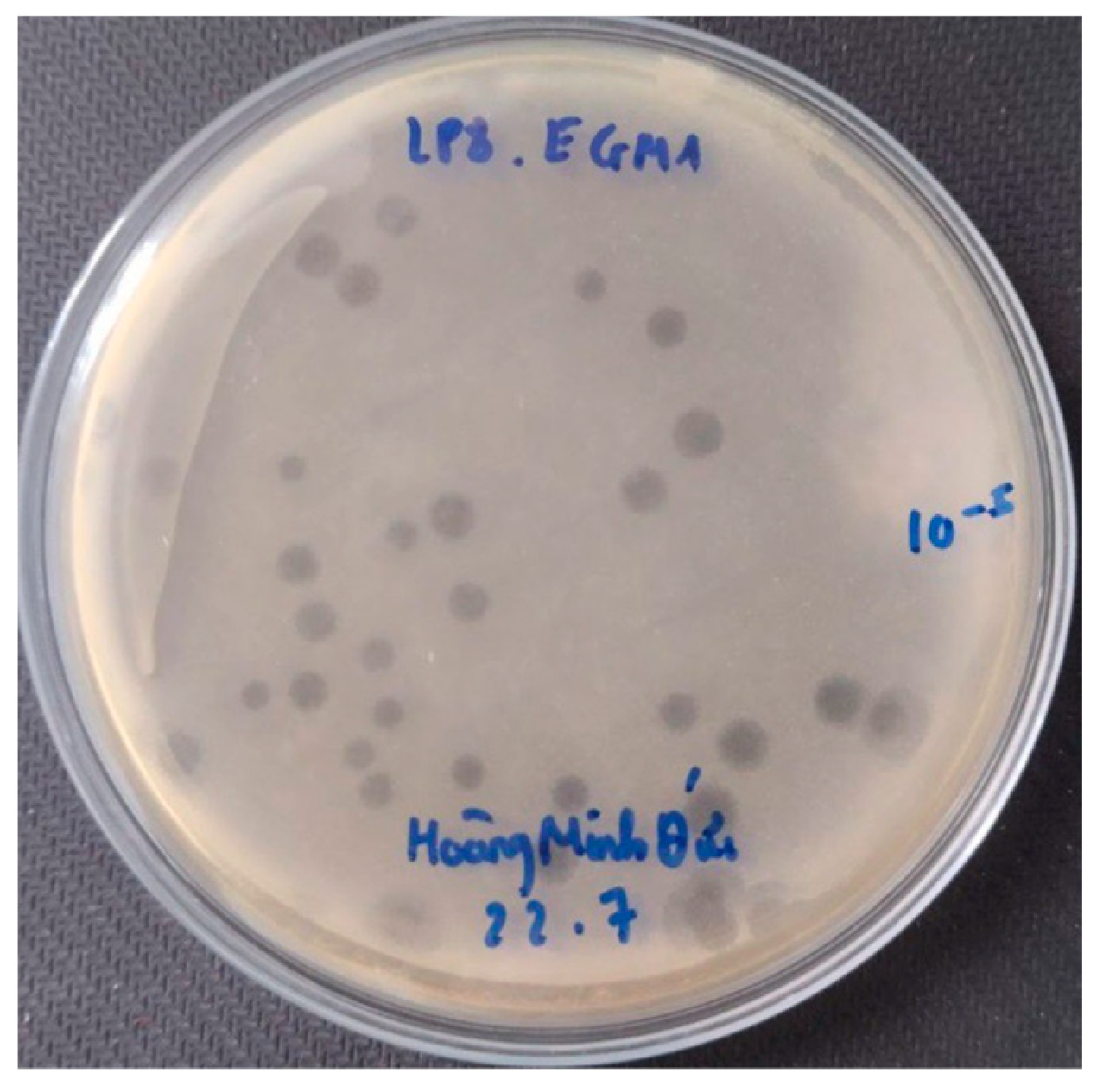
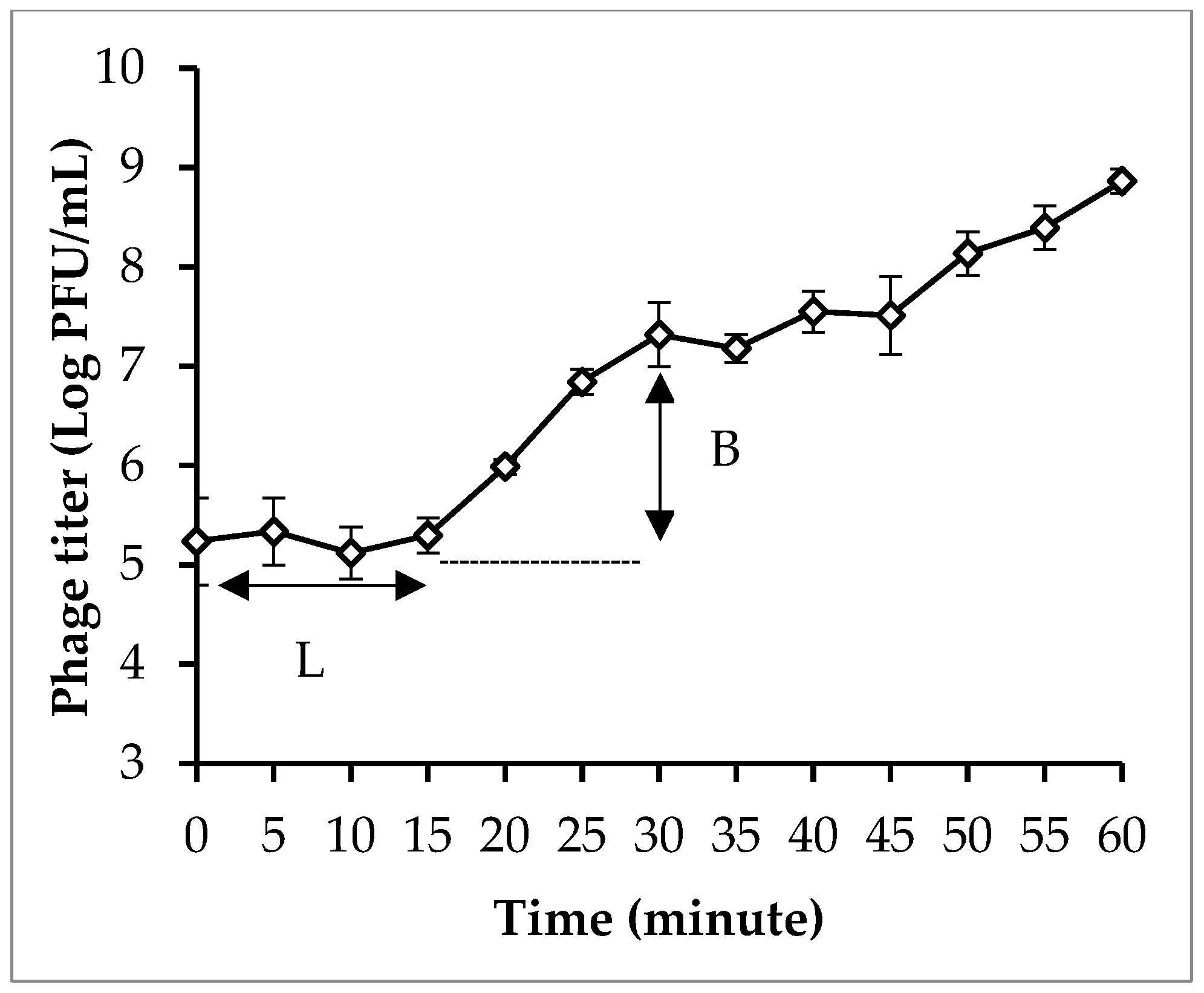
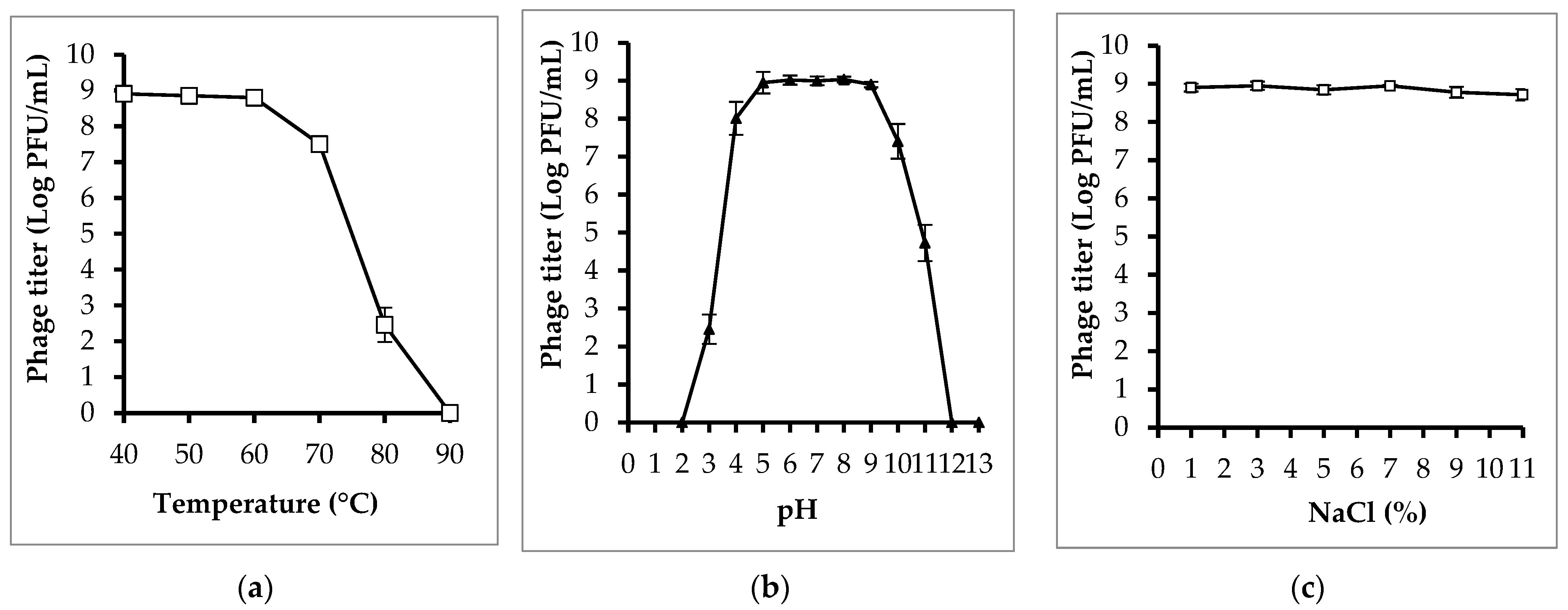
3.4. Phage Isolation and Characterization

A total of eight phages were isolated using E. coli EM148 as a bacterial host in this study. Of the eight isolated phages, six phages (PEM1, PEM2, PEM3, PEM5, PEM6, and PEM8) capable of forming clear plaques and generating stocks of high titer (>109 PFU/mL) were selected for characterization (Figure 1).
The results of the host range show that phage PEM3 had the widest lytic spectrum, lysing 88 (63.3%) of the 139 E. coli isolates tested. On the contrary, phage PEM5 exhibited the narrowest host range, infecting only 9 (6.5%) of the 139 E. coli isolates tested.
The one-step growth curve revealed that phage PEM3 had relatively short latent periods of 15 min and a burst size of 223 PFU/cell in E. coli EM148 (Figure 2).
The results of the stability test revealed that phage PEM3 had relatively good temperature, pH, and NaCl tolerance (Figure 3). The phage titer remained relatively stable at temperatures ranging from 40 °C to 70 °C, pH values from 4 to 10, and NaCl concentrations from 1% to 11%. At 80 °C, infectious phage particles were still detectable; however, the titer was significantly reduced compared to that at 40 °C to 70 °C. When treated at a temperature of 90 °C for 30 min, the phage was completely inactivated. Similarly, phage particles were found to be not infectious when incubated at pH < 3 and pH > 11 for 60 min.

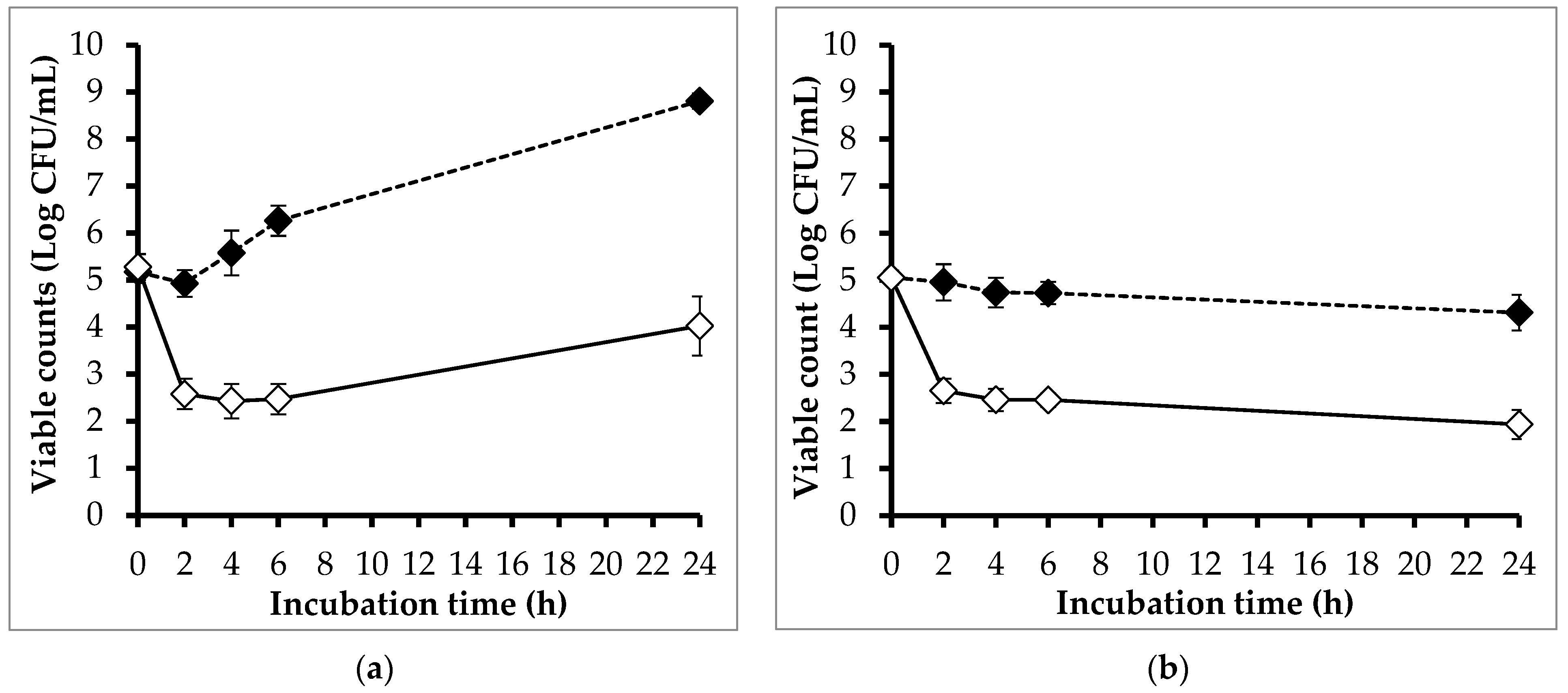
3.5. Efficacy of Phage PEM3 against EM148 in LB Broth and Raw Milk

Figure 4 shows the effect of phage PEM3 on the viability of EM148 grown in LB broth incubated at 24 °C and 4 °C. At 24 °C, the viable counts of EM148 in the controls increased gradually and reached a plateau at 24 h (Figure 4a). On the contrary, the viable counts of EM148 in the treatments were reduced to below a detectable level (<10 CFU/mL) after 2 h of incubation, and the regrowth of EM148 was not observed at the end of the experiment period.
In the trial at 4 °C, the viable counts of EM148 in the controls were found to be unchanged during 24 h of incubation, whereas those in the phage treatments were significantly decreased (Figure 4b). Compared to the controls, the viable count of EM148 in the treatments was reduced by 2.79 log after 2 h of incubation, and this reduction was maintained throughout the experiment.
The effects of phage PEM3 on the viability of EM148 in raw milk stored at 24 °C and 4 °C were also investigated (Figure 5). In treatments at 24 °C, phage PEM3 lowered the viable counts of EM148 by 2.35, 3.15, and 3.79 log at 2, 4, and 6 h, respectively, compared to the controls (Figure 5a). At the end of experiment (24 h), the regrowth of EM148 occurred in the treatments; however, a 4.78 log reduction of the viable counts was still observed when compared to the controls (Figure 5a).
At 4 °C, the viability of EM148 in the controls remained steady and close to the initial inoculum throughout the experimental period (Figure 5b). In contrast, the viable counts of EM148 in the treatments were significantly decreased by 2.31 and 2.37 log after 2 h and 24 h of incubation, respectively, compared to those in the controls (Figure 5b).

4. Discussion

In this study, the prevalence of E. coli in raw milk was 34.75%. This result is in agreement with the findings of Liu et al. (2021) in northern China (34.4%) [29]. However, the E. coli contamination rate in raw milk in our study was lower than those reported in Malaysia (45%), Pakistan (50%), Egypt (52%), Bangladesh (75%), and India (81.1%) [41,42,43,44,45]. On the contrary, the prevalence of E. coli in raw milk found in the present study was higher than that recorded in a study by Awadallah et al. (2016) (22.4%) [46]. In general, our results indicate that the raw milk collected from Hanoi, Vietnam was highly contaminated with E. coli. The presence of E. coli in raw milk does not only indicate fecal contamination of milk but also reveals improper hygiene and sanitary conditions during milking, irregular washing and sterilization of dairy equipment, and infections of dairy cows that are used for milking purposes [47]. In addition, the high prevalence of E. coli in raw milk implies the risk that other enteric pathogens may co-exist [48]. It is worth noting that the differences in E. coli contamination rates among various studies may be due to the differences in locality, sample numbers, sampling seasons, isolation procedures, and milk types.
A majority of E. coli clones are harmless to humans; however, several strains have acquired virulence factors that enable them to cause human diseases. Pathogenic E. coli targeting the human intestine is categorized into six groups, as follows: Shiga-toxin-producing E. coli (STEC); enteropathogenic E. coli (EPEC); enterotoxigenic E. coli (ETEC); enteroaggregative E. coli (EAEC); enteroinvasive E. coli (EIEC); and diffusely adherent E. coli (DAEC) [49,50]. Among these, STEC has been considered as the most important pathogen for the dairy industry since dairy cows are known to harbor STEC in their gastrointestinal tract and excrete the pathogens in their feces [51,52,53]. Also, the teats and udders of animals have been reported to be easily contaminated with feces when cleaning methods and udder hygiene practices are not properly followed, which leads to milk contamination during milking [54]. Another factor making STEC the most important pathogen for the dairy industry is their low dose infection (5–50 cells) compared to those of other E. coli groups [53]. STEC contains various serogroups; however, the O157 serogroup and six other non-O157 serogroups (O26, O103, O111, O121, O45, and O145) have been reported as the most common serogroups associated with STEC outbreaks [55]. The ability of STEC to cause serious human diseases is mainly associated with their production capacity of Stx. Based on their antigen differences, the Stx family has been divided into two groups, Stx1 and Stx2. The Stx2 is more clinically important than Stx1, as Stx2 infection has a higher probability of HUS development compared to either Stx1 or both Stx1 and Stx2 [56]. Another crucial virulence factor for STEC to cause human diseases is the eae gene encoding the outer membrane adhesin, also called intimin, which allows the pathogens to attach to intestinal epithelial cells. It has been reported that eae has frequently been detected in clinical STEC (FAO, 2022). In addition, hemolysin is considered another important virulent factor for STEC. A gene (ehxA) encoding plasmid-carried enterohemolysin is usually found in STEC isolates linked to diarrheal disease and HUS [57]. In this study, 3.6% (5/139) of the isolated E. coli strains were identified as STEC carrying the stx1 gene, corresponding to 1.25% (5/400) of the milk samples, indicating the low contamination rate of STEC in milk. The findings in this study align with those of previous studies from Ireland and the USA reporting 0.8–3.2% of raw milk samples contaminated with STEC [58,59]. A recent review of data from a variety of studies also showed that the prevalence of STEC in raw milk samples in Europe ranged from an undetectable level to 5.7% [60]. The occurrence of STEC in raw milk in the current study was lower than in a study in Iran, which found that 6/150 (4%) raw milk and cheese samples tested positive for STEC [61]. A higher prevalence of STEC was also observed in a study conducted in Egypt, where STEC was present in 36/125 (28.8%) raw milk and dairy samples [62]. Recent studies utilized DNA-based methods to detect stx genes in raw milk. However, it is important to note that the occurrence of stx genes in raw milk does not always indicate the presence of viable cells of STEC, as stx genes can be present in non-pathogenic organisms, in phages, or unlinked with bacterium [63]. Therefore, a culturing method is still needed to confirm the presence of viable STEC cells in raw milk samples. Therefore, it is necessary to differentiate between the prevalence of stx genes and the prevalence of viable STEC cells. The incidence of stx genes in raw milk samples is usually reported to be significantly higher than the prevalence of viable STEC cells [58,59]. In the present study, isolated STEC strains only carried the stx1 gene, and none of the isolates were positive for stx2. This finding is in line with previous studies reporting that stx1 is the predominant gene in E. coli isolates of dairy food origin [64].
The high rate (25.18%) of MDR found in raw milk in this study is reasonable since Vietnam is known to have one of the highest antibiotic resistance rates in Asia. This could be attributed to the significant overuse and misuse of antibiotics in both human and animal health sectors in Vietnam [65,66]. Mastitis, infectious foot, respiratory, reproductive, and diarrhea diseases have been recognized as the most common diseases of dairy cows [67]. Antibiotics, such as penicillin, cephalosporins, and sulfonamides, are often selected to treat these diseases [68]. This could partly explain the high resistance rates to co-trime (27.34%) and ampicillin (27.34%) observed in the present study. Additionally, the E. coli isolated in this study were also highly resistant to streptomycin (25.18%). This may be due to the common practice of using streptomycin in combination with penicillin in treating mastitis and other diseases of dairy cows in Vietnam, as they are known for their synergistic effect, low cost, and viability. These results are consistent with those of previous studies reporting that E. coli isolated from dairy products exhibited high resistance rates to ampicillin, tetracycline, and co-trime [32,61,69]. Another factor contributing to the high antibiotic resistance rate of E. coli isolated from raw milk is the widespread use of antibiotics not only for treating diseases but also for prevention and growth promotion [68]. For example, prophylactic antibiotics through intramammary infusions were applied to 75% of dairy cows in the United States for the prevention of mastitis [70]. The use of antibiotics for disease treatment, prevention, and growth promotion results in residues of antibiotics in milk, consequently leading to the emergence of antibiotic-resistant clones in milk. This is considered the main reason for the presence of MDR in raw milk. However, it cannot be excluded that MDR from bovine feces may directly/indirectly contaminate raw milk during the milking process [50].
The emergence and spread of ESBLEC and colistin-resistant E. coli (CREC) has been recognized as a global health concern [71,72]. In our study, seven ESBLEC and four CREC were isolated from raw milk. This is the first report of the co-occurrence of β-lactamase encoding genes (blaTEM, blaCTX-M-1) and the colistin resistance gene (mcr-1) in E. coli recovered from raw milk in Vietnam. Similarly, Fillioussis et al. (2020) has reported, for the first time, the isolation of mcr-1-postive ESBLEC in raw milk in Greece [73]. A total of 89 E. coli strains were isolated from 400 samples, and 6 out of 89 (6.7%) isolates were phenotypically determined as ESBL producers. All six ESBLEC were positive for mcr-1, five for blaTEM-1, three for blaCTX-M, and three for blaSHV [73]. Recently, the co-occurrence of ESBL genes and mcr-1 in E. coli isolated from raw milk was also reported in China [74]. A total of 2038 milk samples were used to isolate 249 E. coli strains. Among them, five (2%) strains were simultaneously positive for mcr-1 and ESBL genes [9]. Colistin is not the most common drug for treating diseases in dairy cows, suggesting that the mcr-1-positive ESBEC in this study may have originated from bovine feces or the environment, or the use of cephalosporins has resulted in the cross-resistance of cephalosporins and colistin.
Due to growing consumer demand for natural, healthy, and nutritious milk products with a long shelf life, the non-thermal sterilization method capable of controlling pathogens in raw milk is needed to replace pasteurization [75]. Recently, bacteriophages have emerged as promising natural food additives to control foodborne pathogens [76,77,78,79]. In this study, phage PEM3 showed great efficacy in controlling multidrug-resistant E. coli in raw milk stored at 4 °C and 24 °C. The application of phage PEM3 resulted in significant reductions in viable counts of the bacterial host, ranging from 2.31 log to 3.79 log CFU/mL. This is the first report demonstrating that phage was effective in killing mcr-1-positive ESBLEC in raw milk. The findings are in agreement with previous studies evaluating the effect of phages on the viability of E. coli in milk. A study conducted by Mclean et al. (2013) reported that the addition of a phage cocktail containing three phages reduced E. coli ATCC 25922 and E. coli O127:H6 in raw milk incubated at refrigeration temperature (5–9 °C) and 25 °C to under the detection limit [80]. In another study performed by Grygorcewicz et al. (2020), the application of phage ECPS-6 decreased E. coli O157:H7 in raw milk kept at 4 °C and 25 °C by 1.5 log and 4.74 log CFU/mL, respectively [81]. It is noteworthy that the efficacy of phages in reducing the viable cells of bacterial hosts in milk depends on the multiplicity of infection (MOI), the initial inoculum of the bacterial host into milk, the incubation temperature, and the milk properties, thus making it difficult to compare the antibacterial activities of phages across different studies. In our previous studies, several phages against various foodborne pathogens (E. coli, Salmonella, Campylobacter, and S. aureus) have been successfully isolated from different food samples (pork, chicken meat, beef, and chicken organs) [17,22,26,40,82,83]. In this study, phage PEM3 was also recovered from food (raw milk), reinforcing the concept that foods are ideal sources for the isolation of phages against foodborne pathogens.

5. Conclusions

In summary, raw milk in Vietnam was found to be highly contaminated with E. coli. The E. coli isolates showed high resistance rates to co-trime, ampicillin, streptomycin, and tetracycline. This is the first report on the presence of multidrug-resistant E. coli strains harboring blaTEM, blaCTX-M-1 and mcr-1 gene in raw milk in Vietnam. Phage PEM3 effectively reduced the viable counts of multidrug-resistant E. coli isolate (EM148), indicating its potential for controlling MDR-E. coli strains in food.

Author Contributions

Conceptualization, H.M.D.; methodology, H.M.D.; software, H.M.D. and T.T.K.H.; validation, H.M.D. and H.M.S.; formal analysis, T.T.K.H. and H.M.D.; investigation, H.M.D. and H.M.S.; resources, H.M.D.; data curation, H.M.D.; writing—original draft preparation, H.M.D.; writing—review and editing, G.A.F., N.V.T., L.V.H., C.T.T.H. and H.M.S.; visualization, H.M.D.; supervision, H.M.D.; project administration, H.M.D. and H.M.S.; funding acquisition, H.M.D. All authors have read and agreed to the published version of the manuscript.

Funding

This research received no external funding.

Institutional Review Board Statement

Not applicable.

Informed Consent Statement

Not applicable.

Data Availability Statement

The data that support the findings of this study are available from the corresponding author upon reasonable request.

Acknowledgments

This research was conducted in partnership with the Vietnam National University of Agriculture (VNUA) and G.A. Flory Consulting.

Conflicts of Interest

Author Gary A. Flory was employed by the company G.A. Flory Consulting. The remaining authors declare that the research was conducted in the absence of any commercial or financial relationships that could be construed as a potential conflict of interest.

References

  1. Havelaar, A.H.; Kirk, M.D.; Torgerson, P.R.; Gibb, H.J.; Hald, T.; Lake, R.J.; Praet, N.; Bellinger, D.C.; De Silva, N.R.; Gargouri, N. World Health Organization Global Estimates and Regional Comparisons of the Burden of Foodborne Disease in 2010. PLoS Med. 2015, 12, e1001923. [Google Scholar] [CrossRef] [PubMed]
  2. Abebe, E.; Gugsa, G.; Ahmed, M. Review on Major Food-Borne Zoonotic Bacterial Pathogens. J. Trop. Med. 2020, 2020, 4674235. [Google Scholar] [CrossRef] [PubMed]
  3. Kaper, J.B.; Nataro, J.P.; Mobley, H.L.T. Pathogenic Escherichia coli. Nat. Rev. Microbiol. 2004, 2, 123–140. [Google Scholar] [CrossRef] [PubMed]
  4. Minh, S.H.; Kimura, E.; Minh, D.H.; Honjoh, K.; Miyamoto, T. Virulence characteristics of Shiga toxin-producing Escherichia coli from raw meats and clinical samples. Microbiol. Immunol. 2015, 59, 114–122. [Google Scholar] [CrossRef] [PubMed]
  5. Burow, E.; Rostalski, A.; Harlizius, J.; Gangl, A.; Simoneit, C.; Grobbel, M.; Kollas, C.; Tenhagen, B.-A.; Käsbohrer, A. Antibiotic resistance in Escherichia coli from pigs from birth to slaughter and its association with antibiotic treatment. Prev. Vet. Med. 2019, 165, 52–62. [Google Scholar] [CrossRef]
  6. Mcewen, S.A.; Fedorka-Cray, P.J. Antimicrobial Use and Resistance in Animals. Clin. Infect. Dis. 2002, 34 (Suppl. S3), S93–S106. [Google Scholar] [CrossRef] [PubMed]
  7. Anjum, M.F.; Schmitt, H.; Börjesson, S.; Berendonk, T.U.; Donner, E.; Stehling, E.G.; Boerlin, P.; Topp, E.; Jardine, C.; Li, X.; et al. The potential of using E. coli as an indicator for the surveillance of antimicrobial resistance (AMR) in the environment. Curr. Opin. Microbiol. 2021, 64, 152–158. [Google Scholar] [CrossRef] [PubMed]
  8. Saliu, E.M.; Vahjen, W.; Zentek, J. Types and prevalence of extended-spectrum beta-lactamase producing Enterobacteriaceae in poultry. Anim. Health Res. Rev. 2017, 18, 46–57. [Google Scholar] [CrossRef] [PubMed]
  9. Liu, G.; Ali, T.; Gao, J.; Rahman, S.U.; Yu, D.; Barkema, H.W.; Huo, W.; Xu, S.; Shi, Y.; Kastelic, J.P.; et al. Co-Occurrence of Plasmid-Mediated Colistin Resistance (mcr-1) and Extended-Spectrum ß-Lactamase Encoding Genes in Escherichia coli from Bovine Mastitic Milk in China. Microb. Drug Resist. 2020, 26, 685–696. [Google Scholar] [CrossRef]
  10. EFSA. Scientific Opinion on the public health risks related to the consumption of raw drinking milk. EFSA J. 2015, 13, 3940. [Google Scholar] [CrossRef]
  11. Oliver, S.P.; Boor, K.J.; Murphy, S.C.; Murinda, S.E. Food safety hazards associated with consumption of raw milk. Foodborne Pathog. Dis. 2009, 6, 793–806. [Google Scholar] [CrossRef] [PubMed]
  12. Potter, M.E.; Kaufmann, A.F.; Blake, P.A.; Feldman, R.A. Unpasteurized Milk: The Hazards of a Health Fetish. JAMA J. Am. Med. Assoc. 1984, 252, 2048–2052. [Google Scholar] [CrossRef]
  13. Jay-Russell, M.T. Raw (unpasteurized) milk: Are health-conscious consumers making an unhealthy choice? Clin. Infect. Dis. 2010, 51, 1418–1419. [Google Scholar] [CrossRef] [PubMed]
  14. Liu, J.; Zhu, Y.; Jay-Russell, M.; Lemay, D.G.; Mills, D.A. Reservoirs of antimicrobial resistance genes in retail raw milk. Microbiome 2020, 8, 99. [Google Scholar] [CrossRef] [PubMed]
  15. Eldesoukey, I.E.; Elmonir, W.; Alouffi, A.; Beleta, E.I.M.; Kelany, M.A.; Elnahriry, S.S.; Alghonaim, M.I.; Alzeyadi, Z.A.; Elaadli, H. Multidrug-Resistant Enteropathogenic Escherichia coli Isolated from Diarrhoeic Calves, Milk, and Workers in Dairy Farms: A Potential Public Health Risk. Antibiotics 2022, 11, 999. [Google Scholar] [CrossRef] [PubMed]
  16. Lucey, J.A. Raw milk consumption: Risks and benefits. Nutr. Today 2015, 50, 189. [Google Scholar] [CrossRef] [PubMed]
  17. Duc, H.M.; Son, H.M.; Ngan, P.H.; Sato, J.; Masuda, Y.; Honjoh, K.-I.; Miyamoto, T. Isolation and application of bacteriophages alone or in combination with nisin against planktonic and biofilm cells of Staphylococcus aureus. Appl. Microbiol. Biotechnol. 2020, 104, 5145–5158. [Google Scholar] [CrossRef] [PubMed]
  18. Mills, S.; Ross, R.P.; Hill, C. Bacteriocins and bacteriophage; a narrow-minded approach to food and gut microbiology. FEMS Microbiol. Rev. 2017, 41, S129–S153. [Google Scholar] [CrossRef]
  19. Moye, Z.D.; Woolston, J.; Sulakvelidze, A. Bacteriophage Applications for Food Production and Processing. Viruses 2018, 10, 205. [Google Scholar] [CrossRef]
  20. Tagliaferri, T.L.; Jansen, M.; Horz, H.P. Fighting Pathogenic Bacteria on Two Fronts: Phages and Antibiotics as Combined Strategy. Front. Cell. Infect. Microbiol. 2018, 10, 205. [Google Scholar] [CrossRef]
  21. Loc-Carrillo, C.; Abedon, S.T. Pros and cons of phage therapy. Bacteriophage 2011, 1, 111–114. [Google Scholar] [CrossRef] [PubMed]
  22. Duc, H.M.; Son, H.M.; Yi, H.P.S.; Sato, J.; Ngan, P.H.; Masuda, Y.; Honjoh, K.-I.; Miyamoto, T. Isolation, characterization and application of a polyvalent phage capable of controlling Salmonella and Escherichia coli O157:H7 in different food matrices. Food Res. Int. 2020, 131, 108977. [Google Scholar] [CrossRef] [PubMed]
  23. Minh, D.H.; Minh, S.H.; Honjoh, K.I.; Miyamoto, T. Isolation and bio-control of Extended Spectrum Beta-Lactamase (ESBL)-producing Escherichia coli contamination in raw chicken meat by using lytic bacteriophages. LWT 2016, 71, 339–346. [Google Scholar] [CrossRef]
  24. Golkar, Z.; Bagasra, O.; Pace, D.G. Bacteriophage therapy: A potential solution for the antibiotic resistance crisis. J. Infect. Dev. Ctries. 2014, 8, 129–136. [Google Scholar] [CrossRef] [PubMed]
  25. Hong, Y.; Schmidt, K.; Marks, D.; Hatter, S.; Marshall, A.; Albino, L.; Ebner, P. Treatment of Salmonella-Contaminated Eggs and Pork with a Broad-Spectrum, Single Bacteriophage: Assessment of Efficacy and Resistance Development. Foodborne Pathog. Dis. 2016, 13, 679–688. [Google Scholar] [CrossRef] [PubMed]
  26. Huang, H.-H.; Furuta, M.; Nasu, T.; Hirono, M.; Pruet, J.; Duc, H.M.; Zhang, Y.; Masuda, Y.; Honjoh, K.-I.; Miyamoto, T. Inhibition of phage-resistant bacterial pathogen re-growth with the combined use of bacteriophages and EDTA. Food Microbiol. 2021, 100, 103853. [Google Scholar] [CrossRef] [PubMed]
  27. Younis, W.; Hassan, S.; Mohamed, H.M.A. Molecular characterization of Escherichia coli isolated from milk samples with regard to virulence factors and antibiotic resistance. Vet. World 2021, 14, 2410–2418. [Google Scholar] [CrossRef] [PubMed]
  28. Ombarak, R.A.; Hinenoya, A.; Elbagory, A.R.M.; Yamasaki, S. Prevalence and Molecular Characterization of Antimicrobial Resistance in Escherichia coli Isolated from Raw Milk and Raw Milk Cheese in Egypt. J. Food Prot. 2018, 81, 226–232. [Google Scholar] [CrossRef] [PubMed]
  29. Liu, H.; Meng, L.; Dong, L.; Zhang, Y.; Wang, J.; Zheng, N. Prevalence, Antimicrobial Susceptibility, and Molecular Characterization of Escherichia coli Isolated From Raw Milk in Dairy Herds in Northern China. Front. Microbiol. 2021, 12, 730656. [Google Scholar] [CrossRef]
  30. Ngaywa, C.; Aboge, G.O.; Obiero, G.; Omwenga, I.; Ngwili, N.; Wamwere, G.; Wainaina, M.; Bett, B. Antimicrobial resistant Escherichia coli isolates detected in raw milk of livestock in pastoral areas of northern Kenya. Food Control 2019, 102, 173–178. [Google Scholar] [CrossRef]
  31. Ranjbar, R.; Dehkordi, F.S.; Shahreza, M.H.S.; Rahimi, E. Prevalence, identification of virulence factors, O-serogroups and antibiotic resistance properties of Shiga-toxin producing Escherichia coli strains isolated from raw milk and traditional dairy products. Antimicrob. Resist. Infect. Control 2018, 7, 1–11. [Google Scholar] [CrossRef] [PubMed]
  32. Madani, A.; Esfandiari, Z.; Shoaei, P.; Ataei, B. Evaluation of Virulence Factors, Antibiotic Resistance, and Biofilm Formation of Escherichia coli Isolated from Milk and Dairy Products in Isfahan, Iran. Foods 2022, 11, 960. [Google Scholar] [CrossRef] [PubMed]
  33. García-Anaya, M.C.; Sepulveda, D.R.; Sáenz-Mendoza, A.I.; Rios-Velasco, C.; Zamudio-Flores, P.B.; Acosta-Muñiz, C.H. Phages as biocontrol agents in dairy products. Trends Food Sci. Technol. 2020, 95, 10–20. [Google Scholar] [CrossRef]
  34. Ibekwe, A.M.; Lyon, S.R. Microbiological evaluation of fecal bacterial composition from surface water through aquifer sand material. J. Water Health 2008, 6, 411–421. [Google Scholar] [CrossRef] [PubMed]
  35. Bai, J.; Paddock, Z.D.; Shi, X.; Li, S.; An, B.; Nagaraja, T.G. Applicability of a multiplex PCR to detect the seven major Shiga toxin–producing Escherichia coli based on genes that code for serogroup-specific O-antigens and major virulence factors in cattle feces. Foodborne Pathog. Dis. 2012, 9, 541–548. [Google Scholar] [CrossRef] [PubMed]
  36. Bai, J.; Shi, X.; Nagaraja, T.G. A multiplex PCR procedure for the detection of six major virulence genes in Escherichia coli O157:H7. J. Microbiol. Methods 2010, 82, 85–89. [Google Scholar] [CrossRef] [PubMed]
  37. Nguyen, D.T.A.; Kanki, M.; Nguyen, P.D.; Le, H.T.; Ngo, P.T.; Tran, D.N.M.; Le, N.H.; Van Dang, C.; Kawai, T.; Kawahara, R.; et al. Prevalence, antibiotic resistance, and extended-spectrum and AmpC β-lactamase productivity of Salmonella isolates from raw meat and seafood samples in Ho Chi Minh City, Vietnam. Int. J. Food Microbiol. 2016, 236, 115–122. [Google Scholar] [CrossRef] [PubMed]
  38. Rebelo, A.R.; Bortolaia, V.; Kjeldgaard, J.S.; Pedersen, S.K.; Leekitcharoenphon, P.; Hansen, I.M.; Guerra, B.; Malorny, B.; Borowiak, M.; Hammerl, J.A.; et al. Multiplex PCR for detection of plasmid-mediated colistin resistance determinants, mcr-1, mcr-2, mcr-3, mcr-4 and mcr-5 for surveillance purposes. Eurosurveillance 2018, 23, 17-00672. [Google Scholar] [CrossRef] [PubMed]
  39. Borowiak, M.; Fischer, J.; Hammerl, J.A.; Hendriksen, R.S.; Szabo, I.; Malorny, B. Identification of a novel transposon-associated phosphoethanolamine transferase gene, mcr-5, conferring colistin resistance in d-tartrate fermenting Salmonella enterica subsp. enterica serovar Paratyphi B. J. Antimicrob. Chemother. 2017, 72, 3317–3324. [Google Scholar] [CrossRef]
  40. Duc, H.M.; Son, H.M.; Honjoh, K.; Miyamoto, T. Isolation and application of bacteriophages to reduce Salmonella contamination in raw chicken meat. LWT 2018, 91, 353–360. [Google Scholar] [CrossRef]
  41. Ibrahim, A.H.M.; Ali, M.E.E.; Ahmed, M.F.E.; Abdelkhalek, A. Prevalence and Characterization of Escherichia coli in Raw Milk and Some Dairy Products at Mansoura City. J. Adv. Vet. Res. 2022, 12, 363–370. [Google Scholar]
  42. Hussain, A.; Ali, S.; Shakir, H.A.; Qazi, J.I.; Ali, K.; Ullah, N. Prevalence of Escherichia coli in milk and different milk products sold under market conditions at Lahore. Sci. J. Anim. Sci. 2014, 3, 22–26. [Google Scholar]
  43. Bhoomika; Shakya, S.; Patyal, A.; Gade, N.E. Occurrence and characteristics of extended-spectrum β-lactamases producing Escherichia coli in foods of animal origin and human clinical samples in Chhattisgarh, India. Vet. World 2016, 9, 996–1000. [Google Scholar] [CrossRef]
  44. Islam, M.A.; Kabir, S.M.L.; Seel, S.K. Molecular detection and characterization of Escherichia coli isolated from raw milk sold in different markets of Bangladesh. Bangladesh J. Vet. Med. 2016, 14, 271–275. [Google Scholar] [CrossRef]
  45. Jayarao, B.M.; Henning, D.R. Prevalence of foodborne pathogens in bulk tank milk. J. Dairy Sci. 2001, 84, 2157–2162. [Google Scholar] [CrossRef] [PubMed]
  46. Awadallah, M.A.; Ahmed, H.A.; Merwad, A.M.; Selim, M.A. Occurrence, genotyping, shiga toxin genes and associated risk factors of E. coli isolated from dairy farms, handlers and milk consumers. Vet. J. 2016, 217, 83–88. [Google Scholar] [CrossRef]
  47. Ali, A.A.; Abdelgadir, W.S. Incidence of Escherichia coli in raw cow’s milk in Khartoum State. Br. J. Dairy Sci. 2011, 2, 23–26. [Google Scholar]
  48. Ombarak, R.A.; Hinenoya, A.; Awasthi, S.P.; Iguchi, A.; Shima, A.; Elbagory, A.-R.M.; Yamasaki, S. Prevalence and pathogenic potential of Escherichia coli isolates from raw milk and raw milk cheese in Egypt. Int. J. Food Microbiol. 2016, 221, 69–76. [Google Scholar] [CrossRef] [PubMed]
  49. World Health Organization. Control Measures for Shiga Toxin-Producing Escherichia coli (STEC) Associated with Meat and Dairy Products; World Health Organization: Geneva, Switzerland, 2022. [Google Scholar]
  50. Farrokh, C.; Jordan, K.; Auvray, F.; Glass, K.; Oppegaard, H.; Raynaud, S.; Thevenot, D.; Condron, R.; De Reu, K.; Govaris, A.; et al. Review of Shiga-toxin-producing Escherichia coli (STEC) and their significance in dairy production. Int. J. Food Microbiol. 2013, 162, 190–212. [Google Scholar] [CrossRef]
  51. Brown, C.A.; Harmon, B.G.; Zhao, T.; Doyle, M.P. Experimental Escherichia coli O157:H7 carriage in calves. Appl. Environ. Microbiol. 1997, 63, 27–32. [Google Scholar] [CrossRef]
  52. Chapman, P.A.; Malo, A.T.C.; Ellin, M.; Ashton, R.; Harkin, M.A. Escherichia coli O157 in cattle and sheep at slaughter, on beef and lamb carcasses and in raw beef and lamb products in South Yorkshire, UK. Int. J. Food Microbiol. 2001, 64, 139–150. [Google Scholar] [CrossRef] [PubMed]
  53. Bagel, A.; Sergentet, D. Shiga Toxin-Producing Escherichia coli and Milk Fat Globules. Microorganisms 2022, 10, 496. [Google Scholar] [CrossRef]
  54. Ruegg, P.L. Practical food safety interventions for dairy production. J. Dairy Sci. 2003, 86, E1–E9. [Google Scholar] [CrossRef]
  55. Alharbi, M.G.; Al-Hindi, R.R.; Esmael, A.; Alotibi, I.A.; Azhari, S.A.; Alseghayer, M.S.; Teklemariam, A.D. The ‘Big Six’: Hidden Emerging Foodborne Bacterial Pathogens. Trop. Med. Infect. Dis. 2022, 7, 356. [Google Scholar] [CrossRef] [PubMed]
  56. Friedrich, A.W.; Bielaszewska, M.; Zhang, W.-L.; Pulz, M.; Kuczius, T.; Ammon, A.; Karch, H. Escherichia coli Harboring Shiga Toxin 2 Gene Variants: Frequency and Association with Clinical Symptoms. J. Infect. Dis. 2002, 185, 74–84. [Google Scholar] [CrossRef] [PubMed]
  57. Hua, Y.; Zhang, J.; Jernberg, C.; Chromek, M.; Hansson, S.; Frykman, A.; Xiong, Y.; Wan, C.; Matussek, A.; Bai, X. Molecular Characterization of the Enterohemolysin Gene (ehxA) in Clinical Shiga Toxin-Producing Escherichia coli Isolates. Toxins 2021, 13, 71. [Google Scholar] [CrossRef] [PubMed]
  58. Cobbold, R.N.; Davis, M.A.; Rice, D.H.; Szymanski, M.; Tarr, P.I.; Besser, T.E.; Hancock, D.D. Associations between bovine, human, and raw milk, and beef isolates of non-O157 Shiga toxigenic Escherichia coli within a restricted geographic area of the United States. J. Food Prot. 2008, 71, 1023–1027. [Google Scholar] [CrossRef] [PubMed]
  59. Lynch, M.J.; Fox, E.M.; O’Connor, L.; Jordan, K.; Murphy, M. Surveillance of Verocytotoxigenic Escherichia coli in Irish Bovine Dairy Herds. Zoonoses Public Health 2012, 59, 264–271. [Google Scholar] [CrossRef] [PubMed]
  60. Claeys, W.L.; Cardoen, S.; Daube, G.; De Block, J.; Dewettinck, K.; Dierick, K.; De Zutter, L.; Huyghebaert, A.; Imberechts, H.; Thiange, P.; et al. Raw or heated cow milk consumption: Review of risks and benefits. Food Control 2012, 31, 251–262. [Google Scholar] [CrossRef]
  61. Fallah, N.; Ghaemi, M.; Ghazvini, K.; Rad, M.; Jamshidi, A. Occurrence, pathotypes, and antimicrobial resistance profiles of diarrheagenic Escherichia coli strains in animal source food products from public markets in Mashhad, Iran. Food Control 2021, 121, 107640. [Google Scholar] [CrossRef]
  62. Elafify, M.; Khalifa, H.O.; Al-Ashmawy, M.; Elsherbini, M.; El Latif, A.A.; Okanda, T.; Matsumoto, T.; Koseki, S.; Abdelkhalek, A. Prevalence and antimicrobial resistance of Shiga toxin-producing Escherichia coli in milk and dairy products in Egypt. J. Environ. Sci. Health B 2020, 55, 265–272. [Google Scholar] [CrossRef]
  63. EFSA. Technical specifications for the monitoring and reporting of verotoxigenic Escherichia coli (VTEC) on animals and food (VTEC surveys on animals and food). EFSA J. 2009, 7, 1366. [Google Scholar] [CrossRef]
  64. Pradel, N.; Bertin, Y.; Martin, C.; Livrelli, V. Molecular analysis of Shiga toxin-producing Escherichia coli strains isolated from hemolytic-uremic syndrome patients and dairy samples in France. Appl. Environ. Microbiol. 2008, 74, 2118–2128. [Google Scholar] [CrossRef]
  65. Vu, T.V.D.; Do, T.T.N.; Rydell, U.; Nilsson, L.E.; Olson, L.; Larsson, M.; Hanberger, H.; Choisy, M.; Dao, T.T.; van Doorn, H.R.; et al. Antimicrobial susceptibility testing and antibiotic consumption results from 16 hospitals in Viet Nam: The VINARES project 2012–2013. J. Glob. Antimicrob. Resist. 2019, 18, 269–278. [Google Scholar] [CrossRef]
  66. Torumkuney, D.; Kundu, S.; Van Vu, G.; Nguyen, H.A.; Van Pham, H.; Kamble, P.; Lan, N.T.H.; Keles, N. Country data on AMR in Vietnam in the context of community-acquired respiratory tract infections: Links between antibiotic susceptibility, local and international antibiotic prescribing guidelines, access to medicines and clinical outcome. J. Antimicrob. Chemother. 2022, 77, i26–i34. [Google Scholar] [CrossRef] [PubMed]
  67. Oliver, S.P.; Murinda, S.E.; Jayarao, B.M. Impact of antibiotic use in adult dairy cows on antimicrobial resistance of veterinary and human pathogens: A comprehensive review. Foodborne Pathog. Dis. 2011, 8, 337–355. [Google Scholar] [CrossRef] [PubMed]
  68. Virto, M.; Santamarina-García, G.; Amores, G.; Hernández, I. Antibiotics in Dairy Production: Where Is the Problem? Dairy 2022, 3, 541–564. [Google Scholar] [CrossRef]
  69. Zanella, G.N.; Mikcha, J.M.G.; Bando, E.; Siqueira, V.L.D.; MacHinski, M. Occurrence and antibiotic resistance of coliform bacteria and antimicrobial residues in pasteurized cow’s milk from Brazil. J. Food Prot. 2010, 73, 1684–1687. [Google Scholar] [CrossRef] [PubMed]
  70. Landers, T.F.; Cohen, B.; Wittum, T.E.; Larson, E.L. A Review of Antibiotic Use in Food Animals: Perspective, Policy, and Potential. Public Health Rep. 2012, 127, 4–22. [Google Scholar] [CrossRef]
  71. Falgenhauer, L.; Imirzalioglu, C.; Oppong, K.; Akenten, C.W.; Hogan, B.; Krumkamp, R.; Poppert, S.; Levermann, V.; Schwengers, O.; Sarpong, N.; et al. Detection and characterization of ESBL-producing Escherichia coli from humans and poultry in Ghana. Front. Microbiol. 2019, 9, 3358. [Google Scholar] [CrossRef]
  72. Danaei, B.; Sarmastzadeh, T.; Khalili, F.; Yazarlou, F.; Centis, R.; D'Ambrosio, L.; Sotgiu, G.; Migliori, G.B.; Nasiri, M.J. The battle against colistin-resistant E. coli and the need for a one health approach. New Microbes New Infect. 2023, 54, 101161. [Google Scholar] [CrossRef] [PubMed]
  73. Filioussis, G.; Kachrimanidou, M.; Christodoulopoulos, G.; Kyritsi, M.; Hadjichristodoulou, C.; Adamopoulou, M.; Tzivara, A.; Kritas, S.K.; Grinberg, A. Short communication: Bovine mastitis caused by a multidrug-resistant, mcr-1-positive (colistin-resistant), extended-spectrum β-lactamase–producing Escherichia coli clone on a Greek dairy farm. J. Dairy Sci. 2020, 103, 852–857. [Google Scholar] [CrossRef] [PubMed]
  74. Haenni, M.; Poirel, L.; Kieffer, N.; Châtre, P.; Saras, E.; Métayer, V.; Dumoulin, R.; Nordmann, P.; Madec, J.-Y. Co-occurrence of extended spectrum β lactamase and MCR-1 encoding genes on plasmids. Lancet Infect. Dis. 2016, 16, 281–282. [Google Scholar] [CrossRef] [PubMed]
  75. Gutiérrez, D.; Fernández, L.; Rodríguez, A.; García, P. Role of Bacteriophages in the Implementation of a Sustainable Dairy Chain. Front. Microbiol. 2019, 10, 12. [Google Scholar] [CrossRef] [PubMed]
  76. Strauch, E.; Hammerl, J.A.; Hertwig, S. Bacteriophages: New Tools for Safer Food? J. Verbraucherschutz Leb. 2007, 2, 138–143. [Google Scholar] [CrossRef]
  77. Klumpp, J.; Loessner, M.J. New research on bacteriophages and food safety. In Advances in Microbial Food Safety; Elsevier: Amsterdam, The Netherlands, 2013; pp. 321–339. [Google Scholar] [CrossRef]
  78. Mahony, J.; McAuliffe, O.; Ross, R.P.; van Sinderen, D. Bacteriophages as biocontrol agents of food pathogens. Food Biotechnol.–Plant Biotechnol. 2011, 22, 157–163. [Google Scholar] [CrossRef]
  79. Sabour, P.M.; Griffiths, M.W. (Eds.) Bacteriophages in the Control of Food- and Waterborne Pathogens; American Society of Microbiology: Washington, DC, USA, 2010. [Google Scholar] [CrossRef]
  80. McLean, S.K.; Dunn, L.A.; Palombo, E.A. Phage inhibition of Escherichia coli in ultrahigh-temperature-treated and raw milk. Foodborne Pathog. Dis. 2013, 10, 956–962. [Google Scholar] [CrossRef] [PubMed]
  81. Grygorcewicz, B.; Chajęcka-Wierzchowska, W.; Augustyniak, A.; Wasak, A.; Stachurska, X.; Nawrotek, P.; Dołęgowska, B. In-milk inactivation of Escherichia coli O157:H7 by the environmental lytic bacteriophage ECPS-6. J. Food Saf. 2020, 40, e12747. [Google Scholar] [CrossRef]
  82. Duc, H.M.; Zhang, Y.; Hoang, S.M.; Masuda, Y.; Honjoh, K.-I.; Miyamoto, T. The Use of Phage Cocktail and Various Antibacterial Agents in Combination to Prevent the Emergence of Phage Resistance. Antibiotics 2023, 12, 1077. [Google Scholar] [CrossRef]
  83. Furuta, M.; Nasu, T.; Umeki, K.; Hoang, D.M.; Honjoh, K.-I.; Miyamoto, T. Characterization and Application of Lytic Bacteriophages against Campylobacter jejuni Isolated from Poultry in Japan. Biocontrol. Sci. 2017, 22, 213–221. [Google Scholar] [CrossRef]
Figure 1. The clear plaques of phage PEM3.
Pathogens 13 00494 g001
Figure 2. One-step growth curves of phages PEM3 in E. coli EM148. L, latent period; B, burst. Error bars show standard deviations.
Pathogens 13 00494 g002
Figure 3. Effects of temperature (a), pH (b), and NaCl (c) on the stability of phage PEM3. Error bars show standard deviations.
Pathogens 13 00494 g003
Figure 4. Effect of phage PEM3 on the viability of E. coli EM148 in LB broth stored at 24 °C (a) and 4 °C (b). E. coli EM148 was inoculated in 5 mL of LB broth at a final concentration of 105 CFU/mL without (dashed line) and with phage PEM3 at 108 PFU/mL (solid line). Error bars show standard deviations.
Pathogens 13 00494 g004
Figure 5. Effect of phage PEM3 on the viability of E. coli EM148 in raw milk stored at 24 °C (a) and 4 °C (b). E. coli EM148 was inoculated in 5 mL of raw milk at a final concentration of 105 CFU/mL without (dashed line) and with phage PEM3 at 108 PFU/mL (solid line). Error bars show standard deviations.
Pathogens 13 00494 g005
Table 1. Primer for detecting virulence-associated genes.
Target GenePrimerPrimer Sequence (5′–3′)Amplicon
Size (bp)
References
wzxO45O45-FGGGGCTGTCCAGACAGTTCAT890[35]
O45-FTGTACTGCACCCAATGCACCT
wzxO103O103-FGCAGAAAATCAAGGTGATTACG740
O103-RGGTTAAAGCCATGCTCAACG
stx1stx1-FTGTCGCATAGTGGAACCTCA655[36]
stx1-RTGCGCACTGAGAAGAAGAGA
wzqEO121-wzqFO121O121-FTCAGCAGAGTGGAACTAATTTTGT587[35]
O121-RTGAGCACTAGATGAAAAGTATGGCT
wzxO145O145-FTCAAGTGTTGGATTAAGAGGGATT523
O145-RCACTCGCGGACACAGTACC
stx2stx2-FCCATGACAACGGACAGCAGTT477[36]
stx2-RTGTCGCCAGTTATCTGACATTC
wzxO26O26-FAGGGTGCGAATGCCATATT417[35]
O26-RGACATAATGACATACCACGAGCA
eaeeae-FCATTATGGAACGGCAGAGGT375[36]
eae-RACGGATATCGAAGCCATTTG
rfbO157O157-FCAGGTGAAGGTGGAATGGTTGTC296[35]
O157-FTTAGAATTGAGACCATCCAATAAG
wzxO111O111-FTGCATCTTCATTATCACACCA230[35]
O111-RACCGCAAATGCGATAATAACA
ehxAehxA-FGCGAGCTAAGCAGCTTGAAT199[36]
ehxA-RCTGGAGGCTGCACTAACTCC
Table 2. Primers for detecting β-lactamase encoding genes.
Target GenePrimerPrimer Sequence (5′–3′)Amplicon Size (bp)
blaTEMTEM-FGGTCGCCGCATACACTATTCTC372
TEM-RTTTTATCCGCCTCCATCCAGTC
blaSHVSHV-FCCAGCAGGATCTGGTGGACTAC231
SHV-RCCGGGAAGCGCCCTCCAT
blaCTX-M-1CTX-M1-FGAATTAGAGCGGGAGTCGGG588
CTX-M1-RCACAACCCAGGAAGCAGGC
blaCTX-M-2CTX-M2-FGATGGCGACGCTACCCC107
CTX-M2-RCAAGCCGACCTCCCGAAC
blaCTX-M-9CTX-M9-FGTGCAACGGATGATGTTCGC475
CTX-M9-RGAAACGTCTCATCGCCGATC
blaCTX-M-8/25CTX-M8/25-FGCGACCCGCGCGATAC186
CTX-M8/25-RTGCCGGTTTTATCCCCG
Table 3. Primers for the detection of mcr genes.
Target GenePrimerPrimer Sequence (5′–3′)Amplicon Size (bp)Reference
mcr-1mcr1 fAGTCCGTTTGTTCTTGTGGC320[38]
mcr1 rAGATCCTTGGTCTCGGCTTG
mcr-2mcr2 fCAAGTGTGTTGGTCGCAGTT715[38]
mcr2 rTCTAGCCCGACAAGCATACC
mcr-3mcr 3 fAAATAAAAATTGTTCCGCTTATG929[38]
mcr3 rAATGGAGATCCCCGTTTTT
mcr-4mcr4 fTCACTTTCATCACTGCGTTG1116[38]
mcr4 rTTGGTCCATGACTACCAATG
mcr-5mcr5 fATGCGGTTGTCTGCATTTATC1644[39]
mcr 5 rTCATTGTGGTTGTCCTTTTCTG
Table 4. Antibiotic resistance profile of E. coli isolates from raw milk.
GroupName of
Antibiotic
MIC
Breakpoint
(µg/mL)
Number (%) of Resistant StrainsTotal
(n = 139)
Ba Vi
(n = 61)
Gia Lam
(n = 78)
Penicillinampicillin3222 (36.07)16 (20.51)38 (27.34)
Cephalosporinscefotaxime45 (8.2)2 (2.56)7 (5.04)
ceftazidime162 (3.28)1 (1.28)3 (2.16)
cefoxitin321 (1.64)3 (3.85)4 (2.88)
cefepime165 (8.2)2 (2.56)7 (5.04)
Carbapenemsmeropenem4000
Polymyxinscolistin42 (3.28)2 (2.56)4 (2.88)
Tetracyclinestetracycline1621 (34.43)11 (14.1)32 (23.02)
Aminoglycosidesstreptomycin3222 (36.07)13 (16.67)35 (25.18)
gentamicin162 (3.28)1 (1.28)3 (2.16)
Macrolidesazithromycin3213 (21.31)013 (9.35)
Phenicolsflorfenicol166 (9.84)5 (6.41)11 (7.91)
Sulfonamideco-trime4/7627 (44.26)11 (14.1)38 (27.34)
Fluoroquinolonesciprofloxacin42 (3.28)1 (1.28)3 (2.16)
Quinolonesnalidixic acid323 (4.92)2 (2.56)5 (3.6)
Table 5. Antibiotic resistance pattern of E. coli isolates from raw milk.
No. of AntibioticsResistance PhenotypeNumber (%) of Resistance Isolates
Ba Vi (n = 61)Gia lam (n = 78)Total (n = 139)
0-21 (34.43)61 (78.21)82 (58.99)
1SXT4 (6.65)04 (2.88)
FLO2 (3.2802 (1.44)
AMP1 (1.64)01 (0.72)
CST01 (1.28)1 (0.72)
2AZM-SXT6 (9.84)06 (4.320
AMP-STR3 (4.92)03 (2.16)
AMP-FOX02 (2.56)2 (1.44)
TET-SXT2 (3.28)02 (1.44)
STR-SXT1 (1.64)01 (0.72)
3AMP-FOX-STR01 (1.28)1 (0.72)
AMP-STR-CST1 (1.64)01 (0.72)
AMP-STR-TET1 (1.64)01 (0.72)
TET-AZM-SXT1 (1.64)01 (0.72)
4AMP-STR-TET-SXT4 (6.65)5 (6.41)9 (6.47)
AMP-STR-FLO-SXT01 (1.28)1 (0.72)
STR-TET-CST-SXT1 (1.64)01 (0.72)
5AMP-STR-TET-FLO-SXT2 (3.28)4 (5.13)6 (4.32)
AMP-STR-TET-AZM-SXT4 (6.65)04 (2.88)
AMP-CTX-FEP-STR-TET1 (1.64)01 (0.72)
AMP-GEN-STR-TET-SXT01 (1.28)1 (0.72)
FOX-STR-TET-AZM-SXT1 (1.64)01 (0.72)
6AMP-CTX-FEP-CAZ-CIP-NAL1 (1.64)1 (1.28)2 (1.44)
AMP-CTX-FEP-GEN-TET-SXT1 (1.64)01 (0.72)
AMP-GEN-STR-TET-FLO-AZM1 (1.64)01 (0.72)
7AMP-CTX-FEP-STR-TET-CIP-NAL1 (1.64)01 (0.72)
AMP-CTX-FEP-STR-TET-CST-NAL01 (1.28)1 (0.72)
8AMP-CTX-FEP-CAZ-STR-TET-FLO-NAL1 (1.64)01 (0.72)
Resistant ≥ 140 (65.57)17 (21.79)57 (41.01)
MDR21 (34.43)14 (17.95)35 (25.18)
AMP, ampicillin; CTX, cefotaxime; CAZ, ceftazidime; FOX, cefoxitin; FEP, cefepime; CST, colistin, TET, tetracycline; STR, streptomycin; GEN, gentamycin; AZM, azithromycin; FLO, florfenicol; SXT, co-trime; CIP, ciprofloxacin; NAL, nalidixic acid.
Table 6. Phenotypic and genotypic antibiotic resistance of ESBLEC and colistin-resistant isolates.
Isolate IDAntimicrobial Resistance ProfileESBL Screening Testβ-Lactamase
Encoding Genes
mcr Genes
EM2AMP-CTX-FEP-GEN-TET-SXT+blaTEM, blaCTX-M-1ND
EM29AMP-CTX-FEP-CAZ-CIP-NAL+blaTEMND
EM32STR-TET-CST-SXTNDNDmcr-1
EM40AMP-STR-CSTNDNDmcr-1
EM42AMP-CTX-FEP-STR-TET-CIP-NAL+blaTEM, blaCTX-M-1ND
EM72AMP-CTX-FEP-CAZ-STR-TET-FLO-NAL+blaTEM, blaCTX-M-1ND
EM75AMP-CTX-FEP-STR-TET+blaCTX-M-1ND
EM102AMP-CTX-FEP-CAZ-CIP-NAL+blaTEMND
EM144CSTNDNDmcr-1
EM148AMP-CTX-FEP-STR-TET-CST-NAL+blaTEM, blaCTX-M-1mcr-1
ND: Not determined. AMP, ampicillin; CTX, cefotaxime; CAZ, ceftazidime; FEP, cefepime; CST, colistin, TET, tetracycline; STR, streptomycin; GEN, gentamycin; FLO, florfenicol; SXT, co-trime; CIP, ciprofloxacin; NAL, nalidixic acid.
Disclaimer/Publisher’s Note: The statements, opinions and data contained in all publications are solely those of the individual author(s) and contributor(s) and not of MDPI and/or the editor(s). MDPI and/or the editor(s) disclaim responsibility for any injury to people or property resulting from any ideas, methods, instructions or products referred to in the content.

