Article

Isolation, Identification, and Characterisation of a Novel ST2378 Aeromonas hydrophila Strain from Naturally Diseased Frogs, Rana dybowskii

Key Laboratory of Aquatic Animal Diseases and Immune Technology of Heilongjiang Province, Heilongjiang River Fisheries Research Institute, Chinese Academy of Fishery Sciences, Harbin 150070, China
*
Authors to whom correspondence should be addressed.
Pathogens 2024, 13(7), 552; https://doi.org/10.3390/pathogens13070552
Submission received: 11 May 2024 / Revised: 4 June 2024 / Accepted: 18 June 2024 / Published: 30 June 2024
(This article belongs to the Special Issue Aeromonas: Genome, Transmission, Pathogenesis, and Treatment)

Abstract

:
In 2023, Rana dybowskii exhibiting characteristic skin ulcers were found on a farm in northeastern China. Subsequently, two dominant bacteria, Aeromonas hydrophila Rd001 and Acinetobacter johnsonii Rd002, were isolated from naturally infected R. dybowskii. Experimental infection confirmed that Rd001 was the primary pathogen responsible for the disease in R. dybowskii, with a mean lethal dose (LD50) of 6.25 × 10² CFU/g. The virulence genotype of Rd001 was identified as ser+/aha+/lip+/nuc+/hlyA+/aer+/alt+/ast+/act+. Antimicrobial susceptibility testing indicated that Rd001 was sensitive to enrofloxacin, flumequine, and neomycin. MLST analysis showed that Rd001 belonged to a new sequence type of A. hydrophila, named ST2378. This study offered the first comprehensive investigation into the pathogenicity, virulence genotypes, antimicrobial resistance, and genetic traits of A. hydrophila isolated from R. dybowskii, providing a theoretical foundation for preventing and controlling A. hydrophila infections.

1. Introduction

Rana dybowskii, also known as Dybowski’s frog, is an amphibian species native to wooded regions in Korea, Japan, and northeastern China [1]. Amphibians, compared to aquatic and terrestrial animals, inhabit more variable environments, rendering them more susceptible to external environmental influences [2]. In recent decades, the wild populations of these frogs have declined steadily due to environmental degradation and increased capture for consumption and research purposes [3]. The intensive breeding of R. dybowskii started in the 20th century to meet consumer demands. However, with high-density culture and improper management, the development of this industry was severely limited by several bacterial diseases.
Aeromonas hydrophila, a ubiquitous waterborne Gram-negative bacterium [4], has been infecting a wide range of amphibians for over a century, including species like the tiger frog (Rana rugulosa) [5], mountain yellow-legged frog (Rana muscosa) [6], marsh frog (Pelophylax ridibundus) [7], and brown tree frog (Litoria ewingii) [8]. The pathogenicity of A. hydrophila is primarily determined by the presence of multiple virulence factors, such as aerolysin, serine protease, elastase, haemolysin, cytotoxic enterotoxins, lipase, and nuclease [9,10,11]. Variations in virulence genotypes among isolates can result in varying host symptoms and levels of pathology [12]. Therefore, it is crucial to investigate the presence of virulence factors and explore genetic diversity among different A. hydrophila isolates to comprehend their pathogenesis and epidemiology. However, there are limited data available regarding this aspect of A. hydrophila isolated from frogs.
In 2023, a disease characterised by skin ulcers emerged in R. dybowskii on a farm in Heilongjiang Province, China. Two dominant strains, A. hydrophila Rd001 and Acinetobacter johnsonii Rd002, were obtained from the infected R. dybowskii. The experimental challenge confirmed that Rd001 was the primary pathogen responsible for this disease. This study comprises the morphological and biochemical characterisation of the A. hydrophila Rd001, as well as the investigation of its virulence genotypes, drug resistance, and genetic properties in order to develop strategies for disease management.

2. Materials and Methods

2.1. Diseased R. dybowskii and Bacterial Isolation

Diseased R. dybowskii individuals (body weight of 5–10 g) were sampled in a frog farm located in Heilongjiang Province, China (128°43′1.641″ E, 47°45′16.032″ N) and used for bacterial isolation. The diseased R. dybowskii showed clinical signs of skin ulceration in the forelimbs revealing eroded muscles, and some of the affected R. dybowskii showed the same signs on the abdominal skin.
Five symptomatic diseased R. dybowskii were selected for bacterial strain isolation. Following euthanasia by double pithing, the frogs were dissected post skin cleansing with 75% ethyl alcohol. Bacteria were obtained from various organs of each frog (kidney, spleen, and external lesions) using a sterile loop, then cultured on Tryptose Soya Agar (TSA) and incubated at 28 °C for 24 h. The dominant bacteria were screened and re-inoculated on TSA and incubated at 28 °C for 24 h. Single colonies were then picked and inoculated into Tryptose Soy Broth (TSB) medium at 28 °C for 18 h, and then stored in TSB medium containing 20% (v/v) sterile glycerol at −80 °C. The dominant isolates were labelled as Rd001 and Rd002.

2.2. Physiological and Biochemical Characterisation

The purified Rd001 and Rd002 were revived from cryostock into TSA plate and incubated at 28 °C for 24 h. The Gram staining results were observed. Physiological and biochemical indices of this strain were subsequently measured using BD Phoenix M50 reference identification analyser (BD Biosciences, NJ, USA).

2.3. Sequencing and Analysis of 16S rRNA and gyrB Genes

Genomic DNA of Rd001 and Rd002 were extracted using a commercial Kit (TianGen, Beijing, China). The 16S rRNA gene from DNA templates was amplified using the universal primer 27F:5′-AGAGTTTGATCCTGGCTCAG-3′ and 1492R:5′-GGCTACCTTGTTACGACTT-3′. The gyrB gene from DNA templates was amplified using the universal primer gyrB-F:5′-GAAGTCATCATGACCGTTCTGCA(TC)GC(TCAG)GG(TCAG)GG(TCAG)AA(AG)TT(TC)GA-3′ and gyrB-R:5′-AGCAGGGTACGGATGTGCGAGCC(AG)TC(TCAG)AC(AG)TC(TCAG)GC(AG)TC(TCAG)GTCAT-3′. PCR was performed on a thermal cycler (Biometra TAdvanced, Jena, Germany) with 50 µL of reaction mixture, containing 40 ng DNA template, 2 µL of each primer (10 µM), and 25 µL 2×PrimeSTAR Mix (Takara, Shiga, Japan), with DNase-free water comprising the remainder of the mixture. The amplicons were detected and sequenced by Genesoul Technology Company (Harbin, China). BLAST searches of sequences were performed through the NCBI website. Phylogenetic trees were constructed using the neighbour-joining method in MEGA 7.0 software.

2.4. Experimental Infections

2.4.1. R. dybowskii Husbandry

Healthy R. dybowskii (with average body weight of 5.36 ± 1.75 g) were purchased from a frog farm in Heilongjiang Province and transported to our laboratory. Laboratory conditions simulated the natural environment in the wild with water and gravel (20 ± 5 °C), and specimens were temporarily incubated for 7 d before being used for experimental infections. The R. dybowskii were fed with breadworms twice a day. Before the infection experiment, five R. dybowskii were randomly selected for bacteriological examination to verify the absence of the pathogen.

2.4.2. Determination of Median Lethal Dose

In pre-challenge experiment, 12 R. dybowskii were divided into two groups and Rd001 and Rd002 suspensions at a concentration of 1 × 10⁸ CFU/mL were prepared for abdominal subcutaneous injection at a dose of 100 μL/R. dybowskii.
Seventy R. dybowskii were divided equally into 7 groups. The isolate Rd001 was prepared and suspended in sterile PBS buffer at the concentrations from 1 × 10³ to 1 × 10⁸ CFU/mL. R. dybowskii in 6 groups were intraperitoneally injected with the 6 concentrations of Rd001 suspensions at a dose of 100 μL/R. dybowskii. The remaining one group was injected with the same volume of sterile PBS as control. Following the challenge, the R. dybowskii were kept in the simulated environment and fed with breadworms twice a day. Mortality and clinical signs of R. dybowskii in each group were recorded at 9 am and 5 pm daily for 7 d. The mean lethal dose (LD50) of Rd001 was calculated by the improved Karber’s method in IBM SPSS Statistics version 19. The kidney of the infected R. dybowskii were subjected to the re-isolation of tested bacteria.

2.4.3. Single Infection and Co-Infection

Thirty R. dybowskii were divided equally into 3 groups including a single infection group, a co-infection group, and a control group. In the single infection group, R. dybowskii were injected with 100 μL of Rd001 bacterial suspensions (6.09 × 10⁴ CFU/mL). In the co-infection group, injections consisted of 100 μL of Rd001 bacterial suspension (6.09 × 10⁴ CFU/mL) and 100 μL of Rd002 bacterial suspension (1 × 10⁸ CFU/mL). The control group R. dybowskii received 100 μL of sterile PBS injections. Following the challenge, the R. dybowskii were cultured as previously described. Mortality and clinical signs of R. dybowskii in each group were recorded at 9 am and 5 pm daily for 7 d.

2.5. Analysis of Antimicrobial Susceptibility

The broth microdilution method was used to determine the MICs of 8 antimicrobial agents for Rd001 isolate [13]. Ninety-six-well microplates with 8 antimicrobial agents were purchased from Fosundiagnostics (Shanghai, China). The concentrations of 8 antimicrobial agents were enrofloxacin (0.015–32 μg/mL), flumequine (0.125–256 μg/mL), thiamphenicol (0.25–512 μg/mL), florfenicol (0.25–512 μg/mL), sulfamonomethoxine (1–1024 μg/mL), doxycycline (0.06–128 μg/mL), trimethoprim/sulfamethoxazole (0.06/1.2–64/1216 μg/mL), and neomycin (0.125–256 μg/mL), respectively.
The Rd001 isolate was cultured on Mueller–Hinton (MH) agar and placed in a 28 °C incubator for 24 h. The 0.5 McFarland bacterial suspension was created in sterile saline, diluted to approximately 5 × 10⁵ CFU/mL with MH broth, and 50 µL of this suspension was dispensed into individual wells of the microplate. Two wells were added with bacterial suspension only and sterile MH broth only for positive and negative controls, respectively. The microplate was incubated at 28 °C for 24 h. Escherichia coli ATCC 25922 and Staphylococcus aureus ATCC 29213 were used as controls, and all incubated at 37 °C. The MIC was determined by observing the lowest antimicrobial concentration that inhibited the visible growth of the bacterium. MICs obtained are interpreted as sensitive, intermediate, or resistant, based on the criteria listed in the CLSI MIC standard [14].

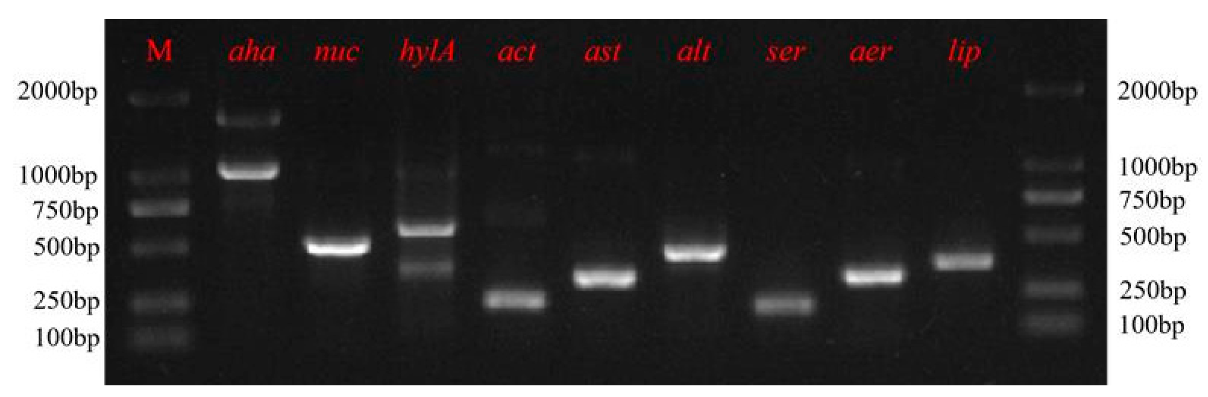
2.6. Screening of Virulence Genes

The presence of 9 virulence genes [aerolysin (aer), serine protease (ser), elastase (aha), haemolysin (hlyA), cytotonic enterotoxins (act, alt, ast), lipase (lip), nuclease (nuc)] were analysed by PCR, and the reaction parameters are summarised in Table 1. The PCR products were separated on a 1% agarose gel that was stained with ethidium bromide (10 μg/mL) in Tris-acetate-EDTA buffer (TAE) using a Power Pac universal Electrophoresis unit (Bio-Rad, CA, USA). The visualisation of the results was carried out using a UV Transilluminator (Bio-Rad, CA, USA) and Quantity-One v4.6.

2.7. Multi Locus Sequence Typing (MLST)

MLST was performed through the amplification of A. hydrophila housekeeping genes (gyrB, groL, gltA, metG, ppsA, recA) [21]. The primers used for the PCR were obtained from the PubMLST-Aeromonas spp. database [22]. The PCR amplification of six pairs of housekeeping genes was performed, and the raw data obtained from sequencing were visualised using DNAman (DNAman v9.0, Lynnon Biosoft, USA) for auditing and splicing. The verified sequences for each of the Rd001 housekeeping genes were analysed within the PubMLST Aeromonas spp. database to assign allelic numbers and defined sequence types (STs). All sequence types are available in the PubMLST database.
The six housekeeping genes of Rd001 were concatenated in the order gyrB-groL-gltA-metG-ppsA-recA, and the ITOL function in PubMLST was applied to compare the isolates in multiple sequences with other type strains in the database. The neighbour-joining method of Interactive Tree of Life v.4 programme (https://itol.embl.de/, accessed on 11 March 2024) was used to construct the evolutionary tree.

3. Results

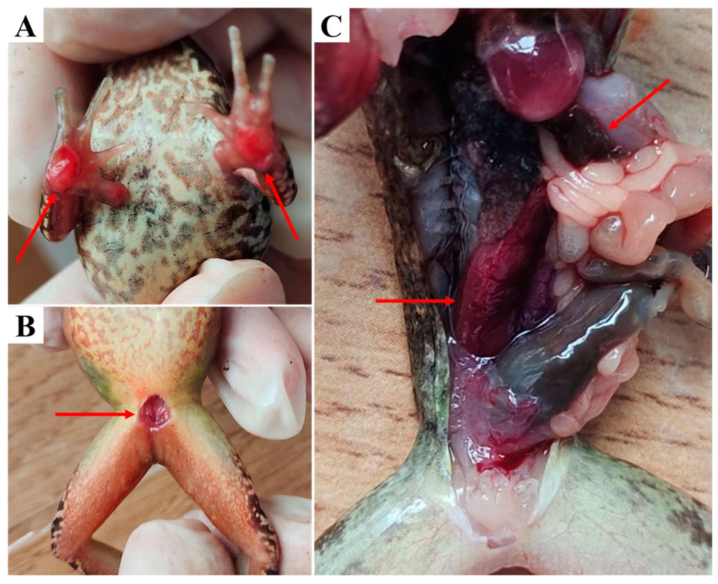
3.1. Clinical Examination

Diseased R. dybowskii from the farm showed reduced activity, reduced feeding and hiding under shelters. Clinical examination showed that the frogs had broken skin on their forelimbs, exposing ulcerated muscles, some also displaying similar signs on the abdomen. Autopsy of the diseased frogs indicated congested and swollen kidney, as well as congested spleen (Figure 1).

3.2. Physiological and Biochemical Characteristics

Rd001 and Rd002 are both Gram-negative bacteria. Rd001 exhibits similar physiological and biochemical responses to A. hydrophila ATCC 35654. While they can both hydrolyse Esculin, Rd001 produces acid from Sucrose, D-Galactose, Maltulose, D-Gluconic acid, and Methyl-B-Glucosid; uses Citrate as its sole carbon source; cannot hydrolyse Urea; and cannot ferment Sorbitol, L-Rhamnose, and D-Melibiose. Notably, Rd001 is able to utilise Adonitol but lacks the ability to ferment L-Arabinose (Table 2).
Rd002 exhibits similar physiological and biochemical responses to A. johnsonii ATCC17909, which can use Adonitol and Citrate as its sole carbon source; cannot hydrolyse Esculin and Urea; and cannot to ferment Sorbitol, Sucrose, D-Galactose, Maltulose, L-Rhamnose, D-Gluconic acid, D-Melibiose, L-Arabinose, and Methyl-B-Glucosid (Table 2).

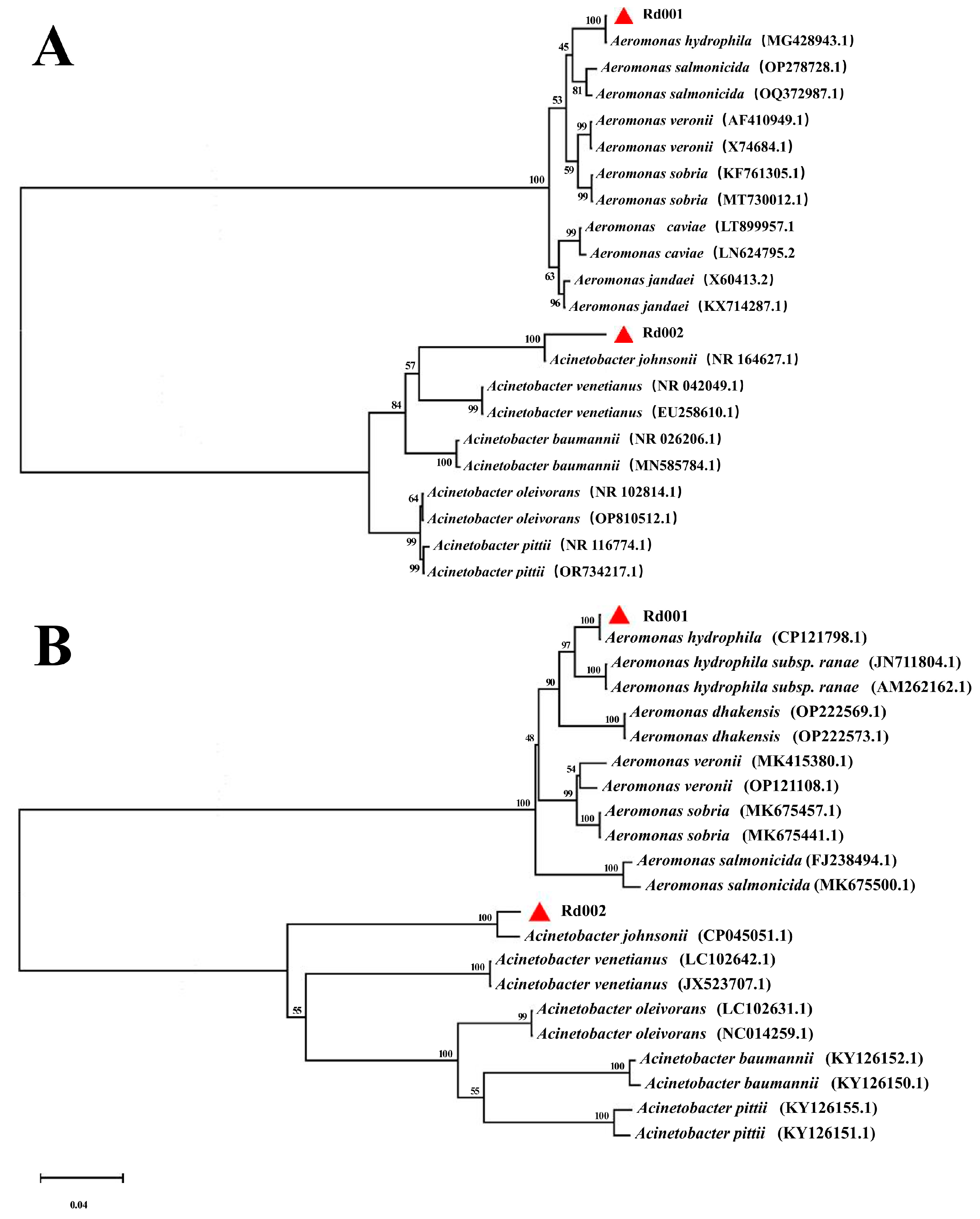
3.3. Sequence Analysis of the 16S rRNA and gyrB Genes

The 16S rRNA gene sequences of Rd001 and Rd002 obtained by PCR amplification have been uploaded at NCBI with GenBank accession numbers PP094559.1 and PP658225, respectively. BLAST results showed that isolate Rd001 shares 100% identity with the A. hydrophila (MG428943.1), and isolate Rd002 shares 99.7% identity with the A. johnsonii (NR164627.1). The gyrB gene sequences of Rd001 and Rd002 obtained by PCR amplification have been uploaded at NCBI with GenBank accession numbers PP894756 and PP894757, respectively. BLAST results showed that isolate Rd001 shares 100% identity with the A. hydrophila (CP121798.1) and isolate Rd002 shares 97.8% identity with the A. johnsonii (CP045051.1). Based on the phylogenetic trees constructed using the 16S rRNA and gyrB genes sequences (Figure 2), isolate Rd001 was grouped with recognised strains of A. hydrophila, while Rd002 was associated with A. johnsonii. The results of BLAST and phylogenetic analysis confirmed that Rd001 is A. hydrophila and Rd002 is A. johnsonii.

3.4. Experimental Infections

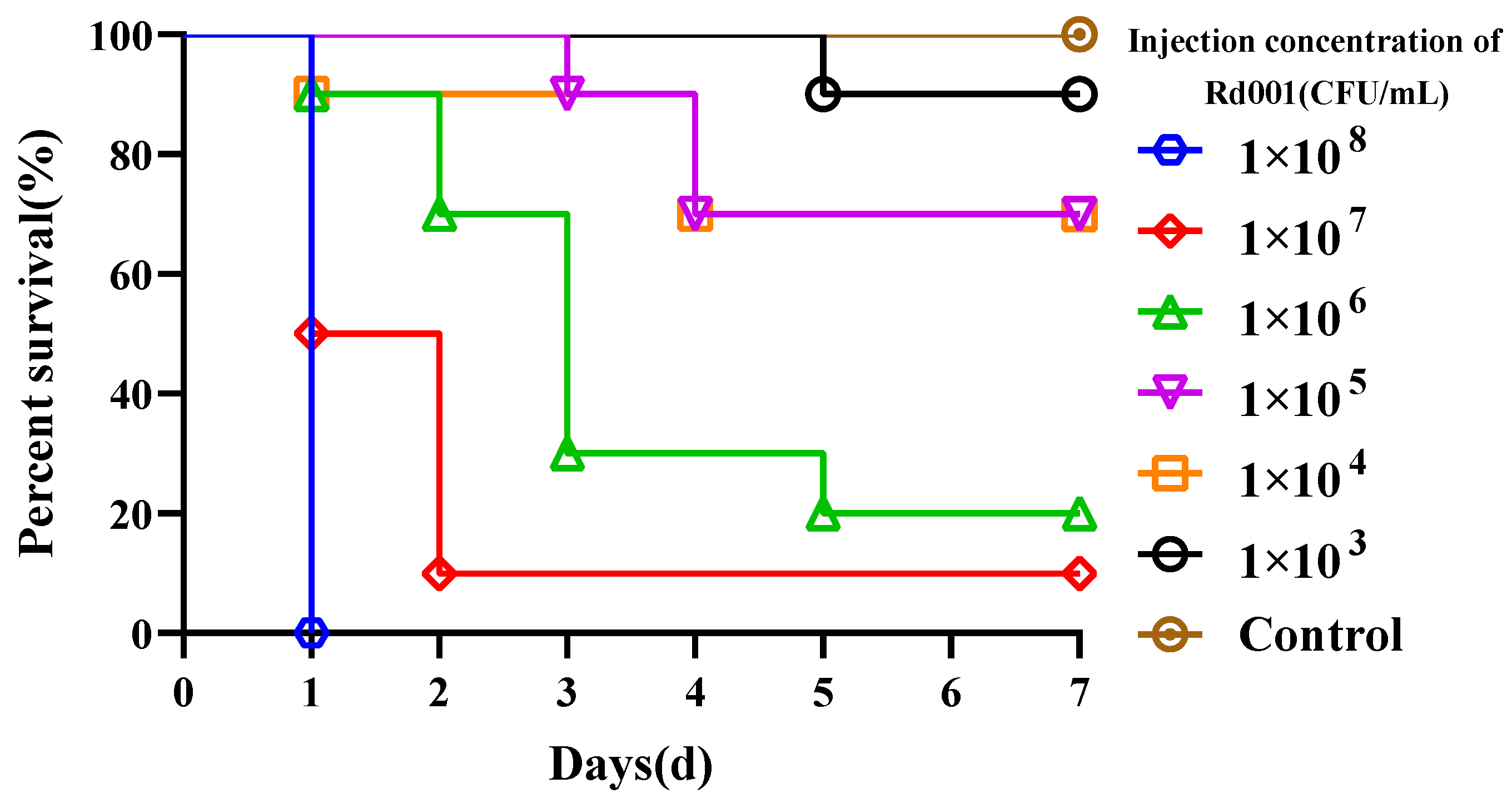
3.4.1. Determination of Median Lethal Dose

In a pre-challenge experiment, R. dybowskii injected with 1 × 10⁸ CFU/mL Rd001 reached 100% mortality within 24 h, whereas R. dybowskii injected with the same dose of Rd002 did not die within 7 d. Subsequently, we determined the LD50 of Rd001 on the R. dybowskii, and there was no mortality in the control group during the experimental period, and the cumulative mortality rates of the frogs injected with Rd001 at concentrations ranging from 1 × 10⁸ to 1 × 10³ CFU/mL were 100%, 90%, 80%, 30%, 30%, and 10%, respectively (Figure 3). The LD50 of Rd001 was calculated as 6.09 × 10⁴ CFU/mL (6.25 × 10² CFU/g). In addition, the bacterium was re-isolated from the kidney of artificially infected R. dybowskii.

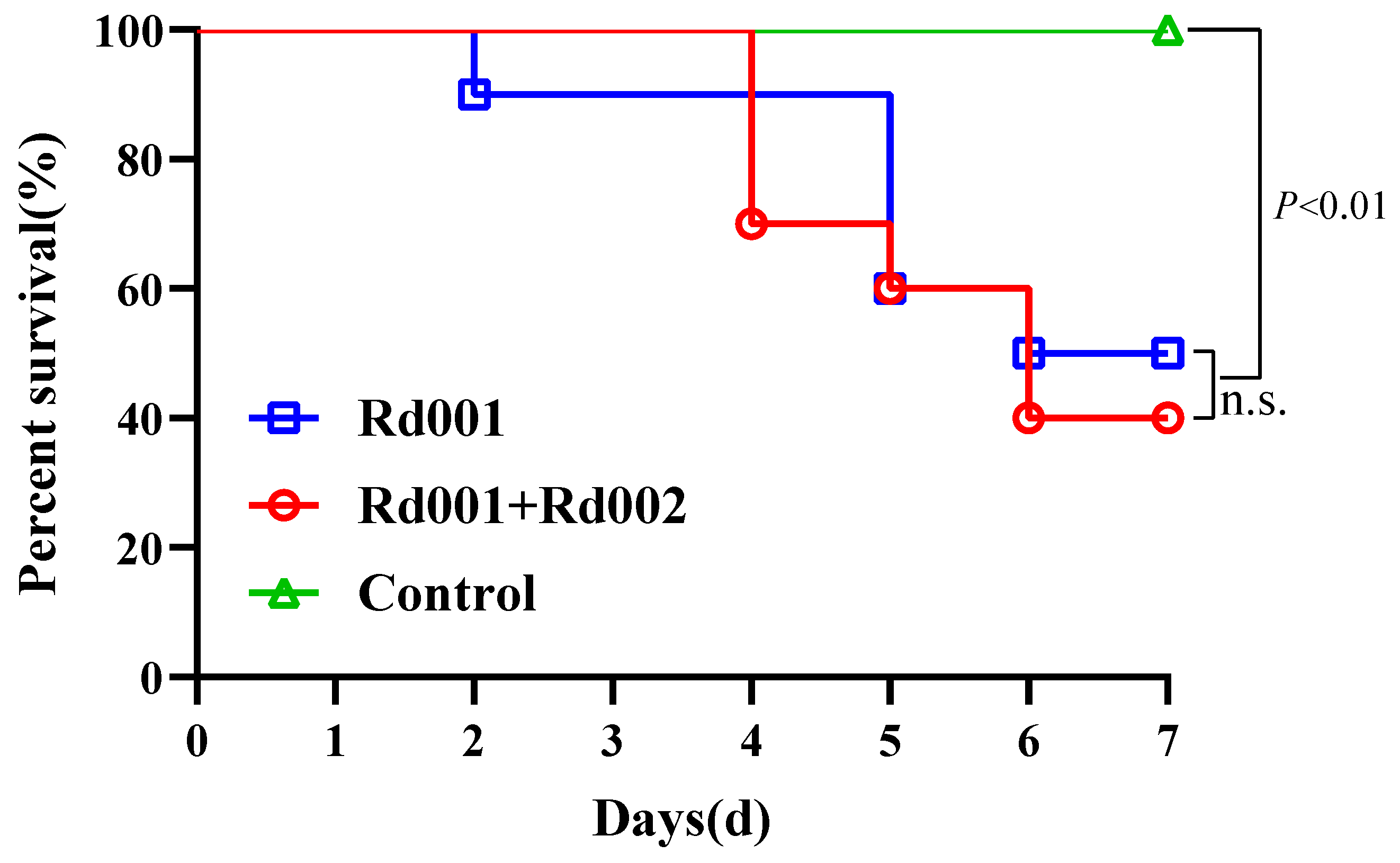
3.4.2. Single Infection and Co-Infection

Throughout the experimental period, no deaths were recorded in the control group. In the co-infection group (Rd001 + Rd002), mortality began on the 3rd day post-challenge, with a cumulative rate of 60% by the 7th day. In the single-infection group (Rd001), mortality commenced on the 2nd day, and by the 7th day, the cumulative mortality rate reached 60% (Figure 4). There was no significant difference in the mortalities between the single-infected and co-infected frogs (p > 0.05).

3.5. Antimicrobial Susceptibility and Virulence Factors

The results of antimicrobial susceptibility of isolate Rd001 were shown in Table 3. The isolate Rd01 was sensitive to enrofloxacin, flumequine, and neomycin with a MIC value of 0.25 μg/mL, 2 μg/mL, and 1 μg/mL, respectively, and intermediated to doxycycline with a MIC value of 8 μg/mL, whereas it exhibited reduced susceptibility to thiamphenicol, florfenicol, sulfamonomethoxine, and trimethoprim/sulfamethoxazole, with MIC values of 512 μg/mL, 512 μg/mL, 512 μg/mL, and 4/76 μg/mL, respectively (Table 3).
As shown in Figure 5, the isolate Rd001 was positive for aer, ser, alt, ast, act, hlyA, nuc, aha, and lip, as seen through the PCR validation of the virulence factors.

3.6. MLST Analysis of Rd001

Six housekeeping genes in Rd001 for MLST analysis were successfully amplified. The allelic profile of Rd001 (gyrB-804, groL-369, gltA-1041, metG-301, ppsA-269, recA-1130) was obtained by sequencing, splicing, and matching. It turned out that no sequence type matching this allelic profile existed in the PubMLST database, so we uploaded our original data and obtained a new sequence type number: ST2378 (Table 4).
A systematic phylogenetic tree was constructed for ST2378, confirming its phylogenetic position within A. hydrophila PubMLST. Among the 16 strains closely related to it, 9 strains (56.25%) were isolated from mammals. Phylogenetic tree results revealed the clustering of ST2378 with ST516 and ST2125, with the closest phylogenetic relationship to ST516 isolated from reptilia in China (Figure 6).

4. Discussion

A. hydrophila is a prevalent pathogen in amphibians, capable of inducing clinical signs like red-leg syndrome and skin ulcers in frogs [25]. Recent reports on A. hydrophila have primarily focused on epidemiological investigations and host response mechanisms [26,27,28], neglecting research on pathogen virulence traits and genetics. This gap has impeded the progress in developing vaccines and control medications to a certain extent.
In this study, we identified the coexistence of A. hydrophila Rd001 and A. johnsonii Rd002 as dominant bacteria in naturally occurring lesions of R. dybowskii, with primary signs presenting as ulcers on the forelimbs and abdomen skin. Abdominal subcutaneous injection infection confirmed that Rd001 was the primary pathogen responsible for the disease of R. dybowskii, with a LD50 of 6.25 × 10² CFU/g in 7 d. Based on the virulence characteristics of Aeromonas spp., Rd001 was classified as highly virulent [29].
Variances in disease signs and mortality resulting from distinct pathogenic A. hydrophila strains could stem from variations in virulence genotypes among isolates, with virulence genotyping recognised in multiple studies as a valuable tool for assessing the pathogenicity of A. hydrophila [30,31]. The Rd001 virulence genotype was identified as ser+/aha+/lip+/nuc+/hlyA+/aer+/alt+/ast+/act+, resembling the highly virulent A. hydrophila strains obtained from fish (Tilapia zillii and Mugil cephalus) in Egypt [32].
The ser and aha encode serine protease and elastase, respectively, both of which are extracellular enzymes that degrade epithelial mucous membranes and can lead to lesions in the skin and internal organs [33]. This contributes to infection establishment and evasion of the host’s initial defence mechanisms [34]. The lip encodes an extracellular lipase that enhances the severity of bacterial infection by modifying the host’s plasma membrane [35]. The aer and hlyA encode aerolysin and haemolysin, respectively, both of which are haemolytic toxins. Aerolysin, often present in pathogenic Aeromonas spp., induces osmotic cell lysis by binding to membrane-specific glycoprotein sites, leading to structural polymerisation [36]. Haemolysin is haemolytic and enterotoxic, binding to and lysing host erythrocytes [37]. Different enterotoxins encoded by the act, act, and ast can induce haemolysis and trigger the production of inflammatory mediators in macrophages and epithelial cells, leading to apoptosis, and elevate cAMP levels in intestinal epithelial cells, disrupting the water–electrolyte balance and causing diarrhoea [38,39].The synergistic action of multiple virulence factors in the strain Rd001 contributes to its survival, reproduction, and spread in the host. This action also leads to the destruction of host tissues and the suppression of the immune response, ultimately exacerbating the infection.
Bacterial drug resistance is a significant factor affecting the susceptibility of hosts to infection [40]. Rd001 showed reduced susceptibility to thiamphenicol, florfenicol, sulfamonomethoxine, and trimethoprim/sulfamethoxazole in an eight-antibiotic test, while being sensitive to enrofloxacin, flumequine, and neomycin. Enrofloxacin, with an MIC of 0.25 μg/mL, effectively inhibited Rd001 growth, thus emerging as the primary drug for the ongoing R. dybowskii outbreak. However, due to limited research on drug administration methods in amphibians and a lack of detailed pharmacokinetic studies, determining a reasonable dosage is challenging, necessitating further investigation.
MLST is frequently used to investigate the epidemiology, pathogenicity, and evolution of A. hydrophila, as well as to trace hosts and geographical origins [21,41]. In this study, the allelic profile of the identified Rd001 did not match the existing PubMLST data, leading to its classification as a novel sequence type: ST2378. The analysis of the phylogenetic tree constructed with existing A. hydrophila data from PubMLST shows that out of the 16 strains closely linked to ST2378, 9 strains (56.25%) are derived from mammals. Importantly, ST2378 is most closely related to ST516, which originates from China and is capable of inducing fatal diarrhoea in Chinese Moccasin (Deinagkistrodon acutus) [42]. Additionally, a high dose of ST516 can result in acute death in mice. Therefore, additional research is necessary to determine if ST2378 presents a public health risk to humans.
In general, A. hydrophila Rd001, isolated from the infected R. dybowskii, was identified as the primary pathogen with high virulence characteristics. MLST analysis suggested that Rd001 had a potential risk of infection in mammals, and relevant findings filled the gap in the genetics of A. hydrophila isolated from R. dybowskii. Given its significance as a major pathogen in frog infections, A. hydrophila requires urgent investigation for optimal drug delivery methods and pharmacokinetics, informed by current drug sensitivity data, and the implementation of infection control measures to limit its spread.

Author Contributions

Conceptualisation: R.Z. and S.L.; formal analysis: R.Z. and D.W.; investigation: R.Z., J.W., Y.W. and G.H.; supervision: D.W. and S.L.; writing—original draft: R.Z.; writing—review and editing: R.Z. and S.L.; funding acquisition: G.H. All authors have read and agreed to the published version of the manuscript.

Funding

This work was supported by the Central Public Interest Scientific Institution Basal Research Fund, CAFS (no. 2023TD45), Government Purchase for Public Service Contract from Ministry of Agriculture and Rural Affairs of China (17230057, 17230180).

Institutional Review Board Statement

The animal study was reviewed and approved by the Committee of the Ethics on Animal Care and Experiments at Heilongjiang River Fisheries Research Institute of Chinese Academy of Fishery Sciences (CAFS20230824-001).

Informed Consent Statement

Not applicable.

Data Availability Statement

Data is contained within the article.

Conflicts of Interest

The authors declare no conflicts of interest.

References

  1. Xu, K.; Zhu, D.-Z.; Wei, Y.; Schloegel, L.M.; Chen, X.-F.; Wang, X.-L. Broad Distribution of Ranavirus in Free-Ranging Rana Dybowskii in Heilongjiang, China. EcoHealth 2010, 7, 18–23. [Google Scholar] [CrossRef] [PubMed]
  2. Morrison, C.; Hero, J. Geographic Variation in Life-history Characteristics of Amphibians: A Review. J. Anim. Ecol. 2003, 72, 270–279. [Google Scholar] [CrossRef]
  3. Carey, C.; Cohen, N.; Rollins-Smith, L. Amphibian Declines: An Immunological Perspective. Dev. Comp. Immunol. 1999, 23, 459–472. [Google Scholar] [CrossRef]
  4. Igbinosa, I.H.; Igumbor, E.U.; Aghdasi, F.; Tom, M.; Okoh, A.I. Emerging Aeromonas Species Infections and Their Significance in Public Health. Sci. World J. 2012, 2012, 625023. [Google Scholar] [CrossRef] [PubMed]
  5. Huys, G.; Pearson, M.; Kämpfer, P.; Denys, R.; Cnockaert, M.; Inglis, V.; Swings, J. Aeromonas hydrophila subsp. ranae subsp. nov., Isolated from Septicaemic Farmed Frogs in Thailand. Int. J. Syst. Evol. Microbiol. 2003, 53, 885–891. [Google Scholar] [CrossRef] [PubMed]
  6. Bradford, D.F. Mass Mortality and Extinction in a High-Elevation Population of Rana Muscosa. J. Herpetol. 1991, 25, 174–177. [Google Scholar] [CrossRef]
  7. Khalifa, A.Y.Z.; AlMalki, M.A.; Bekhet, G.M. Pathological and Mortality Findings Associated with Aeromonas hydrophila from Frog Eggs in Al-Ahsa Region of Saudi Arabia. Aquac. Res. 2021, 52, 1227–1236. [Google Scholar] [CrossRef]
  8. Schadich, E.; Cole, A.L. Pathogenicity of Aeromonas hydrophila, Klebsiella pneumoniae, and Proteus mirabilis to Brown Tree Frogs (Litoria ewingii). Comp. Med. 2010, 60, 114–117. [Google Scholar] [CrossRef] [PubMed]
  9. Rasmussen-Ivey, C.R.; Figueras, M.J.; McGarey, D.; Liles, M.R. Virulence Factors of Aeromonas hydrophila: In the Wake of Reclassification. Front. Microbiol. 2016, 7, 1337. [Google Scholar] [CrossRef]
  10. Allan, B.J.; Stevenson, R.M.W. Extracellular Virulence Factors of Aeromonas hydrophila in Fish Infections. Can. J. Microbiol. 1981, 27, 1114–1122. [Google Scholar] [CrossRef]
  11. Pattanayak, S.; Priyadarsini, S.; Paul, A.; Kumar, P.R.; Sahoo, P.K. Diversity of Virulence-Associated Genes in Pathogenic Aeromonas hydrophila Isolates and Their in Vivo Modulation at Varied Water Temperatures. Microb. Pathog. 2020, 147, 104424. [Google Scholar] [CrossRef] [PubMed]
  12. Bakiyev, S.; Smekenov, I.; Zharkova, I.; Kobegenova, S.; Sergaliyev, N.; Absatirov, G.; Bissenbaev, A. Isolation, Identification, and Characterization of Pathogenic Aeromonas hydrophila from Critically Endangered Acipenser baerii. Aquac. Rep. 2022, 26, 101293. [Google Scholar] [CrossRef]
  13. CLSI. Methods for Broth Dilution Susceptibility Testing of Bacteria Isolated from Aquatic Animals; Approved Guidelinet; CLSI Document VET04-A; Clinical and Laboratory Standards Institute: Berwyn, PA, USA, 2006. [Google Scholar]
  14. CLSI. Performance Standards for Antimicrobial Suceptibility Testing of Bacteria Isolated from Aquatic Animals; Second Informational Supplement; CLSI Document VET03/VET04-S2; Clinical and Laboratory Standards Institute: Berwyn, PA, USA, 2014. [Google Scholar]
  15. Singh, V.; Rathore, G.; Kapoor, D.; Mishra, B.N.; Lakra, W.S. Detection of Aerolysin Gene in Aeromonas hydrophila Isolated from Fish and Pond Water. Indian J. Microbiol. 2008, 48, 453–458. [Google Scholar] [CrossRef] [PubMed]
  16. Nam, I.-Y.; Joh, K. Rapid Detection of Virulence Factors of Aeromonas Isolated from a Trout Farm by Hexaplex-PCR. J. Microbiol. Seoul Korea 2007, 45, 297–304. [Google Scholar] [CrossRef]
  17. Kwon, J.; Kim, S.G.; Kim, S.W.; Yun, S.; Kim, H.J.; Giri, S.S.; Han, S.J.; Oh, W.T.; Park, S.C. A Case of Mortality Caused by Aeromonas hydrophila in Wild-Caught Red-Eyed Crocodile Skinks (Tribolonotus gracilis). Vet. Sci. 2019, 7, 4. [Google Scholar] [CrossRef] [PubMed]
  18. Rozi; Rahayu, K.; Daruti, D.N. Detection and Analysis of Hemolysin Genes in Aeromonas hydrophila Isolated from Gouramy (Osphronemus gouramy) by Polymerase Chain Reaction (PCR). IOP Conf. Ser. Earth Environ. Sci. 2018, 137, 012001. [Google Scholar] [CrossRef]
  19. Li, T.; Raza, S.H.A.; Yang, B.; Sun, Y.; Wang, G.; Sun, W.; Qian, A.; Wang, C.; Kang, Y.; Shan, X. Aeromonas veronii Infection in Commercial Freshwater Fish: A Potential Threat to Public Health. Anim. Open Access J. 2020, 10, 608. [Google Scholar] [CrossRef] [PubMed]
  20. Sen, K.; Rodgers, M. Distribution of Six Virulence Factors in Aeromonas Species Isolated from US Drinking Water Utilities: A PCR Identification. J. Appl. Microbiol. 2004, 97, 1077–1086. [Google Scholar] [CrossRef]
  21. Martino, M.E.; Fasolato, L.; Montemurro, F.; Rosteghin, M.; Manfrin, A.; Patarnello, T.; Novelli, E.; Cardazzo, B. Determination of Microbial Diversity of Aeromonas Strains on the Basis of Multilocus Sequence Typing, Phenotype, and Presence of Putative Virulence Genes. Appl. Environ. Microbiol. 2011, 77, 4986–5000. [Google Scholar] [CrossRef]
  22. Jolley, K.A.; Bray, J.E.; Maiden, M.C.J. Open-Access Bacterial Population Genomics: BIGSdb Software, the PubMLST.Org Website and Their Applications. Wellcome Open Res. 2018, 3, 124. [Google Scholar] [CrossRef]
  23. Schoch, C.L.; Ciufo, S.; Domrachev, M.; Hotton, C.L.; Kannan, S.; Khovanskaya, R.; Leipe, D.; Mcveigh, R.; O’Neill, K.; Robbertse, B.; et al. NCBI Taxonomy: A Comprehensive Update on Curation, Resources and Tools. Database 2020, 2020, baaa062. [Google Scholar] [CrossRef] [PubMed]
  24. Bouvet, P.J.M.; Grimont, P.A.D. Taxonomy of the Genus Acinetobacter with the Recognition of Acinetobacter baumannii Sp. Nov., Acinetobacter haemolyticus sp. nov., Acinetobacter johnsonii sp. nov., and Acinetobacter junii sp. nov. and Emended Descriptions of Acinetobacter calcoaceticus and Acinetobacter lwoffii. Int. J. Syst. Evol. Microbiol. 1986, 36, 228–240. [Google Scholar] [CrossRef]
  25. Lin, H.; Zeng, G.; Yu, Y.; Li, H.; Chen, K.; Qin, Z.; Jiang, B.; Li, W.; Su, Y.; Lin, L.; et al. Acute Septicemia and Diagnostic Evaluation of Aeromonas veronii Infection in American Bullfrogs (Aquarana catesbeiana). Aquaculture 2024, 580, 740349. [Google Scholar] [CrossRef]
  26. Wang, B.; Shao, J.; Qu, L.; Xu, Q.; Zheng, D. The Sequencing of the Key Genes and End Products in the TLR4 Signaling Pathway from the Kidney of Rana dybowskii Exposed to Aeromonas hydrophila. Open Life Sci. 2023, 18, 20220704. [Google Scholar] [CrossRef] [PubMed]
  27. Hill, W.A.; Newman, S.J.; Craig, L.; Carter, C.; Czarra, J.; Brown, J.P. Diagnosis of Aeromonas hydrophila, Mycobacterium Species, and Batrachochytrium dendrobatidis in an African Clawed Frog (Xenopus laevis). J. Am. Assoc. Lab. Anim. Sci. 2010, 49, 215–220. [Google Scholar] [PubMed]
  28. Miller, D.L.; Rajeev, S.; Brookins, M.; Cook, J.; Whittington, L.; Baldwin, C.A. Concurrent Infection with Ranavirus, Batrachochytrium dendrobatidis, and Aeromonas in a Captive Anuran Colony. J. Zoo Wildl. Med. 2008, 39, 445–449. [Google Scholar] [CrossRef]
  29. Mittal, K.R.; Lalonde, G.; Leblanc, D.; Olivier, G.; Lallier, R. Aeromonas hydrophila in Rainbow Trout: Relation between Virulence and Surface Characteristics. Can. J. Microbiol. 1980, 26, 1501–1503. [Google Scholar] [CrossRef]
  30. Senderovich, Y.; Ken-Dror, S.; Vainblat, I.; Blau, D.; Izhaki, I.; Halpern, M. A Molecular Study on the Prevalence and Virulence Potential of Aeromonas spp. Recovered from Patients Suffering from Diarrhea in Israel. PLoS ONE 2012, 7, e30070. [Google Scholar] [CrossRef]
  31. Fernández-Bravo, A.; Kilgore, P.B.; Andersson, J.A.; Blears, E.; Figueras, M.J.; Hasan, N.A.; Colwell, R.R.; Sha, J.; Chopra, A.K. T6SS and ExoA of Flesh-Eating Aeromonas hydrophila in Peritonitis and Necrotizing Fasciitis during Mono- and Polymicrobial Infections. Proc. Natl. Acad. Sci. USA 2019, 116, 24084–24092. [Google Scholar] [CrossRef]
  32. Ayoub, H.F.; Khafagy, A.R.; Esawy, A.M.; El-moaty, N.A.; Alwutayd, K.M.; Mansour, A.T.; Ibrahim, R.A.; Abdel-moneam, D.A.; El-Tarabili, R.M. Phenotypic, Molecular Detection, and Antibiotic Resistance Profile (MDR and XDR) of Aeromonas hydrophila Isolated from Farmed Tilapia zillii and Mugil cephalus. BMC Vet. Res. 2024, 20, 84. [Google Scholar] [CrossRef]
  33. Gonçalves Pessoa, R.B.; de Oliveira, W.F.; Marques, D.S.C.; dos Santos Correia, M.T.; de Carvalho, E.V.M.M.; Coelho, L.C.B.B. The Genus Aeromonas: A General Approach. Microb. Pathog. 2019, 130, 81–94. [Google Scholar] [CrossRef] [PubMed]
  34. Tomás, J.M. The Main Aeromonas Pathogenic Factors. ISRN Microbiol. 2012, 2012, 256261. [Google Scholar] [CrossRef] [PubMed]
  35. Pemberton, J.M.; Kidd, S.P.; Schmidt, R. Secreted Enzymes of Aeromonas. FEMS Microbiol. Lett. 1997, 152, 1–10. [Google Scholar] [CrossRef] [PubMed]
  36. Abrami, L.; Fivaz, M.; Glauser, P.-E.; Sugimoto, N.; Zurzolo, C.; van der Goot, F.G. Sensitivity of Polarized Epithelial Cells to the Pore-Forming Toxin Aerolysin. Infect. Immun. 2003, 71, 739–746. [Google Scholar] [CrossRef] [PubMed]
  37. Takahashi, E.; Ozaki, H.; Fujii, Y.; Kobayashi, H.; Yamanaka, H.; Arimoto, S.; Negishi, T.; Okamoto, K. Properties of Hemolysin and Protease Produced by Aeromonas trota. PLoS ONE 2014, 9, e91149. [Google Scholar] [CrossRef] [PubMed]
  38. Galindo, C.; Sha, J.; Fadl, A.; Pillai, L.; Chopra, A. Host Immune Responses to Aeromonas Virulence Factors. Curr. Immunol. Rev. 2006, 2, 13–26. [Google Scholar] [CrossRef]
  39. Galindo, C.L.; Gutierrez, C., Jr.; Chopra, A.K. Potential Involvement of Galectin-3 and SNAP23 in Aeromonas hydrophila Cytotoxic Enterotoxin-Induced Host Cell Apoptosis. Microb. Pathog. 2006, 40, 56–68. [Google Scholar] [CrossRef] [PubMed]
  40. Tekedar, H.C.; Arick, M.A.I.; Hsu, C.-Y.; Thrash, A.; Blom, J.; Lawrence, M.L.; Abdelhamed, H. Identification of Antimicrobial Resistance Determinants in Aeromonas veronii Strain MS-17-88 Recovered From Channel Catfish (Ictalurus punctatus). Front. Cell. Infect. Microbiol. 2020, 10, 348. [Google Scholar] [CrossRef] [PubMed]
  41. Urwin, R.; Maiden, M.C.J. Multi-Locus Sequence Typing: A Tool for Global Epidemiology. Trends Microbiol. 2003, 11, 479–487. [Google Scholar] [CrossRef]
  42. Liu, J.; Xie, L.; Zhao, D.; Yang, T.; Hu, Y.; Sun, Z.; Yu, X. A Fatal Diarrhoea Outbreak in Farm-Raised Deinagkistrodon Acutus in China Is Newly Linked to Potentially Zoonotic Aeromonas hydrophila. Transbound. Emerg. Dis. 2019, 66, 287–298. [Google Scholar] [CrossRef]
Figure 1. Clinical signs in diseased R. dybowskii. (A) Skin ulceration of the forelimbs, revealing corroded muscles. (B) Abdominal skin was broken and muscles are congested. (C) The kidney and spleen were congested and swollen.
Pathogens 13 00552 g001
Figure 2. Molecular identification of the isolate Rd001 and Rd002. (A) Neighbour-joining phylogenetic tree based on the 16S rRNA gene sequence. (B) Neighbour-joining phylogenetic tree based on the gyrB gene sequence.
Pathogens 13 00552 g002
Figure 3. Cumulative mortalities of R. dybowskii challenged with different concentrations of Rd001.
Pathogens 13 00552 g003
Figure 4. Cumulative mortalities in single-infected (Rd001), co-infected (Rd001 + Rd002), and control group of R. dybowskii at different time points. Comparison of survival curves were performed by a log-rank (Mantel–Cox) test using GraphPad Prism v9.3.1.
Pathogens 13 00552 g004
Figure 5. PCR amplification for nine virulent genes of Rd001. Lane M: DNA marker.
Pathogens 13 00552 g005
Figure 6. Neighbour-joining tree constructed from the tandem nucleotide sequences (gltA-groL-gyrB-metG-ppsA-recA) of six housekeeping genes from 214 A. hydrophila strains (Left). An evolutionary tree of the 17 most closely related strains of Rd001/ST2378 with a dataset including strain labelling, host, and country is presented (Right).
Pathogens 13 00552 g006
Table 1. Primers for PCR detection of 9 virulence genes.
GenePCR Prime Sequence (5′→3′)Product Size (bp)Reference
aerF: ACAGCCAATATGTCGGTGAAG326[15]
R: TCACCTTCTCGCTCAGGC
serF: CACCGAAGTATTGGGTCAGG211[16]
R: GGCTCATGCGTAACTCTGGT
altF:TGACCCAGTCCTGGCACGGC442[17]
R:GGTGATCGATCACCACCAGC
astF:TCTCCATGCTTCCCTTCCACT331[17]
R:GTGTAGGGATTGAAGAGCCG
actF:AGAAGGTGACCACCACCAAGAACA232[17]
R:AACTGACATCGGCCTGAACTC
hlyAF:GGCCGGTGGCCCGAAGATACGGG592[18]
R:GGCGGCGCCGGACGAGACGGGG
nucF:CAGGATCTGAACCGCCTCTATCAGG504[15]
R:GTCCCAAGCTTCGAACAGTTTACGC
ahaF:GGTATTGTATCCCGGCTCTGTT1082[19]
R:CGGTCCATCGTCGTCCATCTTG
lipF: TCTTCTCCGACTGGTTCGG382[20]
R: GTGCCAGGACTGGGTCTT
Table 2. Physiological and biochemical characteristics of Rd001 and Rd002 isolates.
CharacteristicsRd001A. hydrophila ATCC35654 [23]Rd002A. johnsonii ATCC17909 [24]
Gram stain
N-Acetyl-D-glucosamine++
Adonitol++
Citrate+++
Sorbitol
Sucrose++
Glucose++
D-Galactose++
Maltulose++
L-Rhamnose
D-Gluconic acid++
D-Melibiose
L-Arabinose+
Methyl-B-Glucoside++
Urea
Esculin++
Table 3. Drug susceptibility of Rd001 to eight antimicrobial drugs.
GroupAntibioticMIC (μg/mL)Susceptibility
4-quinolonesEnrofloxacin0.25S
Flumequine2S
ChloramphenicolsThiamphenicol512R
Florfenicol512R
SulfonamidesSulfamonomethoxine512R
Trimethoprim/Sulfamethoxazole4/76R
Aminogly cosidesNeomycin1S
TetracyclinesDoxycycline8I
Note: Susceptibility is designated as S = sensitive, I = intermediate, and R = resistant.
Table 4. Multi-locus sequence typing analysis of Rd001.
IsolateRd001
gyrB804
groL369
gltA1041
metG301
ppsA269
recA1130
Assigned ST2378
Disclaimer/Publisher’s Note: The statements, opinions and data contained in all publications are solely those of the individual author(s) and contributor(s) and not of MDPI and/or the editor(s). MDPI and/or the editor(s) disclaim responsibility for any injury to people or property resulting from any ideas, methods, instructions or products referred to in the content.

