Communication

The Diversity of Seed-Borne Fungi Associated with Soybean Grown in Southern Poland

by
Hanna Olszak-Przybyś
* and
Grażyna Korbecka-Glinka
Department of Biotechnology and Plant Breeding, Institute of Soil Science and Plant Cultivation–State Research Institute, ul. Czartoryskich 8, 24-100 Puławy, Poland
*
Author to whom correspondence should be addressed.
Pathogens 2024, 13(9), 769; https://doi.org/10.3390/pathogens13090769
Submission received: 14 June 2024 / Revised: 29 August 2024 / Accepted: 4 September 2024 / Published: 6 September 2024
(This article belongs to the Special Issue Fungal Pathogens of Crops)

Abstract

:
Fungi have the potential to colonize soybean seeds in the field, during their maturation in the pods and after harvest, during storage. The aim of this study was to identify fungi inhabiting soybean seeds after storage with varying germination capacity and to evaluate their chemical composition. The research material consisted of twelve soybean seed lots collected from the fields in southern Poland and stored over winter. The germination percentage of these lots ranged between 20.67% and 81.33%. The seeds were subjected to analyses of the main chemical components and mycological analysis. Fungal isolates were subjected to taxonomic identification using microscopic methods and DNA sequencing (using internal transcribed spacer region and secondary barcoding regions). A total number of 355 fungal isolates from 16 genera were identified, with Aspergillus, Alternaria, and Fusarium being the most common. Species were successfully identified in 94% of isolates. Twelve examined seed lots varied significantly in the number of isolated fungal species (from 1 to 17). Moreover, they also differed in the isolated species composition. Highly significant positive correlation was found between the number of Aspergillus psedudoglaucus isolates and the content of free fatty acids. In turn, the number of Fusarium spp. isolates correlated negatively with protein and nitrogen content. Similarly, highly significant negative correlation was found between the number of all fungal isolates and the 1000-seed weight, indicating that smaller seeds are more vulnerable to fungal infection. The results obtained in this study identify species of fungi which may be responsible for lowering quality of the seeds obtained in southern Poland.

1. Introduction

Soybean [Glycine max (L.) Merrill.] is one of the most valuable oil crops. The global production of this crop in 2022 amounted to 348.8 million tons, which illustrates its enormous economic importance. The main soybean growers are Brazil (120.7 million tons), the United States (116.3 million tons), and Argentina (43.8 million tons). The production in Europe in 2022 amounted to 12.4 million tons, of which approx. 43.8 thousand tons were produced in Poland [1]. Soybean seeds are essential for producing oil or feed for livestock and are a valuable source of protein, oil, vitamins, and minerals for humans [2]. They contain 38% protein, 30% carbohydrates, 18% lipids, and 14% other substances [3]. They are used to produce soy milk, tofu, soy sauce, and paste, which are popular worldwide [4]. Soybean also plays a crucial role in various industries, as it is used in the production of biodiesel, pharmaceuticals, plastics, paints, and cosmetics. Due to its isoflavone content, soybean is believed to have anti-inflammatory, antioxidant, and antifungal properties. Furthermore, fermented soy products have been demonstrated to possess anti-cancer and anti-diabetic properties, and beneficial effects on the cardiovascular system [4,5].
Soybean is threatened by various abiotic and biotic stress factors, which decrease soybean seed quality and yield [6]. According to Roy [7], 80 genera belonging to economically important pathogen groups are associated with soybean diseases. Among them, fungi are one of the most significant biotic factors that adversely affect soybean growth and yield by attacking plants and seeds during their growth and after harvest [2]. Over 150 different species of fungi can inhabit soybean seeds, potentially reducing the nutritional value of seeds and leading to the production of mycotoxins [8]. Fungi that colonize and damage seeds are traditionally divided into field fungi and storage fungi, based on their ecological requirements [9]. Fungi belonging to genera such as Alternaria, Cladosporium, Cercospora, Diaporthe, and Fusarium can invade seeds while still in the field, causing damage before harvest. However, their incidence in the seeds may be significantly reduced during storage [9,10].
Nevertheless, if these field fungi survive in the stored seeds used later as sowing material, they may pose a significant threat to germinating soybean seeds, seedlings, and mature plants. They can reduce seedling emergence in the field and cause many diseases. For example, Alternaria spp. infection may result in leaf spot, pod necrosis, and seed decay, while Diaporthe spp. may cause Phomopsis seed decay, pod and stem blight, and stem canker [8]. In turn, Fusarium spp. may lead to Fusarium seed rot, and pod and stem blight [8].
As storage progresses, the occurrence of field fungi decreases in seeds, while storage fungi begin to colonize the seeds, reducing seed vigor and germination rates [8]. A decline in seed quality is evident through a reduction in the protein and oil content, as well as a rise in the free fatty acid content [8,11]. The predominant storage fungi belong to the genera Aspergillus and Penicillium. The proportion of Aspergillus increases by 1.3% after six months of storage, and by 7.5% after eight months [10]. The quantity of Penicillium in the stored seeds also increases over time, especially after six months of storage [10]. Storage fungi from both genera contribute to seed spoilage and damaged embryos [9]. They may also cause a reduction in the germination level and adverse changes in the shape, color, and biochemical composition of seeds. Seeds with reduced germination capacity are not suitable for sowing. Some of the storage fungi have the ability to produce toxins that are harmful to humans and livestock [2,12,13]. Mycotoxins produced by Aspergillus flavus, Aspergillus parasiticus, and Penicillium spp. can make seeds unsuitable for human and animal consumption. The estimated losses caused by storage fungi amount to more than $20 million a year in the USA [8].
Traditional methods of identifying fungi by recording colony appearance, growth rate, and morphological characteristics can often be misleading due to hybridization, cryptic speciation, and convergent evolution [14,15,16]. Identifying fungi based on morphology can also be challenging, especially when fungal cultures produce a limited number of morphological structures (e.g., conidia) that can be used for identification, or when these structures exhibit highly variable features [16,17]. As a consequence, DNA sequence-based methods have been developed for identifying species. The internal transcribed spacer (ITS) is the most commonly sequenced marker for accurately identifying a wide range of fungi [18,19,20]. Although the ITS region is generally considered a suitable barcode for fungi, it is insufficient for some seed-borne fungi such as Aspergillus, Fusarium and Penicillium. For these genera, it is recommended to use one or more single-copy protein-coding genes for species-level identification [17,21,22,23].
Dominant fungi associated with soybean seeds may differ between geographical regions. For example, in Brazil, Colletotrichum truncatum was identified as the predominant species responsible for anthracnose on soybean. Seeds infected with this pathogen were widely recognized as a major source of disease spread [24,25]. On the other hand, Cortina [26] found that Fusarium was the dominant genus on soybean seeds, accounting for 80–90% of all isolated fungi, while Colletotrichum represented only 5–10%. Research from China reveals that the main fungi isolated from soybean seeds in China belong to the Fusarium genus, with Fusarium fujikuroi being the most prevalent species [27]. In North America, Fusarium species were frequently reported in soybean seeds. The study found that 33% of soybean seed samples were infected with these fungi, and they were isolated from seeds collected from 80% of locations in Kansas [28]. A survey in Turkey revealed that 88% of soybean seeds were contaminated with fungi. The infection rates of Cladosporium spp., Fusarium spp., Penicillium spp., and Aspergillus spp. were 42%, 38% 10%, and 0.9% in soybean seeds, respectively [2].
It is well known that infected seeds can be a source of infection in the field, and can affect seed germination and the development of the disease at a younger stage [8]. Even certified seeds are not guaranteed to be free from seed-borne pathogens [9,10]. Furthermore, soybean sowing material typically comes from stored seeds because several months separate harvest and sowing in the subsequent season. Mycological analyses of the seeds after storage allow for the identification of culturable pathogens that were able to thrive in the stored seeds and could potentially cause plant disease in the field. They also detect storage fungi that potentially affect the seed quality of sowing material. Increased knowledge about seed-borne fungi may be used to develop effective and sustainable practices to reduce the damage they cause.
Many studies have been carried out examining the occurrence of fungi on and in soybean seeds [2,25,26,27], but so far, no such studies have been conducted in southern Poland. In our previous work [29], we characterized fungi isolated from soybean plants grown in one location in the southeast of the country. The aim of this study was to characterize the fungal community in stored soybean seeds originating from multiple fields in southern Poland. To accomplish this, identification of fungi was performed using mycological analyses supported by DNA sequencing of recommended barcoding regions. Germination tests and analyses of chemical composition of the seeds allowed for determining the impact field and storage fungi on soybean seed quality.

2. Materials and Methods

2.1. Seed Collection

Twelve samples of soybean seeds (1.5 kg each) were provided by seed producing company Agroyoumis (Table 1) in 2022, along with the data on the initial (pre-storage) germination percentage (ranging from 40% to 95%).
These seed samples were collected in 2021 from 12 different soybean plantations designated for seed production. These were located in southern Poland in the following four provinces: Dolnośląskie, Lubelskie, Opolskie, and Podkarpackie (Figure 1). All samples were stored in the same warehouse (without temperature or humidity control) for approximately seven months, until April 2022, which is the typical time when soybean is planted in Poland. At that time, the collected seeds were included into our post-storage germination tests and other analyses described below.

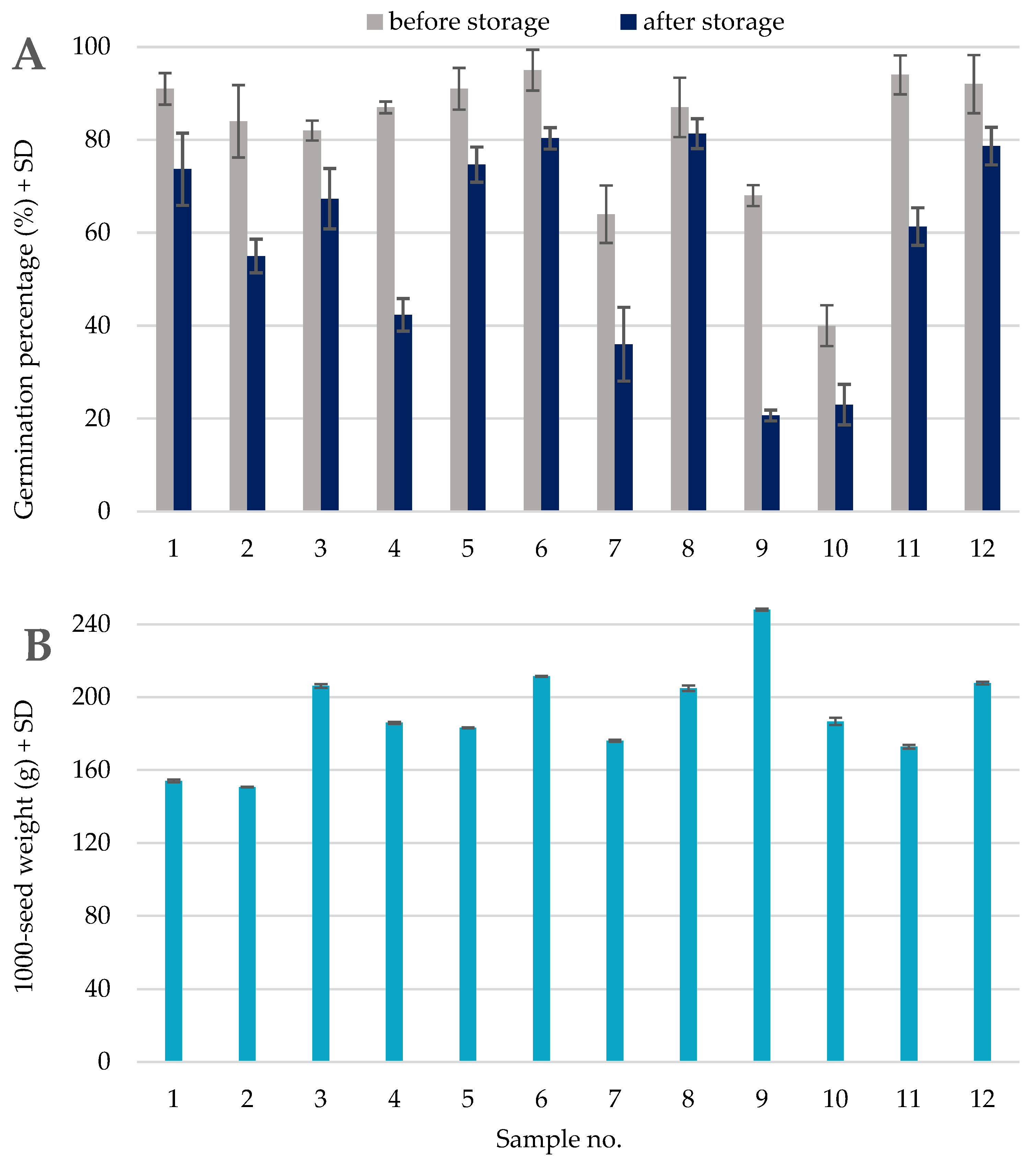
2.2. Germination Test

The methodology used for seed germination assessment followed the guidelines of the International Seed Testing Association (ISTA) [30]. From every seed lot, three replicates of 100 seeds were tested. Seeds from each replicate were placed between moist filter paper rolls, covered with a plastic bag, and germinated in climatic chambers MC-1750 (Snijders Labs, Tilburg, The Netherlands) under the following conditions: 30 °C for 8 h in the light and 20 °C for 16 h in the dark, maintaining 90% humidity throughout. On the 8th day of the incubation period, the number of germinated seedlings was recorded, and the germination percentage was calculated for each seed lot.

2.3. Isolation and Morphological Identification of Fungal Species

One hundred soybean seeds (four replicates of 25 seeds) from each seed lot were used for mycological analysis. Soybean seeds were surface-disinfected with a 1.4% sodium hypochlorite solution for one minute, rinsed three times in sterile water, and dried on sterile tissue paper under airflow in Class II Microbiological Safety Cabinet (NordicSafe®, Esco, Singapore). Finally, the seeds were placed in Petri dishes containing Potato Dextrose Agar (PDA, DifcoTM, Sparks, MD, USA) medium with tetracycline hydrochloride (2.5 mg L−1), in four replicates of 5 Petri dishes each. Five seeds were placed on each Petri dish and then incubated 20 °C for 14 days, with 8 h in the light and 16 h in the dark. The fungi that grew from seeds were isolated by cutting small pieces of the mycelium and transferring them to separate Petri dishes with PDA medium. Pure cultures of individual fungal isolates were used for taxonomic identification. The morphological characterization of the fungal isolates was carried out after incubation on PDA medium at 20 ± 2 °C. Isolates were first characterized by recording their morphological characteristics (color, size, shape; recorded for both sides of the culture) and microscopic features (shape and size of conidia and conidiophores, number of septa) using a NIKON Eclipse 80i microscope (Tokyo, Japan). Then, the recorded data were compared with species descriptions in taxonomic identification keys [31,32]. In this way, most isolates were identified at least to the genus level.

2.4. Molecular Species Identification of Fungal Isolates

DNA sequencing analysis was conducted to identify isolates that were indistinguishable at the genus level under the microscope, and to identify each isolate at the species level. For this purpose, genomic DNA was extracted using a modified CTAB method [33]. The extraction buffer contained the following: 3% w/v CTAB, 100 mM Tris-base, 20 mM EDTA, 1.4 M NaCl, pH = 8. The quality and quantity of the extracted DNA were assessed using a NanoDrop2000 (Thermo Scientific, Wilmington, DE, USA). All isolates were subjected to amplification and sequencing of the ITS region, a widely accepted fungal barcode. For this purpose, primers ITS1 and ITS4 were used [17,18,22]. However, the ITS region is often not variable enough to distinguish some closely related species in genera such as Aspergillus, Fusarium, and Penicillium. Therefore, the Fusarium isolates were identified at the species level by sequencing two highly informative genomic regions: the translation elongation factor (TEF1) and RNA polymerase second largest subunit (RPB2) [23]. For Aspergillus, calmodulin (CaM) was chosen as a recommended additional barcoding marker [17,34,35]. In the case of Penicillium, the beta-tubulin (BenA) gene region was used as a secondary marker [17,21,36].
The initial polymerase chain reaction (PCR) amplification for all studied regions (ITS, TEF1, RPB2, CaM, BenA) was performed in a volume of 25 µL containing 12.5 µL of Platinum Green Hot Start PCR 2× Master Mix (Invitrogen, Vilnus, Lithuania), 0.2 µM of each of the two primers, and 50 ng of DNA. The sequences of specific primers and thermal programs used for amplification of each of the five studied regions are provided in Table 2. PCRs were performed using a of C1000 thermal cycler (Bio-Rad, Singapore).
Subsequently, PCR products were treated with ExoSAP-IT reagent following the manufacturer’s protocol (Applied Biosystems, Vilnus, Lithuania) and then subjected to sequencing. The product of amplification of the TEF1 region was sequenced using internal primers EF3 (5′-GTAAGGAGGASAAGACTCACC-3′) and EF22U (5′-AGGAACCCTTDCCGAGCTC-3′) [23]. The remaining three regions (ITS, CaM, BenA) were sequenced in both directions using the same primers as those used in the initial PCR. Cycle sequencing reactions were performed using Big Dye Terminator v3.1 chemistry (Applied Biosystems, Vilnus, Lithuania) following manufacturer’s recommendations and by means of a Veriti thermal cycler (Applied Biosystems, Singapore). The sequencing products were purified using ethanol/EDTA precipitation and separated on 3500 Genetic Analyzer (Applied Biosystems, Ibaraki, Japan). The sequences were reviewed and edited using Sequencing Analysis software v.6.0 (Applied Biosystems, Foster City, CA, USA). Then, forward and reverse sequences for each genomic region and each isolate were trimmed and assembled into continuous sequences using BioEdit 7.7.1. Representative sequences from each fungal species detected in each tested seed lot were deposited in GenBank. They were also subjected to a search of highly similar sequences in NCBI database (http://blast.ncbi.nlm.nih.gov accessed on 30 May 2024) by nucleotide BLAST. All Fusarium sequences were additionally compared to data in FUSARIUM ID v.3.0. database on the Galaxy platform (http://usegalaxy.eu/datasets/edit accessed on 31 May 2024) [37]. Species for each sequenced isolate was determined based on BLAST results with 99–100% identity.

2.5. Chemical Analysis of Soybean Seeds

The contents (%) of the following soybean seed components were measured: free fatty acids, total oil, protein, nitrogen, and moisture. In addition, the weight of 1000 seeds (1000-seed weight) was assessed for every seed lot. Free fatty acids and total oil content were analyzed by the accredited J.S. Hamilton analysis laboratory, following the established standards: PN-EN ISO 660:2010 (free fatty acids) and PN-EN ISO 659:2010 (total oil content). Meanwhile, protein, nitrogen, and moisture content were assessed by the Main Chemical Laboratory (GLACH, IUNG, Puławy, Poland), which is a research laboratory accredited by the Polish Accreditation Centre. The total nitrogen content in the seeds was determined by the Kjeldahl method, based on mineralization in sulfuric acid [38] and subsequent flow analysis with spectrophotometric detection. Then, protein content (CP) was calculated according to the following formula: CP = N × 6.25.

2.6. Statistical Analysis

All data were processed using Excel (version 1808, Microsoft Office Standard 2019), whereas statistical analyses were carried out using software Statistica version 13.3 (Tibco Software, Palo Alto, CA, USA). From data on germination percentage and thousand-seed weight, averages and standard deviations were calculated for every seed lot. In order to compare germination percentage before and after storage, paired samples t-test was performed. Data averaged over three replicates were subjected to this test.
The non-parametric Spearman correlations were performed to assess the relationship between seed properties (thousand-seed weight, germination percentage, moisture content, nitrogen content, protein content, total oil content, and free fatty acid content) and the number of all fungal isolates obtained from 100 seeds. In addition, the same seed properties were correlated with the number of isolates belonging to genera and species accounting for at least 1% of all obtained isolates. Results of these analyses were reported in a correlation matrix for genera or species for which at least one correlation gave a significant result.

3. Results

The germination percentage before storage ranged between 39.7 and 94.7% (average 81.1%), while after seven months of storage, it varied between 20.7 and 81.3% (average 57.9%; Figure 2). Paired samples t-test confirmed the negative impact of storage on seed germination. The difference in germination percentage assessed at the two time points was highly statistically significant (t = 6.07, df = 11, p < 0.001).
A total of 355 pure isolates were obtained from the twelve seed lots included in this study (Table 3). A combination of mycological analysis with sequencing of the recommended barcoding regions enabled the successful identification of 335 out of 355 isolates (94.4%) to the species level. The obtained DNA sequences were deposited in GenBank under the following accession numbers: PP873103–PP873174. The isolates were assigned to 48 species and 16 genera. The most frequently detected genera were Aspergillus (24.2% of all isolates), Alternaria (22.8%), Fusarium (16.3%), Penicillium (7.0%), Cladosporium (5.9%), Botrytis (4.8%), Epicoccum (3.9%), Diaporthe (3.4%), Rhizopus (2.8%), Sarocladium (2.8%), Stemphylium (2.0%), Boeremia (1.4%), and Periconia (1.4%). Each of the remaining genera, such as Geomyces, Marquandomyces, and Phialophora, accounted for less than one percent of all isolates obtained (Table 3).
Most of the fungi identified in this study belonged to the Aspergillus (As) genus, as confirmed by micromorphology and DNA sequences of ITS and CaM regions. Seven Aspergillus species were successfully identified, with the most common being As. pseudoglaucus, representing 53 isolates, which constituted 14.9% of all isolates and 61.6% of Aspergillus isolates. The second most common species of Aspergillus was As. flavus, which constituted 7.3% of all isolates and 30.2% of Aspergillus isolates. Other Aspergillus species, including As. montevidensis, As. niger, As. niveoglaucus, As. proliferans, and As. repens, accounted for less than one percent of all isolates obtained in this study (Table 3).
The second largest genus identified was Alternaria (Al). It was mainly represented by Al. alternata, which constituted 71 isolates, comprising 20.0% of all isolates. Al. infectoria and Al. tenuissima were represented in less than one percent of all isolates, while other unidentified Alternaria species together accounted for 1.7% of all isolates (Table 3).
The next most common genus identified was Fusarium. A total of 58 isolates were found, accounting for 16.3% of all obtained isolates. Sequences of TEF1 and RBP2 regions enabled the identification of nine Fusarium species (Table 3). The most frequently isolated species was F. graminearum (32.8% of all Fusarium isolates), followed by F. flagelliforme (13.8% of all Fusarium isolates) and F. avenaceum, F. equiseti, and F. tricinctum (each representing 12.1% of all Fusarium isolates). Five or fewer isolates represented each of the remaining Fusarium spp.
The genus Penicillium was represented by nine species, distinguished based on BenA sequences. The most commonly detected Penicillium species was P. brevicompactum (27.0% of all Penicillium isolates). Within the Cladosporium genus, five different species were identified, of which C. cladosporioides was the most frequently detected. However, ITS sequences were insufficient to assign seven Cladosporium isolates to species.
Twelve seed lots included in this study differed with the frequency of isolated fungal species/genera. An overview at the genus level revealed differences in the predominant genera to which the fungal isolates belonged (Figure 3). Alternaria was the most commonly detected genus in seed lot nos. 6, 8, 11, and 12, representing 38.2% to 56.6% of all fungi detected in these samples. The Aspergillus genus was found most frequently in three seed lots, nos. 1, 4, 10, accounting for 52.9%, 73.9%, and 87.9% of the isolates, respectively. The predominant fungi isolated from the remaining seed lots, nos. 2, 3, 7, and 9, represented genera Penicillium, Cladosporium, Fusarium, and Sarocladium, respectively.
The seed samples also differed in the number of isolated fungal species. From sample no. 9, only one species was isolated—Sarocladium mali—while the remaining eleven samples of soybean seeds were colonized by multiple species of fungi (Table S1). A low number of isolates obtained from sample no. 9 could have been caused by bacterial colonization of this seed sample (bacterial growth was observed around the seeds placed on the PDA medium despite the addition of antibiotics to the medium). The highest diversity was observed among fungi isolated from seed sample no. 2, as the isolates represented 17 species belonging to ten genera (Figure 3, Table S1).
Our study revealed statistically significant correlations between the number of Aspergillus or Penicillium isolates obtained and the percentage of free fatty acids in the seeds tested (Table 4). The content of free fatty acids in the seeds increases with the number of isolates of these storage fungi obtained from 100 seeds.
Negative correlations were found between the protein and nitrogen content and the number of the obtained Fusarium isolates, in contrast to the number of Sarocladium isolates, which correlated positively with these seed properties. On the other hand, there was also a relationship between Cladosporium spp. and the higher percentage of germination (Table 4).
We also showed a statistically significant negative correlation between 1000-seed weight and the number of fungal isolates obtained. This result indicates that smaller seeds were more vulnerable to fungal colonization (Table 4).

4. Discussion

Numerous fungal pathogens that infect different parts of the soybean, such as stems, leaves, and roots, also can infect seeds. Not all of them are economically important, but most of them are widespread in the world. Many important soybean pathogens can be transmitted through seeds [8]. In this study, we isolated fungi from soybean seeds that belong to 16 different genera, including Alternaria, Fusarium, Cladosporium, Botrytis, Diaporthe, and Boeremia, which have been associated with soybean diseases such as Alternaria leaf spot, pod necrosis, seed decay, Fusarium seed rot, damping-off, and pod and steam blight [8,39]. These diseases contribute to reduced yield and economic losses. In addition, changes in the appearance of seeds caused by fungi such as Alternaria, Colletotrichum, and Diaporthe can reduce their market value [8].
More than one pathogen may infect the same seed and cause a mixture of symptoms [8]. The vast majority of seed samples analyzed in our study were infected with several genera of fungi. Our results are supported by numerous studies demonstrating the high diversity of fungi isolated from soybean seeds [7,8,39]. However, we also found significant differences in the composition and frequency of fungal species isolated from different seed lots.
In the temperate climate zone, soybean sowing material must be stored for several months between harvest and sowing in the next growing season. Soybean seed quality (germination capacity and chemical composition) may deteriorate during storage due to several biotic and abiotic factors [40]. The germination capacity of the samples analyzed in this study decreased by 6–48% during storage (Figure 2). Before storage, the germination percentage of nine samples exceeded 80%, the required threshold for the seeds used as sowing material. After storage, only two samples met this requirement. Therefore, our results are consistent with previous findings of decreased seed germination due to storage [41]. In the case of samples analyzed in this study, the loss of germination ability can be caused by seed-borne fungi and other microorganisms such as bacteria. The presence of bacteria in the seeds was the most evident in the case of sample no. 9, but we cannot exclude the possibility that other seed samples could also be infected. Moreover, cultivars may differ in their storability because of various genetically determined factors (such as chemical composition of the seeds and resistance to pathogens).
Fungi affecting the quality of stored seeds belong to the genera Aspergillus, Penicillium, Fusarium, and Alternaria. Aspergillus species are dominant and play an important role in the seed biodeterioration [11]. Previous studies have shown that the genus Aspergillus is associated with reduced carbohydrate, protein, and oil content in stored seeds [42]. Furthermore, it causes an increase in free fatty acid content, which is considered a measure of seed deterioration [11]. Aspergillus species can produce lipases that hydrolyze triglycerides to free fatty acids and glycerol, which increases free fatty acid content [42]. Our study confirms the negative impact of the genus Aspergillus on soybean seed quality. We found a positive correlation between the number of Aspergillus isolates and the content of free fatty acids in the seed samples tested. Our results are in agreement with those reported by Dhingra [43]. Moreover, some Aspergillus species further reduce the quality of soybean seeds by producing mycotoxins, such as aflatoxins produced by A. flavus [44].
Next to Aspergillus, Penicillium is the most important genus of storage molds, and it is frequently isolated from soybean seeds. It causes discoloration, rotting, shrinking, seed necrosis, and toxification [7]. Some researchers have demonstrated that Penicillium species significantly reduced the fat content in soybean seeds [45]. Others point to a gradual increase in free fatty acid content in soybean seeds after Penicillium inoculation with an increasing storage period [42]. Our study also showed a positive correlation between the number of Penicillium isolates and free fatty acid content in the soybean seeds. These fungi may also negatively affect soybean seeds by producing mycotoxins [44].
In addition to typical storage fungi, other fungi such as Fusarium and Alternaria have been commonly isolated from soybean seeds [7,11,39]. In our study, Alternaria was the second and Fusarium was the third genus most frequently isolated from the soybean seeds tested, following the Aspergillus genus. Alternaria is generally considered to be a facultative parasite for soybean plants, and it can be detected in up to 95% of freshly harvested soybean seed samples [46]. During storage, Alternaria is gradually replaced by storage fungi, especially by different species of Aspergillus [47].
The cosmopolitan genus Fusarium is very diverse and includes many species. Some can attack soybean seedlings and mature plants as they grow in the field, causing sudden death syndrome, leaf necrosis, damping-off, and root rot [8,48]. Species such as F. acuminatum, F. avenaceum, F. culmorum, F. equiseti, and F. oxysporum are frequently isolated from soybean seeds and pods; they can reduce seed quality and vigor by reducing germination [7,27]. In general, fungi belonging to the Fusarium genus are recognized as field fungi that develop poorly on the seeds after long storage [49]. Carvalho et al. [10] reported a rapid drop in Fusarium incidence after the first two months of storage, after which the rate stabilized at a low level. We detected Fusarium as the third most frequent fungus on soybean seeds stored for eight months. This demonstrates the ability of these fungi to thrive in stored seeds. However, they not only thrive but also contribute to seed deterioration in storage. We found a negative correlation between the number of the obtained Fusarium isolates and the protein content (Table 3). Meriles et al. reported that Fusarium spp. infection of soybean seeds may lead to selective degradation of proteins [50]. These fungi can also reduce seed quality by production of mycotoxins, such as trichothecenes, which are cytotoxic to mammalian cells [44].
Soybean seeds may be colonized by fungi that benefit their quality. Our correlation analyses showed a potential positive effect of Cladosporium spp. on germination percentage. This could be explained by the fact that fungi belonging to this genus can produce gibberellins, hormones responsible for plant growth, especially those that stimulate seed germination [51]. In addition, Cladosporium spp. may act as antagonists, reducing mycelial growth of harmful fungi such as Alternaria alternata, Botrytis cinerea, Rhizoctonia solani, Aspergillus, Fusarium, Penicillium, and Mucor [51]. Interaction among different fungi coexisting in soybean seeds may be responsible for the positive effect of Sarocladium spp. on protein content in our samples of seeds. Only one of the four Sarocladium-containing samples also contained Fusarium (Table S1). Antagonistic interaction between Sarocladium and Fusarium fungi was demonstrated in other studies [52,53]. However, the effects of fungi on the seed properties suggested based on our correlation analyses and possible antagonistic interaction among the isolated fungi should be confirmed in additional experiments (e.g., seed inoculation and dual-culture plate assays).
Our results show that seed-borne fungi can have a negative impact on the germination and quality of soybean seeds. In agricultural practice, these negative effects are mitigated by adequately drying the harvested seeds, selecting only well-germinating seeds for sowing, and using pre-sowing fungicide treatments. These procedures may not protect the sown seed fully [54]. Moreover, the rising prices of fungicides, their negative impact on the environment, and the development of pathogen resistance have stimulated searching for new methods of protecting soybean seeds. Thus, alternative, non-chemical methods have been developed. Physical treatments of the seeds using ultraviolet-C radiation, thermotherapy, or cold plasma have been shown to reduce the incidence of soybean seed-borne fungi [55,56,57]. Other studies demonstrated the antifungal effects of natural substances such as chitosan, plant extracts/powders, or essential oils [58,59,60,61]. Seed treatments with biocontrol agents such as Trichoderma spp. can also reduce the occurrence of fungal diseases in soybean seedlings [62,63]. The isolation of endophytes from soybean seeds and antifungal activity assays allow for the selection of microorganisms that can be used to reduce the impact of seed-borne pathogens [64]. Fungal cultures collected in this study can be used in future research.

Supplementary Materials

The following supporting information can be downloaded at https://www.mdpi.com/article/10.3390/pathogens13090769/s1, Table S1: Number and percentage of fungi isolated from individual seed lots.

Author Contributions

Conceptualization and methodology, H.O.-P. and G.K.-G.; software, H.O.-P.; formal analysis, H.O.-P.; investigation, H.O.-P. and G.K.-G.; writing—original draft preparation, H.O.-P.; writing—review and editing, H.O.-P. and G.K.-G.; supervision, G.K.-G. All authors have read and agreed to the published version of the manuscript.

Funding

This research received no external funding.

Institutional Review Board Statement

Not applicable.

Informed Consent Statement

Not applicable.

Data Availability Statement

Data are contained within the article and Supplementary Materials.

Acknowledgments

We express our gratitude to Barbara Wiewióra and Elżbieta Małuszyńska from The Plant Breeding and Acclimatization Institute—National Research Institute for sharing information on the methodology of mycological analyses and germination tests. We also thank Nataliia Sibirtseva from Argoyoumis company for providing soybean seeds for this research.

Conflicts of Interest

The authors declare no conflicts of interest.

References

  1. Food and Agriculture Statistic Data for Crop and Livestock and Food. Available online: https://www.fao.org/food-agriculture-statistics/data-release/en/ (accessed on 10 June 2024).
  2. Üstün, R.; Cat, A.; Catal, M.; Uzun, B. Identification of seedborne fungi on soybean (Glycine max. L.) seeds grown in Mediterranean region of Turkey. Turk. J. Agric. Res. 2021, 8, 367–373. [Google Scholar] [CrossRef]
  3. Kim, I.S.; Kim, C.H.; Yang, W.S. Physiologically active molecules and functional properties of soybean in human health—A current perspective. Int. J. Mol. Sci. 2021, 22, 4054. [Google Scholar] [CrossRef]
  4. Modgil, R.; Tanwar, B.; Goyal, A.; Kumar, V. Oilseeds: Health Attributes and Food Applications. Soybean (Glycine max); Springer: Singapore, 2021; pp. 1–46. [Google Scholar] [CrossRef]
  5. Shin, D.; Jeong, D. Korean traditional fermented soybean products: Jang. J. Ethn. Foods 2015, 2, 2–7. [Google Scholar] [CrossRef]
  6. Hartman, G.L.; West, E.D.; Herman, T.K. Crops that feed the world 2. Soybean-worldwide production, use, and constraints caused by pathogens and pests. Food Secur. 2011, 3, 5–17. [Google Scholar] [CrossRef]
  7. Roy, K.W.; Baird, R.E.; Abney, T.S. A review of soybean (Glycine max) seed, pod, and flower mycofloras in North America, with methods and a key for identification of selected fungi. Mycopathologia 2001, 150, 15–27. [Google Scholar] [CrossRef]
  8. Hartman, G.L.; Rupe, J.C.; Sikora, E.J.; Domier, L.L.; Davis, J.A.; Steffey, K.L. (Eds.) Compendium of Soybean Diseases and Pests, 5th ed.; APS Press: St. Paul, MN, USA, 2015. [Google Scholar]
  9. Christensen, C.M.; Meronuck, R.A. Quality Maintenance in Stored Grains and Seeds; University Avenue Southeast: Minneapolis, MN, USA, 1986. [Google Scholar]
  10. Carvalho, E.R.; Reis, L.V.; Rocha, D.K.; Penido, A.C.; da Rosa Mavaieie, D.P.; de Fátima Ferreira, V.; Oliveira, J.A. Incidence of fungal species in stored soybean seeds in relation to cooling before packing and to packing material. SCAP 2021, 44, 193–202. [Google Scholar] [CrossRef]
  11. Saxena, N.; Shiva Rani, S.K.; Deepika, M. Biodeterioration of soybean (Glycine max L.) seeds during storage by fungi. IJCMAS 2015, 4, 1118–1126. [Google Scholar]
  12. Park, J.-S.; Lee, K.-R.; Kim, J.-C.; Lim, S.-H.; Seo, J.-A.; Lee, Y.-W. A Hemorrhagic factor (Apicidin) produced by toxic Fusarium isolates from soybean seeds. Appl. Environ. Microbiol. 1999, 65, 126–130. [Google Scholar] [CrossRef]
  13. Barros, G.; Zanon, M.A.; Abod, A.; Oviedo, M.S.; Ramirez, M.L.; Reynoso, M.M.; Torres, A.; Chulze, S. Natural deoxynivalenol occurrence and genotype and chemotype determination of a field population of the Fusarium graminearum complex associated with soybean in Argentina. Food Addit. Contam. Part A 2012, 29, 293–303. [Google Scholar] [CrossRef]
  14. Olson, A.; Stenlid, J. Pathogenic fungal species hybrids infecting plants. Microbes Infect. 2002, 4, 1353–1359. [Google Scholar] [CrossRef]
  15. Kohn, L.M. Mechanisms of fungal speciation. Annu. Rev. Phytopathol. 2005, 43, 279–308. [Google Scholar] [CrossRef] [PubMed]
  16. Hawksworth, D.L. Global species numbers of fungi: Are tropical studies and molecular approaches contributing to a more robust estimate? Biodivers Conserv. 2012, 21, 2425–2433. [Google Scholar] [CrossRef]
  17. Raja, H.A.; Miller, A.N.; Pearce, C.J.; Oberlies, N.H. Fungal identification using molecular tools: A primer for the natural products research community. J. Nat. Prod. 2017, 80, 756–770. [Google Scholar] [CrossRef] [PubMed]
  18. Schoch, C.L.; Seifert, K.A.; Huhndorf, S.; Robert, V.; Spouge, J.L.; Levesque, C.A.; Chen, W. Nuclear ribosomal internal transcribed spacer (ITS) region as a universal DNA barcode marker for Fungi. Proc. Natl. Acad. Sci. USA 2012, 109, 6241–6246. [Google Scholar] [CrossRef] [PubMed]
  19. Kress, W.J.; Erickson, D.L. DNA barcodes: Methods and protocols. Methods Mol. Biol. 2012, 858, 3–8. [Google Scholar] [CrossRef]
  20. Herbert, P.D.; Gregory, T.R. The promise of DNA barcoding for taxonomy. Syst. Biol. 2005, 54, 852–859. [Google Scholar] [CrossRef]
  21. Yin, G.; Zhang, Y.; Pennerman, K.K.; Wu, G.; Hua, S.S.T.; Yu, J.; Jurick, W.M.; Guo, A.; Bennett, J.W. Characterization of Blue Mold Penicillium species isolated from stored Fruits using multiple highly conserved loci. J. Fungi 2017, 3, 12. [Google Scholar] [CrossRef]
  22. White, T.J.; Bruns, T.D.; Lee, S.B.; Taylor, J.W. Amplification and Direct Sequencing of Fungal Ribosomal RNA Genes for Phylogenetics. In PCR Protocols: A Guide to Methods and Applications; Innis, M.A., Gelfand, D.H., Sninsky, J.J., White, T.J., Eds.; Academic Press: New York, NY, USA, 1990; pp. 315–322. [Google Scholar] [CrossRef]
  23. O’Donnell, K.; Whitaker, B.K.; Laraba, I.; Proctor, R.H.; Brown, D.W.; Broders, K.; Kim, H.-S.; McCormick, S.P.; Busman, M.; Aoki, T.; et al. DNA sequence-based identification of Fusarium: A work in progress. Plant Dis. 2022, 106, 1597–1609. [Google Scholar] [CrossRef]
  24. Rogério, F.; Ciampi-Guillardi, M.; Barbieri, M.C.; Braganca, C.A.; Seixas, C.D.; Almeida, A.M.; Massola, N.S., Jr. Phylogeny and variability of Colletotrichum truncatum associated with soybean anthracnose in Brasil. J. Appl. Microbiol. 2017, 122, 402–415. [Google Scholar] [CrossRef]
  25. Dias, M.D.; Dias-Neto, J.J.; Santos, M.D.M.; Formento, A.N.; Bizerra, L.V.A.S.; Fonseca, M.E.N.; Boiteux, L.S.; Café-Filho, A.C. Current status of soybean anthracnose associated with Colletotrichum truncatum in Brazil and Argentina. Plants 2019, 8, 459. [Google Scholar] [CrossRef]
  26. Cortina, J.V.; Theodoro, G.F.; Walker, D.R. Identification of fungi on diseased soybean seeds harvested during a high rainfall period in Mato Grosso Do Sul, Brazil. Biosci. J. 2013, 29, 386–391. [Google Scholar]
  27. Chang, X.; Li, H.; Naeem, M.; Wu, X.; Yong, T.; Song, C.; Liu, T.; Chen, W.; Yang, W. Diversity of the seedborne fungi and pathogenicity of Fusarium species associated with intercropped soybean. Pathogens 2020, 9, 531. [Google Scholar] [CrossRef] [PubMed]
  28. Pedrozo, R. Characterization of Soybean Seedborne Fusarium spp. in the State of Kansas. USA; Kansas State University ProQuest Dissertations Publishing: Manhattan, KS, USA, 2017; Available online: https://krex.k-state.edu/bitstreams/bdc8ecbb-b208-44f6-9adb-fe857bdf628b/download (accessed on 3 September 2024).
  29. Olszak-Przybyś, H.; Korbecka-Glinka, G.; Czubacka, A.; Patkowska, E. Identification of fungi inhabiting underground plant parts of soybean [Glycine max (L.) Merrill] in two developmental stages. Acta Sci. Pol. Hortorum Cultus 2021, 20, 139–149. [Google Scholar] [CrossRef]
  30. ISTA-International Rules for Seed Testing 2022; The International Seed Testing Association: Bassersdorf, Switzerland, 2022; Chapter 5, i-5-56(64).
  31. Leslie, J.F.; Summerell, B.A. The Fusarium Laboratory Manual; Blackwell Publishing: Ames, IA, USA, 2006. [Google Scholar]
  32. Barnett, H.L.; Hunter, B.B. Illustrated Genera of Imperfect Fungi; APS Press: St. Paul, MN, USA, 1988. [Google Scholar]
  33. Doyle, J.J.; Doyle, J.L. A rapid DNA isolation procedure for small quantities of fresh leaf tissue. Phytochem. Bull. 1987, 19, 11–15. [Google Scholar]
  34. Hong, S.-B.; Go, S.-J.; Shin, H.-D.; Frisvad, J.C.; Samson, R.A. Polyphasic taxonomy of Aspergillus fumigatus and related species. Mycologia 2006, 97, 1316–1329. [Google Scholar] [CrossRef]
  35. Lee, S.; Yamamoto, N. Accuracy of the high-throughput amplicon sequencing to identify species within the genus Aspergillus. Fungal Biol. 2015, 119, 1311–1321. [Google Scholar] [CrossRef]
  36. Glass, N.L.; Donaldson, G.C. Development of primers sets designed for use with the PCR to amplify conserved genes from filamentous ascomycetes. Appl. Environ. Microbiol. 1995, 61, 1323–1330. [Google Scholar] [CrossRef]
  37. Torres-Cruz, T.J.; Whitaker, B.; Proctor, R.H.; Laraba, I.; Kim, H.-S.; Brown, D.W.; O’Donnell, K.; Geiser, D.M. FUSARIUM-ID v.3.0: An updated downloadable resource for Fusarium species identification. Plant Dis. 2022, 106, 1610–1616. [Google Scholar] [CrossRef]
  38. Kjeldahl, J.A. New Method for the Determination of Nitrogen in Organic Matter. Z. Anal. Chem. 1883, 22, 366–382. [Google Scholar] [CrossRef]
  39. Escamilla, D.; Rosso, M.L.; Zhang, B. Identification of fungi associated with soybeans and effective seed disinfection treatments. Food Sci. Nutr. 2019, 7, 3194–3205. [Google Scholar] [CrossRef]
  40. Santos, H.O.; Carvalho, M.L.M.; Caldeira, C.M.; Coelho, S.V.B.; Von Pinho, E.V.R.; Oliveira, J.A. Physiological and biochemical aspects of castor beans seeds deterioration stored in different packaging conditions and temperatures. J. Seed Sci. 2016, 38, 241–247. [Google Scholar] [CrossRef]
  41. Kurdikeri, M.B.; Basavaraj, G.T.; Hiremath, N.V.; Aswathanarayan, S.C. Storability of soybean seed under ambient condition. Kar. J. Agric. Sci. 1996, 9, 552–554. [Google Scholar]
  42. Rabie, M.M.; Shady, T.S.M.; El-Shazly, A.E. Effect of growth Aspergillus niger and Penicillium spp. on the oil content and fatty acids composition of stored soybean seeds. J. Agric. Sci. Mansoura Univ. 2002, 27, 6051–6062. [Google Scholar]
  43. Dhingra, O.; Mizubuti, E.S.G.; Napoleao, I.T.; Jham, G. Free fatty acid accumulation and quality loss of stored soybean seeds invaded by Aspergillus ruber. Seed Sci. Technol. SST 2001, 29, 193–203. [Google Scholar]
  44. Sweeney, M.J.; Dobson, A.D.W. Mycotoxin production by Aspergillus, Fusarium and Penicillum species. Int. J. Food. Microbiol. 1998, 43, 141–158. [Google Scholar] [CrossRef]
  45. Rajenda, B.; Kakde, B.; Chavan, A.M. Deteriorative changes in oilseeds due to storage fungi and efficacy of botanicals. Curr. Bot. 2001, 2, 17–22. [Google Scholar]
  46. Medić-Pap, S.; Milošević, M.; Jasnić, S. Soybean seed-borne fungi in the Vojvodina province. Phytopathol. Pol. 2007, 45, 55–67. [Google Scholar]
  47. Bhattacharya, K.; Raha, S. Deteriorative changes of maize, groundnut and soybean seeds by fungi in storage. Mycopathologia 2002, 155, 135–141. [Google Scholar] [CrossRef]
  48. Díaz Arias, M.M.; Leandro, L.; Munkvold, G. Aggressiveness of Fusarium species and impact of root infection on growth and yield of soybean. Phytopathology 2013, 103, 822–832. [Google Scholar] [CrossRef]
  49. Pereira, P.; Nesci, A.; Etcheverry, M. Effects of biocontrol agents on Fusarium verticillioides count and fumonisin content in the maize agroecosystem. Impact on rhizospheric bacterial and fungal groups. Biol. Control 2007, 42, 281–287. [Google Scholar] [CrossRef]
  50. Meriles, J.M.; Lamarque, A.L.; Labuckas, D.O.; Maestri, D.M. Effect of fungal damage by Fusarium spp. and Diaporthe / Phomopsis complex on protein quantity and quality of soybean seed. J. Sci. Food Agric. 2004, 84, 1594–1598. [Google Scholar] [CrossRef]
  51. Răut, I.; Călin, M.; Capră, L.; Gurban, A.-M.; Doni, M.; Radu, N.; Jecu, L. Cladosporium sp. isolate as fungal plant growth promoting agent. Agronomy 2021, 11, 392. [Google Scholar] [CrossRef]
  52. Katati, B.; van Diepeningen, A.D.; Njapau, H.; Kachapulula, P.W.; Zwaan, B.J.; Schoustra, S.E. Niche partitioning association of fungal genera correlated with lower Fusarium and fumonisin-B1 levels in maize. BioControl 2024, 69, 185–197. [Google Scholar] [CrossRef]
  53. Rojas, E.C.; Jensen, B.; Jorgensen, H.J.L.; Latz, M.A.C.; Esteban, P.; Ding, Y.W.; Collinge, D.B. Selection of fungal endophytes with biocontrol potential against Fusarium head blight in wheat. Biol. Control 2020, 144, 9. [Google Scholar] [CrossRef]
  54. Martin, I.; Galvez, L.; Guasch, L.; Palmero, D. Fungal pathogens and seed storage in the dry state. Plants 2022, 11, 3167. [Google Scholar] [CrossRef] [PubMed]
  55. Araujo, R.S.L.; Araujo, M.E.V.; Gomes, F.A.; Barbosa, E.G.; Teixeira, I.R.; Correa, P.C. Ultraviolet-C radiation improves soybean physiological and sanitary quality. N. Z. J. Crop Hortic. Sci. 2020, 48, 213–227. [Google Scholar] [CrossRef]
  56. Santos, L.A.; Faria, C.; Marek, J.; Duhatschek, E.; Martinichen, D. Radiotherapy and thermotherapy as soybean treatments. Appl. Res. Agrotechnol. 2016, 9, 37–44. [Google Scholar] [CrossRef]
  57. Sayahi, K.; Sari, A.H.; Hamidi, A.; Nowruzi, B.; Hassani, F. Application of cold argon plasma on germination, root length, and decontamination of soybean cultivars. BMC Plant Biol. 2024, 24, 23. [Google Scholar] [CrossRef]
  58. Pastucha, A. Chitosan as a compound inhibiting the occurrence of soybean diseases. Acta Sci. Pol. Hortorum Cultus 2008, 7, 41–55. [Google Scholar]
  59. Al Yousef, S.A. Control of seed born mycobiota associated with Glycine max. L. Merr. Seeds by a combination of traditional medicinal plants extracts. Biocell 2021, 45, 1403–1411. [Google Scholar] [CrossRef]
  60. Krishnamurthy, Y.L.; Shashikala, J.; Shankar Naik, B. Antifungal potential of some natural against Aspergillus flavus in soybean seeds during storage. J. Stored Prod. Res. 2008, 44, 305–309. [Google Scholar] [CrossRef]
  61. Parikh, L.; Agindotan, B.O.; Burrows, M.E. Antifungal activity of plant-derived essential oils on pathogens of pulse crops. Plant Dis. 2021, 105, 1692–1701. [Google Scholar] [CrossRef] [PubMed]
  62. Kuchlan, P.; Kuchlan, M.K.; Ansari, M.M. Efficient application of Trichoderma viride on soybean Glycine max (L.) Merrill seed using thin layer polymer coating. Legume Res. 2019, 42, 260–264. [Google Scholar] [CrossRef]
  63. Pimentel, M.F.; Arnao, E.; Warner, E.; Rocha, L.F.; Subedi, A.; Elsharif, N.; Chilvers, M.; Matthiesen, R.; Robertson, A.E.; Bradley, C.A.; et al. Reduction of Phytium damping-off in soybean by biocontrol seed treatment. Plant Dis. 2022, 106, 2403–2414. [Google Scholar] [CrossRef]
  64. Kim, J.; Roy, M.; Ahn, S.H.; Shanmugam, G.; Yang, J.S.; Jung, H.W.; Jeon, J. Culturable endophytes associated with soybean seeds and their potential for suppressing seed-borne pathogens. Plant Pathol. J. 2022, 38, 313–322. [Google Scholar] [CrossRef]
Figure 1. The map of four provinces of Poland with indicated locations of soybean fields where seeds analyzed in this study were collected in 2021 (locations numbered as in Table 1) The provinces are indicated by the following abbreviations: Dol—Dolnośląskie province, Opo—Opolskie province, Pod—Podkarpackie province, Lub—Lubelskie province.
Pathogens 13 00769 g001
Figure 2. Germination percentage and 1000-seed weight of the twelve seed samples collected in four provinces in southern Poland: Dolnośląskie (sample nos. 1–5, 8); Opolskie (no. 6); Podkarpackie (nos. 7, 9–10); and Lubelskie (nos. 11–12). Values represent means of three replicates (±standard deviation). (A) Germination percentage of the seed samples determined before and after seven months of storage in the same warehouse. (B) Thousand-seed weight.
Pathogens 13 00769 g002
Figure 3. Pie charts showing the percentage of dominant fungal genera isolated from twelve soybean seed samples collected in the following four provinces in southern Poland: Dolnośląskie (nos. 1–5, 8); Opolskie (no. 6); Podkarpackie (nos. 7, 9–10); and Lubelskie (nos. 11–12).
Pathogens 13 00769 g003
Table 1. List of soybean seed samples included in analyses in this study, cultivar, and geographic origin (province and field location).
No.CultivarProvinceLocation #
1LajmaDolnośląskieJarząbkowice
2FavoritDolnośląskieMierczyce
3KapralDolnośląskieLubin
4PompeiDolnośląskieJemielno
5PompeiDolnośląskieŻarów
6OrpheusOpolskieBuszyce
7MadlenPodkarpackieKoziarnia
8KapralDolnośląskieMierczyce
9AtlantaPodkarpackieTarnogóra
10KapralPodkarpackieTarnogóra
11BilyavkaLubelskieStężyca
12MavkaLubelskieLubycza Królewska
# The location is shown on the map in Figure 1.
Table 2. Primers and PCR protocols used for initial amplification in the molecular identification of fungi isolated from soybean seeds.
Region Genera of Fungi
Amplified with
This Region
PrimerSequence (5′-3′)Size (bp)PCR Protocol
Used in This Study
Ref.
Internal transcribed spacer (ITS)AllITS1
ITS4
TCCGTAGGTGAACCTGCGG
TCCTCCGCTTATTGATATGC
~60094 °C—5 min; 35 cycles
(94 °C—1 min, 52 °C—1 min, 72 °C—1 min); 72 °C—10 min
[22]
Translation elongation factor 1-alpha (TEF1)FusariumEF1
EF2
ATGGGTAAGGARGACAAGAC
GGARGTACCAGTSATCATG
~70094 °C—2 min; 35 cycles
(94 °C—30 s, 56 °C—90 s, 68 °C—3 min); 68 °C—5 min
[23]
RNA polymerase second largest subunit (RPB2)Fusarium5f2
7cr
GGGGWGAYCAGAAGAAGGC
CCCATRGCTTGYTTRCCCAT
~170094 °C—2 min; 35 cycles
(94 °C—30 s, 56 °C—90 s, 68 °C—3 min); 68 °C—5 min
[23]
Calmodulin (CaM)AspergillusCMD5
CMD6
CCGAGTACAAGGAGGCCTTC
CCGATAGAGGTCATAACGTGG
~58094 °C—1 min; 42 cycles
(94 °C—1 min, 55 °C—30 s, 72 °C—90 s); 72 °C—10 min
[17]
Beta-tubulin (BenA)PenicilliumBt2a
Bt2b
GGTAACCAAATCGGTGCTGCTTTC
ACCCTCAGTGTAGTGACCCTTGGC
~50094 °C—5 min; 35 cycles
(94 °C—45 s, 55 °C—45 s, 72 °C—1 min); 72 °C—7 min
[21]
Table 3. Identity, BLAST result (GenBank ID and percent identity of the matching GenBank record), number, and percentage of fungi isolated from twelve samples of soybean seeds harvested in southern Poland.
Fungal Genus/Species #Top BLAST ResultNumber (and %) of Isolates within GenusNumber (and %) of Isolates within Species
GenBank ID *Percent Identity
Aspergillus spp. 86 (24.2%)
-Aspergillus pseudoglaucus BlochwitzLT671276CaM100% 53 (14.9%)
-Aspergillus flavus LinkOQ181323CaM99.8% 26 (7.3%)
-Aspergillus niger Tiegh.LC794815CaM99.6% 2 (0.6%)
-Aspergillus proliferans G. Sm. LT671146CaM99.1% 2 (0.6%)
-Aspergillus montevidensis Talice & J.A. Mackinnon OQ181343CaM100% 1 (0.3%)
-Aspergillus niveoglaucus Thom & RaperLT671255CaM99.6% 1 (0.3%)
-Aspergillus repens (Corda) Sacc. OR241663CaM100% 1 (0.3%)
Alternaria spp. 81 (22.8%)
-Alternaria alternata (Fr.) Keissl.MT373505ITS100% 71 (20.0%)
-Alternaria tenuissima (Kunze) WiltshireMT212224ITS99.8% 3 (0.9%)
-Alternaria infectoria E.G. SimmonsKR912323ITS99.7% 1 (0.3%)
-Alternaria spp. 6 (1.7%)
Fusarium spp. 58 (16.3%)
-Fusarium graminearum SchwabeOR689618TEF198.8% 19 (5.4%)
-Fusarium flagelliforme J.W. Xia, L. Lombard,
Sand.-Den., X.G. Zhang & Crous
ON292364TEF199.8% 8 (2.3%)
-Fusarium avenaceum (Fr.) Sacc. MK185024TEF1100% 7 (2.0%)
-Fusarium equiseti (Corda) Sacc.DQ842058TEF1100% 7 (2.0%)
-Fusarium tricinctum (Corda) Sacc.MG990939TEF1100% 7 (2.0%)
-Fusarium sambucinum Fuckel (syn. Fusarium cerealis)MH582259TEF1100% 5 (1.4%)
-Fusarium sporotrichioides Sherb.MZ078869TEF1100% 3 (0.9%)
-Fusarium fujikuroi Nirenberg OR933683TEF1100% 1 (0.3%)
-Fusarium redolens Wollenw.HQ731067TEF199.3% 1 (0.3%)
Penicillium spp. 25 (7.0%)
-Penicillium brevicompactum Dierckx MK895703BenA100% 7 (2.0%)
-Penicillium aurantiogriseum DierckxMN031407BenA100% 4 (1.1%)
-Penicillium polonicum K.W. Zaleski MK450898BenA100% 4 (1.1%)
-Penicillium griseofulvum DierckxLC682861BenA100% 3 (0.9%)
-Penicillium melinii ThomKP016760BenA99.8% 2 (0.6%)
-Penicillium adametzii K.W. ZaleskiJN625959BenA100% 1 (0.3%)
-Penicillium bialowiezense K.W. Zaleski MW980921BenA100% 1 (0.3%)
-Penicilium citrinum ThomOR241785BenA100% 1 (0.3%)
-Penicillium freii Frisvad & SamsonOL631579BenA100% 1 (0.3%)
-Penicillium neoechinulatum (Frisvad, Filt. & Wicklow) Frisvad & SamsonMN969388BenA99.8% 1 (0.3%)
Cladosporium spp. 21 (5.9%)
-Cladosporium cladosporioides (Fresen.) G.A. de VriesMN966603ITS100% 9 (2.5%)
-Cladosporium cucumerinum Ellis & ArthurOR008922ITS99.7% 1 (0.3%)
-Cladosporium pseudocladosporioides Bensch,
Crous & U. Braun
MF473221ITS99.0% 1 (0.3%)
-Cladosporium ramotenellum K. Schub., Zalar,
Crous & U. Braun
MT361323ITS99.9% 1 (0.3%)
-Cladosporium rectoides Bensch, H.D. Shin,
Crous & U. Braun
OQ165257ITS99.8% 1 (0.3%)
-Cladosporium uredinicola Speg.MW999339ITS99.7% 1 (0.3%)
-Cladosporium spp. 7 (2.0%)
Botrytis spp. 17 (4.8%)
-Botrytis cinerea Pers.MT250963ITS100% 17 (4.8%)
Epicoccum spp. 14 (3.9%)
-Epicoccum nigrum LinkMN523199ITS99.7% 14 (3.9%)
Diaporthe spp. 12 (3.4%)
-Diaporthe novem J.M. Santos, Vrandečič & A.J.L. PhilipsMZ066809ITS100% 6 (1.7%)
-Diaporthe eres NitschkeMT573478ITS100% 4 (1.1%)
-Diaporthe phaseolorum (Cooke & Ellis) Sacc.
(syn. Diaporthe caulivora)
HM347679ITS100% 1 (0.3%)
-Diaporthe sp.HE774492ITS99.5% 1 (0.3%)
Rhizopus spp. 10 (2.8%)
-Rhizopus stolonifer (Ehrenb.) Vuill.OP437906ITS100% 10 (2.8%)
Sarocladium spp. 10 (2.8%)
-Sarocladium strictum (W. Gams) Summerb. OR346314ITS99.8% 7 (2.0%)
-Sarocladium mali G.Y. Sun & Y.M. HouMF987662ITS99.8% 3 (0.9%)
Stemphylium sp. 7 (2.0%)
-Stemphylium vesicarium (Wallr.) E.G. SimmonsMT629829ITS100% 7 (2.0%)
Boeremia spp. 5 (1.4%)
Boeremia exigua (Desm.) Aveskamp, Gruyter & VerkleyMT397284ITS99.5% 5 (1.4%)
Periconia spp. 5 (1.4%)
-Periconia byssoides PersMH859902ITS99.6% 1 (0.3%)
-Periconia pseudobyssoides Markovsk. & A. KačergiusLC014587ITS99.8% 1 (0.3%)
-Periconia spp. 3 (0.9%)
Geomyces spp. 2 (0.6%)
-Geomyces sp.JX270404ITS99.5% 2 (0.6%)
Marquandomyces (syn. Paecilomyces) sp. 1 (0.3%)
Marquandomyces marquandii (Massee) Samson, Houbraken & Luangsa-ard
(syn. Paecilomyces marquandii)
JN545822ITS99.3% 1 (0.3%)
Phialophora sp. 1 (0.3%)
-Phialophora sp.HQ713777ITS99.6% 1 (0.3%)
Total 355355
# Species identity obtained from the top BLAST result and updated to the current name according to Index Fungorum, if necessary; * in the superscript, next to GenBank accession number, the abbreviated name of the sequenced region is indicated: ITS—internal transcribed spacer, TEF1—translation elongation factor 1-alpha, Cam—calmodulin, BenA—beta-tubulin.
Table 4. Correlation matrix between seed features and numbers of fungal isolates obtained from samples of 100 seeds. Values represent correlation coefficients, and they are marked with asterisks in case of statistical significance (* p < 0.05; ** p < 0.005).
Seed FeaturesFree Fatty Acid Content [%]Total Oil Content [%]Protein Content [%]Nitrogen Content [%]Moisture Content [%]Germination [%]1000-Seed Weight [g]
All fungi0.250.33−0.55−0.55−0.410.06−0.80 **
Aspergillus spp.0.59 *0.33−0.22−0.220.11−0.32−0.57
Aspergillus pseudoglaucus0.76 **0.54−0.56−0.56−0.21−0.23−0.40
Fusarium spp.0.180.57−0.81 **−0.81 **−0.510.23−0.43
Fusarium flagelliforme0.010.18−0.59 *−0.59 *−0.24−0.27−0.38
Penicillium spp. 0.65 *0.14−0.41−0.410.40−0.45−0.35
Penicillium aurantiogriseum0.400.12−0.65 *−0.65 *0.29−0.33−0.50
Cladosporium spp.0.05−0.030.260.26−0.430.70 *−0.07
Sarocladium spp.−0.36−0.480.59 *0.59 *0.08−0.210.03
Disclaimer/Publisher’s Note: The statements, opinions and data contained in all publications are solely those of the individual author(s) and contributor(s) and not of MDPI and/or the editor(s). MDPI and/or the editor(s) disclaim responsibility for any injury to people or property resulting from any ideas, methods, instructions or products referred to in the content.

