Article

Comprehensive Genomic Analysis of Uropathogenic E. coli: Virulence Factors, Antimicrobial Resistance, and Mobile Genetic Elements

by
Kidon Sung
1,*,
Mohamed Nawaz
1,
Miseon Park
1,
Jungwhan Chon
2,
Saeed A. Khan
1,
Khulud Alotaibi
1,† and
Ashraf A. Khan
1
1
Division of Microbiology, National Center for Toxicological Research, U.S. Food and Drug Administration, Jefferson, AR 72079, USA
2
Department of Companion Animal Health, Inje University, Gimhae 50834, Republic of Korea
*
Author to whom correspondence should be addressed.
Current address: Saudi Food and Drug Authority (SFDA), Riyadh 13513, Saudi Arabia.
Pathogens 2024, 13(9), 794; https://doi.org/10.3390/pathogens13090794
Submission received: 9 August 2024 / Revised: 26 August 2024 / Accepted: 4 September 2024 / Published: 13 September 2024
(This article belongs to the Special Issue Antimicrobial Resistance of Pathogens Causing Nosocomial Infections)

Abstract

:
Our whole-genome sequencing analysis of sixteen uropathogenic E. coli isolates revealed a concerning picture of multidrug resistance and potentially virulent bacteria. All isolates belonged to four distinct clonal groups, with the highly prevalent ST131 lineage being associated with extensive antibiotic resistance and virulence factors. Notably, all isolates exhibited multidrug resistance, with some resistant to as many as 12 antibiotics. Fluoroquinolone resistance stemmed primarily from efflux pumps and mutations in gyrase and topoisomerase genes. Additionally, we identified genes encoding resistance to extended-spectrum cephalosporins, trimethoprim/sulfamethoxazole, and various heavy metals. The presence of diverse plasmids and phages suggests the potential for horizontal gene transfer and the dissemination of virulence factors. All isolates harbored genomic islands containing virulence factors associated with adhesion, biofilm formation, and invasion. Genes essential for iron acquisition, flagella biosynthesis, secretion systems, and toxin production were also prevalent. Adding further complexity to understanding the isolates’ genetic makeup, we identified CRISPR-Cas systems. This study underscores the need for continued genomic surveillance in understanding the pathogenic mechanisms and resistance profiles of uropathogenic E. coli to aid in developing targeted therapeutic strategies.

1. Introduction

Urinary tract infections (UTIs) are the most common outpatient infections in the United States, resulting in an estimated 10.5 million physician visits [1]. Escherichia coli stands as the primary microorganism associated with UTIs, accounting for over 70% of cases, followed by Proteus mirabilis, Klebsiella pneumoniae, Enterococcus faecalis, and Staphylococcus saprophyticus [2].
UTIs are highly prevalent, affecting at least 12% of men and 50–60% of women during their lifetimes [3]. Even more people experience asymptomatic bacteriuria, in which bacteria are present in the urine without causing symptoms. Commonly used antibiotics for treating UTIs include fluoroquinolones, nitrofurantoin, fosfomycin, trimethoprim-sulfamethoxazole (TMP-SMX), and β-lactams [4]. The emergence of multidrug-resistant (MDR) uropathogenic E. coli (UPEC) has become a growing challenge in UTI treatment [5]. Fluoroquinolone resistance rates in Europe have reached 22%, and in the USA, approximately 31% of hospitalized patients carry fluoroquinolone-resistant UPEC [6]. The prevalence of MDR extended-spectrum β-lactamase (ESBL)-producing E. coli strains is on the rise, with ESBL-E. coli in bacteriuria increasing from 17 to 24% between 2014 and 2020 [7]. Additionally, most ESBL-positive E. coli strains often showed combined resistance to fluoroquinolones [7].
UPEC possesses a range of virulence factors, enabling it to colonize the urinary tract and evade host defenses. Adhesins, including type 1 fimbriae (fim), P fimbriae (pap), and S fimbriae (sfa), play a crucial role in establishing infection by recognizing receptors on the uroepithelium [8]. UPEC can burrow into the uroepithelium, releasing toxins such as hemolysin (hly), and cytotoxic necrotizing factor 1 (cnf1), to damage cells for nutrients and deploy siderophores to steal iron for growth [9]. Evading the immune system, UPEC multiplies, travels to the kidneys, reattaches with adhesins, and unleashes toxins to colonize and damage renal tissue [8].
Whole-genome sequencing (WGS) has revolutionized the study of bacterial pathogens. Unlike traditional tests, WGS provides a detailed blueprint of a pathogen’s genes, revealing its antibiotic resistance mechanisms, mobile genetic elements (MGEs), virulence factors, multilocus sequence typing, and even its serotype [10]. This powerful tool has been used in recent studies to predict antibiotic susceptibility in various pathogens [10,11,12]. In this study, we leveraged WGS to analyze both the antibiotic resistance and virulence determinants of UPEC.

2. Materials and Methods

2.1. Acquisition of Uropathogenic E. coli

Sixteen E. coli strains were obtained from the Department of Veteran’s Affairs, Minneapolis, MN, and stored at −70 °C in a cryogenic preserving medium with ceramic beads using CRYOBANK tubes (Copan, Murrieta, CA, USA). These strains were then revitalized through overnight growth at 37 °C on trypticase soy agar (TSA) plates supplemented with 5% sheep blood (Thermo Fisher Scientific, Waltham, MA, USA).

2.2. Determination of Antibiotic Susceptibility

A disk-diffusion assay was used according to established criteria from the Clinical and Laboratory Standards Institute on the following antibiotics: aminoglycosides, β-lactams, (e.g., penicillins, cephalosporins, and carbapenems), chloramphenicol, fluoroquinolones, tetracyclines, a macrolide, fosfomycin, trimethoprim/sulfamethoxazole, and polymyxin B [13].

2.3. Effect of Phenylalanine-Arginine β-Naphthylamide (PaβN) on Ciprofloxacin Minimum Inhibitory Concentration (MIC) Using the Broth Microdilution Method

We evaluated the MIC of ciprofloxacin alone and with the efflux inhibitor PaβN (MilliporeSigma, St Louis, MO, USA). Mueller–Hinton broth (Thermo Fisher Scientific) was prepared with ciprofloxacin concentrations ranging from 0.125 to 256 μg/mL in a 96-well plate (Thermo Fisher Scientific). PaβN was added to designated wells to a final concentration of 100 μg/mL. A bacterial suspension of 0.5 McFarland turbidity was added to each well and incubated overnight in a Synergy 2 Multi-Mode Microplate Reader (BIOTEK Instruments, Winooski, VT, USA) at 37 °C with continuous shaking. Bacterial growth was monitored by reading absorbance of 600 nm every 30 min for 24 h. The MIC of ciprofloxacin was determined as the lowest concentration that completely inhibited bacterial growth.

2.4. Quantitation of the Efflux Pump and Outer Membrane Gene Expression via Quantitative Reverse Transcription–Polymerase Chain Reaction (qRT-PCR)

The quantification of efflux pumps via qRT-PCR involved the extraction of total RNA from ciprofloxacin-sensitive (CFT008) and -resistant uropathogenic E. coli isolates using the RNeasy Protect Bacteria Mini Kit (QIAGEN, Germantown, MD, USA). The reverse transcription of one µg of total RNA was carried out using the SuperScript III First-Strand Synthesis SuperMix (Thermo Fisher Scientific) for qRT-PCR. Quantitative PCR was performed on a CFX 96 Touch Real Time PCR machine (Bio-Rad, Hercules, CA, USA). Each gene (including efflux pumps acrA, acrB, ompF, norE, and mdfA; with reference to gapA, Table S1) underwent reactions in a total volume of 20 µL of a reaction mix containing 10 µL of Power SYBR GREEN PCR Master Mix (Thermo Fisher Scientific), 50 ng of cDNA, and 1 µL of each primer (final concentration: 300 nM). All reactions were performed in triplicate. The relative amount of the target transcript, known as the relative quantity, was determined through a comparison with the corresponding standard curve, calculated using this equation: relative quantity = 10([Ct-intercept]/slope). For each sample, the relative quantity was then normalized to that of the reference transcript gapA, leading to a normalized relative quantity calculated as follows: normalized relative quantity = relative quantitytarget transcript/relative quantitygapA. The relative expression of each target gene was then calibrated against the corresponding expression using a ciprofloxacin-susceptible E. coli strain UTI-2 (MIC 0.016 µg/mL), which served as the control.

2.5. Whole-Genome Sequencing

E. coli cultures were grown overnight, after which we extracted their genomic DNA using the DNeasy Blood and Tissue Kit (QIAGEN). Next, DNA libraries were prepared for sequencing with the Nextera XT library prep kit (Illumina, San Diego, CA, USA). WGS was performed on an Illumina MiSeq platform, generating paired-end reads of 251 base pairs each. The raw sequence data were processed using CLC Genomics Workbench 21.0.4 (QIAGEN) for trimming and assembly. These sequencing data are publicly available at the National Center for Biotechnology Information (NCBI) BioProject under accession number PRJNA669151 [14].

2.6. Identification of Multilocus Sequence Types (MLST), Core Genome MLST (cgMLST), Serotype, FimH and FumC Type, Antibiotic Resistance, Mobile Genetic Elements (MGEs), Phages, CRISPR-Cas Systems, and Virulence

The genetic relatedness and serotypes of the E. coli strains were determined using MLST 2.0, cgMLSTFinder 1.2, and SerotypeFinder 2.0, available on the Center for Genomic Epidemiology (CGE) server [15]. Additionally, CHTyper 1.0 was used to identify the types of FimH and FumC proteins [15]. We constructed a phylogenetic tree to visualize the evolutionary relationships among the E. coli strains. The Bacterial Genome Tree tool on the Bacterial and Viral Bioinformatics Resource Center (BV-BRC) platform was used for tree generation, and interactive Tree Of Life (iTOL) v5 was used for editing [16,17].
Multiple databases were employed to identify genes and mutations associated with antibiotic resistance in the E. coli isolates. These databases included the Comprehensive Antibiotic Resistance Database (CARD), Pathosystems Resource Integration Center (PATRIC), the National Database of Antibiotic Resistant Organisms (NDARO), ResFinderPlus, VRprofile2, and Staramr [18,19,20,21,22]. MGEs, which can facilitate the spread of antimicrobial resistance genes, were identified using Mobile Element Finder v1.0.3 (CGE), VRprofile2, Staramr, and mobileOG-db v1.1.3 of Proksee [21,22,23,24]. Additionally, PHASTER was employed to detect phages [25], while the Clustered Regularly Interspaced Short Palindromic Repeats (CRISPR)-Cas system was characterized using CRISPRCasFinder [26]. VRprofile2 was also used to identify genomic islands (GIs) [22]. The Virulence Factors of Pathogenic Bacteria Database (VFDB), the PATRIC curated virulence database (PATRIC_VF), and the Victors database were used to identify virulence factors in the E. coli genomes [27,28,29]. Default settings were used for all software unless otherwise specified.

3. Results

3.1. Phylogenetic, MLST, cgMLST, Serotype, and CH (fimH/fumC) Type Analysis

Analysis of the phylogenetic tree of the UPEC strains revealed the presence of three major phylogroups (Figure 1). The analysis of serotype, MLST, and CH typing revealed distinct patterns among the UPEC isolates (Table 1). The most prominent finding was the predominance of the ST131 lineage, which included 10 of the 16 isolates. This clonal group was strongly associated with O25 serotype, as all ST131 isolates in this study displayed the O25 serogroup and H4 flagellar type. Within the ST131 isolates, there was consistency in CH typing results. Most isolates exhibited fumC40 and fimH30 alleles, which are characteristic of the ST131 lineage.

3.2. Phenotypic Antimicrobial Susceptibility

We evaluated the susceptibility of our UPEC isolates to a range of antibiotics (Table 2). These antibiotics covered various classes, including aminoglycosides, β-lactams, quinolones, macrolides, tetracyclines, and others. While some strains showed susceptibility to streptomycin, imipenem, chloramphenicol, and fosfomycin, we observed a worryingly high prevalence of resistance to several antibiotics. Most isolates showed resistance to gentamicin, kanamycin, ampicillin, cefepime, nalidixic acid, tetracycline, and polymyxin B. Additionally, the MIC for ciprofloxacin exceeded 32 µg/mL for all UPEC isolates. Significantly, all UPEC strains exhibited MDR, with resistance to at least six different antibiotics. Strains 3385 and 4410, each resistant to 12 antibiotics, had the highest level of resistance.
To investigate the potential involvement of efflux pumps in reducing ciprofloxacin susceptibility among the UPEC strains, we employed the efflux pump inhibitor PAβN to assess efflux activity. Nine UPEC strains exhibited a two- to four-fold decrease in ciprofloxacin MIC when PAβN was present (Table 3). Notably, the ciprofloxacin MIC of three isolates (3887, 4410, 4715) displayed a four-fold reduction in the presence of PAβN, highlighting the role of efflux pumps in diminishing ciprofloxacin susceptibility.

3.3. Analysis of Quantification of RNA Expression by qRT-PCR

Table 3 also shows the expression levels of various efflux pump genes in all UPEC isolates. Notably, all strains exhibited the overexpression of at least one efflux pump gene, suggesting a potential role for efflux pumps in antibiotic resistance. Among the individual efflux pumps, acrA and acrB were the most prevalent, with overexpression detected in 15 isolates. Additionally, the overexpression of mdfA and norE was seen in 11 and 15 isolates, respectively. Interestingly, 11 isolates showed overexpression of all four efflux pumps tested (acrA, acrB, mdfA, and norE). Yet, despite the observed overexpression of efflux pump genes, we found no significant correlation between efflux pump activity and ciprofloxacin MICs. Furthermore, we detected a decreased expression of ompF in 15 isolates.

3.4. Genotypic Antimicrobial Analysis of UPEC WGS

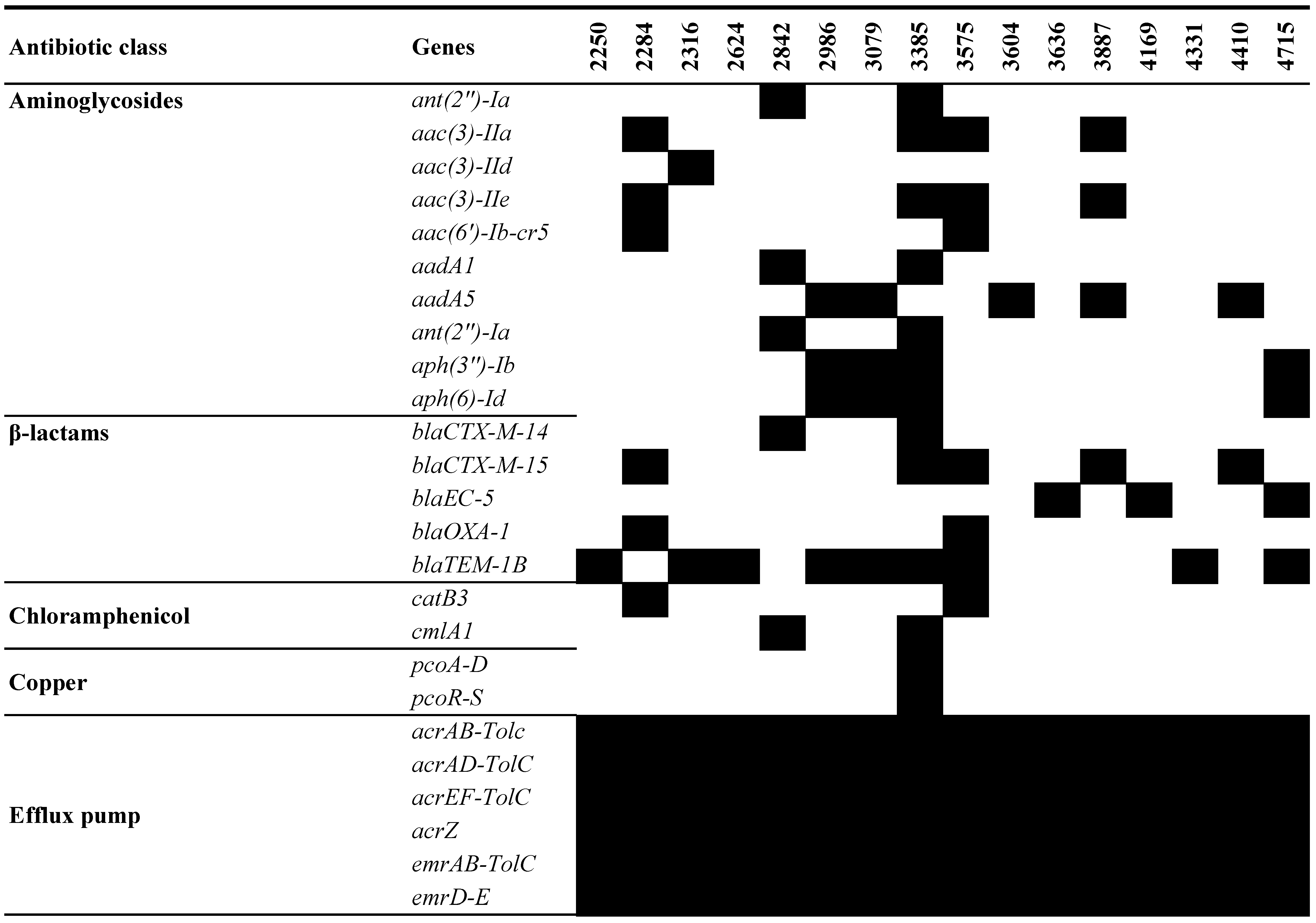
WGS revealed a concerning prevalence of resistance genes in the UPEC isolates. Notably, a significant portion (11 of 16) harbored genes for resistance against at least one aminoglycoside (Figure 2). This resistance was particularly pronounced in seven isolates, which carried combinations more than three aminoglycoside resistance genes. Isolate UPEC 3385 stood out with the highest number of these resistance genes. However, some isolates defied this trend. Five (2250, 2624, 3636, 4169, and 4331) lacked detectable genes for any of the tested aminoglycosides.
All UPEC isolates, except 3604, carried at least one β-lactam resistance gene. The most concerning examples were 3385 and 3575, which displayed a combination of three different β-lactam resistance genes: blaCTX-M-14, blaCTX-M-15, and blaOXA-1, alongside blaTEM-1B. Additionally, two chloramphenicol-resistant genes, catB3 and clmA1, were identified in two UPEC isolates.
The mph(A) gene, conferring low-level macrolide resistance, was found in eight isolates, while six isolates carried the quaternary ammonium compound-resistant gene, qacEΔ1. The hydrogen peroxide-resistant gene, sitABCD, was nearly ubiquitous among all isolates. Isolate 3385 harbored both copper-resistance genes, pcoA-D and pcoR-S, as well as silver-resistance genes, silA-C, silE, silP, and silR-S. Mercury-resistance genes, including merA, merC-E, merP, merR, and merT, were found in three isolates.
Many efflux pump genes were present in all UPEC isolates. Isolate 3385 was found to contain the qnrB1 gene, a plasmid-mediated gene associated with low-level resistance to quinolones. Additionally, tetracycline resistance genes tet(A) and tet(B) were identified in four and three isolates, respectively. Among the UPEC isolates, seven carried dihydrofolate reductase genes dfrA, including variants dfrA14 and dfrA17, encoding enzymes conferring trimethoprim resistance. Furthermore, seven isolates harbored sul1 genes, while four contained sul2 genes, both of which encode sulfonamide resistance.
Thirteen isolates had amino acid substitution in PmrB: 123 (Glu → Asp) associated with chromosome-mediated colistin resistance. Point mutations related to fosfomycin resistance were detected in CyaA: 352 (Ser → Thr), GlpT: 448 (Glu → Lys), PtsI: 25 (Val → Ile), and UhpT: 350 (Glu → Gln), with varying frequencies among the isolates. Moreover, amino acid substitutions associated with quinolones were observed in GyrA, ParC, ParE, and MarR among all isolates, with specific mutations such as Ser83 → Leu and Asp87 → Asn in GyrA and Ser80 → Ile in ParC universally present.

3.5. Analysis of MGEs, Insertion Elements (ISs), Phages, and CRISPR-Cas Systems

WGS unveiled a variety of plasmid replicon types among the UPEC isolates (Table 4). Col(BS512) and IncFIA replicon types were the most frequently identified, being present in all isolates. Each UPEC isolate harbored at least four different replicon types, indicating the prevalence of multiple plasmids within these strains. Additionally, Col156 and IncFIB replicon types were found in 15 isolates, highlighting their common occurrence across the UPEC strains. Conversely, replicon types IncP1, IncX1, IncI1-I(α), and IncQ1 were each identified in only one isolate.
An analysis of transposons revealed several types circulating within the UPEC isolates, with Tn3 and Tn5403 being the most abundant. The presence of additional transposons, such as Tn20 and Tn3000, emphasized the dynamic nature of the UPEC genome and the potential for the mobilization of genetic material. All UPEC isolates harbored MITEEc1, a miniature inverted transposable element belonging to the IS630 family, and an enterobacterial repetitive intergenic consensus (ERIC) sequence, potentially associated with mRNA stability [30].
MGEs in the UPEC isolates carried a formidable arsenal of antibiotic resistance genes. This included genes for resistance against aminoglycosides, (aac(3)-IIa, aadA1, aadA5, ant(2″)-Ia, ant(3″)-Ia, aph(3″)-Ib, and aph(6)-Id), β-lactams, (blaCTX-M-14, blaCTX-M-15, and blaTEM-1B), chloramphenicol (cmlA1), macrolides (mph(A)), quaternary ammonium compounds (qacEΔ1), quinolones (qnrB1), tetracyclines, (tet(A) and tet(B)), and trimethoprim and sulfonamides (dfrA14, dfrA17, sul1, and sul2).
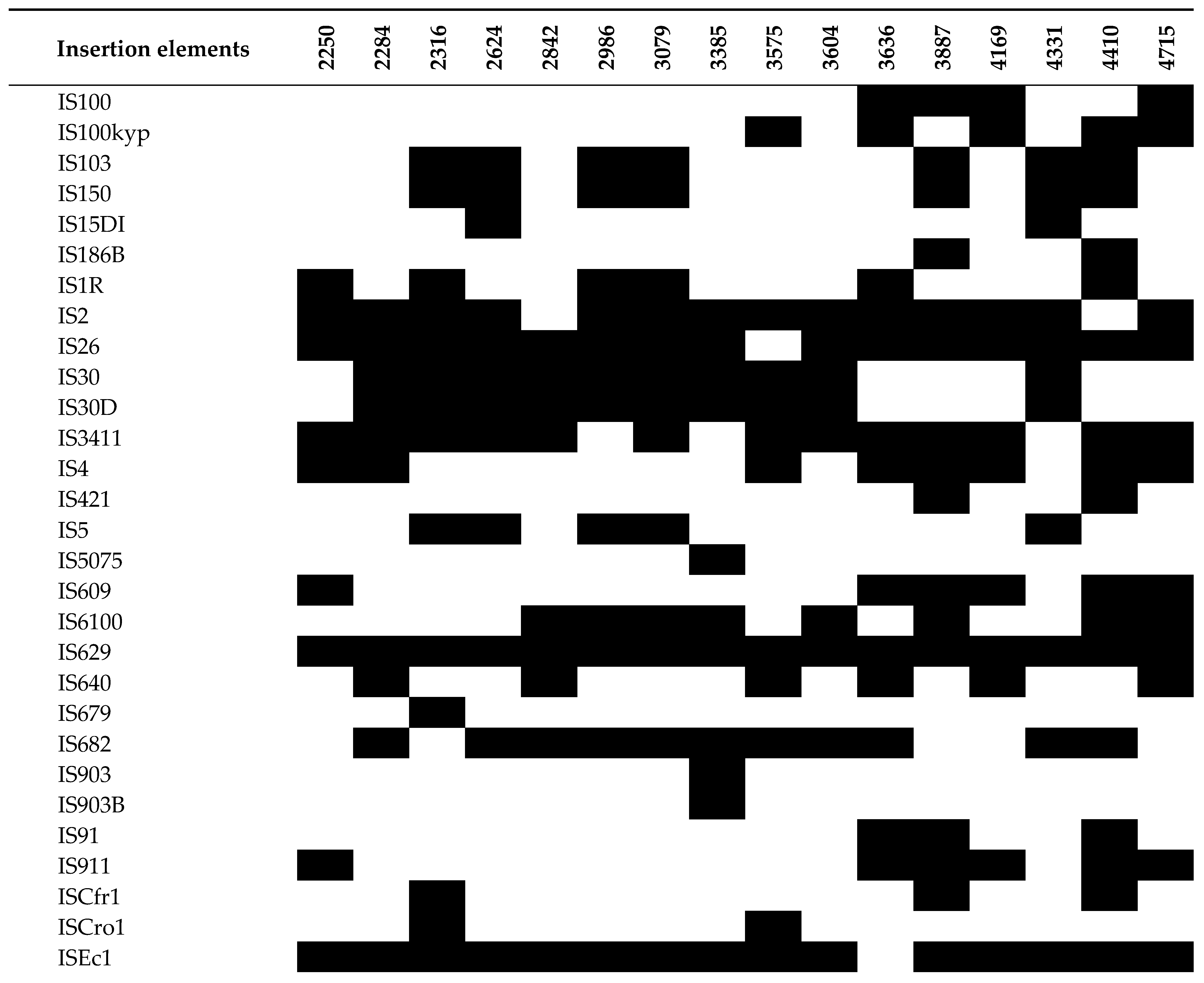
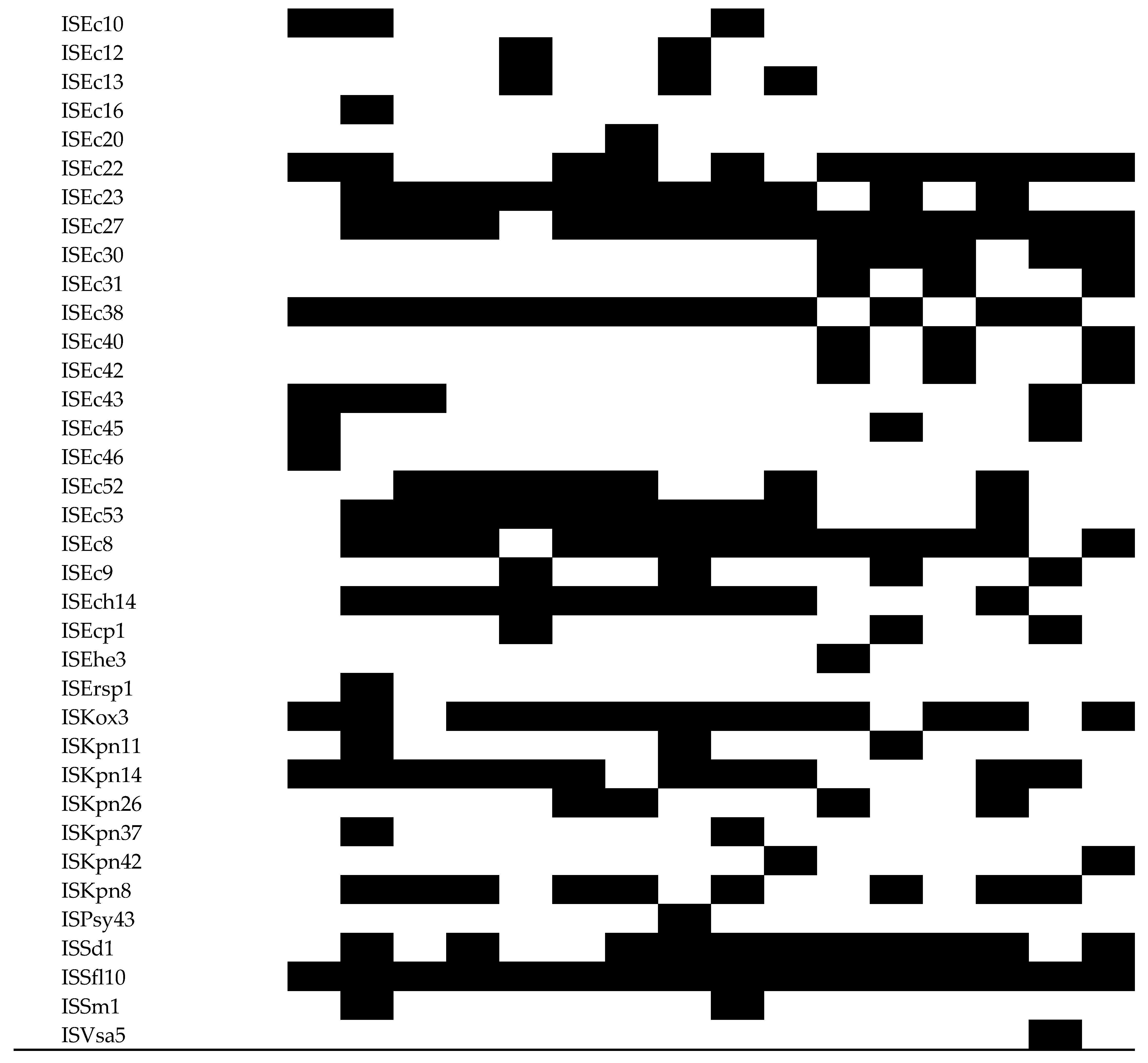
Whole-genome sequencing revealed a surprising abundance and diversity of IS elements within the UPEC isolates (Figure 3). Sixty-five ISs were identified in this study. All isolates harbored a remarkable number, exceeding 18 distinct IS elements each, suggesting a dynamic interplay of MGEs within these strains. Three isolates, 2284, 3887, and 4410, stood out with the most complex profiles, containing a staggering 29 ISs apiece. IS629 and ISSfl10 were remarkably consistent, having been identified in all UPEC isolates. Additionally, IS26 and ISEc1 were found in 15 isolates, followed closely by IS2 and ISEc27, present in 14 isolates.
Phages may bolster antibiotic resistance, environmental resilience, and host-cell adhesion, potentially enhancing pathogenicity. Upon genome analysis for phages, we observed that each UPEC isolate contained a minimum of five phages, with isolate 3575 exhibiting the highest count of 16 phages (Table S2). Shiga toxin 1-converting bacteriophage BP-4795 (PHAGE_Entero_BP_4795_NC_004813) emerged as the most prevalent, identified in 14 isolates. Following BP-4795 in prevalence was Escherichia phage P88 (PHAGE_Entero_P88_NC_026014), found in 13 isolates. Escherichia phage DE3 (PHAGE_Entero_DE3_NC_042057) was detected in 12 isolates, while Pectobacterium phage ZF40 (PHAGE_Pectob_ZF40_NC_019522) was present in 10 isolates. Several phages capable of carrying Shiga toxins (stxA1 and stxB1), including Shiga toxin 1-converting bacteriophage BP-4795 (PHAGE_Entero_BP_4795_NC_004813), Escherichia phage SH2026Stx1 (PHAGE_Escher_SH2026Stx1_NC_049919), Stx2-converting phage Stx2a_F451 (PHAGE_Stx2_c_Stx2a_F451_NC_049924), Enterobacteria phage VT2phi_272 (PHAGE_Entero_VT2phi_272_NC_028656), Enterobacteria phage YYZ-2008 (PHAGE_Entero_YYZ_2008_NC_011356), and Enterobacteria phage phiP27 (PHAGE_Entero_phiP27_NC_003356) were detected.
In addition to Escherichia phages, two Shigella phages capable of carrying Shiga toxins, Shigella phage 75/02 Stx (PHAGE_Shigel_Stx_NC_029120) and Shigella phage POCJ13 (PHAGE_Shigel_POCJ13_NC_025434), were identified. Furthermore, a phage capable of carrying cytolethal distending toxins, Enterobacteria phage fiAA91-ss (PHAGE_Entero_fiAA91_ss_NC_022750), was detected. Phages from Salmonella, Bacillus, Burkholderia, and Pseudomonas were also identified.
Bacteria possess a remarkable immune system, known as “clustered regularly interspaced short palindromic repeats-CRISPR associated” (CRISPR-Cas). This system acts like a shield, protecting bacteria from foreign genetic elements like viruses and plasmids. CRISPR loci were examined in 16 UPEC isolates, revealing a total of 57 confirmed loci (Table 5). Notably, isolate 4410 showed a robust defense mechanism, harboring seven CRISPR loci, while others had at least two. The spacer sequences within each CRISPR locus ranged from 1 to 11, with repeat lengths spanning 23 to 54 bp. The average size of spacers varied between 32 and 60.5 bp, while the overall CRISPR length ranged from 82 to 699 bp. I-E was the most prevalent CRISPR-Cas system subtype observed in the UPEC strains.
Across the strains, 21 distinct consensus-repeat sequences were identified. Notably, the most common sequences we encountered, CGACCCCCACCATGTCAAGGTGGTGCTCTAACCAACTGAGCTA and AGTTCACTGCCGTACAGGCAGCT, were present in nine and seven isolates, respectively. In the CRISPR-Cas system, the cas1 and cas2 genes are crucial components located proximal to the CRISPR locus, essential for its functionality. Hence, their presence was investigated across all CRISPR loci. The analysis revealed that only three UPEC strains—2250, 3887, and 4410—contained both core cas genes, while the remaining 13 strains lacked these genes. Subsequently, the cas genes present in these three strains were further characterized (Figure 4). Additionally, CRISPR-related proteins near CRISPR included endonuclease Cas1, integrase Cas2, helicase Cas3, and proteins Cse1, Cse2, Cas7, Cas5, and Cas6.
All UPEC isolates harbored at least two GIs (Table S3). Notably, isolates 4410 and 4715 exhibited the highest count, each harboring 13 genomic islands. Interestingly, none of the isolates carried genes for antibiotic resistance. However, except for isolates 2842, 3079, and 3887, all isolates possessed virulence genes. These genes were associated with various virulence factors, including type I fimbriae (fimA-C, fimE, and fimI), E. coli common pilus (ECP) operon (ecpA-E and ecpR), curli biogenesis (csgA-G), capsule biogenesis (kpsC-F, kpsS-T, and kpsU), type III secretion system (espL1 and spaP-Q), and hemolysin toxins (hlyA-B and hylC-D).
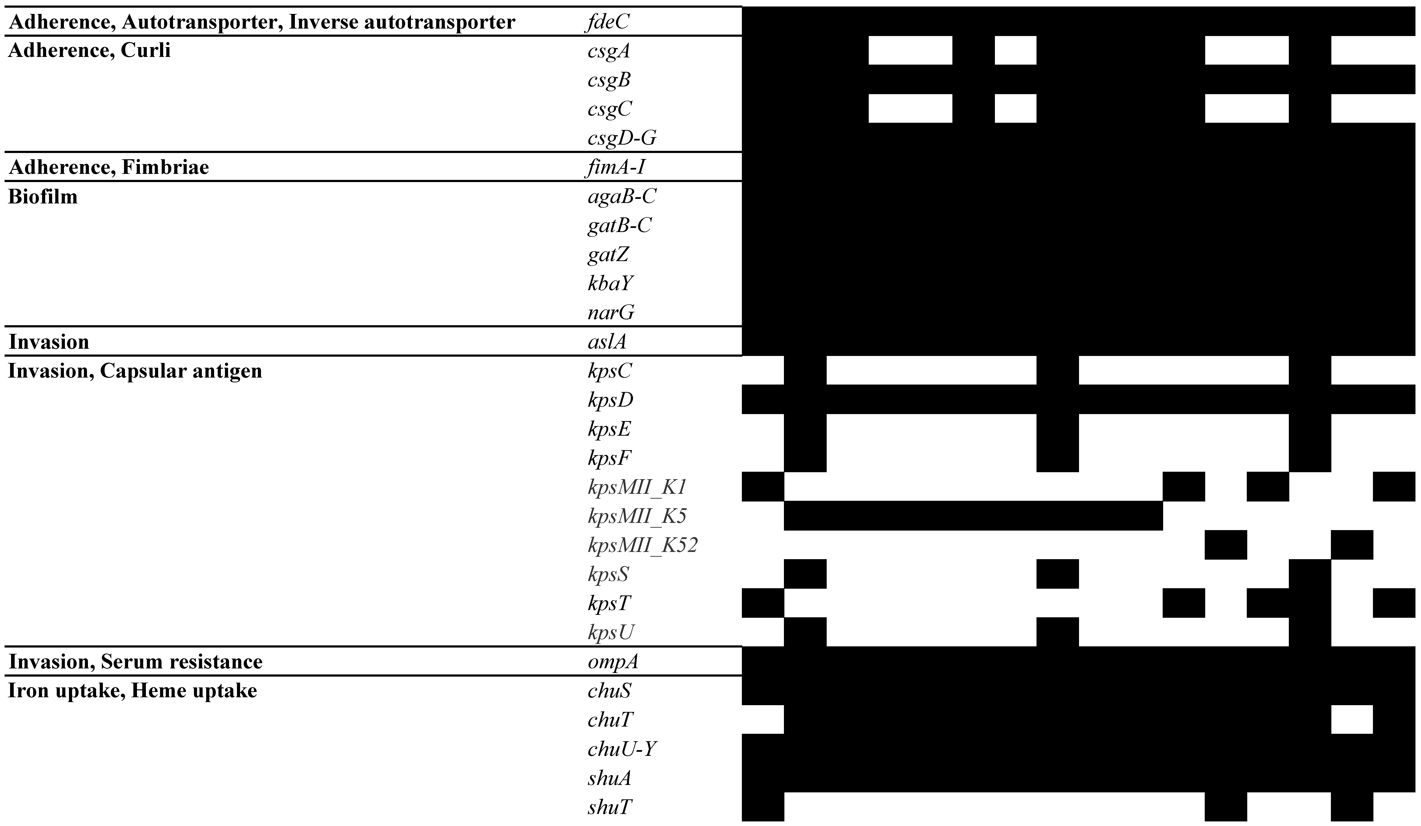
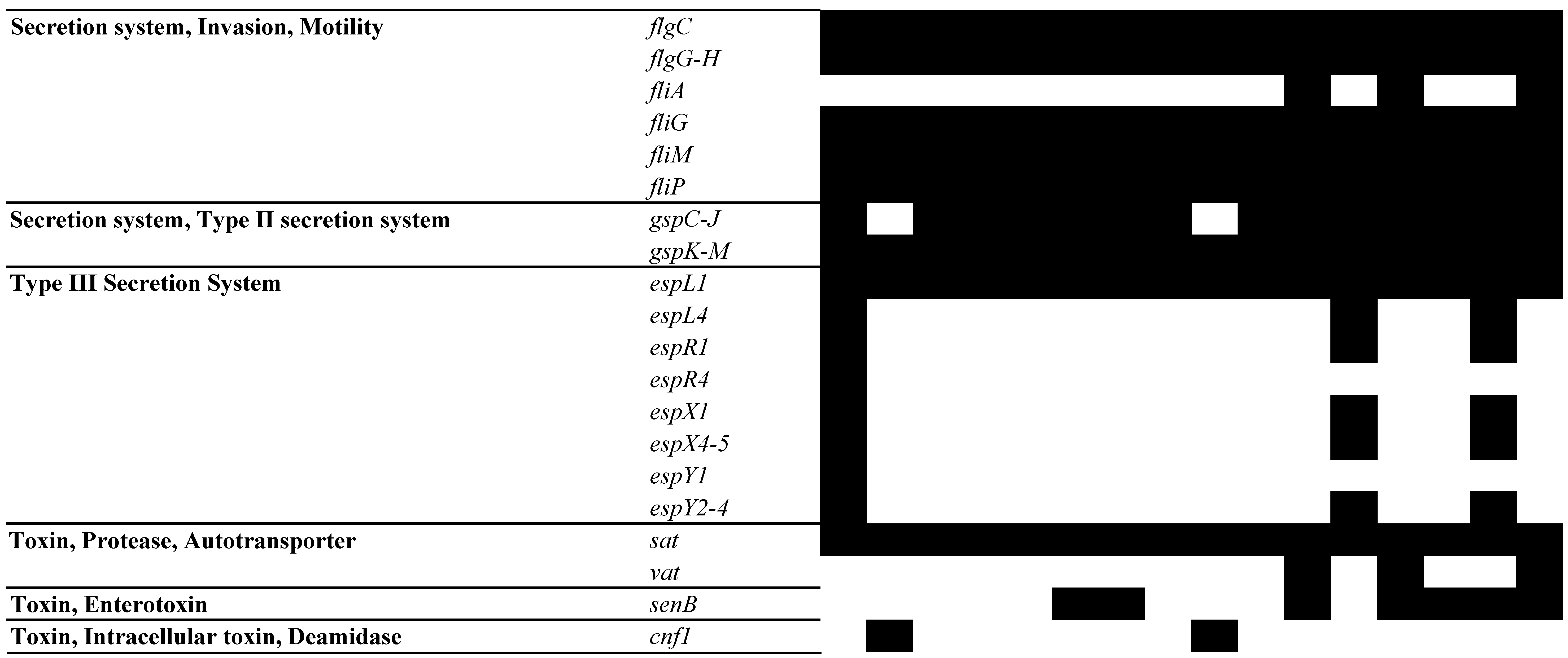
The UPEC isolates displayed a wide array of virulence genes implicated in adherence, invasion, iron uptake, secretion systems, and toxin production (Figure 5). Notably, isolate 2250 exhibited the highest number of virulence genes, followed by 4410 and 2284. Adherence-associated genes were ubiquitous among all UPEC isolates, including those encoding type 1 fimbriae (fimA-C, fimE, and fimI), the E. coli common pilus (ECP) operon (ecpA-E and ecpR), and genes implicated in biofilm formation. However, we observed some variation in the presence of genes for curli biogenesis (csgA-G) and type P fimbriae (papA-K and papX). An analysis of UPEC isolates revealed the presence of several virulence factors associated with invasion. All isolates harbored genes for aslA, ompA, and kpsD. Capsular antigens belonging to group 2 (K1, K5, and K52) were also identified. Interestingly, isolate 4410 stood out in possessing the gene invH, which encodes an invasion protein that plays a critical role in breaching host-cell defenses.
We found a remarkably high prevalence of 36 genes associated with iron acquisition. In fact, all isolates possessed every one of these genes except for three: chuT, shuT, and ybtE. Also, we identified seven genes (flgC, flgG-H, fliA, fliG, fliM, and fliP) essential for flagella production, with all genes present in every UPEC isolate except for fliA. An analysis of secretion system genes, involved in transporting various molecules in and out of bacteria, revealed contrasting distribution patterns. Genes associated with the Type II secretion system (gspC-J, gspK-M) were found in nearly all isolates. Conversely, genes linked to the Type III secretion system specifically linked to the locus of enterocyte effacement (LEE) (espL4, espR1, espR4, espX1, espX4-5, espY1, and espY2-4) were identified exclusively in three isolates (2250, 3887, and 4410). While a secreted autotransporter toxin (sat) was present in all UPEC isolates, genes encoding other toxins, including vacuolating autotransporter toxin (vat), enterotoxin (senB), cytotoxic necrotizing factor 1 (cnf1), and hemolysins (hlyA-D, hlyE), exhibited a more sporadic distribution pattern.

4. Discussion

This comprehensive analysis of UPEC isolates using WGS paints a concerning picture of MDR and potentially virulent bacteria. MLST, serotype, and CH typing analyses revealed four distinct groups within the UPEC isolates studied. Notably, the most prevalent types belonged to ST131, O25:H4, and fumC40/fimH30. The predominance of ST131 among the UPEC isolates aligns with previous reports, as it is recognized as the most prevalent E. coli lineage associated with extraintestinal pathogenic E. coli (ExPEC) infections worldwide [31]. This clonal complex is particularly problematic due to its association with both virulence and antimicrobial resistance. ST131 is an MDR clone commonly associated with resistance to fluoroquinolones, extended-spectrum β-lactams, and carbapenems [32]. Beyond its resistance profile, ST131 is considered a particularly virulent pathogen due to its extensive arsenal of virulence-associated genes, such as papG (P fimbrial adhesins), fimH (type 1 fimbriae), kpsM II (group 2 capsule synthesis), iucD (aerobactin), iutA (aerobactin receptor), fyuA (siderophore yersiniabactin receptor), sat (secreted autotransporter toxin), hlyA (α-hemolysin), and cnf1 (cytotoxic necrotizing factor 1) [33]. These genes encode factors that enhance bacteria’s ability to adhere to host cells, evade the immune system, and cause tissue damage.
The CH typing analysis of our UPEC isolates revealed variability in both fumC and fimh alleles, highlighting genetic diversity within the strains. The most common fumC allele was fumC40, found in the majority of isolates (10 out of 16). For fimh, fimh30 was the most prevalent allele, identified in 10 isolates, while fimh64 and fimh29 were present in fewer isolates. The variability in fimh alleles, alongside fumC typing, underscores the importance of these genes in UPEC-strain differentiation and their role in pathogenicity. The identification of these specific alleles correlates with known UPEC clonal groups, such as ST131, which often harbor fimh30 [34]. The diversity in fimh and fumC typing among our isolates provides insights into their potential virulence.
This study also delved into the mechanisms underpinning the reduced susceptibility of UPEC isolates to fluoroquinolones. By employing PAβN, we uncovered the involvement of efflux pumps in diminishing ciprofloxacin susceptibility in a subset of UPEC strains. Nine isolates exhibited a two-to-four-fold decrease in ciprofloxacin MIC when co-treated with PAβN. Furthermore, our analysis identified multiple-point mutations in gyrA, parC, parE, and marR genes, which likely serve as the primary mechanism driving ciprofloxacin resistance in the UPEC isolates. Intriguingly, we also noted the overexpression of efflux pump genes acrA-B, mdfA, norE, and ompF across the isolates. This observation suggests that efflux pump overexpression acts synergistically with mutations in gyrase and topoisomerase genes, further contributing to the reduced susceptibility of UPEC strains to ciprofloxacin.
The WGS of UPEC isolates revealed a concerning prevalence of key antibiotic resistance genes, particularly blaCTX-M-14, blaCTX-M-15, blaOXA-1, and blaTEM-1B. These genes encode resistance to extended-spectrum cephalosporins, traditionally used to treat UTIs. This finding aligns with observations by Ruppe et al., who reported similar β-lactamase genes, (blaCTX-M-15, blaOXA-1, and blaTEM-1B), in E. coli isolated from community-acquired UTIs in Cambodia [35]. This extensive β-lactam resistance significantly limits the treatment options available for UTIs caused by these specific strains.
This study also identified resistance to trimethoprim-sulfamethoxazole (TMP-SMX) among the analyzed UPEC isolates. SMX is a commonly used antibiotic for treating UTIs, and this finding highlights a significant threat to this critical therapy’s effectiveness. WGS revealed two key resistance mechanisms targeting SMX. First, seven isolates harbored dihydrofolate reductase genes (dfrA), specifically variants dfrA14 and dfrA17. Second, seven isolates contained sul1 genes, while four contained sul2 genes. Additionally, recent investigations have confirmed the presence of dfrA1 and sul1 in UPEC isolates [36,37], providing further evidence of the widespread dissemination of these resistance genes and the resulting threat to the efficacy of SMX in UTI treatment.
The widespread presence of efflux pump genes in all UPEC isolates underscores their significance in mediating multidrug resistance. The diverse repertoire of these genes identified in our UPEC isolates suggests a multifaceted mechanism through which these bacteria evade the effects of antibiotics. Among the efflux pump genes identified, acrAB-tolC, acrAD-tolC, and acrEF-tolC are members of the resistance–nodulation–cell division family of efflux pumps, known for their broad substrate specificity and ability to extrude a wide range of antibiotics, including β-lactams, fluoroquinolones, and tetracyclines [38]. Additionally, mdtABC-tolC, mdtEF-tolC, and mdtM belong to the multidrug and toxic-compound extrusion family of efflux pumps, involved in the extrusion of various antimicrobial compounds [39]. The presence of such efflux pump genes as mexAB-oprM is particularly noteworthy due to their role in conferring resistance to multiple classes of antibiotics, including β-lactams, fluoroquinolones, and macrolides [40]. These efflux pumps contribute to UPEC’s adaptive response to antibiotic exposure and can confer resistance to a broad array of antibiotics.
Some isolates displayed resistance mechanisms against several heavy metals. Notably, isolate 3385 emerged as a standout case, harboring genes for resistance to both copper (pcoA-D and pcoR-S) and silver (silA-C, silE, silP, and silR-S). Additionally, three isolates harbored genes for mercury resistance (merA, merC-E, merP, merR, and merT). The identification of resistance genes targeting heavy metals raises concerns about the potential for UPEC strains to persist in environments with these agents. Copper and silver have antimicrobial properties and are sometimes incorporated into medical devices or wound dressings [41]. The presence of resistance mechanisms against these metals could potentially limit the effectiveness of these strategies for preventing or controlling UPEC infections. Moreover, the co-occurrence of metal resistance genes with antibiotic resistance genes highlights the potential for co-selection, wherein exposure to one type of stressor may inadvertently promote the emergence or dissemination of antibiotic resistance.
Our exploration of plasmid replicon types among the UPEC isolates unveiled a diverse range of replicons, indicating the likely presence of multiple plasmids per isolate. The most prevalent replicon types, Col(BS512) and IncFIA, were consistently detected across all isolates. IncFIA is particularly significant, given its association with conjugative plasmids capable of transferring antibiotic resistance genes among bacteria [42]. Notably, IncF plasmids identified in UPEC ST405 have been implicated in disseminating various antibiotic resistance determinants, including ESBLs and quinolone resistance genes, thereby contributing to the global spread of antibiotic resistance [43].
The presence of Col(BS512) is noteworthy. Known as the Shigella boydii plasmid pBS512 with replicon type FIIA, it is categorized as an invasive plasmid with relation to the type III secretion system [44]. Additionally, the identification of replicons such as IncP1, IncX1, IncI1-I(α), and IncQ1, each found in only one isolate, presents an intriguing aspect. These replicons are less commonly encountered in UPEC, implying potential acquisition events from diverse sources or the presence of unique genetic elements not typically associated with UPEC plasmids.
To our knowledge, this study represents the first comprehensive exploration of phage diversity in UPEC. Our analysis uncovered a wide repertoire of bacteriophages among UPEC isolates, suggesting the potential for phage-mediated genetic exchange and dissemination of virulence factors within these strains. Notably, Shiga toxin 1-converting bacteriophage BP-4795 emerged as the most prevalent, detected in 14 isolates. This phage is known to encode a type III effector that can be translocated into human cells through the locus of an enterocyte effacement-encoded type III secretion system [45]. We identified several phages capable of carrying Shiga toxins, including Shiga toxin 1-converting bacteriophage BP-4795, Escherichia phage SH2026Stx1, Stx2-converting phage Stx2a_F451, Enterobacteria phage VT2phi_272, Enterobacteria phage YYZ-2008, and Enterobacteria phage phiP27. The presence of these phages in UPEC isolates suggests a potential risk factor for the emergence of highly virulent strains capable of causing severe complications. Additionally, the detection of Enterobacteria phage fiAA91-ss, a carrier of cytolethal distending toxin genes, further underscores the complexity of virulence potential encoded via these phages.
The identification of CRISPR-Cas systems in our UPEC isolates sheds light on the potential importance of this adaptive immune system in protecting against foreign genetic elements. We observed variability in spacer-sequence length and composition within CRISPR loci, highlighting diversity among UPEC strains. Our findings on CRISPR-Cas subtype prevalence aligned with previous reports, with the type I-E system being the most dominant [46]. Typically, strains with active CRISPR-Cas systems would be expected to have fewer virulence genes [47]. Lindenstrauss et al. found an inverse correlation between the presence of CRISPR-Cas systems and acquired virulence determinants in Enterococcus faecalis [48]. However, our study presents a contrasting scenario. We identified three UPEC strains (2250, 3887, and 4410) harboring functional type I-E CRISPR-Cas systems, yet these strains also carried a higher number of virulence genes compared to other isolates. This intriguing finding suggests that, in these specific UPEC strains, the presence of type I-E CRISPR-Cas systems might not be directly linked to the acquisition of virulence genes. Adding another layer of complexity, Dang et al. [49] observed that UPEC was less likely than non-pathogenic strains to possess CRISPR loci. This suggests a potential role for CRISPR in acquiring MGEs of E. coli, which may not always translate to virulence genes. These contrasting observations highlight the intricate relationship between CRISPR-Cas systems and virulence factors in UPEC.
The detection of GIs in our UPEC isolates provides significant insights into the genomic architecture and potential virulence of these strains. Our study revealed that all UPEC isolates harbored at least two GIs, with isolates 4410 and 4715 each containing the highest number, 13 GIs. Interestingly, despite the extensive presence of GIs, none of the isolates carried genes for antibiotic resistance within these regions. This finding contrasts with some reports in which GIs have been shown to frequently harbor antibiotic resistance genes, contributing to the MDR phenotype in pathogenic E. coli [50]. The absence of such genes in our isolates’ GIs might indicate a different evolutionary path or selective pressure acting on these UPEC strains, focusing more on virulence than on antibiotic resistance. Except for isolates 2842, 3079, and 3887, all isolates possessed virulence genes within their GIs. These genes were associated with various virulence factors, such as adhesion, biofilm formation, type III secretion system, and hemolysin production; such findings are consistent with GIs’ known role in harboring virulence determinants that enhance E. coli’s pathogenicity [51].
Our study underscores the critical role of adherence-associated virulence genes in UPEC pathogenesis. These genes orchestrate the initial attachment of bacteria to host tissues and subsequent biofilm formation, establishing and perpetuating UTIs [52]. Among the identified genes, those encoding type 1 fimbriae (fimA-C, fimE, and fimI) and the E. coli common pilus operon (ecpA-E and ecpR) were universally present in all UPEC isolates. Type 1 fimbriae are well-established facilitators of UPEC adherence to bladder epithelial cells, promoting colonization and infection [52]. Similarly, the E. coli common pilus operon enhances UPEC’s adhesive and invasive capabilities during interaction with the host bladder [53]. This ubiquity aligns with other reports, including those by Mirzahosseini et al., who identified fimA, fimH, and ecpA as the most common virulence genes in UPEC isolates worldwide [54]. Interestingly, some variability was observed in the presence of genes associated with curli biogenesis (csgA-G) and type P fimbriae (papA-K and papX). Curli are functional amyloid fibers that contribute to biofilm formation and host colonization, while type P fimbriae are involved in adhesion to specific receptors on host cells [52]. This variability in adherence-associated genes highlights the potential for diverse virulence strategies among UPEC isolates.
Beyond the initial attachment to host cells, some UPEC strains possess additional virulence factors that enable them to invade deeper tissues, further complicating UTIs. All isolates were found to harbor the genes ompA and aslA, crucial for bacterial invasion and survival within host tissues. Furthermore, all isolates harbored capsular antigens belonging to group 2, specifically K1, K5, and K52. These capsular antigens provide resistance to phagocytosis via immune cells and serum bactericidal activity, facilitating the establishment and persistence of UTIs [55]. Of particular interest is isolate 4410, which notably possessed the gene invH. This gene is associated with the ability of bacteria to invade epithelial cells, an essential step in the pathogenesis of invasive infections [56]. The presence of invH in this isolate suggests that isolate 4410 may have enhanced invasive capabilities compared to other isolates, potentially influencing its pathogenicity and clinical outcomes.
Iron is an essential nutrient for bacterial growth and virulence, particularly in the iron-limited environment of the urinary tract. A remarkable finding was the high prevalence of genes associated with iron acquisition—36—with all isolates possessing all but three (chuT, shuT, and ybtE). This finding is consistent with previous reports that highlight the importance of iron-acquisition systems in UPEC pathogenicity. Iron-acquisition systems, such as siderophores and heme-uptake mechanisms, allow UPEC to thrive in the host by scavenging iron from host proteins [57].
Flagella are essential for motility and colonization, contributing to UPEC’s ability to ascend the urinary tract and establish infection. Our study identified seven genes essential for flagella production (flgC, flgG-H, fliA, fliG, fliM, and fliP), with all genes being present in every UPEC isolate except for fliA. This gene profile resembles that in the work of Lane et al. [58], who reported that flagellar motility plays a significant role in UPEC pathogenesis. The presence of these genes in nearly all isolates suggests that motility is a key factor in the persistence and virulence of UPEC.
An analysis of secretion system genes revealed contrasting distribution patterns. Genes associated with the Type II secretion system (gspC-J and gspK-M) were found in nearly all isolates. The Type II secretion system is known to be involved in the secretion of a variety of enzymes and toxins that contribute to bacterial virulence [59]. In contrast, genes linked to the Type III secretion system, specifically those associated with the locus of enterocyte effacement (LEE) (espL4, espR1, espR4, espX1, espX4-5, espY1, and espY2-4), were identified exclusively in three isolates (2250, 3887, and 4410). The LEE-encoded Type III secretion system is typically associated with enteropathogenic and enterohemorrhagic E. coli strains, in which it plays a crucial role in delivering effector proteins into host cells, subverting host-cell signaling and immune responses [60].
The toxin genes among UPEC isolates vary significantly in their potential to cause host-tissue damage. Notably, the sat gene was present in all UPEC isolates. Encoding a serine protease autotransporter, it is one of the most frequent virulence genes found in UPEC [61]. In contrast, genes encoding other toxins, such as vat, senB, cnf1, hlyA-D, and hlyE, exhibited more sporadic distribution patterns. This finding aligns with observations by Kim et al., who identified similar toxins in E. coli isolates from hospitalized UTI patients [62]. Interestingly, we found sat to be universally present; Kim et al. reported the hlyE gene in all isolates. This variation in toxin gene profiles suggests that, while all UPEC isolates possess a baseline level of virulence potential due to the ubiquitous presence of sat, the additional virulence factors they harbor may be considerably diverse.
Earlier research has highlighted significant variability in the presence of virulence factors across UPEC isolates, with specific genes like eae, pap, and afa showing a lower prevalence in certain populations. For example, the eae gene was present in only one of 225 UPEC strains, suggesting that not all virulence factors common in other pathogenic E. coli strains are equally prevalent in UPEC [63]. Another study reported that none of their UPEC isolates contained the pap GI gene [64]. Blanco et al. further demonstrated the low prevalence of the afa gene among UPEC isolates. Their study detected afa in only a small subset of isolates from patients with cystitis (4.3%) and asymptomatic bacteriuria (2.4%), highlighting the variability in the presence of this adhesin gene among UPEC strains [65]. These findings underscore that the presence of virulence factors is not uniform across all UPEC isolates, and their distribution can vary significantly based on the genetic background and environmental pressures influencing each strain. This evidence supports the notion that, while certain virulence genes may be prevalent, others can show much lower frequencies, contributing to the observed diversity in virulence potential among UPEC isolates.

5. Conclusions

Our comprehensive analysis of UPEC isolates through WGS paints a concerning picture of MDR and potentially virulent UPEC strains. Widespread resistance to various antibiotics, including fluoroquinolones, extended-spectrum cephalosporins, and TMP-SMX, poses a significant challenge in treating UTIs. The diverse mechanisms of resistance, including efflux pumps, mutations, and plasmid-mediated gene transfer, highlight the adaptability of these bacteria. Furthermore, the presence of virulence factors associated with adhesion, invasion, iron acquisition, and toxin production underscores the potential of these UPEC strains to cause severe UTIs. This study emphasizes the necessity of continued genomic surveillance in understanding UPEC’s pathogenic mechanisms and resistance profiles, thus aiding in the development of targeted therapeutic strategies.

Supplementary Materials

The following supporting information can be downloaded at: https://www.mdpi.com/article/10.3390/pathogens13090794/s1, Table S1: Sequences of oligonucleotide used in this study; Table S2: Predicted phages present in the UPEC isolates. *The score of the region based on the following criteria. Intact (score > 90), Questionable (score 70-90), Incomplete (score < 70); Table S3: Genomic islands and virulence genes in the UPEC isolates.

Author Contributions

Conceptualization, K.S.; Methodology, K.S., J.C. and S.A.K.; Validation, K.S.; Formal analysis, K.S.; Investigation, K.S. and M.P.; Resources, K.S., K.A. and A.A.K.; Data curation, K.S.; Writing—original draft, K.S.; Writing—review & editing, K.S., M.N., M.P., J.C., K.A. and A.A.K.; Supervision, K.S.; Project administration, K.S. and M.N.; Funding acquisition, K.S. All authors have read and agreed to the published version of the manuscript.

Funding

This study was funded by protocol number E07662.01 from the U.S. Food and Drug Administration.

Institutional Review Board Statement

Not applicable.

Informed Consent Statement

Not applicable.

Data Availability Statement

The original contributions presented in the study are included in the article/supplementary material, further inquiries can be directed to the corresponding author.

Acknowledgments

We acknowledge Huizhong Chen and Seong-Jae Kim for reviewing the manuscript and Joanne Berger of the US Food and Drug Administration (FDA) Library for editing a draft of the manuscript. We also extend our appreciation to James Johnson and Brian Johnston of the Minneapolis VA Healthcare System, Minneapolis, Minnesota, USA, for providing the bacterial strain used in this study. This study was funded by the US Food and Drug Administration (FDA).

Conflicts of Interest

The authors declare no conflict of interest.

References

  1. Schappert, S.M.; Rechtsteiner, E.A. Ambulatory medical care utilization estimates for 2007. Vital Health Stat. 13 2011, 169, 1–38. [Google Scholar]
  2. Kahlmeter, G. An international survey of the antimicrobial susceptibility of pathogens from uncomplicated urinary tract infections: The ECO.SENS Project. J. Antimicrob. Chemother. 2003, 51, 69–76. [Google Scholar] [CrossRef] [PubMed]
  3. Foxman, B.; Brown, P. Epidemiology of urinary tract infections: Transmission and risk factors, incidence, and costs. Infect. Dis. Clin. N. Am. 2003, 17, 227–241. [Google Scholar] [CrossRef] [PubMed]
  4. Gupta, K.; Hooton, T.M.; Naber, K.G.; Wullt, B.; Colgan, R.; Miller, L.G.; Moran, G.J.; Nicolle, L.E.; Raz, R.; Schaeffer, A.J.; et al. International clinical practice guidelines for the treatment of acute uncomplicated cystitis and pyelonephritis in women: A 2010 update by the Infectious Diseases Society of America and the European Society for Microbiology and Infectious Diseases. Clin. Infect. Dis. 2011, 52, e103–e120. [Google Scholar] [CrossRef]
  5. Prevention, C.F.D.C.A. Multidrug-resistant E. coli. Available online: https://arpsp.cdc.gov/profile/antibiotic-resistance/mdr-ecoli (accessed on 15 April 2024).
  6. Asadi Karam, M.R.; Habibi, M.; Bouzari, S. Urinary tract infection: Pathogenicity, antibiotic resistance and development of effective vaccines against Uropathogenic Escherichia coli. Mol. Immunol. 2019, 108, 56–67. [Google Scholar] [CrossRef] [PubMed]
  7. Mazzariol, A.; Bazaj, A.; Cornaglia, G. Multi-drug-resistant Gram-negative bacteria causing urinary tract infections: A review. J. Chemother. 2017, 29, 2–9. [Google Scholar] [CrossRef]
  8. Flores-Mireles, A.L.; Walker, J.N.; Caparon, M.; Hultgren, S.J. Urinary tract infections: Epidemiology, mechanisms of infection and treatment options. Nat. Rev. Microbiol. 2015, 13, 269–284. [Google Scholar] [CrossRef]
  9. Oelschlaeger, T.A.; Dobrindt, U.; Hacker, J. Virulence factors of uropathogens. Curr. Opin. Urol. 2002, 12, 33–38. [Google Scholar] [CrossRef]
  10. Bhandari, M.; Poelstra, J.W.; Kauffman, M.; Varghese, B.; Helmy, Y.A.; Scaria, J.; Rajashekara, G. Genomic Diversity, Antimicrobial Resistance, Plasmidome, and Virulence Profiles of Salmonella Isolated from Small Specialty Crop Farms Revealed by Whole-Genome Sequencing. Antibiotics 2023, 12, 1637. [Google Scholar] [CrossRef]
  11. Rizk, A.M.; Elsayed, M.M.; Abd El Tawab, A.A.; Elhofy, F.I.; Soliman, E.A.; Kozytska, T.; Brangsch, H.; Sprague, L.D.; Neubauer, H.; Wareth, G. Phenotypic and genotypic characterization of resistance and virulence in Pseudomonas aeruginosa isolated from poultry farms in Egypt using whole genome sequencing. Vet. Microbiol. 2024, 292, 110063. [Google Scholar] [CrossRef]
  12. Zouharova, M.; Matiasovic, J.; Gebauer, J.; Matiaskova, K.; Nedbalcova, K. Survey of Genotype Diversity, Virulence, and Antimicrobial Resistance Genes in Mastitis-Causing Streptococcus uberis in Dairy Herds Using Whole-Genome Sequencing. Pathogens 2023, 12, 1378. [Google Scholar] [CrossRef] [PubMed]
  13. CLSI Standard M07; Methods for Dilution Antimicrobial Susceptibility Tests for Bacteria That Grow Aerobically, 11th ed. Clinical and Laboratory Standards Institute: Wayne, PA, USA, 2019.
  14. Alotaibi, K.; Khan, A.A.; Marasa, B.; Khan, S.A.; Johnson, J.; Johnston, B.; Sung, K.; Nawaz, M. Draft Genome Sequences of Sixteen Fluoroquinolone-Resistant Extraintestinal Escherichia coli Isolates from Human Patients. Microbiol. Resour. Announc. 2022, 11, e0000322. [Google Scholar] [CrossRef]
  15. Center for Genomic Epidemiology. Available online: https://www.genomicepidemiology.org/services/ (accessed on 20 February 2024).
  16. Letunic, I.; Bork, P. Interactive Tree of Life (iTOL) v5: An online tool for phylogenetic tree display and annotation. Nucleic Acids Res. 2021, 49, W293–W296. [Google Scholar] [CrossRef]
  17. Olson, R.D.; Assaf, R.; Brettin, T.; Conrad, N.; Cucinell, C.; Davis, J.J.; Dempsey, D.M.; Dickerman, A.; Dietrich, E.M.; Kenyon, R.W.; et al. Introducing the Bacterial and Viral Bioinformatics Resource Center (BV-BRC): A resource combining PATRIC, IRD and ViPR. Nucleic Acids Res. 2023, 51, D678–D689. [Google Scholar] [CrossRef]
  18. Antonopoulos, D.A.; Assaf, R.; Aziz, R.K.; Brettin, T.; Bun, C.; Conrad, N.; Davis, J.J.; Dietrich, E.M.; Disz, T.; Gerdes, S.; et al. PATRIC as a unique resource for studying antimicrobial resistance. Brief. Bioinform. 2019, 20, 1094–1102. [Google Scholar] [CrossRef]
  19. Zankari, E.; Hasman, H.; Cosentino, S.; Vestergaard, M.; Rasmussen, S.; Lund, O.; Aarestrup, F.M.; Larsen, M.V. Identification of acquired antimicrobial resistance genes. J. Antimicrob. Chemother. 2012, 67, 2640–2644. [Google Scholar] [CrossRef] [PubMed]
  20. McArthur, A.G.; Waglechner, N.; Nizam, F.; Yan, A.; Azad, M.A.; Baylay, A.J.; Bhullar, K.; Canova, M.J.; De Pascale, G.; Ejim, L.; et al. The comprehensive antibiotic resistance database. Antimicrob. Agents Chemother. 2013, 57, 3348–3357. [Google Scholar] [CrossRef]
  21. Bharat, A.; Petkau, A.; Avery, B.P.; Chen, J.C.; Folster, J.P.; Carson, C.A.; Kearney, A.; Nadon, C.; Mabon, P.; Thiessen, J.; et al. Correlation between Phenotypic and In Silico Detection of Antimicrobial Resistance in Salmonella enterica in Canada Using Staramr. Microorganisms 2022, 10, 292. [Google Scholar] [CrossRef] [PubMed]
  22. Wang, M.; Goh, Y.X.; Tai, C.; Wang, H.; Deng, Z.; Ou, H.Y. VRprofile2: Detection of antibiotic resistance-associated mobilome in bacterial pathogens. Nucleic Acids Res. 2022, 50, W768–W773. [Google Scholar] [CrossRef] [PubMed]
  23. Johansson, M.H.K.; Bortolaia, V.; Tansirichaiya, S.; Aarestrup, F.M.; Roberts, A.P.; Petersen, T.N. Detection of mobile genetic elements associated with antibiotic resistance in Salmonella enterica using a newly developed web tool: MobileElementFinder. J. Antimicrob. Chemother. 2021, 76, 101–109. [Google Scholar] [CrossRef]
  24. Brown, C.L.; Mullet, J.; Hindi, F.; Stoll, J.E.; Gupta, S.; Choi, M.; Keenum, I.; Vikesland, P.; Pruden, A.; Zhang, L. mobileOG-db: A Manually Curated Database of Protein Families Mediating the Life Cycle of Bacterial Mobile Genetic Elements. Appl. Environ. Microbiol. 2022, 88, e0099122. [Google Scholar] [CrossRef] [PubMed]
  25. Arndt, D.; Grant, J.R.; Marcu, A.; Sajed, T.; Pon, A.; Liang, Y.; Wishart, D.S. PHASTER: A better, faster version of the PHAST phage search tool. Nucleic Acids Res. 2016, 44, W16–W21. [Google Scholar] [CrossRef] [PubMed]
  26. Couvin, D.; Bernheim, A.; Toffano-Nioche, C.; Touchon, M.; Michalik, J.; Neron, B.; Rocha, E.P.C.; Vergnaud, G.; Gautheret, D.; Pourcel, C. CRISPRCasFinder, an update of CRISRFinder, includes a portable version, enhanced performance and integrates search for Cas proteins. Nucleic Acids Res. 2018, 46, W246–W251. [Google Scholar] [CrossRef]
  27. Wattam, A.R.; Davis, J.J.; Assaf, R.; Boisvert, S.; Brettin, T.; Bun, C.; Conrad, N.; Dietrich, E.M.; Disz, T.; Gabbard, J.L.; et al. Improvements to PATRIC, the all-bacterial Bioinformatics Database and Analysis Resource Center. Nucleic Acids Res. 2017, 45, D535–D542. [Google Scholar] [CrossRef] [PubMed]
  28. Chen, L.; Yang, J.; Yu, J.; Yao, Z.; Sun, L.; Shen, Y.; Jin, Q. VFDB: A reference database for bacterial virulence factors. Nucleic Acids Res. 2005, 33, D325–D328. [Google Scholar] [CrossRef] [PubMed]
  29. Sayers, S.; Li, L.; Ong, E.; Deng, S.; Fu, G.; Lin, Y.; Yang, B.; Zhang, S.; Fa, Z.; Zhao, B.; et al. Victors: A web-based knowledge base of virulence factors in human and animal pathogens. Nucleic Acids Res. 2019, 47, D693–D700. [Google Scholar] [CrossRef] [PubMed]
  30. Wilson, L.A.; Sharp, P.M. Enterobacterial repetitive intergenic consensus (ERIC) sequences in Escherichia coli: Evolution and implications for ERIC-PCR. Mol. Biol. Evol. 2006, 23, 1156–1168. [Google Scholar] [CrossRef]
  31. Manges, A.R.; Geum, H.M.; Guo, A.; Edens, T.J.; Fibke, C.D.; Pitout, J.D.D. Global Extraintestinal Pathogenic Escherichia coli (ExPEC) Lineages. Clin. Microbiol. Rev. 2019, 32, e00135-18. [Google Scholar] [CrossRef]
  32. Pitout, J.D.D.; Peirano, G.; Chen, L.; DeVinney, R.; Matsumura, Y. Escherichia coli ST1193: Following in the Footsteps of E. coli ST131. Antimicrob. Agents Chemother. 2022, 66, e0051122. [Google Scholar] [CrossRef]
  33. Nicolas-Chanoine, M.H.; Blanco, J.; Leflon-Guibout, V.; Demarty, R.; Alonso, M.P.; Canica, M.M.; Park, Y.J.; Lavigne, J.P.; Pitout, J.; Johnson, J.R. Intercontinental emergence of Escherichia coli clone O25:H4-ST131 producing CTX-M-15. J. Antimicrob. Chemother. 2008, 61, 273–281. [Google Scholar] [CrossRef]
  34. Kondratyeva, K.; Salmon-Divon, M.; Navon-Venezia, S. Meta-analysis of Pandemic Escherichia coli ST131 Plasmidome Proves Restricted Plasmid-clade Associations. Sci. Rep. 2020, 10, 36. [Google Scholar] [CrossRef] [PubMed]
  35. Ruppe, E.; Hem, S.; Lath, S.; Gautier, V.; Ariey, F.; Sarthou, J.L.; Monchy, D.; Arlet, G. CTX-M beta-lactamases in Escherichia coli from community-acquired urinary tract infections, Cambodia. Emerg. Infect. Dis. 2009, 15, 741–748. [Google Scholar] [CrossRef] [PubMed]
  36. Rahman, M.M.; Hossain, M.M.K.; Rubaya, R.; Halder, J.; Karim, M.E.; Bhuiya, A.A.; Khatun, A.; Alam, J. Association of Antibiotic Resistance Traits in Uropathogenic Escherichia coli (UPEC) Isolates. Can. J. Infect. Dis. Med. Microbiol. 2022, 2022, 4251486. [Google Scholar] [CrossRef]
  37. Somorin, Y.M.; Weir, N.M.; Pattison, S.H.; Crockard, M.A.; Hughes, C.M.; Tunney, M.M.; Gilpin, D.F. Antimicrobial resistance in urinary pathogens and culture-independent detection of trimethoprim resistance in urine from patients with urinary tract infection. BMC Microbiol. 2022, 22, 144. [Google Scholar] [CrossRef]
  38. Anes, J.; McCusker, M.P.; Fanning, S.; Martins, M. The ins and outs of RND efflux pumps in Escherichia coli. Front. Microbiol. 2015, 6, 587. [Google Scholar] [CrossRef] [PubMed]
  39. Alav, I.; Kobylka, J.; Kuth, M.S.; Pos, K.M.; Picard, M.; Blair, J.M.A.; Bavro, V.N. Structure, Assembly, and Function of Tripartite Efflux and Type 1 Secretion Systems in Gram-Negative Bacteria. Chem. Rev. 2021, 121, 5479–5596. [Google Scholar] [CrossRef]
  40. Lopez, C.A.; Travers, T.; Pos, K.M.; Zgurskaya, H.I.; Gnanakaran, S. Dynamics of Intact MexAB-OprM Efflux Pump: Focusing on the MexA-OprM Interface. Sci. Rep. 2017, 7, 16521. [Google Scholar] [CrossRef]
  41. Saint, S.; Veenstra, D.L.; Sullivan, S.D.; Chenoweth, C.; Fendrick, A.M. The potential clinical and economic benefits of silver alloy urinary catheters in preventing urinary tract infection. Arch. Intern. Med. 2000, 160, 2670–2675. [Google Scholar] [CrossRef]
  42. Carattoli, A. Plasmids and the spread of resistance. Int. J. Med. Microbiol. 2013, 303, 298–304. [Google Scholar] [CrossRef]
  43. Yao, M.; Zhu, Q.; Zou, J.; Shenkutie, A.M.; Hu, S.; Qu, J.; He, Z.; Leung, P.H.M. Genomic Characterization of a Uropathogenic Escherichia coli ST405 Isolate Harboring bla (CTX-M-15)-Encoding IncFIA-FIB Plasmid, bla (CTX-M-24)-Encoding IncI1 Plasmid, and Phage-Like Plasmid. Front. Microbiol. 2022, 13, 845045. [Google Scholar] [CrossRef]
  44. Johnson, T.J.; Nolan, L.K. Pathogenomics of the virulence plasmids of Escherichia coli. Microbiol. Mol. Biol. Rev. 2009, 73, 750–774. [Google Scholar] [CrossRef]
  45. Creuzburg, K.; Recktenwald, J.; Kuhle, V.; Herold, S.; Hensel, M.; Schmidt, H. The Shiga toxin 1-converting bacteriophage BP-4795 encodes an NleA-like type III effector protein. J. Bacteriol. 2005, 187, 8494–8498. [Google Scholar] [CrossRef] [PubMed]
  46. Yin, S.; Jensen, M.A.; Bai, J.; Debroy, C.; Barrangou, R.; Dudley, E.G. The evolutionary divergence of Shiga toxin-producing Escherichia coli is reflected in clustered regularly interspaced short palindromic repeat (CRISPR) spacer composition. Appl. Environ. Microbiol. 2013, 79, 5710–5720. [Google Scholar] [CrossRef] [PubMed]
  47. Ogura, Y.; Ooka, T.; Iguchi, A.; Toh, H.; Asadulghani, M.; Oshima, K.; Kodama, T.; Abe, H.; Nakayama, K.; Kurokawa, K.; et al. Comparative genomics reveal the mechanism of the parallel evolution of O157 and non-O157 enterohemorrhagic Escherichia coli. Proc. Natl. Acad. Sci. USA 2009, 106, 17939–17944. [Google Scholar] [CrossRef]
  48. Lindenstrauss, A.G.; Pavlovic, M.; Bringmann, A.; Behr, J.; Ehrmann, M.A.; Vogel, R.F. Comparison of genotypic and phenotypic cluster analyses of virulence determinants and possible role of CRISPR elements towards their incidence in Enterococcus faecalis and Enterococcus faecium. Syst. Appl. Microbiol. 2011, 34, 553–560. [Google Scholar] [CrossRef] [PubMed]
  49. Dang, T.N.; Zhang, L.; Zollner, S.; Srinivasan, U.; Abbas, K.; Marrs, C.F.; Foxman, B. Uropathogenic Escherichia coli are less likely than paired fecal E. coli to have CRISPR loci. Infect. Genet. Evol. 2013, 19, 212–218. [Google Scholar] [CrossRef]
  50. Schmidt, H.; Hensel, M. Pathogenicity islands in bacterial pathogenesis. Clin. Microbiol. Rev. 2004, 17, 14–56. [Google Scholar] [CrossRef]
  51. Dobrindt, U.; Hochhut, B.; Hentschel, U.; Hacker, J. Genomic islands in pathogenic and environmental microorganisms. Nat. Rev. Microbiol. 2004, 2, 414–424. [Google Scholar] [CrossRef]
  52. Luthje, P.; Brauner, A. Virulence factors of uropathogenic E. coli and their interaction with the host. Adv. Microb. Physiol. 2014, 65, 337–372. [Google Scholar] [CrossRef]
  53. Saldana, Z.; De la Cruz, M.A.; Carrillo-Casas, E.M.; Duran, L.; Zhang, Y.; Hernandez-Castro, R.; Puente, J.L.; Daaka, Y.; Giron, J.A. Production of the Escherichia coli common pilus by uropathogenic E. coli is associated with adherence to HeLa and HTB-4 cells and invasion of mouse bladder urothelium. PLoS ONE 2014, 9, e101200. [Google Scholar] [CrossRef]
  54. Mirzahosseini, H.K.; Najmeddin, F.; Najafi, A.; Ahmadi, A.; Sharifnia, H.; Khaledi, A.; Mojtahedzadeh, M. Correlation of biofilm formation, virulence factors, and phylogenetic groups among Escherichia coli strains causing urinary tract infection: A global systematic review and meta-analysis. J. Res. Med. Sci. 2023, 28, 66. [Google Scholar] [CrossRef] [PubMed]
  55. Sarkar, S.; Ulett, G.C.; Totsika, M.; Phan, M.D.; Schembri, M.A. Role of capsule and O antigen in the virulence of uropathogenic Escherichia coli. PLoS ONE 2014, 9, e94786. [Google Scholar] [CrossRef] [PubMed]
  56. Miyazaki, J.; Ba-Thein, W.; Kumao, T.; Akaza, H.; Hayashi, H. Identification of a type III secretion system in uropathogenic Escherichia coli. FEMS Microbiol. Lett. 2002, 212, 221–228. [Google Scholar] [CrossRef] [PubMed]
  57. Garcia, E.C.; Brumbaugh, A.R.; Mobley, H.L. Redundancy and specificity of Escherichia coli iron acquisition systems during urinary tract infection. Infect. Immun. 2011, 79, 1225–1235. [Google Scholar] [CrossRef]
  58. Lane, M.C.; Lockatell, V.; Monterosso, G.; Lamphier, D.; Weinert, J.; Hebel, J.R.; Johnson, D.E.; Mobley, H.L. Role of motility in the colonization of uropathogenic Escherichia coli in the urinary tract. Infect. Immun. 2005, 73, 7644–7656. [Google Scholar] [CrossRef]
  59. Sandkvist, M. Type II secretion and pathogenesis. Infect. Immun. 2001, 69, 3523–3535. [Google Scholar] [CrossRef]
  60. De Souza Santos, M.; Orth, K. The Role of the Type III Secretion System in the Intracellular Lifestyle of Enteric Pathogens. Microbiol. Spectr. 2019, 7, 10. [Google Scholar] [CrossRef] [PubMed]
  61. Freire, C.A.; Silva, R.M.; Ruiz, R.C.; Pimenta, D.C.; Bryant, J.A.; Henderson, I.R.; Barbosa, A.S.; Elias, W.P. Secreted Autotransporter Toxin (Sat) Mediates Innate Immune System Evasion. Front. Immunol. 2022, 13, 844878. [Google Scholar] [CrossRef]
  62. Kim, B.; Kim, J.H.; Lee, Y. Virulence Factors Associated with Escherichia coli Bacteremia and Urinary Tract Infection. Ann. Lab. Med. 2022, 42, 203–212. [Google Scholar] [CrossRef]
  63. Abe, C.M.; Salvador, F.A.; Falsetti, I.N.; Vieira, M.A.; Blanco, J.; Blanco, J.E.; Blanco, M.; Machado, A.M.; Elias, W.P.; Hernandes, R.T.; et al. Uropathogenic Escherichia coli (UPEC) strains may carry virulence properties of diarrhoeagenic E. coli. FEMS Immunol. Med. Microbiol. 2008, 52, 397–406. [Google Scholar] [CrossRef]
  64. Yun, K.W.; Kim, H.Y.; Park, H.K.; Kim, W.; Lim, I.S. Virulence factors of uropathogenic Escherichia coli of urinary tract infections and asymptomatic bacteriuria in children. J. Microbiol. Immunol. Infect. 2014, 47, 455–461. [Google Scholar] [CrossRef] [PubMed]
  65. Blanco, M.; Blanco, J.E.; Alonso, M.P.; Mora, A.; Balsalobre, C.; Munoa, F.; Juarez, A.; Blanco, J. Detection of pap, sfa and afa adhesin-encoding operons in uropathogenic Escherichia coli strains: Relationship with expression of adhesins and production of toxins. Res. Microbiol. 1997, 148, 745–755. [Google Scholar] [CrossRef] [PubMed]
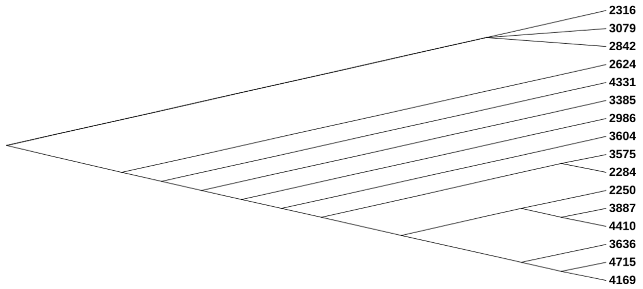
Figure 1. Phylogenetic tree of the UPEC isolates.
Pathogens 13 00794 g001
Figure 2. Antimicrobial resistance determinants in the UPEC isolates. The figure shows the presence (black for antimicrobial resistance gene) or absence (blank) of antimicrobial resistance genes.
Pathogens 13 00794 g002aPathogens 13 00794 g002b
Figure 3. Insertion elements (ISs) in the UPEC isolates. The figure shows the presence (black for antimicrobial resistance gene) or absence (blank) of antimicrobial resistance genes.
Pathogens 13 00794 g003aPathogens 13 00794 g003b
Figure 4. Schematic view of CRISPR-Cas in UPEC isolate 2250.
Pathogens 13 00794 g004
Figure 5. Virulence determinants in the UPEC isolates. The figure shows the presence (black) or absence (blank) of virulence genes.
Pathogens 13 00794 g005aPathogens 13 00794 g005bPathogens 13 00794 g005cPathogens 13 00794 g005dPathogens 13 00794 g005e
Table 1. MLST, cgMLST, serotype, CH type analysis of the UPEC isolates.
2250228423162624284229863079338535753604363638874169433144104715
Multilocus sequence typingadk18535353535353535353143514533514
fumC106404040404040404040143714403714
gyrB17474747474747474747102910472910
icd6131313131313131313200252001325200
mdh53636363636363636361741736417
purA52828282828282828287572857
recA4292929292929292929107310297310
ST393131131131131131131131131131119340511931314051193
cgMLSTcgST152,922152,915152,921152,912152,913152,924152,918152,917152,920152,91172,154152,916152,923152,914158,995152,919
Sero typingO typeO25O25O25O25O25O25O25O25O25O25O75O102O75O25O102O75
H typeH1H4H4H4H4H4H4H4H4H4H5H6H5H4H6H5
CH typingFumC106404040404040404040143714403714
FimH54303030303030303030642964302964
Table 2. Frequency of phenotypic antibiotic resistance among sixteen UPEC isolates.
ResistantIntermediateSusceptible
AZR727
GEN1510
KAN1051
STR4012
AMP1600
CEF1600
CFP646
IMI547
CHL0016
NAL1600
TET1331
DOX646
FOS0016
TMP-SMX718
POL1501
AZR: azithromycin, GEN: gentamicin, KAN: kanamycin, STR: streptomycin, AMP: ampicillin, CEF: cefepime, CFP: cefoperazone, IMI: imipenem, CHL: chloramphenicol, NAL: nalidixic acid, TET: tetracycline, DOX: doxycycline, FOS: fosfomycin, TMP-SMX: trimethoprim/sulfamethoxazole, POL: polymyxin B.
Table 3. Ciprofloxacin MIC with and without the inhibitor PaβN and gene expression of efflux pump genes and outer membrane proteins in the UPEC isolates.
Overexpression of Efflux Pump GenesAlterations of Outer Membrane Protein
StrainsCiprofloxacinCiprofloxacin + PAβN (100 ug/mL)acrAacrBmdfAnorEompF
225032323.184.9714.614.592.24
22842561281.903.202.913.09−1.43
23162562562.932.853.283.77−1.29
262464641.621.34−1.651.76−2.75
284264641.501.943.423.12−1.95
29862561281.711.89−1.352.12−2.61
3079128641.353.011.561.98−2.17
33852561281.701.43−1.671.58−2.77
3575256128−1.16−1.17−1.481.53−2.58
36041281282.282.691.863.43−2.73
36362561282.834.294.332.43−1.07
3887256643.544.924.854.02−14.61
41692562563.094.182.883.03−1.40
43312562562.121.761.742.57−2.09
4410128323.914.061.182.08−3.64
4715256641.181.32−2.45−1.57−3.18
Table 4. Mobile genetic elements carrying antibiotic resistance genes in the UPEC isolates. Bold: common.
StrainsPlasmidComposite TransposonsTransposonsMiniature Inverted RepeatsAntibiotic Resistance Genes
2250Col(BS512), Col156, IncFIA, IncFIB, IncP1cn_5813_IS911TnpA_Tn3, TnpR_Tn3MITEEc1 blaTEM-1B
2284Col(BS512), Col156, IncFIA, IncFIB, IncFIIcn_4876_IS629, cn_15494_IS629Tn5403, TnpA_Tn5403, TnpA_TnAs1, TnpR_Tn5403MITEEc1aac(3)-Ila, tet(A)
2316Col(BS512), Col(MG828), Col156, IncFIA, IncFIB, IncFII MITEEc1
2624Col(BS512), Col(MG828), Col156, IncFIA, IncFII TnpA_Tn3MITEEc1
2842Col(BS512), Col156, IncFIA, IncFIB, IncFII MITEEc1ant(2″)-Ia, ant(3″)-Ia, blaCTX-M-14, cmlA1, mph(A), qacE, sul1
2986Col(BS512), Col156, IncFIA, IncFIB, IncFII MITEEc1aadA5, dfrA17, mph(A), qacE, sul1
3079Col(BS512), Col(MG828), Col156, IncFIA, IncFIB, IncFII MITEEc1aadA5, aph(3″)-Ib, aph(6)-Id, dfrA17, mph(A), qacE, sul2, tet(A)
3385Col(BS512), Col156, IncFIA, IncFIB(K), IncFII Tn5403, TnpA_ISPa38, TnpA_Tn3, TnpA_Tn3000, TnpA_Tn5403, TnpR_Tn3, TnpR_Tn5403MITEEc1aadA1, ant(2″)-Ia, aph(3″)-Ib, aph(6)-Id, blaCTX-M-15, blaTEM-1B, cmlA1, qacE, qnrB1, sul2
3575Col(BS512), Col(MG828), Col156, IncFIA, IncFIB, IncFII, IncX1 Tn5403, TnpA_Tn5403, TnpR_Tn5403MITEEc1
3604Col(BS512), IncFIA, IncFIB, IncFII Tn5403, TnpA_Tn5403, TnpR_Tn20, TnpR_Tn5403MITEEc1aadA5, dfrA17, mph(A), qacE, sul1
3636Col(BS512), Col156, IncFIA, IncFIB, IncI1-I(α) MITEEc1
3887Col(BS512), Col(MG828), Col156, IncFIA, IncFIB, IncFIIcn_3324_ISEc1TnpA_Tn3MITEEc1aac(3)-Ila, aadA5, blaCTX-M-15, dfrA17, mph(A), qacE, sul1
4169Col(BS512), Col156, IncFIA, IncFIBcn_1487_IS629, cn_1794_IS26, cn_8908_IS911, cn_8908_ISEc31 MITEEc1
4331Col(BS512), Col156, IncFIA, IncFIB, IncFII MITEEc1 blaTEM-1B
4410Col(BS512), Col156, IncFIA, IncFIB cn_3324_ISEc1, cn_5813_IS911TnpA_Tn3, TnpR_Tn3MITEEc1aadA5, blaCTX-M-15, dfrA17, mph(A), qacE, sul1
4715Col(BS512), Col(MG828), Col156, IncFIA, IncFIB, IncQ1 MITEEc1dfrA14, tet(B)
Table 5. CRISPR-Cas system in the UPEC isolates.
StrainsTypeCas GenesCRISPR Consensus RepeatNumbers of SpacerRepeat LengthMean Size of SpacersCRISPR Length
2250I, I-Ecas3, cse1, cse2, cas7, cas5, cas6, cas1, cas2CGCGTCTTATCAGGCCTACGAGTTCGGTGCTGTGTAGGTCGGATAAGGCGTTCA15442149
GAGTTCCCCGCGCCAGCGGGGATAAACCG32932211
GTGTTCCCCGCGTCAGCGGGGATAAACCG112932699
CGACCCCCACCATGTCAAGGTGGTGCTCTAACCAACTGAGCTA14338123
GTTCACTGCCGTACAGGCAGCTTAGAAA32832207
2284NDNDGCCGGATGCGGCGTGAACGCCTTATCCGGCCTACAAAAGAAATGCAG14748141
AGTTCACTGCCGTACAGGCAGCT1233782
CGACCCCCACCATGTCAAGGTGGTGCTCTAACCAACTGAGCTA14338123
2316Icas3AGTTCACTGCCGTACAGGCAGCT1233782
AATGCCTGATGCGACGCTTGTCGCGTCTTATCATGCCTACAAGT14457144
2624Icas3GCCGGATGCGGCGTGAACGCCTTATCCGGCCTACAAAAGAAATGCAG14748141
CCACCTTTTTTACCTGCTTCAGATGC1264091
AGTTCACTGCCGTACAGGCAGCT1233782
2842Icas3CGACCCCCACCATGTCAAGGTGGTGCTCTAACCAACTGAGCTA14338123
ATCTGCCTGTACGGCAGTGAACT1233782
2986NDNDCCACCTTTTTTACCTGCTTCAGATGC1264091
ATCTGCCTGTACGGCAGTGAACT1233782
3079Icas3CGACCCCCACCATGTCAAGGTGGTGCTCTAACCAACTGAGCTA14338123
AGTTCACTGCCGTACAGGCAGCT1233782
GCCGGATGCGGCGTGAACGCCTTATCCGGCCTACAAAAGAAATGCAG14748141
3385NDNDGCCGGATGCGGCGTGAACGCCTTATCCGGCCTACAAAAGAAATGCAG14748141
AGTTCACTGCCGTACAGGCAGCT1233782
CGACCCCCACCATGTCAAGGTGGTGCTCTAACCAACTGAGCTA14338123
3575Icas3GCCGGATGCGGCGTGAACGCCTTATCCGGCCTACAAAAGAAATGCAG14748141
AGTTCACTGCCGTACAGGCAGCT1233782
CCACCTTTTTTACCTGCTTCAGATGC1264091
3604NDNDCGACCCCCACCATGTCAAGGTGGTGCTCTAACCAACTGAGCTA14338123
GCCGGATGCGGCGTGAACGCCTTATCCGGCCTACAAAAGAAATGCAG14748141
AGTTCACTGCCGTACAGGCAGCT1233782
3636Icas3GTTCACTGCCGTACAGGCAGCTTAGAAA22832147
CCGAGCCGTAGGCCGGATAAGGCGTTCACGC13156117
CGACCCCCACCATGTCAAGGTGGTGCTCTAACCAACTGAGCTA14338123
TGAACGCCTTATCCGACCTACACAGCACTGAACTCGTAGGCCTGATAAGACGCG15442149
3887I, I-Ecas3, cse1, cse2, cas7, cas5, cas6, cas1, cas2TGAACGCCTTATCCGACCTACACAGCACTGAACTCGTAGGCCTGATAAGACGCG15442149
GTGTTCCCCGCGCCAGCGGGGATAAA92635574
CCACCTTTTTTACCTGCTTCAGATGC1264091
CAGCGTCGCATCAGGCATTGTGCACGATTGCCGGATGCGGCGTGAACGCCTT15247150
4169Icas3TTTCTAAGCTGCCTGTACGGCAGTGAAC22832147
ACGCTGCCGCGTCTTATCGGGCCTACAAAAGTTCTGAACCGT14249132
TTGATTGCCGGATGCGGCACGAGTGCCTTATCCGGCCTAC24060.5240
CGCGTCTTATCAGGCCTACGAGTTCGGTGCTGTGTAGGTCGGATAAGGCGTTCA15442149
CCGAGCCGTAGGCCGGATAAGGCGTTCACGC13156117
CGACCCCCACCATGTCAAGGTGGTGCTCTAACCAACTGAGCTA14338123
4331NDNDCCACCTTTTTTACCTGCTTCAGATGC1264091
ATCTGCCTGTACGGCAGTGAACT1233782
4410I, I-Ecas3, cse1, cse2, cas7, cas5, cas6, cas1, cas2AAGGCGTTCACGCCGCATCCGGCAATCGTGCATAATGCCTGATGCGACGCTG15247150
CCACCTTTTTTACCTGCTTCAGATGC1264091
GTGTTCCCCGCGCCAGCGGGGATAAA92635574
TGAACGCCTTATCCGACCTACACAGCACTGAACTCGTAGGCCTGATAAGACGCG15442149
CGACCCCCACCATGTCAAGGTGGTGCTCTAACCAACTGAGCTA14338123
CCCAATTAAGTGAAGAGCAGGTGACGAAGTTACTGCATCGCAAA14458145
GAGTTCCCCGCGCCAGCGGGGATAAACCG102932638
4715NDNDTTTGTAGGCCTGATAAGACGCGCCAGCGTCGCATCAGGC13949126
GTTCACTGCCGTACAGGCAGCTTAGAAA22832147
GTAGGCCGGATAAGGCACTTGTGCCGCATCCGGCA13557126
TGAACGCCTTATCCGACCTACACAGCACTGAACTCGTAGGCCTGATAAGACGCG15442149
ACGCTGCCGCGTCTTATCGGGCCTACAAAAGTTCTGAACCGT14249132
Disclaimer/Publisher’s Note: The statements, opinions and data contained in all publications are solely those of the individual author(s) and contributor(s) and not of MDPI and/or the editor(s). MDPI and/or the editor(s) disclaim responsibility for any injury to people or property resulting from any ideas, methods, instructions or products referred to in the content.