Review

The Main Arboviruses and Virus Detection Methods in Vectors: Current Approaches and Future Perspectives

by
Amanda Montezano Cintra
1,†,
Nathália Mayumi Noda-Nicolau
1,†,
Milena Leite de Oliveira Soman
1,
Pedro Henrique de Andrade Affonso
1,
Guilherme Targino Valente
2 and
Rejane Maria Tommasini Grotto
1,2,*
1
Multiuser Central Laboratory, School of Agricultural Sciences, São Paulo State University (UNESP), Botucatu 18618-687, Brazil
2
Clinical Hospital of School of São Paulo State University, São Paulo State University (UNESP), Botucatu 18618-970, Brazil
*
Author to whom correspondence should be addressed.
These authors contributed equally to this work.
Pathogens 2025, 14(5), 416; https://doi.org/10.3390/pathogens14050416
Submission received: 5 April 2025 / Revised: 16 April 2025 / Accepted: 23 April 2025 / Published: 25 April 2025
(This article belongs to the Section Viral Pathogens)

Abstract

:
Arthropod-borne viruses (arboviruses) represent a growing concern for global public and veterinary health, with cases reported across all continents. This review presents a broad overview of the geographic distribution of arboviruses transmitted by insect vectors, emphasizing the importance of early viral detection as a cornerstone of surveillance and outbreak preparedness. Special attention is given to the phenomenon of zoonotic spillover, where viruses maintained in natural transmission cycles often involving wildlife reservoirs and arthropod vectors cross into human populations, triggering emergent or re-emergent outbreaks. This article discusses key arboviral families of medical and veterinary significance, including Togaviridae, Flaviviridae, Nairoviridae, Phenuiviridae, Peribunyaviridae, and Orthomyxoviridae, highlighting their molecular and structural characteristics. These features are essential for guiding the development and implementation of specific and sensitive detection strategies. In addition, this work provides a comparative analysis of diverse laboratory methodologies for viral detection in vectors. From serological assays and viral isolation to advanced molecular tools and next-generation sequencing, we explore their principles, practical applications, and context-dependent advantages and limitations. By compiling this information, we aim to support researchers and public health professionals in selecting the most appropriate tools for vector surveillance, ultimately contributing to improved response strategies in the face of arboviral threats.

1. Introduction

Arthropod-borne pathogens contribute to more than 17% of infectious diseases, affecting millions of people around the world each year and playing a pivotal role in the emergence of new human pathogens. Dengue, the most prevalent arboviral disease, results in approximately 90 million cases and about 40,000 deaths a year [1]. Emerging arboviruses such as Phlebovirus riftense (Rift Valley fever virus), Alphavirus mayaro (Mayaro fever virus), Orthoflavivirus nilense (West Nile fever virus), Alphavirus chikungunya (Chikungunya fever virus), and Orthoflavivirus encephalitidis (tick-borne encephalitis virus) have also garnered scientific attention as public health concerns [2].
Arboviruses are a diverse group of more than 500 viruses transmitted by arthropod vectors such as mosquitoes and ticks [3] present on several continents (Figure 1). In the Southeast Asian region, the presence of Orthoflavivirus denguei, or dengue fever virus (DENV), and Orthoflavivirus japonicum, or Japanese encephalitis virus (JEV), probably due to anthropogenic actions and geographic changes, is the result of a “spillover” of natural zoonotic pathogens into the human population [2,4].
The South American region experiences a high incidence of infections caused by arboviruses, Mayaro fever virus (MAYV) and Orthoflavivirus flavi, or yellow fever virus (YFV), are predominantly found in specific biomes and population densities of rural areas; however, due to the constant urbanization of these places, there is a high risk of an increase in cases and epidemics [5,6]. DENV incidence in South American countries shows periodic oscillations [7]. According to the Pan American Health Organization (PAHO) report on the epidemiological situation of dengue in the Americas alerts, DENV cases in the year 2024 increased by 283% compared to the average of the last 5 years. With Brazil leading the highest indices of infected individuals, the detection of all four different viral serotypes in the country is noteworthy [8]. Beyond the diseases caused by the dengue fever virus, the Chikungunya fever virus (CHIKV) is also prevalent in South America, especially in Brazil, thus posing a challenge up to the present time [9]. The Orthoflavivirus zikaense, or Zika virus (ZIKV), has been very present in South America since 2015, and currently, with the lack of specificity of clinical presentation between cases and the complexity of laboratory diagnosis in a context of co-circulation, detecting ZIKV presents many challenges for the region’s surveillance system [6].
Rift Valley fever virus (RVFV) is endemic on the African continent and triggers large-scale outbreaks, typically associated with climatic patterns and rainfall, resulting in significant impacts on both human and animal populations [10]. The virus has spread to regions like Saudi Arabia, Madagascar, and other islands in the Indian Ocean, such as Mayotte and Comoros. Effectively managing RVFV requires addressing a range of unique challenges, involving coordination among various sectors, including public health, animal welfare, agriculture, and biosecurity [11]. Similarly, yellow fever virus (YFV) is endemic, primarily in Sub-Saharan Africa, with 274 probable cases reported by 12 countries between 2021 and 2022 [12]. The Crimean–Congo hemorrhagic fever virus (CCHFV) is also a major concern for both human and veterinary health due to its high lethality. Additionally, like many other arboviruses, environmental factors play a key role in the spread of the disease. As CCHFV is primarily transmitted by ticks, increasing human–animal interactions pose significant risks for transmission to human populations. Currently, the virus is distributed across large parts of Africa, Europe, and Asia [13]. A study on various RNA viral types highlighted the presence of unknown Thogotovirus (THOV) sequences in mosquitoes in Senegal, emphasizing the insufficient data on circulating RNA viruses. Thogotoviruses can infect a wide range of vertebrate hosts, including birds, rodents, cattle, and humans, and they remain a growing health concern in the region [14].
In the North American region, epidemiological concerns revolve around Venezuelan equine encephalitis virus (VEEV), West Nile fever virus (WNV), and La Crosse virus (LACV). VEEV first appeared in southern Texas in 1971, following an epidemic in Mexico. This infection is highly debilitating for humans and can be fatal to horses. However, the prompt actions taken by authorities, including the approval of a vaccine and mosquito control strategies, have significantly reduced the virus’s impact in the region [15]. WNV was initially identified on the African continent and has since spread to Africa, Europe, Asia, and North America. In the USA, the number of cases fluctuates annually, with nearly 44,000 confirmed and probable cases reported between 1999 and 2015 [16]. The transmission of LACV is closely linked to climatic factors and urbanization and is considered a significant pathogen with the potential to spread or emerge in new areas across North America. However, further scientific research is necessary to better understand human infections. At present, no specific treatment for LACV exists, and prevention efforts mainly focus on minimizing mosquito exposure [17].
In Europe, the circulation of WNV has been recognized since the 2000s and has been continuously monitored. Data indicate a spread of the virus on the continent and the need for vaccines to protect populations that may be at risk [18]. Tick-borne encephalitis virus (TBEV) is also a disease present on the continent, specifically in Italy, Czech Republic, and Lithuania. Studies show that despite the high incidence of the disease, there is a lack of action to combat it, either through vaccines or effective behavioral practices aimed at avoiding contact with the vector [19,20].

2. Arboviruses with Veterinary Importance and the “Spillover” of Natural Zoonotic Pathogens into the Human Population

The spread of viruses from other animals to humans and their risk of triggering epidemics is widely known; however, the reverse, called repercussion, is underestimated. This process can establish permanent enzootic cycles in new regions, in addition to creating a reservoir of future pandemics. Despite this, the understanding of the dynamics of enzootic arbovirus cycles is limited [21]; as multiple viruses, vectors, and hosts are involved in the transmission of arboviruses, the identification of vertebrate reservoirs is complicated [22]. The transmission of enzootic and epizootic diseases to humans occurs due to various factors, including intrinsic characteristics of the hosts, pathogens, and vectors; environmental factors such as changes in ecology and urbanization; and phylogenetic distances between the hosts, among others [1]. The accurate prediction and determination of zoonotic pathogen dissemination patterns require transdisciplinary research that integrates ecological, epidemiological, virological, immunological, demographic, and sociological data on a global scale. Only through the integrated analysis of these data using advanced computational approaches will it be possible to gain a deeper understanding of this phenomenon [23].
Among the arboviruses mentioned here, those of veterinary importance and primarily transmitted by mosquitoes include MAYV, RVFV, VEEV, WNV, JEV, and LACV, while TBEV and THOV, also present in zoonotic cycles, are transmitted mainly by ticks. MAYV was first detected in the serum of forestry workers in Trinidad [24], but it is known to infect a broad range of invertebrate and vertebrate hosts [25,26]. The virus’s transmission cycle primarily involves the Haemagogus janthinomys mosquito and non-human primates as primary hosts, with rodents, reptiles, and birds serving as secondary hosts [27,28]. RVFV, which has historically been present in Kenya and Tanzania, is maintained in a zoonotic cycle among African buffalo and transmitted by over 30 species of mosquitoes [29,30].
The VEE complex consists of seven species and numerous subvariants [31], with its distribution varying across the New World. Rodents act as reservoirs in a cycle involving mosquitoes of the Culex subgenus (Melanoconion), primarily in humid tropical forests and swampy areas, leading to fatal human cases [32]. WNV, maintained in a zoonotic cycle by Culex mosquitoes and passerine birds, causes outbreaks in both horses and humans. Some viral strains are found worldwide, while strain 2 remains enzootic in Africa [33]. Public health measures will continue to focus on mosquito control, education, and, hopefully, new therapeutic developments [34].
The JEV serocomplex includes WNV, Murray Valley encephalitis virus, and St. Louis encephalitis virus (SLEV). JEV was first documented in Japan in the 1800s, with its cycle maintained by Culex mosquitoes, pigs, horses, and humans [35]. The LACV virus is transmitted by Aedes triseriatus mosquitoes that breed in tree holes and containers, with eastern squirrels acting as amplifying hosts. The introduction of Ae. albopictus may alter the virus’s ecology and increase the likelihood of new variants emerging [36,37].
TBEV is primarily transmitted to humans through infected ticks, circulating between these ticks and various animals, including deer and small mammals, with infection rates correlating to tick activity [38]. For transmission to occur, a high level of viremia is required, which is typically found in mammals such as sheep, goats, horses, dogs, and rodents. Humans, however, do not develop sufficient viremia to transmit the virus to ticks and are considered dead-end hosts [39]. Similarly, THOV and Dhori virus (DHOV), also transmitted by ticks, cause a range of symptoms from mild febrile illness to meningoencephalitis. Animal reservoirs for both include banded mongooses, rodents, dromedaries, and livestock [40,41,42,43].

3. The Importance of Viral Detection in Insect Vectors for Predicting Emerging and Re-Emerging Viruses into Populations

Early detection of emerging and re-emerging arboviruses is a challenge for the medical and veterinary community worldwide, requiring a combination of factors and studies from various areas such as studies of the biology of vectors and enzootic hosts, meteorological and climatic surveillance models, and Geographic Information Systems. Due to growing urbanization and climate change, including increases in temperature and humidity, it is expected that there will be a greater spread of vectors and, consequently, a greater number of circulating arboviruses around the world, such as the growing expansion of Ae. aegypti mosquitoes into temperate regions [44].
A key factor for the early epidemiological surveillance of arboviruses is the use of adult mosquitoes using saliva collections with sugar bait; however, the infection rates of the vector are typically low by this route, limiting the sensitivity of the testing methods [45]. Testing engorged mosquitoes for both pathogens and antibodies is being explored as an alternative to direct vertebrate blood testing, which includes human patients and other reservoir animals [46]. With the decreasing costs of next-generation sequencing, metagenomics has become attractive for surveillance using various types of samples, allowing not only the detection of known sequences of mosquito-borne viruses, but also emerging viruses through de novo assembly [47]. In short, surveillance using potential vectors could provide vital information on circulation for emergency preparedness. Given finite surveillance resources, prioritization is key and is usually given to virus families with a history of emergencies, with the potential for transmission by urban mosquitoes, or viruses with high lethality rates. However, some resources must also be targeted broadly, as obscure viruses with no previous history of epidemics can emerge unexpectedly.
Beyond mosquitoes, ticks are increasingly recognized as important vectors in the detection of emerging arboviruses. Advances in next-generation sequencing have enabled the identification of novel tick-borne viruses associated with febrile illnesses in humans, highlighting their role in early warning systems for viral emergence.
In 2025, the Xue-Cheng virus (XCV), a novel orthonairovirus, was identified in northeastern China during surveillance of febrile patients with recent tick exposure [48]. Similarly, the Yezo virus (YEZV), initially reported in Japan in 2021, has since been detected in China, raising concerns about its broader distribution [49]. In another case, the Wetland virus (WELV) was discovered following severe illness in a patient bitten by a tick in Inner Mongolia [50]. These findings underscore the value of proactive viral surveillance in ticks, not only to better understand zoonotic transmission cycles but also to anticipate potential public health threats from emerging pathogens. Metagenomic approaches, in particular, have proven essential in these discoveries, allowing the unbiased detection of known and novel viruses directly from clinical and environmental samples, and playing a critical role in expanding our understanding of tick-borne viral diversity.

4. Different Virus Families Responsible from the Main Arboviruses

Arthropod-borne viruses, named arboviruses, are composed of RNA viruses with more than 500 described species, of which approximately 150 species are associated with diseases, belonging to six viral families: Togaviridae, Flaviviridae, Nairoviridae, Phenuiviridae, Peribunyaviridae, and Orthomyxoviridae. Considering that all of these viral families have different characteristics, the understanding behind structural and genomic features of these viruses is of paramount importance for accurate diagnosis. For this purpose, the main virus characteristics of each family are organized in Table 1, and, below, we describe the major features of these six viral families.
Alphavirus is the only genus in the family Togaviridae; the main viruses of this genus in terms of human pathogenicity are CHIKV and MAYV. They share several common characteristics, and their surfaces have the presence of glycoprotein spikes covering the alphavirion [51]. Their genome encodes different proteins that are important for replication-related and virus assembly-related functions; four non-structural proteins, which are nsP1, nsP2, nsP3, and nsP4; and six structural proteins, including capsid, E3, E2, 6K, TF, and E1. Their replication complex is formed in the plasma membrane of infected vertebrate cells by merging the virus membrane with the host endosomal membrane.
The subgenus Orthoflavivirus belongs to the family Flaviviridae and includes several important mosquito-borne viruses such as ZIKV, DENV, JEV, WNV, and YFV. They are all small and lipidic enveloped viruses, and basic capsid, which is composed of a single protein. The envelope surface contains two virus-encoded membrane-associated proteins. Viruses belonging to this genus contain three structural proteins: capsid, envelope protein (E) and either prM or M, for immature virions and mature virions, respectively; and seven nonstructural proteins: NS1, NS2A, NS2B, NS3, NS4A, NS4B, and NS5. Their replication occurs in the cytoplasm, more specifically in membrane vesicles derived from the endoplasmic reticulum, followed by the assembly of the virus and its export out of the cell by the vesicle transport pathway.
Orthonairovirus belongs to the family Nairoviridae; one of the most significant viruses in this genus, considering its human pathogenicity, is CCHFV. It has a spherical shape and is enveloped with glycoprotein spikes. They are responsible for encoding three different proteins: nucleoprotein, the viral glycoprotein precursor (GPC), and the large protein (L), which has RNA-directed RNA polymerase, helicase, and endonuclease domains. Its replication occurs in the cytoplasm, when the virus attaches to the host receptors by a glycoprotein dimer, and it is endocytosed into vesicles to the host cell.
The genus Phlebovirus belongs to the family Phenuiviridae; it includes one of the most significant pathogens for humans, the RVFV. They are enveloped spherical viruses, and their glycoproteins at the envelope surface are arranged in an icosahedral (T = 12) lattice symmetry. The genome is capable of encoding six proteins. A characteristic of all phleboviruses is the ambisense coding strategy of the S genome segment: the N protein is encoded in the negative-sense orientation on the S segment, while the NSs protein is encoded in the positive sense. However, both proteins are translated from separate subgenomic mRNAs that are transcribed from genomic or antigenomic RNA [52].
The Orthobunyaviruses belong to the family Peribunyaviridae and include several viruses like the LACV, Jamestown Canyon virus, and Snowshoe hare virus, which are named the California Encephalitis Virus Complex. They are enveloped, having spherical or pleomorphic shape [53]. The envelope of their surface has spikes composed of two glycoproteins called Gn and Gc. The genome of this genus is segmented in three molecules (S, M, and L) of single-stranded negative sense RNA and they are not polyadenylated. The S, M, and L segments are responsible for encoding the nucleocapsid protein (N), envelope glycoproteins (Gn and Gc), and the RNA-directed RNA polymerase and endonuclease (L), respectively; as well as the non-structural proteins (NSs and NSm). During cell invasion, Orthobunyaviruses attach to host receptors using Gn-Gc glycoprotein dimer, which leads to their endocytosis into the vesicles membrane; then, transcription and replication occur in the cytoplasm.
The genus Thogotovirus is classified within the Orthomyxoviridae family. Thogotovirus was one of the first viruses identified in this genus and was originally isolated from ticks in Kenya. They are enveloped and spherical viruses. They are responsible for encoding six to seven proteins from six distinct ORFs. During cell invasion, the virus attaches to the sialic acid receptor through the GP protein and is endocytosed by the host cell, followed by endosome acidification, which induces fusion of the virus membrane with the vesicle membrane. Subsequently, the encapsulated RNA segments migrate to the nucleus when transcription occurs.

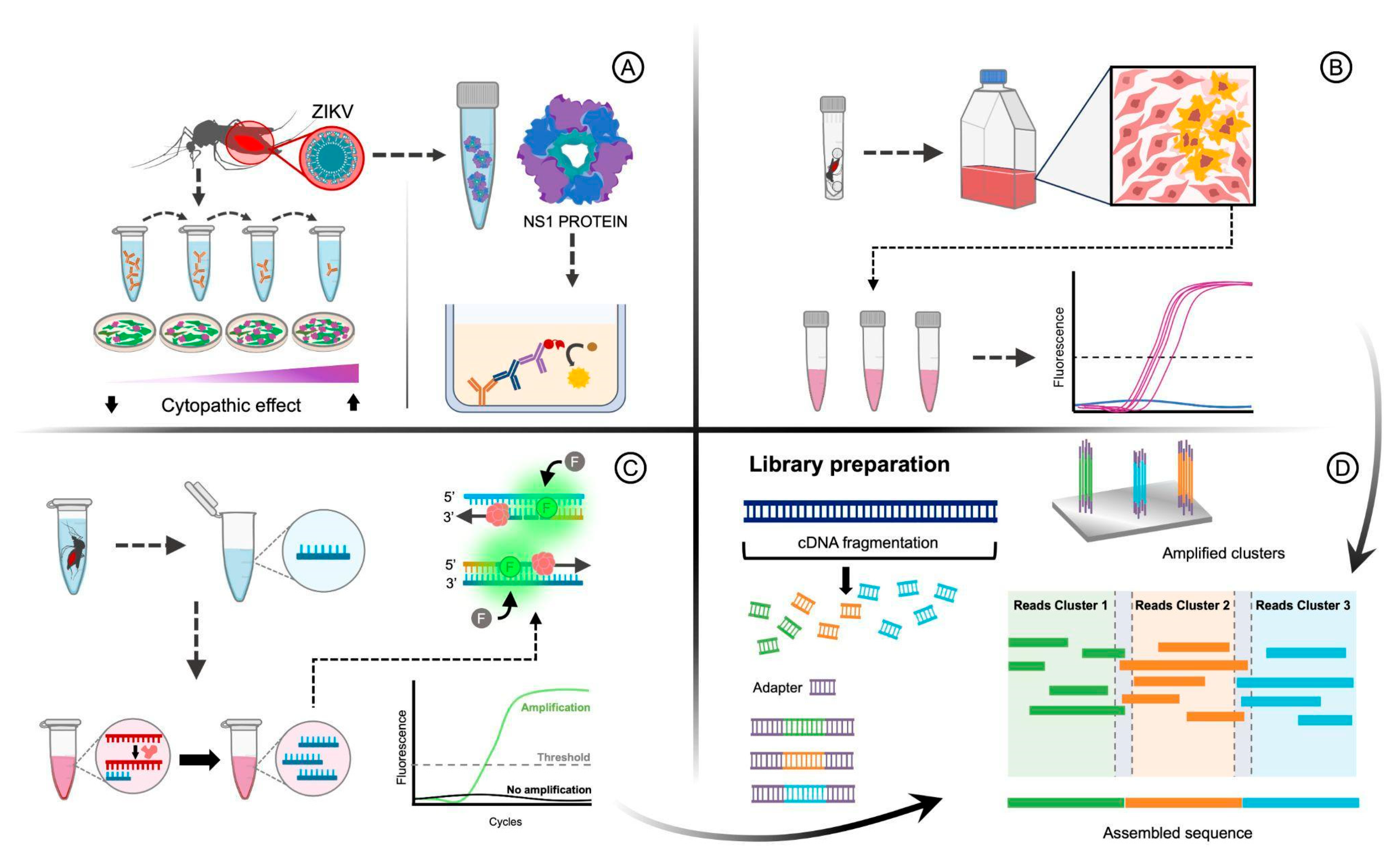
5. Methodologies to Detect Arboviruses in Vector

Since arboviruses are transmitted primarily by mosquitoes and can cause a range of debilitating diseases in humans, including dengue fever, Zika virus infection, chikungunya infection, and yellow fever, the detection of these viruses in mosquito populations is critical for effective disease surveillance, control, and prevention [54]. Identification of arboviruses in mosquitoes provides an early warning system for potential disease outbreaks [55]. Timely detection allows health authorities to respond quickly with targeted interventions, preventing the escalation of infections and reducing the burden on healthcare systems [54].
Identifying the virus in mosquitoes is important for studying the intricate transmission cycles between them, providing better knowledge of the factors influencing disease spread, such as mosquito species, behavior, and environmental conditions [56]. Furthermore, the detection of arboviruses is relevant in vector control strategies, because accurate detection data guide the development and implementation of effective mosquito control strategies. These strategies include targeted application of insecticides, biological control agents, and genetic modification of mosquitoes to reduce their ability to transmit viruses [57].
Detecting arboviruses in mosquitoes contributes to the development of vaccines and treatments, enabling us to study the genetic makeup and characteristics of the detected viruses to design vaccines that target specific strains and variants [58]. Continuous monitoring of arboviruses in mosquitoes allows public health agencies to track disease trends, assess the effectiveness of control measures, and adapt strategies as needed. This surveillance is crucial for evidence-based decision making [57].
Over the years, several methodologies have been developed to identify and characterize arboviruses in mosquitoes, each contributing to a comprehensive understanding of disease transmission dynamics [59]. This article explores some of the primary methodologies used for virus detection in mosquitoes, such as serological assays, virus isolation, molecular techniques, next-generation sequencing (NGS), and combined methods.
Serological assays are essential tools for the detection of viral antigens or antibodies in mosquitoes (Figure 2A). These techniques indicate whether mosquitoes have been exposed to a particular virus, providing information on the prevalence of infections in mosquito populations. Mosquito samples can be used to detect virus-specific antibodies using various techniques. Enzyme-linked immunosorbent assays (ELISAs) and plaque reduction neutralization tests (PRNTs) are commonly used techniques. ELISA can identify virus-specific antigens in mosquito homogenates, while PRNT measures the presence of neutralizing antibodies in mosquito. Although relatively simple, these methods require prior knowledge of the virus being targeted and might lack sensitivity in certain contexts. PRNT is the “gold standard” of serological tests [60] and then could be used to identify virus-specific antibodies in different animal tissues. Gyawali et al. were able to detect Ross River Virus antibodies in small volumes of blood present in mosquito abdomens using the PRNT method, indicating the host exposure to arbovirus infection [61].
ELISA is an immunological method that detects and quantifies specific viral antigens in mosquito samples. This technique utilizes antibodies that bind to viral proteins, producing a measurable signal. ELISA is sensitive and widely used for the large-scale screening of mosquito populations to identify the presence of specific viruses. The ELISA method has been used to detect ZIKV [62] and DENV [63] in mosquitoes by targeting the nonstructural protein 1 (NS1) protein; additionally, this test was sensitive enough to detect the CHIK antigen from a small fraction of an infected mosquito homogenate [64], and it can also detect DENV and JEV antigens in mosquitoes Aedes aegypti and Ae. albopictus [65].
Another amplified ELISA procedure has been developed to detect the Western equine encephalitis virus (WEEV) in mosquitoes, with a sensitivity of detecting 10 plaque-forming units or greater of WEEV [66]. Furthermore, the use of antibodies against viral nucleoproteins and glycoproteins has been found to facilitate the detection of RVFV in mosquito tissues [67]. In another study [68], the authors developed two monoclonal antibodies that can detect and isolate a range of known and novel viruses in mosquito samples that identify proteins involved in viral RNA replication, and incorporated them into a high-throughput, economical ELISA-based screening system for the detection and discovery of viruses from mosquito populations.
In addition, an electrochemical impedimetric method using antibody-conjugated sensor electrodes has been developed to detect ZIKV, DENV, and CHIKV with high sensitivity and selectivity [69]. Furthermore, an antigen detection assay, not virus-specific antibodies, named The Rapid Analyte Measurement Platform (RAMP®), was designed to detect all DENV serotypes in mosquito pools. The data showed that it can detect geographically distinct strains of all four DENV serotypes in virus-spiked mosquito pools that contain at least 4.3 log10 plaque forming units/mL [70].
However, methodologies based on virus isolation involve the attempt to culture and propagate viruses present in mosquito samples, infecting susceptible cells with virus-containing mosquito samples, enabling researchers to isolate and identify viruses based on their cytopathic effects. This classical technique remains valuable for the discovery and characterization of viruses. However, viral isolation can be time-consuming, labor-intensive, and may yield low viral titers, limiting its practicality in large-scale surveillance [71].
Mosquito homogenates are inoculated into susceptible cell lines, and if the virus is viable, it will replicate within cells, leading to observable cytopathic effects. Virus isolation allows virus characterization and is essential for studying virus–host interactions (Figure 2B). Mosquitoes were used for virus isolation in several studies; for example, in Indonesia, a large surveillance of mosquitoes and mosquito-borne viruses was carried out, resulting in the isolation of insect Orthoflavivirus, Banna virus (BAV), new permutotetravirus, and alphamesoniviruses [72]. In Hunan Province, China, mosquito-borne arboviruses were isolated from various species of mosquitoes, including Akabane virus, JEV, and Tibet orbivirus [73]. In Tokyo, Japan, a comparison study of DENV isolation methods using different cell lines showed higher isolation rates using FcγR-expressing BHK cells [74]. BAV was also isolated from Anopheles sinensis mosquitoes in Hubei, China, revealing a new genotype [75]. In Yunnan Province, China, the Nam Dinh virus was isolated from mosquitoes using Aedes albopictus C6/36 cells [76]. The Vero cell plaque assay was reliable for detecting live WNV in pools containing up to 200 mosquitoes [77]. Furthermore, in another study, the authors described how to handle, process, and screen field-collected mosquitoes for infectious viruses by Vero cell culture assay. They isolated nine different viruses from mosquitoes collected in Connecticut, USA, and among these, five are known to cause human disease [78]. These studies highlight the importance of mosquito surveillance and virus isolation to understand the diversity and circulation of mosquito-borne viruses.
Molecular techniques have revolutionized the detection of viruses in mosquitoes by enabling the identification of specific viral nucleic acids. Reverse transcription polymerase chain reaction (RT-PCR) and quantitative real-time PCR (RT-qPCR) are widely used for their precision and sensitivity (Figure 2C). However, RT-qPCR is more commonly used because it is faster and allows multiple virus to be tested simultaneously and quantifies the virus load. Several arboviruses were detected in mosquitoes using RT-qPCR, such as RVFV [79,80]; MAYV [81]; Ilheus virus (ILHV) [82]; DENV, CHIKV, and ZIKV [83,84]; JEV [85]; and WNV [77,86].
The multiplex RT-qPCR assay has been described in several works. Some authors using primer and probe specifics simultaneously detected ZIKV, USUV, WNV, and CHIKV in large mosquito pools [87]. Another work describes the development of two different multiplex RT-qPCR assays, one for detecting YFV, JEV, WNV, and SLEV, and the other to detect DENV1, -2, -3, and -4 in mosquito pools [88]. In this sense, another study used a multiplex RT-qPCR assay to detect and serotype DENV in individual mosquito samples [89]. Rademan et al. developed a one-step multiplex real-time RT-PCR assay for the detection and distinction of Spondweni and ZIKV [90]. Furthermore, group-specific primers to detect Orthoflavivirus and Alphaviruses were designed to detect this genus virus in the mosquito pool [91]. In addition, Villinger et al. [92] identified specific arbovirus sequences from Orthoflavivirus, Alphavirus, Orthonairovirus, Phlebovirus, Orthobunyavirus, and Thogotovirus genera in mosquito pools using multiplex high-resolution melting analysis. Furthermore, RT-qPCR could be used as a screening before Sanger sequencing using universal primers for a specific genus such as Orthoflavivirus [93,94,95], Alphavirus [94,95], and Orthobunyavirus [95].
These assays have shown high sensitivity and specificity, allowing the simultaneous detection of multiple viral targets in mosquito samples and viral quantification [96]. However, they require well-equipped laboratories and skilled personnel, making them less suitable for resource-limited settings. Thus, the reverse transcription-loop-mediated isothermal amplification (RT-LAMP) method has been developed and validated for detecting viruses in mosquitoes and provides an alternative to PCR. This method requires simpler equipment and yields results rapidly, making it advantageous for field settings. The RT-LAMP allows rapid and sensitive detection of ZIKV [97,98,99] and BAV [100]. Another study used an LAMP-based virus-derived DNA (vDNA-LAMP) detection assay to successfully detect ZIKV in crude DNA purified from infected cultured cells and Aedes mosquitoes [101]. However, the sensitivity and specificity of a commercially available WNV real-time RT-LAMP assay were tested, and only 70 of 94 pools positive for real-time RT-PCR were also positive for this test [102]. On the other hand, in another study, the authors compared qRT-PCR and 2 RT-LAMP assays (based on different primer design approaches), for the detection of African and Asian/American lineages of ZIKV isolates, and showed that RT-LAMP detected 100% of the samples while RT-qPCR detected 88.88% of the samples [103].
Next-generation sequencing (NGS) has revolutionized virus detection in various samples, including mosquitoes. This high-throughput sequencing technique can identify known and novel viruses by sequencing millions of DNA or RNA strands simultaneously. NGS allows researchers to explore the entire virome of mosquito populations, providing valuable insights into viral diversity and potential emerging pathogens (Figure 2D). Mosquitoes were collected and subjected to metagenomic next-generation sequencing to detect viruses in several studies [104,105,106,107,108]. Three significantly novel viruses, with two being highly prevalent in A. vexans nipponii, were discovered using metagenomic analysis [109]. Moreover, metatranscriptomic sequencing was able to detect more than 70 known and novel viral species in mosquitoes [110]. Additionally, the metagenomic arbovirus detection approach used detected more than 88 viruses and found evidence of novel ZIKV variants circulating in the local mosquito population; hence, it can be a useful tool for identifying epidemic-causing arboviruses and collecting phylogenetic information on their source [111]. Furthermore, a PCR-based NGS protocol was developed, and the method allows the sequencing of lineage 2 WNV, without the need for cell culture isolation, even in cases of low virus titers [112].
Moreover, in molecular methodology, some authors have been using arrays system to detect virus in mosquitoes. The Lawrence Livermore Microbial Detection Array (LLMDA) can be able to detect multiple arboviruses of public health importance, including viruses belonging to the Orthoflavivirus, Alphavirus, and Orthobunyavirus genera [113]. Therefore, another study used a high-throughput chip array to detect arboviruses in mosquitoes, specifically identifying the presence of CHIKV in Iran [114]. Other authors developed a multiplexed Luminex array panel to detect medically important arboviruses in mosquitoes, including those of the Flaviviridae, Togaviridae, and Bunyaviridae families [115]. In this sense, another multiplexed Luminex array can detect and discriminate ZIKV, CHIKV, and DENV in mosquitoes [116]. These arrays can be valuable tools for the surveillance and management of arboviral diseases, allowing the detection of multiple viruses in the same mosquito pool and the identification of potential ecological associations between different viruses.
Lastly, there is a non-invasive analytical technique, named near-infrared spectroscopy (NIRS), that has been employed to identify viruses in mosquitoes. This methodology is based on the interaction between near-infrared light and the biological components present in the mosquito, allowing the detection of viral infections. Remarkably, NIRS is a rapid, reagent-free, and cost-effective tool that can be used as a non-invasive technique to detect and differentiate mosquito infections with ZIKV and DENV [117]. However, there are limited data on this methodology, and robust models must be developed and validated prior to use. As this methodology has the ability to predict infection based on specific chemical compounds or the actual pathogen, improving its predictive accuracy could be an excellent strategy in the identification of arboviruses [118].
As outlined in this review article, there are numerous methods available for detecting arboviruses, and the most suitable approach for each situation depends on several factors, including turnaround time, required equipment, advantages and limitations of each technique, and most importantly, the specific objective of the study. To facilitate understanding and provide a comprehensive overview of the main characteristics of these methodologies, we compiled a summary table (Table 2) highlighting their key features.
In summary, when rapid field detection of arboviruses is needed, RT-LAMP and RAMP are the preferred methods due to their speed, portability, and visual readouts. While NIRS offers the fastest screening capability, it tends to have lower specificity. Conversely, RT-qPCR remains the gold standard for high-sensitivity diagnostics and is extensively employed in outbreak surveillance. In cases involving the discovery of novel viruses or genomic characterization, NGS is indispensable despite its high cost and complexity. Additionally, LLMDA is a valuable tool for detecting multiple pathogens simultaneously, particularly in biodefense scenarios or during investigations of unknown outbreaks. In contexts requiring large-scale surveillance, ELISA offers a cost-effective solution for antibody detection, while electrochemical impedimetric biosensors provide portable, real-time results. Finally, PRNT and virus isolation are essential for confirming infectious viruses, though they require biosafety-level laboratories and extended processing times.

6. Conclusions

The detection of viruses in mosquitoes is crucial for understanding disease transmission dynamics and developing effective control measures. A combination of molecular, serological, and culture-based techniques provides comprehensive information on the virome of mosquito populations. Moreover, this study highlighted the importance of developing multiplex assays that allow the simultaneous detection of multiple viral targets in mosquito samples and viral quantification with high sensitivity and specificity, such as RT-qPCR and array systems. Advanced methodologies like NGS and metagenomics have opened new avenues in mosquito virology by enabling the identification of emerging and novel viruses. With ongoing research and technological advancements, these methodologies will continue to play a vital role in the surveillance and management of mosquito-borne diseases.

Author Contributions

Conceptualization, A.M.C. and N.M.N.-N.; investigation, A.M.C., N.M.N.-N., M.L.d.O.S. and P.H.d.A.A.; writing—original draft preparation, A.M.C., N.M.N.-N. and M.L.d.O.S.; writing—review and editing, G.T.V. and R.M.T.G.; supervision, R.M.T.G. All authors have read and agreed to the published version of the manuscript.

Funding

This research was funded by PROPG/UNESP through call nº 01/2025, São Paulo Research Foundation FAPESP (grant number 2022/02261-5) and by National Council for Scientific and Technological Development—CNPq.

Institutional Review Board Statement

Not applicable.

Informed Consent Statement

Not applicable.

Data Availability Statement

All data produced during this study are included in the published article.

Conflicts of Interest

The authors declare no conflicts of interest.

References

  1. WHO Fact Sheet: Vector-Borne Diseases. Available online: https://www.who.int/news-room/fact-sheets/detail/vector-borne-diseases (accessed on 14 August 2022).
  2. Kading, R.C.; Brault, A.C.; Beckham, J.D. Global Perspectives on Arbovirus Outbreaks: A 2020 Snapshot. Trop. Med. Infect. Dis. 2020, 5, 142. [Google Scholar] [CrossRef] [PubMed]
  3. Young, P.R. Arboviruses: A Family on the Move. In Advances in Experimental Medicine and Biology; Springer: New York, NY, USA, 2018; Volume 1062, pp. 1–10. [Google Scholar]
  4. Young, K.I.; Medwid, J.T.; Azar, S.R.; Hu, R.M.; Drumm, H.; Co, L.L.; Pitts, R.J.; Buenemann, M.; Vasilakis, N.; Perera, D.; et al. Identification of Mosquito Bloodmeals Collected in Diverse Habitats in Malaysian Borneo Using COI Barcoding. Trop. Med. Infect. Dis. 2020, 5, 51. [Google Scholar] [CrossRef]
  5. Lorenz, C.; Freitas Ribeiro, A.; Chiaravalloti-Neto, F. Mayaro Virus Distribution in South America. Acta Trop. 2019, 198, 105093. [Google Scholar] [CrossRef] [PubMed]
  6. Amraoui, F.; Pain, A.; Piorkowski, G.; Vazeille, M.; Couto-Lima, D.; de Lamballerie, X.; Lourenço-de-Oliveira, R.; Failloux, A.B. Experimental Adaptation of the Yellow Fever Virus to the Mosquito Aedes Albopictus and Potential Risk of Urban Epidemics in Brazil, South America. Sci. Rep. 2018, 8, 14337. [Google Scholar] [CrossRef] [PubMed]
  7. Mugabe, V.A.; Borja, L.S.; Cardoso, C.W.; Weaver, S.C.; Reis, M.G.; Kitron, U.; Ribeiro, G.S. Changes in the Dynamics of Dengue Incidence in South and Central America Are Possibly Due to Cross-Population Immunity after Zika Virus Epidemics. Trop. Med. Int. Health 2021, 26, 272–280. [Google Scholar] [CrossRef]
  8. PAHO. Report on the Epidemiological Situation of Dengue in the Americas; PAHO: Washington, DC, USA, 2024; Volume 143, pp. 1–3. [Google Scholar]
  9. Cunha, M.S.; Costa, P.A.G.; Correa, I.A.; de Souza, M.R.M.; Calil, P.T.; da Silva, G.P.D.; Costa, S.M.; Fonseca, V.W.P.; da Costa, L.J. Chikungunya Virus: An Emergent Arbovirus to the South American Continent and a Continuous Threat to the World. Front. Microbiol. 2020, 11, 1297. [Google Scholar] [CrossRef]
  10. Linthicum, K.J.; Britch, S.C.; Anyamba, A. Rift Valley Fever: An Emerging Mosquito-Borne Disease. Annu. Rev. Entomol. 2016, 61, 395–415. [Google Scholar] [CrossRef]
  11. Grossi-Soyster, E.N.; LaBeaud, A.D. Rift Valley Fever: Important Considerations for Risk Mitigation and Future Outbreaks. Trop. Med. Infect. Dis. 2020, 5, 89. [Google Scholar] [CrossRef]
  12. Chopra, H.; Patel, N.; Sethi, Y.; Emran, T.B. Resurgence of Yellow Fever in Africa in 2022: A Glance on Protective Measures. Int. J. Surg. 2023, 109, 112–114. [Google Scholar] [CrossRef]
  13. Sorvillo, T.E.; Rodriguez, S.E.; Hudson, P.; Carey, M.; Rodriguez, L.L.; Spiropoulou, C.F.; Bird, B.H.; Spengler, J.R.; Bente, D.A. Towards a Sustainable One Health Approach to Crimean–Congo Hemorrhagic Fever Prevention: Focus Areas and Gaps in Knowledge. Trop. Med. Infect. Dis. 2020, 5, 113. [Google Scholar] [CrossRef]
  14. Temmam, S.; Monteil-Bouchard, S.; Robert, C.; Baudoin, J.P.; Sambou, M.; Aubadie-Ladrix, M.; Labas, N.; Raoult, D.; Mediannikov, O.; Desnues, C. Characterization of Viral Communities of Biting Midges and Identification of Novel Thogotovirus Species and Rhabdovirus Genus. Viruses 2016, 8, 77. [Google Scholar] [CrossRef] [PubMed]
  15. Mclean, R.G. Letter to the Editor: Venezuelan Equine Encephalitis Virus 1B Invasion and Epidemic Control—South Texas, 1971. Trop. Med. Infect. Dis. 2020, 5, 104. [Google Scholar] [CrossRef] [PubMed]
  16. Clark, M.B.; Schaefer, T.J. West Nile Virus; StatPearls Publishing LLC: St. Petersburg, FL, USA, 2023. [Google Scholar]
  17. Harding, S.; Greig, J.; Mascarenhas, M.; Young, I.; Waddell, L.A. La Crosse Virus: A Scoping Review of the Global Evidence. Zoonoses Public Health 2019, 66, 739–758. [Google Scholar] [CrossRef] [PubMed]
  18. Vilibic-Cavlek, T.; Savic, V.; Petrovic, T.; Toplak, I.; Barbic, L. Emerging Trends in the Epidemiology of West Nile and Usutu Virus Infections in Southern Europe. Front. Vet. Sci. 2019, 6, 437. [Google Scholar] [CrossRef]
  19. Riccò, M.; Gualerzi, G.; Ranzieri, S.; Ferraro, P.; Bragazzi, N.L. Knowledge, Attitudes, Practices (KAP) of Italian Occupational Physicians towards Tick Borne Encephalitis. Trop. Med. Infect. Dis. 2020, 5, 117. [Google Scholar] [CrossRef]
  20. Beauté, J.; Spiteri, G.; Warns-Petit, E.; Zeller, H. Tick-Borne Encephalitis in Europe, 2012 to 2016. Euro Surveill. 2018, 23, 1800201. [Google Scholar] [CrossRef]
  21. Campos, R.K.; Rossi, S.L.; Tesh, R.B.; Weaver, S.C. Zoonotic Mosquito-Borne Arboviruses: Spillover, Spillback, and Realistic Mitigation Strategies. Sci. Transl. Med. 2023, 15, eadj2166. [Google Scholar] [CrossRef]
  22. Kuno, G.; Mackenzie, J.S.; Junglen, S.; Hubálek, Z.; Plyusnin, A.; Gubler, D.J. Vertebrate Reservoirs of Arboviruses: Myth, Synonym of Amplifier, or Reality? Viruses 2017, 9, 185. [Google Scholar] [CrossRef]
  23. Escudero-Pérez, B.; Lalande, A.; Mathieu, C.; Lawrence, P. Host–Pathogen Interactions Influencing Zoonotic Spillover Potential and Transmission in Humans. Viruses 2023, 15, 599. [Google Scholar] [CrossRef]
  24. Anderson, C.R.; Downs, W.G.; Wattley, G.H.; Ahin, N.W.; Reese, A.A. Mayaro Virus: A New Human Disease Agent. II. Isolation from Blood of Patients in Trinidad, B.W.I. Am. J. Trop. Med. Hyg. 1957, 6, 1012–1016. [Google Scholar] [CrossRef]
  25. Mezencio, J.M.S.; de Souza, W.; Fonseca, M.E.F.; Rebello, M.A. Ultrastructural Study of Mayaro Virus Replication in BHK-21 Cells. Arch. Virol. 1990, 114, 229–235. [Google Scholar] [CrossRef] [PubMed]
  26. Mezencio, J.M.S.; de Souza, W.; Fonseca, M.E.F.; Rebello, M.A. Replication of Mayaro Virus in Aedes Albopictus Cells: An Electron Microscopic Study. Arch. Virol. 1989, 104, 299–308. [Google Scholar] [CrossRef] [PubMed]
  27. Celone, M.; Potter, A.M.; Han, B.A.; Beeman, S.P.; Okech, B.; Forshey, B.; Dunford, J.; Rutherford, G.; Mita-Mendoza, N.K.; Estallo, E.L.; et al. A Geopositioned and Evidence-Graded Pan-Species Compendium of Mayaro Virus Occurrence. Sci. Data 2023, 10, 460. [Google Scholar] [CrossRef]
  28. Hoch, A.L.; Peterson, N.E.; Leduc, J.W.; Pinheiro, F.P. An Outbreak of Mayaro Viru Disease in Belterra, Brazil. Am. J. Trop. Med. Hyg. 1981, 30, 689–698. [Google Scholar] [CrossRef]
  29. Meegan, J.; Le Guenno, B.; Ksiazek, T.; Jouan, A.; Knauert, F.; Digoutte, J.P.; Peters, C.J. Rapid Diagnosis of Rift Valley Fever: A Comparison of Methods for the Direct Detection of Viral Antigen in Human Sera. Res. Virol. 1989, 140, 59–65. [Google Scholar] [CrossRef]
  30. Linthicum, K.J.; Davies, F.G.; Kairo, A.; Bailey, C.L. Rift Valley Fever Virus (Family Bunyaviridae, Genus Phlebovirus). Isolations from Diptera Collected during an Inter-Epizootic Period in Kenya. J. Hyg. 1985, 95, 197–209. [Google Scholar] [CrossRef]
  31. Weaver, S.C.; Ferro, C.; Barrera, R.; Boshell, J.; Navarro, J.C. Venezuelan Equine Encephalitis. Annu. Rev. Entomol. 2004, 49, 141–174. [Google Scholar] [CrossRef]
  32. Johnson, K.M.; Shelokov, A.; Peralta, P.H.; Dammin, G.J.; Young, N.A. Recovery of Venezuelan Equine Encephalomyelitis in Panamá. Am. J. Trop. Med. Hyg. 1968, 17, 432–440. [Google Scholar] [CrossRef]
  33. Kramer, L.D.; Styer, L.M.; Ebel, G.D. A Global Perspective on the Epidemiology of West Nile Virus. Annu. Rev. Entomol. 2008, 53, 61–81. [Google Scholar] [CrossRef]
  34. Weaver, S.C.; Reisen, W.K. Present and Future Arboviral Threats; Springer: Cham, Switzerland, 2010; Volume 85, ISBN 4097472429. [Google Scholar]
  35. Brès, P. Impact of Arboviruses on Human and Animal Health, 1st ed.; CRC Press: Boca Raton, FL, USA, 1986; ISBN 9780429280221. [Google Scholar]
  36. Nasci, R.S.; Moore, C.G.; Biggerstaff, B.J.; Panella, N.A.; Liu, H.Q.; Karabatsos, N.; Davis, B.S.; Brannon, E.S. La Crosse Encephalitis Virus Habitat Associations in Nicholas County, West Virginia. J. Med. Entomol. 2000, 37, 559–570. [Google Scholar] [CrossRef]
  37. Cheng, L.L.; Rodas, J.D.; Schultz, K.T.; Christensen, B.M.; Yuill, T.M.; Israel, B.A. Potential for Evolution of California Serogroup Bunyaviruses by Genome Reassortment in Aedes Albopictus. Am. J. Trop. Med. Hyg. 1999, 60, 430–438. [Google Scholar] [CrossRef]
  38. Pustijanac, E.; Buršić, M.; Talapko, J.; Škrlec, I.; Meštrović, T.; Lišnjić, D. Tick-Borne Encephalitis Virus: A Comprehensive Review of Transmission, Pathogenesis, Epidemiology, Clinical Manifestations, Diagnosis, and Prevention. Microorganisms 2023, 11, 1634. [Google Scholar] [CrossRef] [PubMed]
  39. Ijaz, S.; Bhatti, M.F.E.; Shahid, S.; Faiz, A.; Asad, K.; Arshad, M.; Mushtaq, A. Tick-Borne Encephalitis—A Threat to Life. One Health Triad 2023, 3, 8–11. [Google Scholar] [CrossRef]
  40. Butenko, A.M.; Leshchinskaia, E.V.; Semashko, I.V.; Donets, M.A.; Mart’ianova, L.I. Dhori Virus—A Causative Agent of Human Disease. 5 Cases of Laboratory Infection. Vopr. Virusol. 1987, 32, 724–729. [Google Scholar]
  41. Ogen-Odoi, A.; Miller, B.R.; Happ, C.M.; Maupin, G.O.; Burkot, T.R. Isolation of Thogoto Virus (Orthomyxoviridae) from the Banded Mongoose, Mongos Mungo (Herpestidae), in Uganda. Am. J. Trop. Med. Hyg. 1999, 60, 439–440. [Google Scholar] [CrossRef]
  42. Mateo, R.I.; Xiao, S.-Y.; Lei, H.; Da Rosa, A.P.A.T.; Tesh, R.B. Dhori Virus (Orthomyxoviridae: Thogotovirus) Infection in Mice: A Model of the Pathogenesis of Severe Orthomyxovirus Infection. Am. J. Trop. Med. Hyg. 2007, 76, 785–790. [Google Scholar] [CrossRef]
  43. Al-Khalifa, M.S.; Diab, F.M.; Khalil, G.M. Man-Threatening Viruses Isolated from Ticks in Saudi Arabia. Saudi Med. J. 2007, 28, 1864–1867. [Google Scholar]
  44. Kraemer, M.U.G.; Reiner, R.C.; Brady, O.J.; Messina, J.P.; Gilbert, M.; Pigott, D.M.; Yi, D.; Johnson, K.; Earl, L.; Marczak, L.B.; et al. Past and Future Spread of the Arbovirus Vectors Aedes Aegypti and Aedes Albopictus. Nat. Microbiol. 2019, 4, 854–863. [Google Scholar] [CrossRef]
  45. Hall-Mendelin, S.; Hewitson, G.R.; Genge, D.; Burtonclay, P.J.; De Jong, A.J.; Pyke, A.T.; Van Den Hurk, A.F. FTA Cards Facilitate Storage, Shipment, and Detection of Arboviruses in Infected Aedes Aegypti Collected in Adult Mosquito Traps. Am. J. Trop. Med. Hyg. 2017, 96, 1241–1243. [Google Scholar] [CrossRef]
  46. Štefanić, S.; Grimm, F.; Mathis, A.; Winiger, R.; Verhulst, N.O. Xenosurveillance Proof-of-Principle: Detection of Toxoplasma Gondii and SARS-CoV-2 Antibodies in Mosquito Blood Meals by (Pan)-Specific ELISAs. Curr. Res. Parasitol. Vector-Borne Dis. 2022, 2, 100076. [Google Scholar] [CrossRef]
  47. Ko, K.K.K.; Chng, K.R.; Nagarajan, N. Metagenomics-Enabled Microbial Surveillance. Nat. Microbiol. 2022, 7, 486–496. [Google Scholar] [CrossRef] [PubMed]
  48. Zhang, M.-Z.; Bian, C.; Ye, R.-Z.; Cui, X.-M.; Chu, Y.-L.; Yao, N.-N.; Xu, X.-W.; Ye, J.-L.; Chen, L.; Yang, J.-H.; et al. Human Infection with a Novel Tickborne Orthonairovirus Species in China. N. Engl. J. Med. 2025, 392, 200–202. [Google Scholar] [CrossRef] [PubMed]
  49. Zhang, M.Z.; Bian, C.; Ye, R.Z.; Cui, X.M.; Yao, N.N.; Yang, J.H.; Chu, Y.L.; Su, X.L.; Wu, Y.F.; Ye, J.L.; et al. A Series of Patients Infected with the Emerging Tick-Borne Yezo Virus in China: An Active Surveillance and Genomic Analysis. Lancet Infect. Dis. 2024, 25, 390–398. [Google Scholar] [CrossRef] [PubMed]
  50. Zhang, X.-A.; Ma, Y.-D.; Zhang, Y.-F.; Hu, Z.-Y.; Zhang, J.-T.; Han, S.; Wang, G.; Li, S.; Wang, X.; Tang, F.; et al. A New Orthonairovirus Associated with Human Febrile Illness. N. Engl. J. Med. 2024, 391, 821–831. [Google Scholar] [CrossRef]
  51. Chen, R.; Mukhopadhyay, S.; Merits, A.; Bolling, B.; Nasar, F.; Coffey, L.L.; Powers, A.; Weaver, S.C.; Consortium, I.R. ICTV Virus Taxonomy Profile: Togaviridae. J. Gen. Virol. 2018, 99, 761–762. [Google Scholar] [CrossRef]
  52. Giorgi, C.; Accardi, L.; Nicoletti, L.; Gro, M.C.; Takehara, K.; Hilditch, C.; Morikawa, S.; Bishop, D.H. Sequences and Coding Strategies of the S RNAs of Toscana and Rift Valley Fever Viruses Compared to Those of Punta Toro, Sicilian Sandfly Fever, and Uukuniemi Viruses. Virology 1991, 180, 738–753. [Google Scholar] [CrossRef]
  53. Martin, M.L.; Lindsey-Regnery, H.; Sasso, D.; Mccormick, J.B.; Palmer, E. Distinction Between Bunyaviridae Genera by Surface Structure and Comparison with Hantaan Virus Using Negative Stain Electron Microscopy. Arch. Virol. 1985, 86, 17–28. [Google Scholar] [CrossRef]
  54. Girard, M.; Nelson, C.B.; Picot, V.; Gubler, D.J. Arboviruses: A Global Public Health Threat. Vaccine 2020, 38, 3989–3994. [Google Scholar] [CrossRef]
  55. Hussain-Alkhateeb, L.; Ramírez, T.R.; Kroeger, A.; Gozzer, E.; Runge-Ranzinger, S. Early Warning Systems (EWSs) for Chikungunya, Dengue, Malaria, Yellow Fever, and Zika Outbreaks: What Is the Evidence? A Scoping Review. PLoS Negl. Trop. Dis. 2021, 15, e0009686. [Google Scholar] [CrossRef]
  56. Bolling, B.G.; Weaver, S.C.; Tesh, R.B.; Vasilakis, N. Insect-Specific Virus Discovery: Significance for the Arbovirus Community. Viruses 2015, 7, 4911–4928. [Google Scholar] [CrossRef]
  57. Fouet, C.; Kamdem, C. Integrated Mosquito Management: Is Precision Control a Luxury or Necessity? Trends Parasitol. 2019, 35, 85–95. [Google Scholar] [CrossRef] [PubMed]
  58. Carvalho, V.L.; Long, M.T. Perspectives on New Vaccines against Arboviruses Using Insect-Specific Viruses as Platforms. Vaccines 2021, 9, 263. [Google Scholar] [CrossRef] [PubMed]
  59. Petit, M.J.; Shah, P.S. Mapping Arbovirus-Vector Interactions Using Systems Biology Techniques. Front. Cell. Infect. Microbiol. 2019, 9, 440. [Google Scholar] [CrossRef] [PubMed]
  60. Kuno, G. Serodiagnosis of Flaviviral Infections and Vaccinations in Humans. Adv. Virus Res. 2003, 61, 3–65. [Google Scholar] [CrossRef]
  61. Gyawali, N.; Murphy, A.K.; Hugo, L.E.; Devine, G.J. A Micro-PRNT for the Detection of Ross River Virus Antibodies in Mosquito Blood Meals: A Useful Tool for Inferring Transmission Pathways. PLoS ONE 2020, 15, e0229314. [Google Scholar] [CrossRef]
  62. Zhang, L.; Du, X.; Chen, C.; Chen, Z.; Zhang, L.; Han, Q.; Xia, X.; Song, Y.; Zhang, J. Development and Characterization of Double-Antibody Sandwich ELISA for Detection of Zika Virus Infection. Viruses 2018, 10, 634. [Google Scholar] [CrossRef]
  63. Cheng, L.; Liu, W.; Li, H.; Su, M.P.; Wu, S.; Chen, H.-W.; Pan, C.-Y.; Tsai, J.-J.; Chen, C.-H. Releasing Intracellular NS1 from Mosquito Cells for the Detection of Dengue Virus-Infected Mosquitoes. Viruses 2020, 12, 1105. [Google Scholar] [CrossRef]
  64. Hundekar, S.L.; Thakare, J.P.; Gokhale, M.D.; Barde, P.V.; Argade, S.V.; Mourya, D.T. Development of Monoclonal Antibody Based Antigen Capture ELISA to Detect Chikungunya Virus Antigen in Mosquitoes. Indian J. Med. Res. 2002, 115, 144–148. [Google Scholar]
  65. Sithiprasasna, R.; Strickman, D.; Innis, B.L.; Linthicum, K.J. ELISA for Detecting Dengue and Japanese Encephalitis Viral Antigen in Mosquitoes. Ann. Trop. Med. Parasitol. 2017, 88, 397–404. [Google Scholar] [CrossRef]
  66. Artsob, H.; Spence, L.; Bishai, F.; Kurjanczyk, L.; Sekla, L. Amplified ELISA for the Detection of Western Equine Encephalitis Virus from Mosquitoes in Manitoba, Canada. J. Virol. Methods 1987, 18, 233–242. [Google Scholar] [CrossRef]
  67. Gregor, K.M.; Michaely, L.M.; Gutjahr, B.; Rissmann, M.; Keller, M.; Dornbusch, S.; Becker, S.; Eiden, M.; Spitzbarth, I.; Baumgärtner, W.; et al. Rift Valley Fever Virus Detection in Susceptible Hosts with Special Emphasis in Insects. Sci. Rep. 2021, 11, 9822. [Google Scholar] [CrossRef] [PubMed]
  68. Brien, C.A.O.; Hobson-Peters, J.; Yam, A.W.Y.; Colmant, A.M.G.; McLean, B.J.; Prow, N.A.; Watterson, D.; Hall-Mendelin, S.; Warrilow, D.; Ng, M.-L.; et al. Viral RNA Intermediates as Targets for Detection and Discovery of Novel and Emerging Mosquito-Borne Viruses. PLoS Negl. Trop. Dis. 2015, 9, e0003629. [Google Scholar] [CrossRef]
  69. Nasrin, F.; Tsuruga, K.; Utomo, D.I.S.; Chowdhury, A.D.; Park, E.Y. Design and Analysis of a Single System of Impedimetric Biosensors for the Detection of Mosquito-Borne Viruses. Biosensors 2021, 11, 376. [Google Scholar] [CrossRef] [PubMed]
  70. Burkhalter, K.L.; Savage, H.M. Laboratory Evaluation of the Rapid Analyte Measurement Platform Assay to Detect Dengue Virus in Mosquito Pools. J. Am. Mosq. Control Assoc. 2021, 37, 152–156. [Google Scholar] [CrossRef]
  71. Ramírez, A.L.; Van Den Hurk, A.F.; Meyer, D.B.; Ritchie, S.A. Searching for the Proverbial Needle in a Haystack: Advances in Mosquito-Borne Arbovirus Surveillance. Parasites Vectors 2018, 11, 320. [Google Scholar] [CrossRef]
  72. Kuwata, R.; Torii, S.; Shimoda, H.; Ishijima, K.; Yonemitsu, K.; Minami, S.; Kuroda, Y.; Tatemoto, K.; Tran, N.T.B.; Takano, A.; et al. Mosquito-Borne Viruses, Insect-Specific Flaviviruses (Family Flaviviridae, Genus Flavivirus), Banna Virus (Family Reoviridae, Genus Seadornavirus), Bogor Virus (Unassigned Member of Family Permutotetraviridae), and Alphamesoniviruses 2 and 3 (Family Mesoniviridae, Genus Alphamesonivirus) Isolated from Indonesian Mosquitoes. J. Vet. Med. Sci. 2020, 82, 1030–1041. [Google Scholar] [CrossRef]
  73. Cao, Y.; Fu, S.; Song, S.; Cai, L.; Zhang, H.; Gao, L.; Cao, L.; Li, M.; Gao, X.; He, Y.; et al. Isolation and Genome Phylogenetic Analysis of Arthropod-Borne Viruses, Including Akabane Virus, from Mosquitoes Collected in Hunan Province, China. Vector-Borne Zoonotic Dis. 2019, 19, 62–72. [Google Scholar] [CrossRef]
  74. Moi, M.L.; Kobayashi, D.; Isawa, H.; Sasaki, T.; Saijo, M.; Kurane, I.; Sawabe, K.; Takasaki, T. Dengue Virus Isolation in Mosquito Aedes Albopictus Captured During an Outbreak in Tokyo, 2014, by a Method Relying on Antibody-Dependent Enhancement Mechanism Using FcγR-Expressing BHK Cells. Vector-Borne Zoonotic Dis. 2016, 16, 810–812. [Google Scholar] [CrossRef]
  75. Xia, H.; Liu, H.; Zhao, L.; Atoni, E.; Wang, Y.; Yuan, Z. First Isolation and Characterization of a Group C Banna Virus (BAV) from Anopheles Sinensis Mosquitoes in Hubei, China. Viruses 2018, 10, 555. [Google Scholar] [CrossRef]
  76. Lei, W.; Guo, X.; Fu, S.; Feng, Y.; Song, J.; Zhou, H.; Liang, G. Isolation and Identification of the Nam Dinh Virus from Mosquitoes on the China-Laos-Myanmar Border. Bing Du Xue Bao Chin. J. Virol. 2016, 32, 782–789. [Google Scholar]
  77. Sutherland, G.L.; Nasci, R.S. Detection of West Nile Virus in Large Pools of Mosquitoes. J. Am. Mosq. Control Assoc. 2007, 23, 389–395. [Google Scholar] [CrossRef] [PubMed]
  78. Armstrong, P.M.; Andreadis, T.G.; Finan, S.L.; Shepard, J.J.; Thomas, M.C. Detection of Infectious Virus from Field-Collected Mosquitoes by Vero Cell Culture Assay. J. Vis. Exp. 2011, 52, e2889. [Google Scholar] [CrossRef]
  79. Ratovonjato, J.; Olive, M.-M.; Tantely, L.M.; Andrianaivolambo, L.; Tata, E.; Razainirina, J.; Jeanmaire, E.; Reyn, J.-M.; Elissa, N. Detection, Isolation, and Genetic Characterization of Rift Valley Fever Virus from Anopheles (Anopheles) Coustani, Anopheles (Anopheles) Squamosus, and Culex (Culex) Antennatus of the Haute Matsiatra Region, Madagascar. Vector-Borne Zoonotic Dis. 2011, 11, 753–759. [Google Scholar] [CrossRef] [PubMed]
  80. Mwaengo, D.; Lorenzo, G.; Iglesias, J.; Warigia, M.; Sang, R.; Bishop, R.P.; Brun, A. Detection and Identification of Rift Valley Fever Virus in Mosquito Vectors by Quantitative Real-Time PCR. Virus Res. 2012, 169, 137–143. [Google Scholar] [CrossRef]
  81. de Curcio, J.S.; Salem-Izacc, S.M.; Neto, L.M.P.; Nunes, E.B.; Anunciação, C.E.; de Paula Silveira-Lacerda, E. Detection of Mayaro Virus in Aedes Aegypti Mosquitoes Circulating in Goiânia-Goiás-Brazil. Microbes Infect. 2022, 24, 104948. [Google Scholar] [CrossRef]
  82. da Silva Pessoa Vieira, C.J.; de Andrade, C.D.; Kubiszeski, J.R.; da Silva, D.J.F.; Barreto, E.S.; Massey, A.L.; Canale, G.R.; Bernardo, C.S.S.; Levi, T.; Peres, C.A.; et al. Detection of Ilheus Virus in Mosquitoes from Southeast Amazon, Brazil. Trans. R. Soc. Trop. Med. Hyg. 2019, 113, 424–427. [Google Scholar] [CrossRef]
  83. Burkhalter, K.L.; Savage, H.M. Detection of Zika Virus in Desiccated Mosquitoes by Real-Time Reverse Transcription PCR and Plaque Assay. Emerg. Infect. Dis. 2017, 23, 680–681. [Google Scholar] [CrossRef]
  84. Barnarda, T.R.; Wanga, A.B.; Sagan, S.M. A Highly Sensitive Strand-Specific Multiplex RT-QPCR Assay for Quantitation of Zika Virus Replication. J. Virol. Methods 2022, 307, 114556. [Google Scholar] [CrossRef]
  85. Pantawane, P.B.; Dhanze, H.; Ravi Kumar, G.V.P.P.S.; Grace, M.R.; Dudhe, N.C.; Bhilegaonkar, K.N. TaqMan Real-Time RT-PCR Assay for Detecting Japanese Encephalitis Virus in Swine Blood Samples and Mosquitoes. Anim. Biotechnol. 2019, 30, 267–272. [Google Scholar] [CrossRef]
  86. Hadfield, T.L.; Turell, M.; Dempsey, M.P.; David, J.; Park, E.J. Detection of West Nile Virus in Mosquitoes by RT-PCR. Mol. Cell. Probes 2001, 15, 147–150. [Google Scholar] [CrossRef]
  87. Tang, Z.; Yamada, H.; Kraupa, C.; Canic, S.; Busquets, N.; Talavera, S.; Jiolle, D.; Vreysen, M.J.B.; Bouyer, J.; Abd-Alla, A.M.M. High Sensitivity of One-Step Real-Time Reverse Transcription Quantitative PCR to Detect Low Virus Titers in Large Mosquito Pools. Parasites Vectors 2020, 13, 460. [Google Scholar] [CrossRef] [PubMed]
  88. Chao, D.; Davis, B.S.; Chang, G.J. Development of Multiplex Real-Time Reverse Transcriptase PCR Assays for Detecting Eight Medically Important Flaviviruses in Mosquitoes. J. Clin. Microbiol. 2007, 45, 584–589. [Google Scholar] [CrossRef] [PubMed]
  89. Balingit, J.C.; Carvajal, T.M.; Obata, M.S.; Gamboa, M.; Nicolasora, A.D.; Sy, A.K.; Oshitani, H.; Watanabe, K. Surveillance of Dengue Virus in Individual Aedes Aegypti Mosquitoes Collected Concurrently with Suspected Human Cases in Tarlac City, Philippines. Parasites Vectors 2020, 13, 594. [Google Scholar] [CrossRef] [PubMed]
  90. Rademan, R.; Markotter, W.; Paweska, J.T.; van Vuren, P.J. Multiplex Real-Time RT-PCR for Detection and Distinction of Spondweni and Zika Virus. J. Virol. Methods 2019, 266, 72–76. [Google Scholar] [CrossRef]
  91. Yang, C.; Chen, C.; Su, C.; Teng, H.; Lu, L.; Lin, C.; Wang, C.; Shu, P.; Huang, J.; Wu, H. Screening of Mosquitoes Using SYBR Green I-Based Real-Time RT-PCR with Group-Specific Primers for Detection of Flaviviruses and Alphaviruses in Taiwan. J. Virol. Methods 2010, 168, 147–151. [Google Scholar] [CrossRef]
  92. Villinger, J.; Mbaya, M.K.; Ouso, D.; Kipanga, P.N. Arbovirus and Insect-Specific Virus Discovery in Kenya by Novel Six Genera Multiplex High-Resolution Melting Analysis. Mol. Ecol. Resour. 2017, 17, 466–480. [Google Scholar] [CrossRef]
  93. Guarido, M.M.; Govender, K.; Riddin, M.A.; Schrama, M.; Gorsich, E.E.; Brooke, B.D.; Paulo, A.; Almeida, G.; Venter, M. Detection of Insect-Specific Flaviviruses in Mosquitoes (Diptera: Culicidae) in Northeastern Regions of South Africa. Viruses 2021, 13, 2148. [Google Scholar] [CrossRef]
  94. Hoyos-López, R.; Suaza-Vasco, J.; Rúa-Uribe, G.; Uribe, S.; Gallego-Gómez, J.C. Molecular Detection of Flaviviruses and Alphaviruses in Mosquitoes (Diptera: Culicidae) from Coastal Ecosystems in the Colombian Caribbean. Mem. Inst. Oswaldo Cruz 2016, 111, 625–634. [Google Scholar] [CrossRef]
  95. Fang, Y.; Tambo, E.; Xue, J.; Zhang, Y.; Zhou, X.-N.; Khater, E.I.M. Detection of DENV-2 and Insect-Specific Flaviviruses in Mosquitoes Collected from Jeddah, Saudi Arabia. Front. Cell. Infect. Microbiol. 2021, 11, 626368. [Google Scholar] [CrossRef]
  96. Lura, T.; Su, T.; Thieme, J.; Brown, M.Q. A Validated Triplex RT-QPCR Protocol to Simultaneously Detect Chikungunya, Dengue and Zika Viruses in Mosquitoes. J. Vector Borne Dis. 2022, 59, 198–205. [Google Scholar] [CrossRef]
  97. Siriyasatien, P.; Wacharapluesadee, S.; Kraivichian, K.; Suwanbamrung, C.; Sutthanont, N.; Cantos-Barreda, A.; Phumee, A. Development and Evaluation of a Visible Reverse Transcription-Loop-Mediated Isothermal Amplification (RT-LAMP) for the Detection of Asian Lineage ZIKV in Field-Caught Mosquitoes. Acta Trop. 2022, 236, 106691. [Google Scholar] [CrossRef] [PubMed]
  98. Lopez-Jimena, B.; Bakheit, M.; Bekaert, M.; Harold, G.; Frischmann, S.; Fall, C.; Diagne, C.T.; Faye, O.; Faye, O.; Sall, A.A.; et al. Development and Validation of Real-Time RT-LAMP Assays for the Specific Detection of Zika Virus. In Zika Virus; Humana: New York, NY, USA, 2020; pp. 147–164. [Google Scholar]
  99. Silva, S.J.R.; Paiva, M.H.S.; Guedes, D.R.D.; Krokovsky, L.; De Melo, F.L.; Silva, M.A.L.; Silva, A.; Ayres, C.F.J.; Pena, L.J. Development and Validation of Reverse Transcription Loop-Mediated Isothermal Amplification (RT-LAMP) for Rapid Detection of ZIKV in Mosquito Samples from Brazil. Sci. Rep. 2019, 9, 4494. [Google Scholar] [CrossRef] [PubMed]
  100. Xia, H.; Zhao, N.; Zhao, L.; Wang, Y.; Zhao, W.; Yuan, Z. Rapid Detection of Banna Virus by Reverse Transcription-Loop-Mediated Isothermal Ampli Fi Cation (RT-LAMP). Int. J. Infect. Dis. 2019, 78, 93–98. [Google Scholar] [CrossRef] [PubMed]
  101. Aonuma, H.; Iizuka, I.; Li, J.; Ote, M.; Tajima, S.; Saijo, M.; Chen, C.-H.; Kanuka, H. LAMP Detection of Virus-Derived DNA of Zika Virus in Vector Mosquito. Front. Trop. Dis. 2022, 3, 759375. [Google Scholar] [CrossRef]
  102. Burkhalter, K.L.; Keefe, M.O.; Holbert-Watson, Z.; Green, T.; Savage, H.M.; Markowski, D.M. Laboratory and Field Evaluations of a Commercially Available Commercially Real-Time Loop-Mediated Isothermal Amplification Assay for the Detection of West Nile Virus in Mosquito Pools. J. Am. Mosq. Control Assoc. 2021, 37, 256–262. [Google Scholar] [CrossRef]
  103. Diagne, C.T.; Faye, M.; Lopez-Jimena, B.; El Wahed, A.A.; Loucoubar, C.; Fall, C.; Mencatelli, G.; Faye, O.; Faye, O.; Weidmann, M.; et al. Comparative Analysis of Zika Virus Detection by RT-QPCR, RT-LAMP, and RT-RPA. In Zika Virus; Humana: New York, NY, USA, 2020; pp. 165–179. ISBN 9781071605806. [Google Scholar]
  104. Huang, D.; Ma, H.; Zhao, L.; Wang, X.; Huang, Y.; Wang, F.; Yuan, Z.; Xia, H. Mosquito-Associated Virus Isolation from Field-Collected Mosquitoes. J. Vis. Exp. 2022, 186, e63852. [Google Scholar] [CrossRef]
  105. Thomas, K.M.; Pelletier, N.J.; França, C.M.B. Using Metagenomics to Detect West Nile Virus in Mosquitoes Collected in Oklahoma. Bios 2022, 93, 139–148. [Google Scholar] [CrossRef]
  106. Hameed, M.; Khan, S.; Xu, J.; Zhang, J.; Wnag, X.; Di, D.; Chen, Z.; Anwar, M.N.; Wahaab, A.; Ma, X.; et al. Detection of Japanese Encephalitis Virus in Mosquitoes from Xinjiang during Next Generation Sequencing Arboviral Surveillance. Transbound. Emerg. Dis. 2021, 68, 467–476. [Google Scholar] [CrossRef]
  107. Ribeiro, G.D.O.; Julio, F.; Monteiro, C.; Octavio, M.; Rego, S.; Soares, E.; Ribeiro, D.A.; De Castro, D.F.; Caseiro, M.M.; Souza, S.; et al. Detection of RNA-Dependent RNA Polymerase Sequencing in Aedes Aegypti and Culex Quinquefasciatus Mosquitoes from Brazil. Viruses 2019, 11, 147. [Google Scholar] [CrossRef]
  108. Duarte, M.A.; Campos, F.S.; Neto, O.F.A.; Silva, L.A.; Silva, A.B.; Aguiar, T.C.; Santos, R.N.; Souza, U.J.B.; Alves, G.B.; Melo, F.L.; et al. Identification of Potential New Mosquito-Associated Viruses of Adult Aedes Aegypti Mosquitoes from Tocantins State, Brazil. Braz. J. Microbiol. 2022, 53, 51–62. [Google Scholar] [CrossRef]
  109. Sanborn, M.A.; Klein, T.A.; Kim, H.; Fung, C.K.; Figueroa, K.L.; Yang, Y.; Asafo-Adjei, E.A.; Jarman, R.G.; Hang, J. Metagenomic Analysis Reveals Three Novel and Prevalent Mosquito Viruses from a Single Pool of Aedes Vexans Nipponii Collected in the Republic of Korea. Viruses 2019, 11, 222. [Google Scholar] [CrossRef] [PubMed]
  110. Batson, J.; Dudas, G.; Haas-Stapleton, E.; Kistler, A.L.; Li, L.M.; Logan, P.; Ratnasiri, K.; Retallack, H. Single Mosquito Metatranscriptomics Identifies Vectors, Emerging Pathogens and Reservoirs in One Assay. eLife 2021, 10, e64943. [Google Scholar] [CrossRef] [PubMed]
  111. Thannesberger, J.; Rascovan, N.; Eisenmann, A.; Klymiuk, I.; Zittra, C.; Fuehrer, H.P.; Manning, T.S.; Gittens-St. Hilaire, M.; Austin, S.; Landis, R.C.; et al. Viral Metagenomics Reveals the Presence of Novel Zika Virus Variants in Aedes Mosquitoes from Barbados. Parasites Vectors 2021, 14, 343. [Google Scholar] [CrossRef] [PubMed]
  112. Chaintoutis, S.C.; Papadopoulou, E.; Melidou, A.; Papa, A.; Dovas, C.I. A PCR-Based NGS Protocol for Whole Genome Sequencing of West Nile Virus Lineage 2 Directly from Biological Specimens. Mol. Cell. Probes 2019, 46, 101412. [Google Scholar] [CrossRef]
  113. Martin, E.; Borucki, M.K.; Thissen, J.; Garcia-Luna, S.; Hwang, M.; De Valdez, W.; Jaing, C.J.; Hamer, G.L.; Frank, M. Mosquito-Borne Viruses and Insect-Specific Viruses Revealed in Field-Collected Mosquitoes by a Monitoring Tool Adapted from a Microbial Detection Array. Appl. Environ. Microbiol. 2019, 85, e01202-19. [Google Scholar] [CrossRef]
  114. Bakhshi, H.; Mousson, L.; Moutailler, S.; Vazeille, M.; Piorkowski, G.; Zakeri, S.; Raz, A.; De Lamballerie, X.; Dinparast-Djadid, N.; Failloux, A.-B. Detection of Arboviruses in Mosquitoes: Evidence of Circulation of Chikungunya Virus in Iran. PLoS Negl. Trop. Dis. 2021, 14, e0008135. [Google Scholar] [CrossRef]
  115. Glushakova, L.G.; Bradley, A.; Bradley, K.M.; Alto, B.W.; Hoshika, S.; Hutter, D.; Sharma, N.; Yang, Z.; Kim, M.; Benner, S.A. High-Throughput Multiplexed XMAP Luminex Array Panel for Detection of Twenty Two Medically Important Mosquito-Borne Arboviruses Based on Innovations in Synthetic Biology. J. Virol. Methods 2015, 214, 60–74. [Google Scholar] [CrossRef]
  116. Glushakova, L.G.; Alto, B.W.; Kim, M.; Hutter, D.; Bradley, A.; Bradley, K.M.; Burkett-Cadena, N.D.; Benner, S.A. Multiplexed Kit Based on Luminex Technology and Achievements in Synthetic Biology Discriminates Zika, Chikungunya, and Dengue Viruses in Mosquitoes. BMC Infect. Dis. 2019, 19, 418. [Google Scholar] [CrossRef]
  117. Fernandes, J.N.; Santos, L.M.B.; Chouin-Carneiro, T.; Pavan, M.G.; Garcia, G.A.; David, M.R.; Beier, J.C.; Dowell, F.E.; Maciel-de-Freitas, R.; Sikulu-Lord, M.T. Rapid, Noninvasive Detection of Zika Virus in Aedes Aegypti Mosquitoes by Near-Infrared Spectroscopy. Sci. Adv. 2018, 4, eaat0496. [Google Scholar] [CrossRef]
  118. Sikulu-Lord, M.; Maciel-de-Freitas, R. Application of Infrared Techniques for Characterisation of Vector-Borne Disease Vectors. In Infrared Spectroscopy—Perspectives and Applications; IntechOpen: London, UK, 2022; pp. 1–18. [Google Scholar]
Figure 1. Map of arbovirus incidence. The figure highlights incident viruses and viruses related to the spillover of zoonotic pathogens from natural transmission cycles to the human population. It indicates the incidence of arboviruses in the following regions: Southeast Asia (blue), South America (purple), the African continent (yellow), North America (green), and Europe (pink). Arboviruses covered include Orthoflavivirus denguei (DENV), Orthoflavivirus japonicum (JEV), Alphavirus mayaro (MAYV, Alphavirus chikungunya (CHIKV), Orthoflavivirus flavi (YFV), Crimean–Congo hemorrhagic fever virus (CCHFV), Phlebovirus riftense (RVFV), Venezuelan equine encephalitis virus (VEEV), Orthoflavivirus nilense (WNV), Orthoflavivirus encephalitidis (TBEV), Orthoflavivirus zikaense (ZIKV), La Crosse virus (LACV), and Thogotovirus (THOV).
Pathogens 14 00416 g001
Figure 2. This is an illustration regarding the techniques we will address in this article: (A) serological assays, (B) virus isolation, (C) molecular techniques, (D) next-generation sequencing (NGS), and combined methods. The serological assays (A) are represented in two different techniques in the figure. The left side represents the plaque reduction neutralization test (PRNT) technique, which measures the presence of neutralizing antibodies in mosquito through dilution methods and inoculation into responsive cell cultures for the detection of specific viruses, observed through cytopathic effects. On the right side of the figure, the enzyme-linked immunosorbent assays (ELISAs) are depicted; they aim to identify virus-specific antigens in mosquito homogenates, involving the fixation of these proteins on a plate and detection through enzymatic reactions, resulting in a color change indicating the presence of the protein of interest (in this case, the presence or absence of the virus). The virus isolation (B) is represented in the figure from infected vector mosquitoes followed by homogenization of the insects, filtration of the material to remove debris, inoculation of the filtrate into cell cultures to enable viral replication, identification and confirmation of viral infection, isolation of the virus from the cultures, and molecular characterization of the virus for precise identification. Molecular techniques (C) are represented by RT-qPCR for the diagnosis of arboviruses in vector mosquitoes, starting with sample preparation and viral RNA extraction. Following conversion of viral RNA into complementary DNA (cDNA) through reverse transcription, the cDNA is amplified using qPCR with specific primers for the arbovirus in order to quantify the viral load in the sample. The final figure represents the NGS technique and combined methods (D) for arbovirus identification in vector mosquito samples, which involves several steps. Initially, vector mosquitoes are collected and species are identified. Samples are prepared, and viral nucleic acid is extracted and converted into cDNA. This material is used to create a sequencing library (a process that may include tagging viral genetic material with adapter insertion and indexing), which is then subjected to next-generation sequencing, generating sequence reads. The reads are processed by bioinformatic analysis to assemble viral sequences and identify the present arbovirus, often by comparison with reference sequences.
Pathogens 14 00416 g002
Table 1. The main virus characteristics of Togaviridae, Flaviviridae, Nairoviridae, Phenuiviridae, Peribunyaviridae, and Orthomyxoviridae.
Viral FamilyShapeDiameterGenomeGenome Size
TogaviridaeSpherical70 nM+ssRNA9.7–11.8 kb
FlaviviridaeSpherical50 nM+ssRNA9.2–11 kb
NaioroviridaeSpherical80–120 nM3 segments −ssRNASmall 2 kb
Medium 5 kb
Large 12 kb
PhenuiviridaeSpherical80–120 nM3 segments −ssRNASmall 1.7 kb
Medium 3.2 kb
Large 6.4 kb
PeribunyaviridaeSpherical80–120 nM3 segments −ssRNASmall 1 kb
Medium 4 kb
Large 6.8 kb
OrthomyxoviridaeSpherical80–120 nM6 segments −ssRNA10 kb
Notes: +ssRNA (positive-sense single-stranded RNA); −ssRNA negative-sense single-stranded RNA.
Table 2. Arbovirus detection methods in mosquitoes: comparative features.
MethodTimeEquipmentAdvantagesDisadvantagesMain Applications
ELISA2–4 hMicroplate reader High-throughput, detects proteins Lower sensitivity Antibody detection, surveillance studies
PRNT5–7 daysCell culture, biosafety laboratory Gold standard for neutralizing antibodiesLabor-intensive, requires live virus Differentiates viable viruses, serological surveys
Electrochemical Impedimetric30 minPortable sensor Label-free, real-time, portableRequires sensor optimizationField biosensors, smart traps
RAMP15–30 minWater bath/heat block (37–42 °C) Faster than LAMP, room temp possibleLess validated, limited commercial kits Rapid field diagnostic
Virus isolation3–14 daysCell culture, biosafety laboratory Confirms infectious virus, gold standard for viability Slow, laborious, biosafety risks Research, vaccine development
RT-qPCR1–3 hReal-time PCR machineQuantitative, high sensitivity, multiplex capable Expensive reagents Outbreak monitoring, diagnostic
RT-LAMP30–60 minHeat block Isothermal, Field-deployable, visual readout Primer design complex, false positives Point-of-care testing, field surveillance
NGS1–3 daysSequencer, bioinformatics Comprehensive genomic data, novel pathogenHigh cost, complex data analysis Virus discovery
LLMDA6–24 hMicroarray scanner Detects thousands of pathogens simultaneously Slow, expensive Biodefense, unknown pathogens
NIRS1–5 min Portable NIR spectrometer No sample preparation, rapid, reagent-free Lower specificity, needs calibration Mass mosquito screening
Disclaimer/Publisher’s Note: The statements, opinions and data contained in all publications are solely those of the individual author(s) and contributor(s) and not of MDPI and/or the editor(s). MDPI and/or the editor(s) disclaim responsibility for any injury to people or property resulting from any ideas, methods, instructions or products referred to in the content.

