Article

Candidatus Mycoplasma Haemoalbiventris’ and Tick-Borne Pathogens in Black-Eared Opossum (Didelphis aurita) from Southeastern Brazil

by
Andrés Maurício Ortega Orozco
1,
Lucas Drumond Bento
1,
Pollyanna Cordeiro Souto
1,
Fabricia Modolo Girardi
1,
Bárbara Cristina Félix Nogueira
1,
Ricardo Seiti Yamatogi
1,
Artur Kanadani Campos
1,
Carolyn Cray
2,
Fabiano Montiani-Ferreira
3,
Flávia Carolina Meira Collere
4,
Thállitha Samih Wischral Jayme Vieira
4,
Rafael Felipe da Costa Vieira
4,5,*,† and
Leandro Abreu da Fonseca
1,*,†
1
Veterinary Departament, Universidade Federal de Viçosa, Viçosa 36570-900, Brazil
2
Division of Comparative Pathology, University of Miami Miller School of Medicine, Miami, FL 33136, USA
3
Department of Veterinary Medicine, Universidade Federal do Paraná, Curitiba 80035-050, Brazil
4
Vector-Borne Diseases Laboratory, Department of Veterinary Medicine, Universidade Federal do Paraná, Curitiba 80035-050, Brazil
5
Global One Health initiative (GOHi), The Ohio State University, Columbus, OH 43210, USA
*
Authors to whom correspondence should be addressed.
These authors contributed equally to this work.
Microorganisms 2022, 10(10), 1955; https://doi.org/10.3390/microorganisms10101955
Submission received: 30 August 2022 / Revised: 26 September 2022 / Accepted: 28 September 2022 / Published: 30 September 2022
(This article belongs to the Special Issue Vector-Borne Infections in Wildlife)

Abstract

:
The black-eared opossum (Didelphis aurita) is a South American synanthropic marsupial. The presence of opossums in domestic spaces is relevant in the One-Health context since they are hosts of pathogens and ectoparasites that may affect the health of domestic animals and humans. In this study, we aim to determine the occurrence of hemoplasmas and selected tick-borne pathogens in free-ranging black-eared opossums, along with their molecular characterization, hematological and biochemical evaluation and factors associated with infection, in the municipality of Viçosa, State of Minas Gerais, southeastern Brazil. Thirty black-eared opossums were trapped between March 2021 and June 2022. Ectoparasites were collected. Hematological and biochemical analyses were performed. DNA from EDTA-blood samples were analyzed by PCR and qPCR assays. By molecular analyses, ‘Candidatus Mycoplasma haemoalbiventris’ was the most prevalent hemoparasite (73.3%), followed by Hepatozoon sp. (22.2%). Significant differences were observed in the number of platelets, and in the concentration of protein and globulins in the animals infected by ‘Ca. M. haemoalbiventris’ when compared with the negative group. This is the first report of ‘Ca. M. haemoalbiventris’ infection in D. aurita.

1. Introduction

The black-eared opossum (Didelphis aurita) is a synanthropic marsupial belonging to the genus Didelphis (Linnaeus, 1758) that inhabits the eastern region of Brazil, and is found mainly in the Atlantic Forest. Its distribution occurs as far as southeastern Paraguay and northeastern Argentina [1,2]. The presence of these marsupials in domestic spaces is relevant to the One-Health context since they are hosts of pathogens as bacteria, virus, protozoan and helminths that affect the health of domestic and wild animals and humans [3,4,5]. Data on hemotropic mycoplasmas (hemoplasmas) and tick-borne pathogens in opossums (Didelphis sp.) is scarce. Two species of hemoplasmas are known to infect Didelphis, ‘Candidatus Mycoplasma haemodidelphis’, detected in the Virginia opossum (Didelphis virginiana) from USA [6], and ‘Candidatus Mycoplasma haemoalbiventris’, detected in white-eared opossum (Didelphis albiventris) from Brazil [7,8,9,10].
In the Americas, different tick-borne pathogens (TBP) have been described to infect Didelphis sp. In 1973, Ayala described Hepatozoon didelphidys in the common opossum (Didelphis marsupialis) in Colombia [11] and De Thoisy et al. [12] visualized Hepatozoon sp. gamonts in the blood smear of D. marsupialis and D. albiventris in the French Guyana. In the United States, Hepatozoon sp. has been molecularly detected in the blood of D. virginiana [13]. In Brazil, Da Silva et al. [14] and Colle et al. [15] also identified Hepatozoon sp. in D. albiventris and D. marsupialis, respectively. Babesia sp. have been found in D. albiventris [16] and in D. marsupialis from Brazil [15,17]. Additionally, novel ehrlichial agents have been described in D. albiventris, Ehrlichia sp. strain Natal in the Brazilian northeastern region [18] and Ehrlichia sp. in Campo Grande, Mato Grosso do Sul State [19], central-western Brazil.
In Brazil, different tick species have been reported to parasitize D. albiventris and D. aurita such as Amblyomma sculptum, Amblyomma dubitatum, and Ixodes loricatus [20,21,22]. Although different TBP have been described infecting Didelphis sp. [5,13,14,15,16,19,23,24], information on hemoplasmas and TBP on D. aurita still are scarce. In addition, the impact that these microorganisms may have on the health of affected individuals as well as the possible changes on hematological and biochemical profile remain unknown. Therefore, the aims of this study were (i) to determine the occurrence of hemoplasmas and selected TBP, (ii) to identify tick species parasitizing animals, and (iii) to evaluate the hematological and biochemical profile in free-ranging D. aurita, in the municipality of Viçosa, State of Minas Gerais, southeastern Brazil.

2. Material and Methods
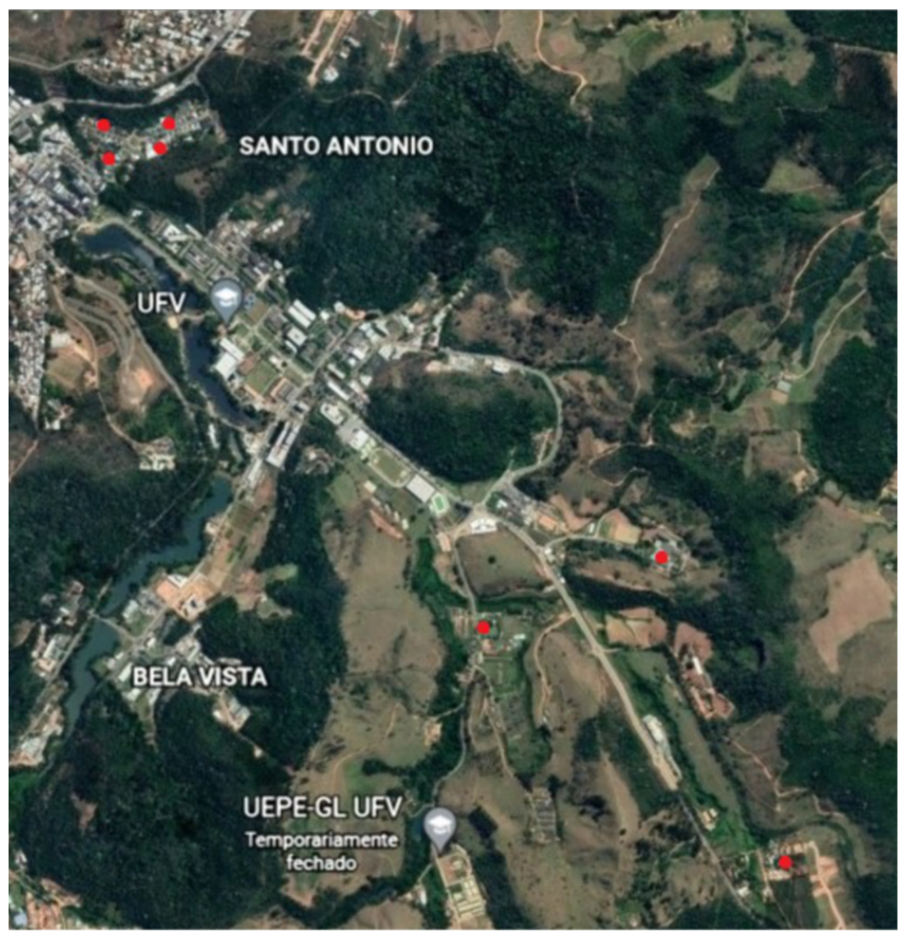
2.1. Study Area

The study was carried out in the Viçosa municipality (22°45′14″ S and 42°52′55″ W), Minas Gerais State, in the Southeast Region of Brazil (Figure 1). The municipality has an area of 299,418 km2 and a has a humid subtropical climate, category Cwa by Kӧppen and Geiger, with an average temperature of 20.4 °C and average rainfall of 1251 mm/year [25].

2.2. Sampling

A total of 30 D. aurita were captured using Tomahawk traps baited with sardines, cornmeal, and bananas. Sampling was performed between March 2021 and June 2022. The animals were physically restrained for clinical examination [26] and blood sampling. Blood samples (up to 2 mL) were collected by venipuncture of the coccygeal lateral vein [27] using sterile EDTA-coated tubes (Becton & Dickinson Co, Franklin Lakes, NJ, USA) for hematological analyses, and later kept at −20 °C until PCR testing. Additionally, two milliliters were placed into tubes without anticoagulant (Becton & Dickinson Co, Franklin Lakes, NJ, USA) and kept at room temperature (25 °C) until visible clot formation. The samples were then centrifuged at 1500 × g for 5 min, serum separated and kept at −20 °C for biochemical analyses.
The animals also were submitted to a visual inspection for presence of ectoparasites (ticks and fleas). Ticks were removed using a commercial Hook and kept in 70% ethanol labeled tube for identification according to previously described morphological taxonomic keys [28].

2.3. Hematological and Biochemical Analyses

For complete blood cell count (CBC) the samples were analyzed with an automatic cell counter (URIT 3000®, MHLab®, Sao Paulo, Brazil). The packed cell volume (PCV) was determined by the micro-hematocrit technique, with animals considered anemic when <0.31 L/L [29]. Total plasma protein (TPP) concentration was determined by refractometer. For leukocyte evaluation, blood smears were stained using a Romanowsky-type stain (Panotico: Instant Prov (Newprov®, Pinhais, Paraná, Brazil) and examined using light microscopy (CX40, Olympus, Tokyo, Japan) adapted to a camera connected to the ToupView software (ToupTek, Zhejiang, China) under 1000× magnification. CBC data were expressed through the mean and standard deviation (SD).
The serum biochemical parameters evaluated were albumin (ALB; bromocresol green), total protein (TP; biuret method), Alanine Aminotransferase (ALT; method: UV kinetic), Aspartate Aminotransferase (AST; method: UV kinetic), Phosphatase Alkaline (PA; Method: optimized kinetic), Gamma glutamyl transferase (GGT; kinetic method), urea (method: kinetic) and creatinine (method: alkaline picrate) obtained through spectrophotometry in an automatic biochemical analyzer (HumaStar 200®, In Vitro Diagnóstica, Belo Horizonte, Brazil) with specific commercial reagents. The results obtained were expressed through the mean and SD.

2.4. Morphometry

The length, width and area of the structures and cells were measured using Image-Pro Plus 4.5 software (Media Cybernetcs1, Silver Spring, MD, USA).

2.5. DNA Extraction

DNA from 200 μL whole blood was extracted using a commercial kit (MagMAXTM CORE Nucleic Acid Purification Kit, TX, USA), according to the manufacturer’s instructions. Ultrapure water was used in parallel as a negative control to monitor cross contamination. To monitor DNA extraction, a conventional PCR assay targeting a fragment of the mammalian endogenous gene glyceraldehyde-3-phosphate dehydrogenase (gapdh) [30] was performed in all samples.

2.6. PCR Assays

Black-eared opossums’ DNA samples were initially screened using a universal hemoplasma SYBR green real-time PCR (qPCR) assay, as previously described [31]. Samples with threshold cycle (CT) value < 32 were considered positive [32] and then submitted to conventional PCR assays targeting a fragment (900 bp) of the 16S rRNA [33,34] and (800 pb) of the 23S rRNA genes of hemoplasmas [35]. Nuclease-free water and Mycoplasma ovis DNA obtained from a naturally infected goat (Capra hircus) [35] were used as negative and positive controls, respectively.
Moreover, black-eared opossums’ DNA samples were also screened by conventional PCR assays targeting a fragment (667 bp) of the 18S rRNA gene of Hepatozoon spp. [26] and a fragment (349 bp) of the dsb gene of Ehrlichia spp. [36]. Hepatozoon canis and Ehrlichia canis DNA obtained from naturally infected dogs and nuclease-free water were used as positive and negative controls, respectively.

2.7. Sequencing and Phylogenetic Analysis

Amplicons (∼900 bp) obtained from two hemotropic Mycoplasma sp.-positive samples were sequenced in both directions by Sanger method, with nucleotide sequences of the 16S rRNA submitted to GenBank® database (accession numbers: OP279616, OP279617). The partial sequences of the 16S rRNA gene of hemotropic Mycoplasma sp. were aligned with other hemotropic Mycoplasma sp. species available in GenBank® using MAFFT 7.110 [37] on the Guidance 2 server [38]. Phylogenetic analyses on the 16S rRNA gene was performed based on Bayesian inference using the Beast 1.8.0 package [39]. Three independent runs of 100,000,000 generations of Monte Carlo Markov Chain (MCMC) were performed, with one sampling per 10,000 generations and a 10% burn in. The substitution model was estimated as GTR + I + G based on the Akaike information criterion (AIC) using jModeltest 2.1.10 [40]. Reconstructions were visualized using the FigTree 1.4.4 software [41]. The 16S rRNA gene tree was rooted with Mycoplasma pneumoniae (CP039761). Moreover, a haplotype network for each fragment was estimated in the PopArt 1.7 [42] using the median-joining method [43].
Amplicons (740 bp) obtained from five Hepatozoon sp.-positive samples were sequenced in both directions by Sanger method, with nucleotide sequences of the 18S rRNA submitted to GenBank® database (accession numbers: OP267564–OP267568). The partial sequences of the 18S rRNA gene of Hepatozoon sp. were aligned with other Hepatozoon species available in GenBank® using MAFFT 7.110 [37] on the Guidance 2 server [38]. Phylogenetic analyses on the 18S rRNA gene was performed based on Bayesian inference using the Beast 1.8.0 package [39]. Three independent runs of 100,000,000 generations of Monte Carlo Markov Chain (MCMC) were performed, with one sampling per 10,000 generations and a 10% burn in. The substitution model was estimated as GTR + G based on the AIC using jModeltest 2.1.10 [40]. Reconstructions were visualized using the FigTree 1.4.4 software [41]. The 18S rRNA gene tree was rooted with Plasmodium vivax (XR_003001206). Moreover, a haplotype network for each fragment was estimated in the PopArt 1.7 [42] using the median-joining method [43].

2.8. Statistical Analysis

To compare the means, t-test (red blood cells, hemoglobin, PCV, MCH, MCHC, RDW, platelets, ALB, TP, globulins, albumin:globulin ratio, creatinine, glucose) was used. Data normality was verified by the Shapiro-Wilk test. Non-parametric analyzes for non-normal variables (MVC, white blood cells, neutrophils, lymphocytes, monocytes, band cells, basophils, length, width, area, TP, fibrinogen, PA, ALT, AST, urea, gamma-glutamyl transferase) were analyzed using the Mann-Whitney test. A significance level of 5% was considered for all analyses. A Fisher’s exact test was used to determine whether individual variables (gender, age group and presence of ectoparasites) was associated with the infection. Odds ratio (OR), 95% confidence interval and p-values were calculated for each variable. Results were considered significantly different when p < 0.05. Data were compiled and analyzed in the software GraphPad Prism (version 9.0, Graphpad, San Diego, CA, USA).

2.9. Ethical Aspects

The study was approved by the Ethics Committee for the Use of Animals-CEUA/UFV by process number 30/2021 under the regulations of the Chico Mendes Institute for Biodiversity Conservation (ICMBio, license number 64930-3).

3. Results

Out of the 30 animals captured, 15 (50.0%) were adult females, four (13.3%) sub-adult females, eight (26.6%) adult males and three (10.0%) sub-adult males (Figure 2). A total of 14/30 (46.66%; 95% CI = 30.23–63.86) black-eared opossums were infested by ticks at the time of sampling. Ticks were identified as I. loricatus (seven F and three M), Amblyomma ovale (three nymphs), A. dubitatum (one nymph) and Amblyomma sp. (21 larvae).

3.1. Hematological and Biochemical Analyses

The mean and range values obtained in the CBC and biochemical analyses of black-eared opossums are summarized in Table 1. A total of three/30 (10.0%; 95% CI = 3.46–25.62) black-eared opossums were anemic.
During blood smears evaluation, hemoplasma-like structures were visualized on the surface of red blood cells (RBCs) from one/30 (3.33%; 95% CI = 0.05–16.67) black-eared opossums (Figure 3). Additionally, 15/30 (50.0%; 95% CI = 33.15–66.85) animals showed Hepatozoon sp. gamonts on blood smears evaluation (Figure 4).
Hepatozoon sp. gamonts found on blood smears presented variations: they were mainly oval, but it was also possible to identify some with a more rounded shape and others with a banana-like shape (Figure 4A–D). The mean and SD measures of Hepatozoon sp. gamonts were length: 10.89 ± 0.92 µm; width: 5.29 ± 0.97 µm; area: 48.51 ± 7.72 µm. When present, the pleomorphic nucleus mean measures were length: 3.90 ± 1.05 µm and width: 2.03 ± 0.81 µm. Measurements were performed on Hepatozoon sp. gamonts inside RBCs and those free on blood smears. When RBCs were infected with Hepatozoon sp. gamonts, cells showed the following mean and SD measures length: 11.7 ± 0.74; Width: 6.83 ± 1.19 µm; Area: 64.53 ± 7.93 µm. RBCs that were not infected presented the following mean and SD measures length: 7.88 ± 0.71; Width: 7.40 ± 0.64 µm; Area of 45.74 ± 7.63 µm. Measures of RBCs infected with Hepatozoon sp. gamonts differed significantly from non-infected RBCs in the length (p < 0.0001), width (p < 0.0001) and area (p < 0.0001).

3.2. PCR Assays

The gapdh gene was consistently amplified in all black-eared opossum samples. A total of 22/30 (73.3%; 95% CI = 55.55–85.82) animals were positive for hemotropic Mycoplasma sp. by the qPCR. The animal that hemoplasma-like structures were visualized on blood smear evaluation has also tested positive by qPCR. Sequencing of the 16S rRNA gene fragments from two hemoplasma-positive black-eared opossums revealed 98.87–98.96% identity with ‘Ca. M. haemodidelphis’ detected in D. albiventris from Brazil (AF178676). Phylogenetic analyses on the 16S rRNA gene of the hemoplasma detected herein clustered together with those of the Mycoplasma suis group. Hemoplasma sequences obtained from D. aurita clustered together with ‘Ca. M. haemoalbiventris’ sequences previously detected in D. albiventris from southern Brazil (Figure 5).
Twelve out of 30 (40%; 95% CI = 24.59–57.68) D. aurita tested positive for Hepatozoon sp. by PCR. Seven out of 15 (46.67%; 95% CI = 24.81–69.88) animals that showed Hepatozoon sp. gamonts on blood smears evaluation also tested positive for Hepatozoon sp. by PCR. A total of eight/30 (26.67% 95% CI = 14.18–44.45) tested positive for hemoplasmas and Hepatozoon sp. by molecular assays, however animals did not show any clinical signs of infection. Sequencing of the 18S rRNA gene fragments from five Hepatozoon-positive D. aurita showed 96.2–96.4% identity with Hepatozoon colubri detected in the aesculapian snake (Zamenis lineatus) from Iran (MN723844.1). Phylogenetic analysis inferred by Bayesian inference and based on a 740 bp alignment (Figure 6) demonstrate the close relationship of the D. aurita Hepatozoon sp. with Hepatozoon sp. genotypes detected in D. marsupialis from Brazil (GenBank® accession no. MK257775).
Two out of 30 (6.66%; 95% CI = 1.85–21.32) animals tested positive for Ehrlichia sp. by PCR. Sequencing of the dsb gene fragment (165 bp) from one Ehrlichia-positive D. aurita showed 99.38% identity with Ehrlichia chaffeensis detected in Hyalomma excavatum tick from Egypt (MN372100) and Odocoileus virginianus from USA (MK611628).

3.3. Statistical Analysis

In hemoplasma-positive D. aurita, the number of platelets were lower (p < 0.05) and the TPP concentration showed a significant increase (p < 0.05) (Table 2). In the biochemical analyses, a similar result was evidenced. The concentration of TP and globulins, as well activity of Alkaline Phosphatase (AP) showed significant increased (p < 0.05) in the hemoplasma-positive animals (Table 2).
In the animals infected by Hepatozoon sp. no significant differences were observed for hematological analyses and in the biochemical analyses, only creatinine showed a significant increase (p < 0.05) in the positive group. No significant associations (p > 0.05) were found between gender, age group or presence of ectoparasites and positivity to hemoplasmas or Hepatozoon sp. (Table 3).

4. Discussion

To the best of authors knowledge, this is the first study on hemoplasmas in D. aurita. Previous studies have reported ‘Ca. M. haemoalbiventris’ in D. albiventris from different Brazilian regions and biomes with prevalence data ranging from 32.5–87.5% by PCR [7,8,9,10]. Herein, 73.3% D. aurita tested positive for ‘Ca. M. haemoalbiventris’ by qPCR. Additionally, ‘Ca. M. haemoalbiventris’ was observed by light microscopy of black-eared opossum’s blood smear as small basophilic epierythrocytic structures of coccoid shape, individually attached and often one per erythrocyte (Figure 3).
The main laboratory alterations were observed in hemoplasma-positive animals. Messick et al. [6] stated that infection by ’Ca. M. haemodidelphis’ produces severe anemia in D. virginiana. In the present study, this was not evident and the values of PCV, hemoglobin and RBCs were higher than those previously reported in D. aurita and D. albiventris [27], and lower than the values obtained by Lewis [44]. Herein, an association between hemoplasma-positive animals and a decrease in the number of platelets was observed. A previous study has described that one of the mechanisms involved in thrombocytopenia in cases of Mycoplasma pneumoniae infection would be related to the production of antibodies against platelets which would induce their destruction by the immune system [45]. In the present study an increased TPP concentration in hemoplasma-positive animals was observed (p < 0.05), that may be related to the antigenic stimulation of pathogens detected [46,47,48]. Whether ‘Ca. M. haemoalbiventris’ is capable of producing a clinical disease similar to ’Ca. M. haemodidelphis’ in D. virginiana [6] remains to be fully established.
In the biochemical analysis, in the hemoplasmas group, we had similar results as observed in the CBC. The concentration of TP (p < 0.05) and globulins (p < 0.05) were higher in animals infected with ‘Ca. M. haemoalbiventris’ than in the negative groups. The mean value of TP obtained in this study was higher at 5.6 ± 0.6 g/dL described by Carvalho Do Nascimento and Horta [27] and the 7.1 g/dL of Lewis [44]. The increase in TP concentration may be a consequence of hyperglobulinemia [46,47,48]. The main proteins present in serum are albumin and globulins. Albumin is a hepatic synthesis protein and among its functions are the transport of substances and the regulation of oncotic pressure. Globulins correspond to a diverse group of proteins such as immunoglobulins, complement system proteins, fibrinogen and others [47].
One of the main causes of hyperglobulinemia are inflammatory processes, either acute or chronic, which can result as a consequence of an increase in one or more globulins [46]. In this case, the infected opossums could have an increase in globulins by the antigenic stimulation that the hemoplasmas produce in the organism of the affected individuals.
The activity of AP was higher (p < 0.05) in the animals that were not infected with ‘Ca. M. haemoalbiventris. Interpreting AP results can be a challenge. The only available reference [27] showed mean values of 11.9 ± 4.2 U/L, much lower than ours. In the physical or laboratory evaluation, it was not possible to identify a cause of those commonly associated (hepatobiliary disease, osteoblastic activity or cortisol induction) to increases AP activity [47] that could justify the variations, therefore, perhaps it is a case of a statistical difference, but not a biological.
In the present study, 50% D. aurita presented Hepatozoon sp. gamonts by light microscopy of blood smears and 40% tested positive by the PCR targeting the 18S rRNA gene of Hepatozoon species. Sequencing confirmed that animals were infected by Hepatozoon sp. closely related to Hepatozoon sp. detected in D. marsupialis from Brazil [15]. Previous studies have reported different Hepatozoon species in marsupials [15,23,49,50] and H. canis in D. albiventris from São Paulo State, southeastern Brazil [14]. Additionally, putative new Hepatozoon sp. have been found in D. marsupialis [15] and D. albiventris from Brazil [19], with an overall prevalence of 45.16% (14/31) and 2.3% (1/43), respectively. Phylogenetic analysis showed that four Hepatozoon sp. sequences detected in D. aurita herein clustered apart from Hepatozoon sp. detected in D. marsupialis from Brazil. Future studies should be performed to elucidate and characterize Hepatozoon sp. infecting black-eared opossums from Brazil.
The morphometric analyzes of the Hepatozoon sp. gamonts detected herein were similar to those described for H. didelphydis [51] found as intra-erythrocytic and free parasites, with spherical or ovoid form with 8–10 × 4–6 µm in the blood from one male black-eared opossum (n = 50) in the city of São João de Meriti, Rio de Janeiro State, southeastern Brazil. In Colombia, in the Eastern Llanos region, Ayala et al. [11] described two types of Haemogregarines. In the first case, D. marsupialis, Philander opossum and Metachirus nudicaudatus showed H. didelphydis on stained blood smears with an average size of 10.5 × 5.0 µm in 10 animals, similar to our findings. In the second group, gamonts of an unidentified Haemogregarine were visualized in the blood smear of D. marsupialis at a different capture site with an average measurement of 11.7 × 6.0 µm (n = 20). According to the authors, the main difference was that in the second group, the RBCs were considerably hypertrophied, and in the first group they only showed a slight increase. In a similar way in the present study, the RBCs that were infected with gamonts were also hypertrophied when compared with not infected cells.
De Thoisy et al. [12] also described two types of Hepatozoon sp. in mammals from French Guyana. In the first case, infecting erythrocytes of D. marsupialis, P. opossum and M. nudicaudatus which increased the red cells size, with a prevalence of 25%, 57% and 5% respectively and that could be related to H. didelphydis, perhaps the same species described by Ayala et al., [11]. In that study, D. albiventris had an infection prevalence of 17%. In this species, the gamonts had measurements of 11.2 ± 1.2 × 8.0 ± 1.1 μm and a pleomorphic nucleus of 7.7 ± 0.9 × 5.1 ± 0.4 μm and according to the authors, it could be a new species. In the study carried out by Da Silva et al. [14] was found in the blood smear of one animal a Hepatozoon gamont that presented 8.88 × 5.17 μm and the nucleus 3.84 × 3.25 μm. In the other hand, André et al. [19] did not identified any gamont in the blood smears of the D. albiventris evaluated.
In the animals infected with Hepatozoon sp. there was no difference in the CBC parameters even when the degree of parasitemia varied among individuals, from a few to countless gamonts (Figure 4A–D). The only difference in the biochemical analysis in the Hepatozoon sp. group was observed in the creatinine value, which was higher in the positive group. The mean value of creatinine was practically identical to the 0.05 ± 0.6 mg/dL mentioned by Carvalho Do Nascimento e Horta [27] used as a reference. In the absence of significant differences in the CBC or biochemical analyzes, it is valid to consider the hypothesis that this Hepatozoon sp. is well adapted to the host because it does not produce an evident clinical disease. To better evaluate this hypothesis, an investigation with a greater number of opossums should be carried out. Perhaps the infection may be similar to dogs with H. canis, and in agreement with what was proposed by Baneth et al., [52] who suggest that this protozoan is well adapted to dogs that, in case of producing disease, it will be subclinical or mild.

5. Conclusions

A high occurrence of ‘Ca. M. haemoalbiventris’ and Hepatozoon sp. was found in D. aurita from the municipality of Viçosa, Minas Gerais State. Infection was not associated with sex, age group and/or the presence of ectoparasites. Infection by ‘Ca. M. haemoalbiventris’ was associated with lower number of platelets, an increase in TPP, TP and globulins concentration. Whether infection by these agents may cause clinical disease in D. aurita remains to be fully established.

Author Contributions

Conceptualization, A.M.O.O., C.C., R.F.d.C.V. and L.A.d.F.; methodology, A.M.O.O., F.C.M.C., T.S.W.J.V., R.F.d.C.V. and L.A.d.F.; formal analysis, A.M.O.O., F.C.M.C., T.S.W.J.V., R.F.d.C.V. and L.A.d.F.; investigation, A.M.O.O., C.C., L.D.B., P.C.S., F.M.G., B.C.F.N., R.S.Y., A.K.C., F.M.-F., F.C.M.C., T.S.W.J.V., R.F.d.C.V. and L.A.d.F.; resources, C.C., R.F.d.C.V. and L.A.d.F.; data curation, A.M.O.O., F.C.M.C., T.S.W.J.V., R.F.d.C.V. and L.A.d.F.; writing—original draft preparation, A.M.O.O., F.C.M.C., T.S.W.J.V., R.F.d.C.V. and L.A.d.F. writing—review and editing, A.M.O.O., C.C., L.D.B., P.C.S., F.M.G., B.C.F.N., R.S.Y., A.K.C., F.M.-F., F.C.M.C., T.S.W.J.V., R.F.d.C.V. and L.A.d.F.; visualization, A.M.O.O., C.C., R.F.d.C.V. and L.A.d.F.; supervision, R.F.d.C.V. and L.A.d.F.; project administration, L.A.d.F.; funding acquisition, R.F.d.C.V. and L.A.d.F. All authors have read and agreed to the published version of the manuscript.

Funding

This research was funded by Fundação de Amparo à Pesquisa do Estado de Minas Gerais (FAPEMIG), grant number 5.273/15. Andrés Ortega and Flavia Collere were sponsored by a fellowship from the Coordenaçaão de Aperfeiçoamento de Pessoal de Nível Superior (CAPES) at the time of this study. Brazilian National Council of Scientific and Technological Development (CNPq) provided a Research Productivity (PQ) fellowship to Fabiano Montiani-Ferreira (CNPq—304715/2019-0) and Rafael Vieira (CNPq—313161/2020-8).

Institutional Review Board Statement

The animal study protocol was approved by the Institutional Ethics Committee for the Use of Animals—CEUA/UFV (process number 30/2021), and it was approved also from the Information and Authorization System on Biodiversity (SISBIO) of the Brazilian Institute of Environment and Renewable Natural Resources (IBAMA) under license number 64930-3.

Informed Consent Statement

Not applicable.

Data Availability Statement

Nucleotide sequences obtained in the present study can be found at Genbank data base (https://www.ncbi.nlm.nih.gov/genbank/, accessed on 29 August 2022) under accession numbers: OP279616, OP279617, OP267564–OP267568.

Acknowledgments

This study is part of a PhD degree for Andrés Ortega at the Universidade Federal de Viçosa.

Conflicts of Interest

The authors declare no conflict of interest.

References

  1. Gardner, A.L. Mammals of South America, Volume 1: Marsupials, Xenarthrans, Shrews, and Bats; University of Chicago Press: Chicago, IL, USA, 2008; ISBN 978-0-226-28242-8. [Google Scholar]
  2. Rossi, R.; Bianconi, G.; Pedro, W.A. Ordem Didelphimorphia. In Mamíferos do Brasil; Universidade Estadual de Londrina: Londrina, Brazil, 2006; pp. 34–37. [Google Scholar]
  3. Bezerra-Santos, M.A.; Fontes, C.S.; Nogueira, B.C.F.; Yamatogi, R.S.; Ramos, R.A.N.; Galhardo, J.A.; Furtado, L.F.V.; Rabelo, É.M.L.; de Araújo, J.V.; Campos, A.K. Gastrointestinal Parasites in the Opossum Didelphis aurita: Are They a Potential Threat to Human Health? J. Parasit. Dis. 2020, 44, 355–363. [Google Scholar] [CrossRef] [PubMed]
  4. Bermúdez, S.E.; Gottdenker, N.; Krishnavajhala, A.; Fox, A.; Wilder, H.K.; González, K.; Smith, D.; López, M.; Perea, M.; Rigg, C.; et al. Correction: Synanthropic Mammals as Potential Hosts of Tick-Borne Pathogens in Panama. PLoS ONE 2019, 14, e0226195. [Google Scholar] [CrossRef] [PubMed]
  5. Bitencourt, M.M.; Bezerra, A.M.R. Infection Agents of Didelphidae (Didelphimorphia) of Brazil: An Underestimated Matter in Zoonoses Research. Mammalia 2022, 86, 105–122. [Google Scholar] [CrossRef]
  6. Messick, J.B.; Walker, P.G.; Raphael, W.; Berent, L.; Shi, X. “Candidatus Mycoplasma haemodidelphidis” sp. nov., “Candidatus Mycoplasma haemolamae” sp. nov. and Mycoplasma haemocanis comb. nov., Haemotrophic Parasites from a Naturally Infected Opossum (Didelphis virginiana), Alpaca (Lama pacos) and Dog (Canis familiaris): Phylogenetic and Secondary Structural Relatedness of Their 16S RRNA Genes to Other Mycoplasmas. Int. J. Syst. Evol. Microbiol. 2002, 52, 693–698. [Google Scholar] [CrossRef]
  7. de Oliveira, R.P.A.; Collere, F.C.M.; Ferrari, L.D.R.; Coradi, V.d.S.; Soares, N.d.A.; Leandro, A.d.S.; de Oliveira, W.F.; Galvão, S.R.; Kafka, R.; Delai, R.M.; et al. ‘Candidatus Mycoplasma haemoalbiventris’ and Tick-Borne Pathogens Screening in White-Eared Opossums (Didelphis albiventris) from Curitiba and Foz Do Iguaçu Cities, Paraná State, Southern Brazil. Rev. Bras. Parasitol. Vet. 2021, 30, e009721. [Google Scholar] [CrossRef] [PubMed]
  8. Pontarolo, G.H.; Kühl, L.F.; Pedrassani, D.; Campos, M.; Borges Figuereido, F.; Valente Marinho, J.D.; Gonçalves, L.R.; André, M.R.; Wischral, T.S.; Vieira, J.; et al. ‘Candidatus Mycoplasma Haemoalbiventris’, a Novel Hemoplasma Species in White-eared Opossums (Didelphis Albiventris) from Brazil. Transbound Emerg. Dis. 2021, 68, 565–572. [Google Scholar] [CrossRef] [PubMed]
  9. Massini, P.F.; Drozino, R.N.; Otomura, F.H.; Mongruel, A.C.B.; Valente, J.D.M.; Toledo, M.J.d.O.; Martins, T.F.; Vidotto, O.; Vieira, T.S.W.J.; Vieira, R.F.d.C. Detection of Hemotropic Mycoplasma sp. in White-Eared Opossums (Didelphis albiventris) from Southern Brazil. Rev. Bras. Parasitol. Vet. 2019, 28, 797–801. [Google Scholar] [CrossRef]
  10. Gonçalves, L.R.; Herrera, H.M.; Nantes, W.A.G.; Santos, F.M.; Porfírio, G.E.d.O.; Barreto, W.T.G.; de Macedo, G.C.; Assis, W.d.O.; Campos, J.B.V.; da Silva, T.M.V.; et al. Genetic Diversity and Lack of Molecular Evidence for Hemoplasma Cross-Species Transmission between Wild and Synanthropic Mammals from Central-Western Brazil. Acta Trop. 2020, 203, 105303. [Google Scholar] [CrossRef]
  11. Ayala, S.C.; D’Alessandro, A.; Mackenzie, R.; Angel, D. Hemoparasite Infections in 830 Wild Animals from the Eastern Llanos of Colombia. J. Parasitol. 1973, 59, 52–59. [Google Scholar] [CrossRef]
  12. de Thoisy, B.; Michel, J.C.; Vogel, I.; Vié, J.C. A Survey of Hemoparasite Infections in Free-Ranging Mammals and Reptiles in French Guiana. J. Parasitol. 2000, 86, 1035–1040. [Google Scholar] [CrossRef]
  13. Allen, K.E.; Yabsley, M.J.; Johnson, E.M.; Reichard, M.V.; Panciera, R.J.; Ewing, S.A.; Little, S.E. Novel Hepatozoon in Vertebrates from the Southern United States. J. Parasitol. 2011, 97, 648–653. [Google Scholar] [CrossRef] [PubMed]
  14. da Silva, M.R.L.; Fornazari, F.; Demoner, L.d.C.; Teixeira, C.R.; Langoni, H.; O’Dwyer, L.H. Didelphis albiventris Naturally Infected with Hepatozoon canis in Southeastern Brazil. Ticks Tick Borne Dis. 2017, 8, 878–881. [Google Scholar] [CrossRef] [PubMed]
  15. Colle, A.C.; de Mendonça, R.F.B.; Maia, M.O.; Freitas, L.d.C.; Witter, R.; Marcili, A.; de Aguiar, D.M.; Muñoz-Leal, S.; Labruna, M.B.; Rossi, R.V.; et al. Molecular Survey of Tick-Borne Pathogens in Small Mammals from Brazilian Amazonia. Rev. Bras. Parasitol. Vet. 2019, 28, 592–604. [Google Scholar] [CrossRef] [PubMed]
  16. Gonçalves, L.R.; Paludo, G.; Bisol, T.B.; Perles, L.; de Oliveira, L.B.; de Oliveira, C.M.; da Silva, T.M.V.; Nantes, W.A.G.; Duarte, M.A.; Santos, F.M.; et al. Molecular Detection of Piroplasmids in Synanthropic Rodents, Marsupials, and Associated Ticks from Brazil, with Phylogenetic Inference of a Putative Novel Babesia sp. from White-Eared Opossum (Didelphis albiventris). Parasitol. Res. 2021, 120, 3537–3546. [Google Scholar] [CrossRef]
  17. Soares, H.S.; Marcili, A.; Barbieri, A.R.M.; Minervino, A.H.H.; Moreira, T.R.; Gennari, S.M.; Labruna, M.B. Novel Piroplasmid and Hepatozoon Organisms Infecting the Wildlife of Two Regions of the Brazilian Amazon. Int. J. Parasitol. Parasites Wildl. 2017, 6, 115–121. [Google Scholar] [CrossRef]
  18. Lopes, M.G.; Muñoz-Leal, S.; de Lima, J.T.R.; Fournier, G.F.d.S.R.; Acosta, I.d.C.L.; Martins, T.F.; Ramirez, D.G.; Gennari, S.M.; Labruna, M.B. Ticks, Rickettsial and Erlichial Infection in Small Mammals from Atlantic Forest Remnants in Northeastern Brazil. Int. J. Parasitol. Parasites Wildl. 2018, 7, 380–385. [Google Scholar] [CrossRef]
  19. André, M.R.; Calchi, A.C.; Perles, L.; Gonçalves, L.R.; Uccella, L.; Lemes, J.R.B.; Nantes, W.A.G.; Santos, F.M.; Porfírio, G.E.d.O.; Barros-Battesti, D.M.; et al. Novel Ehrlichia and Hepatozoon Genotypes in White-Eared Opossums (Didelphis albiventris) and Associated Ticks from Brazil. Ticks Tick-Borne Dis. 2022, 13, 102022. [Google Scholar] [CrossRef]
  20. Miziara, S.R.; Paiva, F.; Andreotti, R.; Koller, W.W.; Lopes, V.A.; Pontes, N.T.; Bitencourt, K. Ocorrência de Ixodes loricatus Neumann, 1899 (Acari: Ixodidae) parasitando Didelphis albiventris (Lund, 1841), (Didelphimorphia: Didelphidae), em Campo Grande, MS. Rev. Bras. Parasitol. Vet. 2008, 17, 158–160. [Google Scholar] [CrossRef]
  21. Horta, M.C.; Labruna, M.B.; Pinter, A.; Linardi, P.M.; Schumaker, T.T.S. Rickettsia Infection in Five Areas of the State of São Paulo, Brazil. Mem. Inst. Oswaldo Cruz 2007, 102, 793–801. [Google Scholar] [CrossRef]
  22. Bezerra-Santos, M.A.; Nogueira, B.C.F.; Yamatogi, R.S.; Ramos, R.A.N.; Galhardo, J.A.; Campos, A.K. Ticks, Fleas and Endosymbionts in the Ectoparasite Fauna of the Black-Eared Opossum Dipelphis aurita in Brazil. Exp. Appl. Acarol. 2020, 80, 329–338. [Google Scholar] [CrossRef]
  23. Sousa, K.C.M.D.; Calchi, A.C.; Herrera, H.M.; Dumler, J.S.; Barros-Battesti, D.M.; Machado, R.Z.; André, M.R. Anaplasmataceae Agents among Wild Mammals and Ectoparasites in Brazil. Epidemiol. Infect. 2017, 145, 3424–3437. [Google Scholar] [CrossRef] [PubMed]
  24. Guimarães, A.; Raimundo, J.M.; da Silva, A.T.; Carpintero, F.M.; Pires, J.R.; Benevenute, J.L.; Machado, R.Z.; André, M.R.; Baldani, C.D. Detection of a Putative Novel Genotype of Ehrlichia sp. from Opossums (Didelphis aurita) from Brazil. Rev. Bras. Parasitol. Vet. 2018, 28, 140–144. [Google Scholar] [CrossRef] [PubMed]
  25. Climate-Data.Org. Viçosa Climate: Average Temperature, Weather by Month, Viçosa Weather Averages. Available online: https://en.climate-data.org/south-america/brazil/minas-gerais/vicosa-25021/ (accessed on 16 August 2022).
  26. Inokuma, H.; Okuda, M.; Ohno, K.; Shimoda, K.; Onishi, T. Analysis of the 18S rRNA Gene Sequence of a Hepatozoon Detected in Two Japanese Dogs. Vet. Parasitol. 2002, 106, 265–271. [Google Scholar] [CrossRef]
  27. Carvalho do Nascimento, C.; Horta, M.C. Didelphimorphia (Gambá e Cuíca). In Tratado de Animais Selvagens: Medicina Veterinária; Roca: São Paulo, Brazil, 2014; Volume 1, ISBN 978-85-277-2648-1. [Google Scholar]
  28. Barros-Battesti, D.M.; Arzua, M.; Bechara, G.H. Carrapatos de importância médico-veterinária da região neotropical: Um guia ilustrado para identificação de espécies; Vox/ICTTD-3/Butantan: São Paulo, Brezil, 2006; 223p, ISBN 85-99909-01-0. [Google Scholar]
  29. Casagrande, R.A.; Cesar, M.d.O.; Horta, M.C.; Rossi, S.; Teixieira, R.H.; Matushima, E.R. Perfil hematológico de gambás Didelphis aurita e D. albiventris do Estado de São Paulo, Brasil. Acta Scientiarum. Biol. Sci. 2009, 31, 185–189. [Google Scholar] [CrossRef]
  30. Birkenheuer, A.J.; Levy, M.G.; Breitschwerdt, E.B. Development and Evaluation of a Seminested PCR for Detection and Differentiation of Babesia gibsoni (Asian Genotype) and B. canis DNA in Canine Blood Samples. J. Clin. Microbiol. 2003, 41, 4172–4177. [Google Scholar] [CrossRef]
  31. Willi, B.; Meli, M.L.; Lüthy, R.; Honegger, H.; Wengi, N.; Hoelzle, L.E.; Reusch, C.E.; Lutz, H.; Hofmann-Lehmann, R. Development and Application of a Universal Hemoplasma Screening Assay Based on the SYBR Green PCR Principle. J. Clin. Microbiol. 2009, 47, 4049–4054. [Google Scholar] [CrossRef]
  32. Vieira, R.F.d.C.; Vidotto, O.; Vieira, T.S.W.J.; Guimaraes, A.M.S.; dos Santos, A.P.; Nascimento, N.C.; dos Santos, N.J.R.; Martins, T.F.; Labruna, M.B.; Marcondes, M.; et al. Molecular Investigation of Hemotropic Mycoplasmas in Human Beings, Dogs and Horses in a Rural Settlement in Southern Brazil. Rev. Inst. Med. Trop. São Paulo 2015, 57, 353–357. [Google Scholar] [CrossRef]
  33. Machado, C.A.L.; Vidotto, O.; Conrado, F.O.; Santos, N.J.R.; Valente, J.D.M.; Barbosa, I.C.; Trindade, P.W.S.; Garcia, J.L.; Biondo, A.W.; Vieira, T.S.W.J.; et al. Mycoplasma ovis Infection in Goat Farms from Northeastern Brazil. Comp. Immunol. Microbiol. Infect. Dis. 2017, 55, 1–5. [Google Scholar] [CrossRef]
  34. Hoelzle, K.; Winkler, M.; Kramer, M.M.; Wittenbrink, M.M.; Dieckmann, S.M.; Hoelzle, L.E. Detection of ‘Candidatus Mycoplasma haemobos’ in Cattle with Anaemia. Vet. J. 2011, 187, 408–410. [Google Scholar] [CrossRef]
  35. Mongruel, A.C.B.; Spanhol, V.C.; Valente, J.D.M.; Porto, P.P.; Ogawa, L.; Otomura, F.H.; Marquez, E.d.S.; André, M.R.; Vieira, T.S.W.J.; Vieira, R.F.d.C. Survey of Vector-Borne and Nematode Parasites Involved in the Etiology of Anemic Syndrome in Sheep from Southern Brazil. Rev. Bras. Parasitol. Vet. 2020, 29, 1–12. [Google Scholar] [CrossRef]
  36. Parola, P.; Roux, V.; Camicas, J.-L.; Baradji, I.; Brouqui, P.; Raoult, D. Detection of Ehrlichiae in African Ticks by Polymerase Chain Reaction. Trans. R. Soc. Trop. Med. Hyg. 2000, 94, 707–708. [Google Scholar] [CrossRef]
  37. Katoh, K.; Standley, D.M. MAFFT Multiple Sequence Alignment Software Version 7: Improvements in Performance and Usability. Mol. Biol. Evol. 2013, 30, 772–780. [Google Scholar] [CrossRef] [PubMed]
  38. Sela, I.; Ashkenazy, H.; Katoh, K.; Pupko, T. GUIDANCE2: Accurate Detection of Unreliable Alignment Regions Accounting for the Uncertainty of Multiple Parameters. Nucleic Acids Res. 2015, 43, W7–W14. [Google Scholar] [CrossRef] [PubMed]
  39. Drummond, A.J.; Suchard, M.A.; Xie, D.; Rambaut, A. Bayesian Phylogenetics with BEAUti and the BEAST 1.7. Mol. Biol. Evol. 2012, 29, 1969–1973. [Google Scholar] [CrossRef] [PubMed]
  40. Darriba, D.; Taboada, G.L.; Doallo, R.; Posada, D. JModelTest 2: More Models, New Heuristics and Parallel Computing. Nat. Methods 2012, 9, 772. [Google Scholar] [CrossRef]
  41. Rambaut, A. FigTree; University of Edinburgh, Institute of Evolutionary Biology: Edinburgh, UK, 2016. [Google Scholar]
  42. Leigh, J.W.; Bryant, D. Popart: Full-Feature Software for Haplotype Network Construction. Methods Ecol. Evol. 2015, 6, 1110–1116. [Google Scholar] [CrossRef]
  43. Bandelt, H.J.; Forster, P.; Röhl, A. Median-Joining Networks for Inferring Intraspecific Phylogenies. Mol. Biol. Evol. 1999, 16, 37–48. [Google Scholar] [CrossRef]
  44. Lewis, J.H. Comparative Hematology: Studies on Opossums Didelphis Marsupialis (virginianus). Comp. Biochem. Physiol. Part A Physiol. 1975, 51, 275–280. [Google Scholar] [CrossRef]
  45. Aviner, S.; Miskin, H.; London, D.; Horowitz, S.; Schlesinger, M. Mycoplasma Pneumonia Infection: A Possible Trigger for Immune Thrombocytopenia. Indian J. Hematol. Blood Transfus. 2011, 27, 46–50. [Google Scholar] [CrossRef]
  46. Scott, M.A.; Stockham, S.L. Fundamentals of Veterinary Clinical Pathology; John Wiley & Sons: Hoboken, NJ, USA, 2013; ISBN 978-1-118-68607-2. [Google Scholar]
  47. Thrall, M.A.; Weiser, G.; Allison, R.W.; Campbell, T.W. Veterinary Hematology and Clinical Chemistry; John Wiley & Sons: Hoboken, NJ, USA, 2012; ISBN 978-1-118-37840-3. [Google Scholar]
  48. Kaneko, J.J.; Harvey, J.W.; Bruss, M.L. Clinical Biochemistry of Domestic Animals; Academic Press: Cambridge, MA, USA, 2008; ISBN 978-0-08-056882-9. [Google Scholar]
  49. Wicks, R.M.; Spencer, P.B.S.; Moolhuijzen, P.; Clark, P. Morphological and Molecular Characteristics of a Species of Hepatozoon Miller, 1908 (Apicomplexa: Adeleorina) from the Blood of Isoodon Obesulus (Marsupialia: Peramelidae) in Western Australia. Syst. Parasitol. 2006, 65, 19–25. [Google Scholar] [CrossRef]
  50. Merino, S.; Vásquez, R.A.; Martínez, J.; Celis-Diez, J.L.; Gutiérrez-Jiménez, L.; Ippi, S.; Sánchez-Monsalvez, I.; Martínez-De La Puente, J. Molecular Characterization of an Ancient Hepatozoon Species Parasitizing the ‘Living Fossil’ Marsupial ‘Monito Del Monte’ Dromiciops Gliroides from Chile. Biol. J. Linn. Soc 2009, 98, 568–576. [Google Scholar] [CrossRef]
  51. Silva, O.d.’U.; Arantes, J.B. Sobre Uma Hemogregarina Da Gamba. Memórias Do Inst. Oswaldo Cruz 1918, 8, 61–63. [Google Scholar] [CrossRef]
  52. Baneth, G.; Mathew, J.S.; Shkap, V.; Macintire, D.K.; Barta, J.R.; Ewing, S.A. Canine Hepatozoonosis: Two Disease Syndromes Caused by Separate Hepatozoon spp. Trends Parasitol. 2003, 19, 27–31. [Google Scholar] [CrossRef]
Figure 1. Black-eared opossum (Didelphis aurita) capture sites, Viçosa municipality, Minas Gerais State, southeastern Brazil.
Microorganisms 10 01955 g001
Figure 2. A black-eared opossum (Didelphis aurita), male, adult. (A) Inside the Tomahawk trap. (B) During physical restraint.
Microorganisms 10 01955 g002
Figure 3. Light microscopy image of Romanowsky-type-stained peripheral blood smear from black-eared opossum (Didelphis aurita), showing small basophilic and round structures attached to erythrocytes (arrows). Sample from adult female black-eared opossum (1000× magnification). Bar = 10 µm.
Microorganisms 10 01955 g003
Figure 4. Light microscopy image of Romanowsky-type-stained peripheral blood smear from black-eared opossum (Didelphis aurita), showing (A,B) intra-erythrocyte Hepatozoon sp. gamonts (arrow), and (C,D) free (between cells). Sample from female adult (A,B,D) and a male adult (C) black-eared opossum (1000× magnification). Bar = 10 µm.
Microorganisms 10 01955 g004
Figure 5. Phylogenetic tree based on partial sequences of the 16S rRNA gene (850 bp), showing the relationship between the hemotropic Mycoplasma sp. detected in the black-eared opossums (Didelphis aurita) from this study and other hemoplasmas. Mycoplasma pneumoniae was used as outgroup. The GenBank accession number is in parentheses after the species name and origin of each agent. Bayesian inferences were carried out applying the GTR + I + G model and 1000 bootstrap replicates for all analyses.
Microorganisms 10 01955 g005
Figure 6. Phylogenetic tree based on partial sequences of the 18S rRNA gene (740 bp), showing the relationship between the Hepatozoon sp. detected in the black-eared opossums (Didelphis aurita) from this study and other Hepatozoon species. Mycoplasma pneumoniae was used as outgroup. The GenBank accession number is in parentheses after the species name and origin of each agent. Bayesian inferences were carried out applying the GTR + I + G model and 1000 bootstrap replicates for all analyses.
Microorganisms 10 01955 g006
Table 1. Mean, standard deviation and range of complete blood count (CBC) and serum biochemical results of black-eared opossums (Didelphis aurita) (n = 30).
Erythrocytes
AnalyteMean ± SDRange (Min-Max)
Red blood cells (×106/µL)4.31 ± 1.061.68–7.33
Hemoglobin (g/dL)11.77 ± 1.995.80–15.60
PCV (%)38.15 ± 6.4719.00–50.30
Total protein (g/dL)7.41 ± 0.855.80–9.60
MVC (fl)84.00 ± 8.17 65.50–110.10
MCH (pg)28.47 ± 5.2816.23–39.82
MCHC (%)30.91 ± 1.8426.90–34.23
RDW (%)10.02 ± 0.698.70–11.40
Fibrinogen (g/dL)0.23 ± 0.090.20–0.60
Leukocytes
AnalyteMean ± SDRange (Min-Max)
Leukocytes /µL14,596.67 ± 6200.585800–36,700
Neutrophils /µL6755.37 ± 3614.522300–17,616
Lymphocytes /µL5306.87 ± 2499.46456–11,233
Monocytes /µL416.43 ± 493.1073–2202
Band /µL60.33 ± 1580–734
Eosinophils /µL1790.77 ± 1603.630–6606
Basophils /µL202.30 ± 246.690–1088
Platelets
AnalyteMean ± SDRange (Min-Max)
Platelets ×103/µL284.96 ± 173.2332–716
Serum biochemical analyses
AnalyteMean ± SDRange (Min-Max)
Albumin (g/dL)3.02 ± 0.441.85–3.75
Total protein (g/dL)7.58 ± 1.125.49–9.33
Globulins (g/dL)4.56 ± 1.202.77–7.48
Albumin:globulin ratio0.70 ± 0.200.24–1.09
AP (U/L)1009.30 ± 855.14223.00–3345.00
ALT (U/L)65.82 ± 48.1929.80–226.40
AST (U/L)192.93 ± 162.553.00–994.00
Urea (mg/dL)71.37 ± 17.4450.00–292.00
Creatinine (mg/dL)0.49 ± 0.190.00–0.76
GGT (U/L)31.00 ± 11.5818.00–65.00
Glucose (mg/dL)120.71 ± 46.7644.80–256.60
SD: standard deviation; PA: alkaline phosphatase; ALT: alanine aminotransferase; AST: aspartate aminotransferase; GGT: gamma glutamyl transferase.
Table 2. Significant differences * (p < 0.05) in the concentration of some of the analytes evaluated of D. aurita with hemoplasma infection.
Hematology
Analyte NegativePositivep-Value
Red blood cells (×106/µL)4.596 ± 1.2594.199 ± 0.9890.3734
Hemoglobin (g/dL)11.65 ± 0.8911.81 ± 2.280.8465
PCV (%)38.51 ± 2.9038.02 ± 7.410.8570
Total protein (g/dL)6.800 ± 0.59 7.800 ± 0.790.0083 *
MVC (fl)85.75 ± 7.884.40 ± 8.340.9542
MCH (pg)27.53 ± 6.6728.81± 4.810.5642
MCHC (%)30.34 ± 2.5131.12 ± 1.550.3111
RDW (%)10.36 ± 0.649.900 ± 0.670.1034
Fibrinogen (g/dL)0.2000 ± 0.070.2000 ± 0.09>0.9999
Leukocytes /µL13,250 ± 4724.6913,000 ± 6751.730.9817
Neutrophils /µL5636 ± 3729.826341± 3647.430.6622
Lymphocytes /µL4655 ± 1857.245265 ± 2733.69>0.9999
Monocytes /µL187.0 ± 235.15243.5 ± 551.480.3156
Band /µL72.75 ± 148.5155.81 ± 164.580.8620
Eosinophils /µL1514 ± 1729.281320 ± 1598.270.9451
Basophils /µL167.0 ± 227.87165.5 ± 258.27>0.9999
Platelets ×103/µL452,500 ± 184,945224,045 ± 124,3440.0006 *
Serum biochemical analyses
Albumin (g/dL)2.779 ± 0.153.005 ± 0.390.1271
Total protein (g/dL)6.370 ± 0.567.559 ± 1.030.0048 *
Globulins (g/dL)3.591 ± 0.604.555 ± 1.110.0285 *
Albumin:globulin ratio0.7935 ± 0.130.7035 ± 0.200.2579
AP (U/L)1932 ± 827.10857.0 ± 706.910.0021 *
ALT (U/L)46.90 ± 11.7655.00 ± 48.090.1154
AST (U/L)117.5 ± 152.61233.5 ± 229.330.2371
Urea (mg/dL)78.65 ± 19.1973.50 ± 59.990.7214
Creatinine (mg/dL0.4600 ± 0.150.4591 ± 0.190.9905
GGT (U/L)27.50 ± 7.3427.00 ± 12.460.6525
Glucose (mg/dL)126.4 ± 51.13126.4 ± 69.340.9994
Table 3. Prevalence of hemotropic mycoplasmas and Hepatozoon sp. in D. aurita within each variable studied, Viçosa, Minas Gerais, Brazil.
Hemotropic Mycoplasmas
Variable+/nOR (95% CI)p-Value
Gender
Male7/11 (63.33)0.4667 (0.1066–2.036)0.4172
Female15/19 (78.94)
Age group
Adult17/23 (73.91)1.1333 (0.1858–5.956)>0.9999
Sub adult5/7 (71.42)
Presence of ectoparasites
Ectoparasite17/21 (80.95)1.7000 (0.2578–11,58)0.6219
Without ectoparasite5/9 (55.55)
Hepatozoon sp.
Variable+/nOR (95% CI)p-Value
Gender
Male3/10 (30.00)2.0000 (0.3769–10.13)0.6382
Female3/17 (17.64)
Age group
Adult5/20 (25.00)2.0000 (0.2494–27.07)>0.9999
Sub adult1/7 (14.28)
Presence of ectoparasites
Ectoparasite4/19 (21.05)0.8000 (0.1267–5.119)>0.9999
Publisher’s Note: MDPI stays neutral with regard to jurisdictional claims in published maps and institutional affiliations.

