Article

Ralstonia solanacearum Facing Spread-Determining Climatic Temperatures, Sustained Starvation, and Naturally Induced Resuscitation of Viable but Non-Culturable Cells in Environmental Water

by
Belén Álvarez
1,2,†,
María M. López
1 and
Elena G. Biosca
2,*
1
Departamento de Bacteriología, Instituto Valenciano de Investigaciones Agrarias (IVIA), 46113 Valencia, Spain
2
Departamento de Microbiología y Ecología, Universitat de València (UV), 46100 Valencia, Spain
*
Author to whom correspondence should be addressed.
Present address: Departamento de Investigación Aplicada y Extensión Agraria, Instituto Madrileño de Investigación y Desarrollo Rural, Agrario y Alimentario (IMIDRA), 28800 Madrid, Spain.
Microorganisms 2022, 10(12), 2503; https://doi.org/10.3390/microorganisms10122503
Submission received: 31 October 2022 / Revised: 6 December 2022 / Accepted: 12 December 2022 / Published: 16 December 2022
(This article belongs to the Special Issue Plant Pathogenic Microorganisms: State-of-the-Art Research in Spain)

Abstract

:
Ralstonia solanacearum is a bacterial phytopathogen affecting staple crops, originally from tropical and subtropical areas, whose ability to survive in temperate environments is of concern under global warming. In this study, two R. solanacearum strains from either cold or warm habitats were stressed by simultaneous exposure to natural oligotrophy at low (4 °C), temperate (14 °C), or warm (24 °C) temperatures in environmental water. At 4 °C, the effect of temperature was higher than that of oligotrophy, since R. solanacearum went into a viable but non-culturable (VBNC) state, which proved to be dependent on water nutrient contents. Resuscitation was demonstrated in vitro and in planta. At 14 °C and 24 °C, the effect of oligotrophy was higher than that of temperature on R. solanacearum populations, displaying starvation-survival responses and morphological changes which were stronger at 24 °C. In tomato plants, starved, cold-induced VBNC, and/or resuscitated cells maintained virulence. The strains behaved similarly regardless of their cold or warm areas of origin. This work firstly describes the natural nutrient availability of environmental water favoring R. solanacearum survival, adaptations, and resuscitation in conditions that can be found in natural settings. These findings will contribute to anticipate the ability of R. solanacearum to spread, establish, and induce disease in new geographical and climatic areas.

1. Introduction

The plant pathogen Ralstonia solanacearum is a relevant species and former constituent of the R. solanacearum species complex [1,2,3]. It causes severe wilt disease and economic losses in solanaceous and other basic crops for human consumption worldwide as well as in important ornamentals [4,5,6,7,8]. The pathogen frequently cannot be effectively controlled due to its high pathogenic potential and persistence in natural settings. Bacterial wilt control in the field has frequently been addressed by conventional methods, mainly agrochemicals and/or cultural practices, with variable results, and often with environmental impact [9,10]. Alternatively, biological control methods are being explored, such as bacteriophage-based treatments. The use of lytic bacteriophages may be an eco-sustainable strategy because of their specificity and bactericidal activity, although until now no bacteriophage-based product is commercially available against R. solanacearum [10]. Therefore, this pathogen poses a threat to the maintenance of global food security. In fact, the species has a quarantine status in the European Union (EU), the USA and Canada [5,11,12], and is considered a priority pathogen in agriculture for control and containment [13]. A major concern is that R. solanacearum seems to hold great potential for geographical expansion even under environmentally unsuitable conditions, as it appears to infect plants and persist during variable periods in soil or surface water as a free-living form or associated to plant material or non-host roots [4,14,15,16,17]. This is despite the exposure to abiotic stresses compromising the endurance of the bacterium, such as sustained oligotrophy and sub-optimal temperatures. In water systems, it can be consistently detected for years after its introduction, maintaining pathogenicity [16,17,18,19,20,21,22]. This creates a problem for farmers, as water is a scarce resource, particularly under the current conditions of global warming. According to EU and other countries’ regulations, there is a ban on the irrigation of host plants with R. solanacearum-contaminated water as long as the bacterium is detected. The procedure for detection is mainly based on the molecular identification of R. solanacearum colonies isolated from the water. Global climatic changes are thought to increase extreme climate events and the prevalence of abiotic stresses. It is therefore decisive to understand the impact of prevailing environmental factors on R. solanacearum persistence in watercourses to be able to foresee changes regarding water-borne dissemination of the pathogen [6,8,12] and colonization of new geographical and climatic areas.
In that sense, knowledge about the effects of environmental temperatures on microorganisms is crucial to understand bacterial growth and adaptations facing global warming, as well as pathogen virulence and expression of symptoms in the plant [23]. Further, increased temperature is frequently associated with increased severity of the bacterial wilt disease [14,24,25]. Likewise, in watercourses, R. solanacearum population levels seasonally varied according to a range of temperatures [15,16,17], and persistence was also variable in agricultural water microcosms [26]. R. solanacearum, considered a pathogen of tropical and subtropical regions, is capable of causing bacterial wilt in temperate latitudes [27]. However, results from epidemiological studies are contradictory regarding the ability of R. solanacearum to survive in cold conditions away from plants [28]. Although the pathogen becomes viable but non-culturable (VBNC) in pure water by prolonged exposure to 4 °C [26,28,29], the fact that up to date the dynamics of this process has not been monitored in more realistic environmental water microcosms limits the extent to which the results can be extrapolated to the field [28].
Not only temperature, but also the limited nutrient availability characteristic of environmental water has long been claimed to affect bacterial survival in natural settings [30,31]. Weather events are likely to increase the impact of nutrient availability on bacterial communities in the environment. Without climate adaptation strategies, bacteria will probably have to either disseminate or stay and face starvation. R. solanacearum can persist in water for different periods [26,29,32,33,34,35], remaining pathogenic up to four years in environmental water at a favorable temperature through strategies, such as starvation-survival responses, the VBNC state, transition to coccoid cells, and aggregation [6], mechanisms evolved by non-sporulating bacteria facing adverse environmental conditions [30,31,36,37]. However, the effects of the simultaneous exposure to environmental abiotic stresses, such as different climatic temperatures and starvation on this pathogen survival and adaptation capability in natural environmental water have not yet been clarified.
Furthermore, after induction to the VBNC state by exposure to low temperature, transition to a fully culturable and pathogenic state or resuscitation may occur in favorable environmental conditions [37,38], although evidence for this requires a clear-cut distinction between true resuscitation (reversion from non-culturability to culturability) and regrowth (multiplication of a few culturable cells that had remained undetected). In fact, attempts to resuscitate cold-induced VBNC R. solanacearum in water have been carried out by the addition of hydrogen peroxide-degrading compounds, such as catalase to standard solid media to release the pathogen from oxidative stress [28,29]. However, R. solanacearum resuscitation by a simple reversal of low temperature has not been documented so far, despite being the main environmental VBNC inducing factor in natural settings, and a key factor in bacterial wilt outbreaks. This finding could have relevant epidemiological consequences under global warming, since increases in water temperature could lead to increased geographical expansion and/or incidence of water-borne infections resulting from resuscitated cells of this phytopathogenic bacterial species.
This work addressed, for the first time, the simultaneous effect of different temperatures and starvation on stress induction of two R. solanacearum strains (former R. solanacearum phylotype II) from either cold or warm habitats, and their responses in environmental water microcosms. Further, the capability of these R. solanacearum strains to resuscitate and keep pathogenic under conditions that can be found in natural settings was also firstly demonstrated. Knowledge derived from this work will help to foresee tendencies in R. solanacearum persistence and dissemination in aquatic systems within the frame of global warming, as well as their capability for establishment and disease induction in new geographical and climatic areas.

2. Materials and Methods

2.1. Bacterial Strains and Culture Conditions

Two bacterial strains of the present species R. solanacearum [2,3] isolated from either warm or cold habitats were used: strain IVIA-1602.1, from a diseased potato tuber from Canary Islands (Spain), and strain IPO-1609, from a diseased potato plant from The Netherlands [29], both are race 3, biovar 2 of the former R. solanacearum phylotype II. They were kept at −80 °C in a 30% (v/v) glycerol medium and routinely grown on the non-selective Yeast Peptone Glucose Agar (YPGA) [39] for 72 h at 29 °C. In stress induction assays, bacterial culturability was tested on YPGA and the Semiselective Medium South Africa (SMSA) agar developed for R. solanacearum isolation [40] after incubation at 29 °C for 72 h. SMSA medium was also used to re-isolate the pathogen from the host (tomato plants). Both media are frequently used for R. solanacearum isolation, since the colonies of the pathogen can be easily recognized as typically smooth. YPGA contains filtered-sterilized glucose, from which R. solanacearum produces a large amount of extracellular polysaccharide. Colonies are fluidal with pearly cream-white whorls. With respect to SMSA, semiselectivity is mainly based on the action of four antibiotics (penicillin, polymyxin, chloramphenicol and bacitracin), triphenyl-tetrazolium chloride, and crystal violet. Colonies are fluidal with reddish whorls.

2.2. Characteristics of Environmental and Distilled Water Samples

River water samples were collected according to [16] from four different locations in Spain, and nutrient contents were separately determined for each of them. In the different water samples, organic matter levels were from 2 to 3.73% (w/v), and the main ion concentrations ranged as follows (values per liter): Na+, 9.7–9.9 mg; K+, 2.1–2.9 mg; Ca2+, 10.1–13.0 mg; Mg2+, 3.9–5.0 mg; dissolved Fe, 0.24–0.27 mg; Mn, 0.06–0.11 mg; Cu, <0.024 mg; dissolved Zn2+, <0.018 mg; CO32−, <1.8 mg; NO3−, 4.37–5.93 mg; P2O5, 0.374–0.583 mg, and Cl, 9.4–11.4 mg. Salt contents in the samples were correspondent with conductivity values from 151 to 168 µSiemens/cm at 20 °C, and pH values were from 7.48 to 7.83. Distilled water, used for comparative purposes in some assays, had no organic matter and only trace mineral ions: at 20 °C, conductivity was ≤20 µSiemens/cm, and pH value was 7. All water samples were stored in the refrigerator.

2.3. Preparation and Monitoring of Stressed R. solanacearum in Water Microcosms

All water samples were autoclaved and filtered through 0.22-µm-pore-size membranes and used for microcosm preparation and inoculation with either of the strains IVIA-1602.1 or IPO-1609 at a range of 5 × 10⁶–1 × 10⁷ CFU (colony-forming units)/mL similarly to [6]. To induce stressed R. solanacearum populations, cells in microcosms were incubated at 4 °C, 14 °C, and 24 °C without shaking for 40 days or until loss of culturability. The temperature of 4 °C was selected because it had induced the VBNC state in R. solanacearum (former R. solanacearum phylotype II) in non-environmental pure water [26,28,29]. The temperatures of 14 °C and 24 °C were within the range in which R. solanacearum had been detected in environmental water [16,17]. Initially, microcosms were prepared with each of the four river water samples from different locations, and the survival of R. solanacearum monitored at 4 °C and 24 °C. Based on the results obtained, one river water sample was selected for a comparative study on the survival of R. solanacearum in microcosms of river water versus distilled water at 4 °C, 14 °C, and 24 °C. Microcosms from river water samples and distilled water were prepared in triplicate.
Sampling from each microcosm was performed at inoculation time (day 0) and at 1, 2, 4, 8, 14, 28, and 40 days post-inoculation (dpi) to monitor:

2.3.1. Total, Viable, and Culturable Bacterial Populations

Microscopic counts of total and viable R. solanacearum cells were done by a direct viable count (DVC) method [41], extended to 16 h [26] and subsequent staining with either the polyclonal antiserum 1546-H IVIA against R. solanacearum or acridine orange [16,42]. Plate counts of culturable cells were done on two media, the general YPGA and the semiselective SMSA, both recommended by EU legislation to isolate the pathogen from environmental samples [43]. R. solanacearum colonies for culturable cell counts were confirmed by PCR as described [43] with primers Ps-1 and Ps-2 based on the sequence of the 16S rRNA gene.

2.3.2. Cell Morphology

Bacterial cell shape was observed by specific immunofluorescence staining with the polyclonal antiserum 1546-H IVIA against R. solanacearum [16]. Cell morphology was observed with a Nikon Eclipse E800 microscope at a magnification of ×1000. Pictures were taken with an adapted digital camera DXM1200 using ACT-1 version 2.62 software, and no processing of the images was performed. At each sampling time and for each temperature and environmental water microcosm, the number of bacilli and/or cocci from at least 20 random fields was counted (approximately 300 cells).

2.3.3. Pathogenicity

The ability of starved R. solanacearum cells incubated in the environmental water microcosms at 4 °C, 14 °C and 24 °C to induce disease was tested from each triplicate microcosm at each sampling time on groups of 72 tomato plants cv. ‘Roma’ aged three weeks (two plants per microcosm, six plants per water sample at each temperature). Inoculations were performed by injecting into the stem volumes of 10 µL directly taken from the microcosms. Plant inoculations were carried out according to EU Legislation [43]. Positive and negative controls were performed on groups of 12 tomato plants cv. ‘Roma’ (six plants per positive/negative control) at each sampling time. In the case of positive controls, inoculations were performed by injecting 10 µL of a freshly growing cell suspension from either of the two R. solanacearum strains. Each suspension was previously washed and adjusted in sterile 10 mM phosphate buffered saline solution (PBS), pH 7.2 (NaCl, 8 g/L; PO4H2Na·2H2O, 0.4 g/L; PO4HNa2·12H2O, 2.7 g/L) to OD600nm = 0.1 (approximately 108 CFU/mL), and diluted to a final concentration of about 107 CFU/mL. In the case of negative controls, inoculations were done by injecting 10 µL of sterile 10 mM PBS. Incubation of the plants and monitoring of disease symptoms were performed in a growth chamber (16 h light, 8 h dark; 26 °C) under quarantine conditions. The pathogen was re-isolated from the wilting plants by cutting 2–3 cm of the stems above the inoculation point, and plating the obtained extracts onto SMSA. The colonies were PCR-identified as described [43] with primers Ps-1 and Ps-2 based on the sequence of the 16S rRNA gene. Stems from inoculated non-wilted plants were processed in the same way.

2.4. Resuscitation of R. solanacearum Populations from the VBNC State Induced in Environmental Water Microcosms

Assays for resuscitation were performed in three different conditions with VBNC R. solanacearum populations from the environmental water microcosms at 4 °C in triplicate. To determine if the appearance of culturable cells was due to true resuscitation instead of regrowth of a few remaining culturable but undetected cells, serial ten-fold dilutions were carried out with the VBNC cells, as described [44,45]. The first series of ten-fold dilutions was performed when the microcosms were containing initially approximately 106 viable cells/mL and <10 culturable cells (CFU)/mL, until reaching concentrations of 10−2 viable cells/mL and <10−7 culturable cells (CFU)/mL. From then and prior to the resuscitation assays, non-culturability was tested at each sampling time by plating volumes of 1 mL directly taken from the microcosms. Resuscitation assays were performed with aliquots (1–10 mL) taken from each environmental water microcosm and their ten-fold dilutions, both in vitro and in planta, according to three different procedures, as follows:

2.4.1. By Enrichment in a Modified Wilbrink (WB) Broth [16]

Direct aliquots and their ten-fold dilutions in WB broth were incubated at 29 °C with shaking (200 r.p.m.) until appearance of turbidity or for at least one week. Moreover, additional aliquots were taken, transferred to WB broth, and maintained at 4 °C without shaking. Sampling was at time 0 and once a week during a month. To check culturability from turbid tubes, streaks were plated onto YPGA. To test non-culturability from non-turbid tubes, 100-µL volumes were plated onto YPGA. Colonies appeared on plates were PCR-identified. To prove pathogenicity of the colonies on plates, bacterial suspensions were prepared from them and 10-µL volumes were stem-inoculated onto tomato plants (two plants per suspension). Moreover, 10-µL volumes were directly taken from the turbid tubes and inoculated onto stems (two plants per tube) for pathogenicity tests of the cells. Plants were processed for re-isolation and identification of the bacterial pathogen as abovementioned.

2.4.2. By Temperature Upshift in Environmental Water

Briefly, 10-mL aliquots and their ten-fold dilutions in sterile environmental water were incubated at 24 °C without shaking. Sampling was at time zero and each two weeks during a month. To check culturability, 100-µL volumes from the dilutions were daily plated onto YPGA until appearance of colonies or for at least one week. The colonies were PCR-identified and their pathogenicity tested as abovementioned. Moreover, pathogenicity of the cells in the ten-fold dilutions was proved by inoculating 10-µL volumes directly from the dilutions onto the stems (two plants per dilution), which were processed as described.

2.4.3. In Planta

From direct aliquots and their ten-fold dilutions in sterile environmental water, 10-µL volumes were stem-inoculated onto tomato plants aged three weeks (two plants per dilution). Sampling was at time 0 and over one month. Plants were processed for re-isolation and identification of the pathogen as abovementioned.

2.5. Statistical Analysis

Survival assays were performed at three incubation temperatures with environmental water sample and distilled water in triplicate R. solanacearum-inoculated microcosms. Total, viable, and culturable data of R. solanacearum cell counts were normalized by log-transformation, and mean values analyzed by a linear regression model considering the following factors: incubation temperature, type of water (environmental or distilled), period of incubation, media, and bacterial strain. Differences among means of coccoid percentages at the three temperatures were estimated by variance analysis (ANOVA). A p value <0.05 was defined as significant.

3. Results

3.1. R. solanacearum Goes into a Nutrient-Dependent Cold-Induced VBNC State in Environmental Water

At low (4 °C) temperature, in environmental or distilled water (Figure 1), total populations of the strain IVIA-1602.1 of R. solanacearum remained above their initial inoculation numbers throughout the 40-day experiments, while viability was slightly lower, with declines approximately from 25–30 dpi in both types of water. In contrast, culturable bacterial populations significantly decreased (p < 0.05) about one log unit up until eight and four dpi for river and distilled water respectively, pointing out a proportion of cells sensitive to low-temperature conditions. Thereafter, progressively and significant stronger losses in culturability occurred, with values below detection level (101 CFU/mL) by 40 ± 7 and 20 ± 3 days, depending on the water sample, in river and distilled water, respectively (Figure 1). These drops in culturable counts with high numbers of cells still viable indicated a majority of the populations becoming VBNC. The strain IVIA-1602.1 displayed similar trends in the microcosms of the other environmental water samples at 4 °C, only with differences in non-culturability between environmental and distilled water (p < 0.05). The non-selective medium YPGA and the semiselective SMSA medium yielded similar results for each of the water samples (p > 0.05).
At temperate (14 °C) and warm (24 °C) temperatures, trends in total, viable and culturable populations were similar (p > 0.05) (Figure 1 and Figure S1), and so only those at 24 °C have been plotted in Figure 1. At both temperatures, total populations of the strain IVIA-1602.1 remained above 107 cells/mL in environmental water and around this value in distilled water, and viability was slightly lower in both types of water for the 40-day experiments. During the period, culturability remained roughly at 107 CFU/mL in environmental water while in distilled water culturable cells stabilized below this value (Figure 1). Assays with the other water samples yielded analogous results, also on both media (p > 0.05).
Similarity in trends of culturable data from the microcosms of the four water samples inoculated with the R. solanacearum strains could be observed by the statistical analyses. For comparative purposes, increments of culturable data at 4 °C and 24 °C were jointly calculated with respect to the initial value and plotted with time to assess the effect of water sample (Figure 2, left) and the effect of media (Figure 2, right).
Population dynamics of total, viable, and culturable cells of the strain IPO-1609 of R. solanacearum were similar to those of the strain IVIA-1602.1 (p > 0.05) in triplicate microcosms from environmental water samples (Figure S2).

3.2. R. solanacearum Changes Their Shape in Environmental Water with Increased Temperatures

Cells of the strain IVIA-1602.1 were examined in triplicate environmental water sample microcosms at 4 °C, 14 °C, and 24 °C. Data are plotted in Figure 3.
At 4 °C and throughout the 40-day experiments, bacterial cells showed the typical R. solanacearum bacillar morphology and coccoid cells were seldom detected, with a frequency <1% depending on the water sample (Figure 3).
At 14 °C, a great majority of R. solanacearum cells kept bacillar shape. Coccoids were observed in a low proportion, with constant percentages around a value between 1–3% throughout the first 28 days, and then a slight increase up to values ranging 2–6% by 40 dpi, depending on the water sample (Figure 3).
At 24 °C R. solanacearum bacilli remained a majority but, coccoid cells were more frequent: depending on the water sample, percentages by the first week were around 8–10%, progressively increasing to 13–16% by the second week, and then up to 22–32% by 28 dpi which stabilized to the end of the 40 days (Figure 3).
Among the low, temperate, and warm temperatures, the average percentage of coccoids significantly increased with temperature (p < 0.05). Cell shape of the strain IPO-1609 showed the same trends in one-off trials at each of the three temperatures.

3.3. Starved and/or Cold-Induced VBNC R. solanacearum in Environmental Water Keeps Virulent in Planta

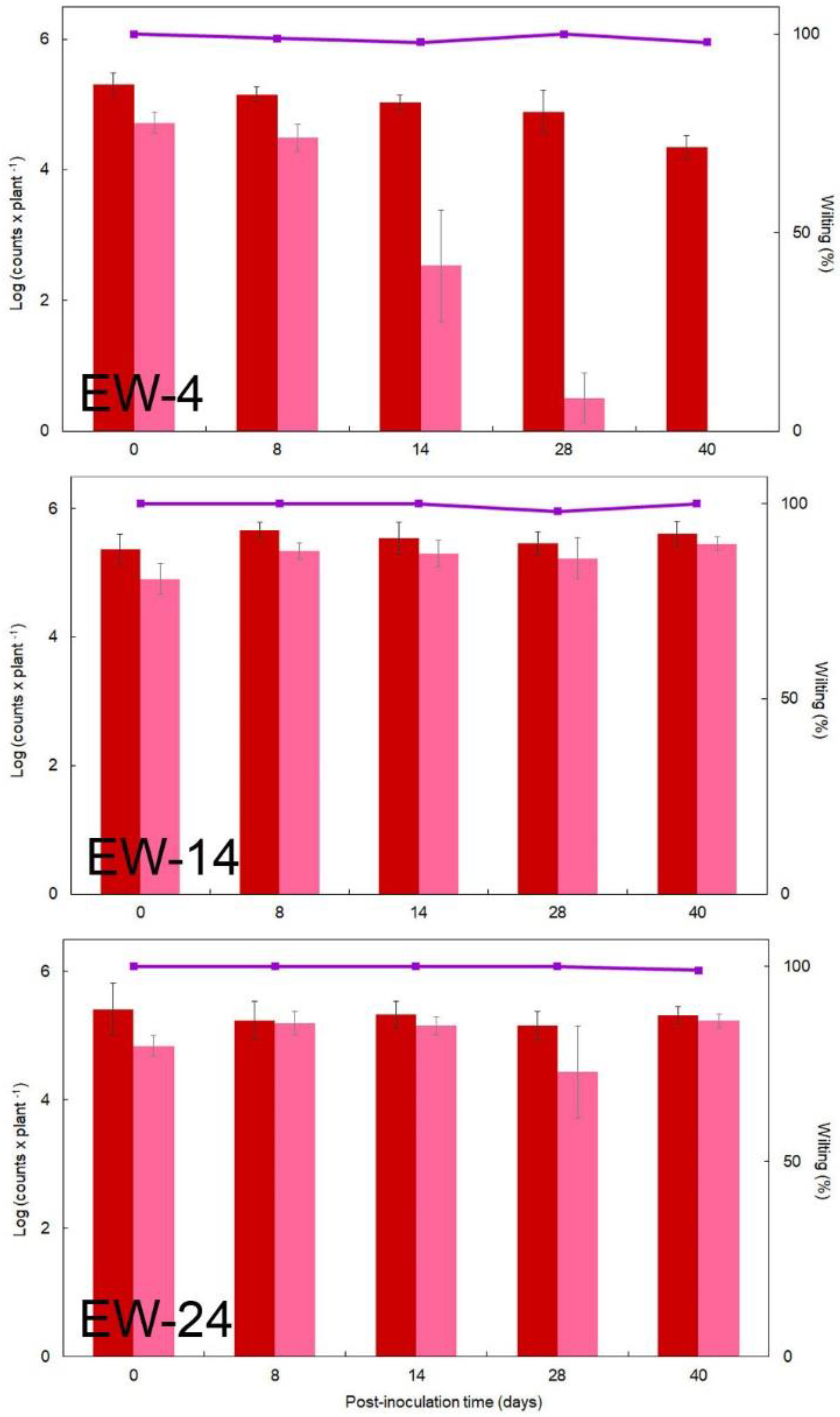
During the 40-day periods of incubation at 4 °C, 14 °C, and 24 °C in the water microcosms under starvation conditions, aliquots were taken at different times to be inoculated in planta. R. solanacearum cells of strain IVIA-1602.1 incubated at 4 °C and inoculated in tomato stems induced disease in 98–100% of the plants (Figure 4). Similar wilting percentages were obtained with cells from microcosms at 14 °C and at 24 °C (Figure 4) and were comparable to those of the strain IPO-1609 in one-off trials. At the three temperatures and depending on the water sample, viable R. solanacearum cells inoculated per plant were about 105 throughout the 40-day sampling periods, and only from approximately 28 dpi at 4 °C there was a slight decline to values around 104 viable cells per plant (Figure 4). At 4 °C, culturable cells inoculated per plant ranged from 105 to 104 in the initial dpi depending on the water sample. Then, they were decreasing until 104–103 CFU per plant by the first week, and progressively to <10 CFU per plant by 28 dpi and to undetectable levels by 40 dpi (Figure 4). At 14 °C and 24 °C, culturable cells were 105–104 per plant throughout the sampling periods (Figure 4). Plants started to show symptoms within 8–11 dpi and completely wilted within four weeks. The pathogens were re-isolated on SMSA agar from the diseased plants and PCR-identified. Positive control plants yielded 100% wilting. Negative control plants did not show any symptoms.

3.4. R. solanacearum Resuscitates from the Cold-Induced VBNC State in Environmental Water and Is Fully Pathogenic in the Host

Assays carried out to assess the resuscitation capability of the VBNC R. solanacearum cells of strain IVIA-1602.1 yielded similar results in triplicate microcosms from environmental water samples. Data are summarized in Table 1, in the three different conditions. The viability of the VBNC cells when the microcosms were containing approximately 106 viable cells/mL and <10 culturable cells (CFU)/mL is illustrated in Figure 5.

3.4.1. By Enrichment in WB Broth

From cold-induced VBNC cells of R. solanacearum strain IVIA-1602.1, and after the temperature upshift with shaking and nutrients, monitoring was of: (i) turbidity by R. solanacearum growth in the direct aliquots and their serial ten-fold dilutions, (ii) culturability on YPGA, and (iii) pathogenicity in the host. These were observed in all the direct aliquots and their serial ten-fold dilutions up to 10−6, corresponding to 1 VBNC cell/mL, at time 0 of the VBNC induction of the R. solanacearum populations (Table 1). Thereafter, the resuscitation capability of these VBNC cells in the microcosms was decreasing with time until reaching about two orders of magnitude by one month from the VBNC induction. Time for resuscitation (estimated as time for observation of turbidity by R. solanacearum growth) was 24 h for direct aliquots, 36 h for dilutions 10−1, 10−2, and 10−3, 48 h for dilutions 10−4 and 10−5, and four days for dilutions 10−6. These rates of growth were maintained throughout the experimental period. Culturability of the cells in the turbid dilutions was positive in all cases, and colonies were PCR-identified as R. solanacearum. However, cells from aliquots in WB broth maintained at 4 °C without shaking remained non-culturable. Pathogenicity assays were positive in all plants, either when inoculated directly from the turbid dilutions or from the colonies on the plates. The pathogen was re-isolated from the wilted plants and PCR-identified. Sensitivity of the detection of resuscitated R. solanacearum cells from the VBNC state was 1 VBNC cell/mL.

3.4.2. By Temperature Upshift in Environmental Water

From cold-induced VBNC cells of R. solanacearum strain IVIA-1602.1 and after the temperature upshift, monitoring was performed regarding: (i) culturability on YPGA and (ii) pathogenicity in the host. These were observed in cells from the direct aliquots and their serial ten-fold dilutions up to 10−4, corresponding to 102 VBNC cells/mL, and up to 10−3, corresponding to 103 VBNC cells/mL, respectively, at time 0 of the VBNC induction (Table 1). Then, the resuscitation capability of the cells in the microcosms was decreasing with time until about three orders of magnitude by one month from the induction (Table 1). Time for resuscitation (estimated as time for observation of culturability after plating) was 48 h from the temperature upshift. This was observed after plating 100-µL volumes from the same direct aliquots incubated during 24 h, 48 h and 72 h from the temperature upshift, and then on plates for 3 days at 29 °C. Sampling at 24 h yielded no growth on the plates, sampling at 48 h turned out in countable colonies, and continuous bacterial growth was observed after sampling at 72 h from the temperature upshift, reaching in all cases 106 CFU/mL. Colonies were PCR-identified as R. solanacearum. Pathogenicity assays to test cells from the colonies were positive in all plants. Pathogenicity assays to test cells from the direct aliquots and their serial ten-fold dilutions were positive in at least one of the two inoculated plants per microcosm. The pathogen was re-isolated and PCR-identified from the wilted plants. Sensitivity of the detection of resuscitated R. solanacearum cells from the VBNC state was 102 VBNC cells/mL.

3.4.3. In Planta

From cold-induced VBNC cells of R. solanacearum strain IVIA-1602.1 and after the temperature upshift in the host, pathogenicity was monitored. This was observed in cells from the direct aliquots and their serial ten-fold dilutions up to 10−3, corresponding to 103 VBNC cells/mL (10 VBNC cells/plant), at time 0 of the VBNC induction, and then the resuscitation capability was decreasing until about one order of magnitude by one month from the induction (Table 1). The pathogen was re-isolated and PCR-identified from the wilted plants. Sensitivity of the detection of resuscitated R. solanacearum cells from the VBNC state was 103 VBNC cells/mL.

4. Discussion

To anticipate the spread of the disease, current prevention and control strategies against bacterial wilt should take into account knowledge on the potential behavior of R. solanacearum in response to global warming. In this work, adaptations by strains of the pathogen from different climatic regions were observed under exposure to environmental temperatures in oligotrophic freshwater, which allowed survival without losing wilting capacity.
With respect to low temperatures, this is the first report of viable R. solanacearum populations induced to the VBNC state in environmental water, in conditions more approaching those of natural settings. Previous work described either: (i) a loss in R. solanacearum culturability under low temperature in natural or distilled, ultrapure water, but without determining the viability of the bacterial populations, thus without confirming the presence of VBNC cells [26,27,32,46]; or (ii) the VBNC induction in distilled, ultrapure, non-environmental water [28,29]. The fact that other cold-adapted water bacteria are not likely to be cold-induced VBNC [47], contrarily to what was observed in this work with R. solanacearum from different climates, suggests that this pathogen is not naturally cold-adapted, even when introduced to cold habitats. Thus, this work demonstrated that, in R. solanacearum, low temperature plays a major role than starvation in inducing the VBNC state (Figure 6), contrarily to what has been reported for other bacteria [47]. However, starvation-induced stress proteins could have protected R. solanacearum from temperature damage, since it was less vulnerable to cyclic cold stress in pure water than in host tissue [27]. Moreover, the VBNC R. solanacearum cold-induction period occurred more slowly in environmental water, pointing to an effect of water nutrient contents, namely trace organic matter and some dissolved salts available for the cells but absent in distilled water, and so nutrient concentrations not supporting R. solanacearum growth would act as an additional stress contributing to the cold-induced VBNC state. In the field, latent VBNC cells maintain structure, biology, and significant gene expression, and global climate change might be resuscitating them when low temperature is the inducing factor, leading to increased outbreaks [48]. Similar to R. solanacearum, a lower mineral salt concentration markedly shortened the VBNC Vibrio parahaemolyticus induction period [49]. Bacterial species, such as V. vulnificus and Aeromonas hydrophila, also behaved similarly to R. solanacearum under low-temperature and nutrient-limiting conditions [50,51], whilst others, such as Campylobacter jejuni and Erwinia amylovora, displayed different responses [52,53,54,55].
At temperate and warm temperatures in environmental water, R. solanacearum populations displayed starvation-survival responses as described at 24 °C [6] and similar in terms of population levels. Lack of unculturability was in agreement with previous work reporting the isolation and persistence of the pathogen in environmental water at temperatures allowing R. solanacearum multiplication [15,16,17,26]. The presence of organic matter and salts in environmental water contributed to stimulate R. solanacearum survival, similarly to Aerobacter aerogenes [56], E. amylovora [53,54], and Leuconostoc mesenteroides [57], where trace minerals facilitated culturability, since mineral salts can affect not only cell growth, but also cell survival during nutrient limitation conditions [31].
Morphological changes are a visible indicator of adaptation to the environment [31,54,58]. Starved R. solanacearum cells transformed from the typical bacilli into coccoids, since shape rounding off and size reduction allow nutrients to be sequestered more efficiently [31]. This was observed in different proportions according to temperature. Although cells entering the VBNC state often exhibit dwarfing [37,59], R. solanacearum coccoids were seldom observed during this process, probably because low temperature rapidly causes decrease in R. solanacearum metabolism and uptake of water nutrients, with constitutive expression of genes associated with survival and stress response for a stable maintenance of their transcript level [60]. Likewise, copper-induced VBNC R. solanacearum cells were unchanged in size [61]. Therefore, at both starvation- and survival-inducing temperatures, the transition to coccoids would be mostly influenced by nutrient limitation and to a lesser extent by low temperature, as reported elsewhere [62]. At these two temperatures, the proportions of coccoids differed, with significantly higher numbers at warm temperature, probably to improve the speed for exchange of material with the surrounding environment to hold a faster energy-consuming metabolism, which becomes a requirement at elevated temperatures [58,63]. Thus, in natural nutrient-deprived environments, the stress of oligotrophy would be less intense for the pathogen at temperatures around 14 °C than at values nearer to the optimum as 24 °C, and so temperature would be modulating this adaptation to oligotrophy (Figure 6), acting on cell metabolism rate and nutrient requirement frequency. Moreover, in the presence of indigenous microbiota, R. solanacearum survived longer at 14 °C than at 24 °C in oligotrophic environmental water [35], and the culturability of R. solanacearum strain IPO-1609 was favored at 12 °C and 20 °C rather than at 28 °C in agricultural water in both the presence and absence of other aquatic microorganisms [26]. Similar to R. solanacearum, a number of bacterial species decreased their sizes with increasing environmental temperatures [51,52,63]. Notwithstanding, this cannot be considered a general bacterial behavior [47,50,58,63].
Although R. solanacearum has frequently been described as cold tolerant [14,28], the strains introduced to either cold or warm areas were apparently better temperate-adapted than cold-adapted as considered [12], and similarly to [28], where data indicated that R. solanacearum had no special adaptation to survive cold temperatures in water under controlled conditions. Likewise, the cold-water-adapted Vibrio tasmaniensis did not enter the VBNC state at 4 °C while the warm-water-adapted V. shiloi did [47].
R. solanacearum resuscitation from the cold-induced VBNC state was observed after stress removal by placement of the VBNC cells in three different favorable conditions, including the host plant, and all of them implying, at least, an upshift in temperature. In enrichment conditions, culturability on solid medium and pathogenicity of the resuscitated R. solanacearum cells from the dilutions were both confirmed. Restoration of culturability was more dependent on the temperature upshift and shaking than the presence of nutrients, since VBNC cells in enrichment liquid medium at 4 °C were not able to form colonies, and so they maintained their VBNC status. In environmental water and after the temperature upshift, culturability and pathogenicity of the resuscitated cells were similarly confirmed, the only resuscitation-inducing factor here being the temperature upshift, which has not been reported for R. solanacearum up until now. That would explain the seasonal variation of R. solanacearum populations in environmental water [16,17]. If temperature is so critical, increases in water temperature corresponding to rising global surface temperatures will likely lead to a wider geographic distribution of R. solanacearum and a higher incidence of infections in planta resulting from resuscitated cells of the pathogen, as it is being observed in Vibrio species [48]. Likewise, a simple reversal of temperature was sufficient to allow the resuscitation of other bacterial species [37,38,59]. In contrast, it was not effective to resuscitate the close R. pseudosolanacearum (former R. solanacearum phylotype I) in soil and water [60,64], since the addition of hydrogen peroxide-degrading compounds, such as catalase or sodium pyruvate, was necessary. The number of bacterial cells resuscitated by temperature upshift in environmental water was equal to the initial inoculum, similarly to [37,44,60]. Resuscitation in planta of the VBNC R. solanacearum cells was evidenced by the occurrence of wilting symptoms, and progressively declined over time, accordingly to [29]. Virulence in tomato plants was also observed in revived cells of R. pseudosolanacearum after exiting a cold-induced VBNC state in pure water [64]. In all the three different resuscitation conditions, the resuscitated R. solanacearum cells displayed similar phenotypes to the original culturable cells, including virulence in tomato plants, as described [60]. Moreover, also in the three different conditions, a decrease in the proportion of VBNC cells capable of resuscitation occurred over time, this process being dependent on the age of the VBNC cells, as stated [38]. In that respect, several authors agree to consider the existence of gradual stages within the VBNC state, namely a reversible non-culturable stage where cells can be resuscitated and an irreversible non-culturable stage, where cells cannot be resuscitated, although they keep respiratory activity [28,60,64] (Figure 6). Among these conditions, in this work, the enrichment was the most effective for R. solanacearum resuscitation and the most sensitive for their detection. This is probably because it combines a temperature upshift with nutrients and shaking, which supplies with oxygen and disperses oxidative compounds (peroxides, other free radicals) accumulated extra- or intracellularly either produced in the cells in response to low temperature stress or commonly present in rich culture media [37,65]. Reversal of adverse VBNC-state-inducing factors can be efficiently applied to the detection of R. solanacearum resuscitated cells.
On the basis of all these results, starved and/or cold-stressed and/or cold-induced VBNC R. solanacearum cells could be present in environmental water, being a threat to secure crop production as they are not easily detected [61], can survive cold temperature fluctuations [27], and can revert to a fully pathogenic state just by a temperature upshift, which can be favored within the frame of global climate change conditions. All of these R. solanacearum survival forms maintained their capacity for in planta multiplication and colonization, causing disease symptoms in the host, as observed elsewhere for similar time periods [6,16,29]. Not only can global warming contribute to the pathogen spread and virulence, but crops resistant to R. solanacearum at moderate temperatures can also become more susceptible at high ambient temperatures [14,24], increasing the probability of infections. The bacterial wilt disease is most severe on plants at temperature values ranging from 25 °C to 35 °C [14,28,66].
On the other hand, taking into account the temperature interval of 4–10 °C applied by EU legislation [43] to transport suspected water samples, temperatures above 4 °C up to around 10 °C would be more advisable than 4 °C, since cultivation-based methods are required to confirm pathogen detection. For the inspection of environmental samples, it should be determined whether to also test for these VBNC cells [64] to improve the sensitivity of the detection. This is a relevant point since the early detection of R. solanacearum in irrigation water and its eradication would contribute to improve any integrated management program of the bacterial wilt disease [61].
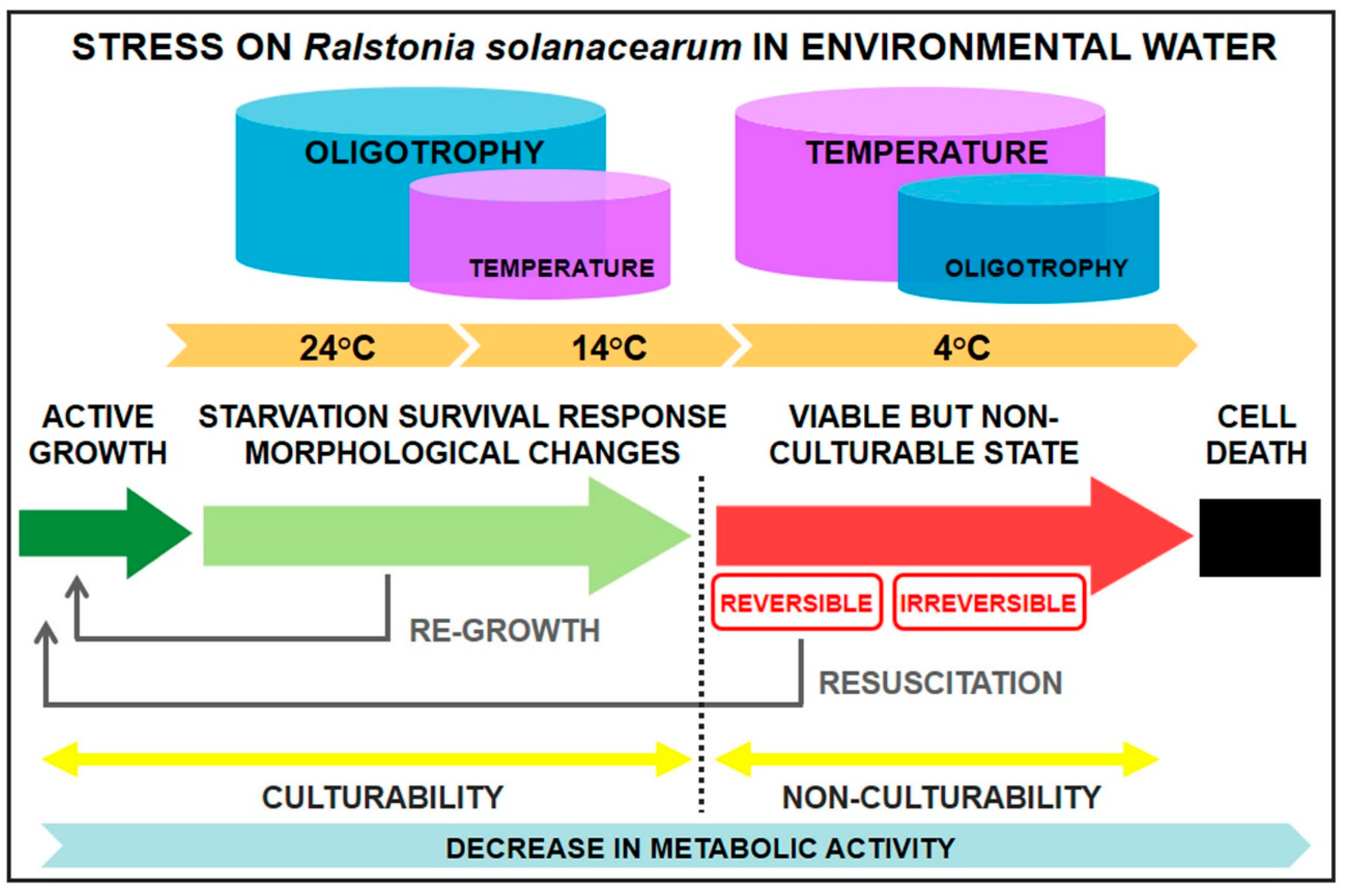
Overall, R. solanacearum strains from either cold or warm origin were able to adapt to a combined effect of temperature and oligotrophy. At low temperature, the delay in the induction of the VBNC state in environmental water suggested a protective effect of water nutrient contents on bacterial cells and pointed out the relevance of performing survival studies in conditions better approaching those in the environment. At temperate and warm temperatures, adaptations to oligotrophy were starvation–survival responses and morphological changes influenced by temperature. It appeared that, when temperature was the main stress (cold conditions), nutrient deprivation acted as an additional stress, contributing to accelerate the effect of temperature, and conversely, when oligotrophy was the main stress (temperate and warm conditions), temperature increased the effect of oligotrophy (Figure 6). In all conditions, R. solanacearum cells remained pathogenic and capable of resuscitation by a simple reversal of temperature.
Figure 6. Influence of temperature and oligotrophy on Ralstonia solanacearum stress induction in environmental water and stress responses by the bacterium. Adapted to R. solanacearum from a proposed model by [67]. See text for details.
Microorganisms 10 02503 g006
Under circumstances of global warming, understanding R. solanacearum adaptations to environmental abiotic stresses can help to design strategies to prevent and control their spread and dissemination in waterways and other natural settings. This is particularly important in the case of R. solanacearum-contaminated water, since it cannot be used for irrigation, contributing to the global problem of the increased water scarcity in the environment due to climate change, which has serious implications, among others, for food production and health.

Supplementary Materials

The following supporting information can be downloaded at: https://www.mdpi.com/article/10.3390/microorganisms10122503/s1, Figure S1: Effect of temperature under nutrient-limiting conditions on survival of Ralstonia solanacearum strain IVIA-1602.1 during 40-day periods in environmental water at 14 °C. Total (), viable (), and culturable cells on SMSA () and YPGA () media. Points are mean ± standard deviation of triplicate microcosms; Figure S2: Effect of low temperature under nutrient-limiting conditions on survival of Ralstonia solanacearum strain IPO-1609 during 40-day periods in water. Microcosms of environmental water at: 4 °C (top), 14 °C (middle), and 24 °C (bottom). Total (), viable (), and culturable cells on SMSA () and YPGA () media. Points are mean ± standard deviation of triplicate microcosms.

Author Contributions

Conceptualization, B.Á., M.M.L. and E.G.B.; methodology, B.Á., M.M.L. and E.G.B.; validation, B.Á., M.M.L. and E.G.B.; formal analysis, B.Á. and E.G.B.; investigation, B.Á., M.M.L. and E.G.B.; resources, M.M.L. and E.G.B.; writing—original draft preparation, B.Á.; writing—review and editing, B.Á., M.M.L. and E.G.B.; supervision, M.M.L. and E.G.B.; project administration, M.M.L. and E.G.B.; funding acquisition, M.M.L. and E.G.B. All authors have read and agreed to the published version of the manuscript.

Funding

This research was funded by the EU project QLK 3-CT-2000-01598, the Spanish project RTA2015-00087-C02 financed by MCIN/AEI/10.13039/501100011033, the Spanish Instituto Nacional de Investigación y Tecnología Agraria y Alimentaria (INIA), and “ERDF A way of making Europe”, as well as the Laboratory of Reference for Plant Pathogenic Bacteria of the Spanish Ministry of Agriculture (MAPA) at Instituto Valenciano de Investigaciones Agrarias (IVIA), the University of Valencia (UV) project UV-INV-AE 112-66196, and the University of Valencia-Research Support to BACPLANT group GIUV2015-219.

Data Availability Statement

Not applicable.

Acknowledgments

The authors thank JL Palomo and the Consejería de Agricultura de Castilla-León for sending water samples, JD van Elsas for strain IPO-1609, and E Carbonell, J Pérez (Instituto Valenciano de Investigaciones Agrarias, Moncada, Valencia, Spain) and JL Díez (Hospital Universitario y Politécnico La Fe, Valencia, Spain) for the statistical analyses.

Conflicts of Interest

The authors declare no conflict of interest. The funders had no role in the design of the study; in the collection, analyses, or interpretation of data; in the writing of the manuscript; or in the decision to publish the results.

References

  1. Fegan, M.; Prior, P. How Complex Is the “Ralstonia solanacearum Species Complex”? In Bacterial wilt Disease and the Ralstonia solanacearum Species Complex; Allen, C., Prior, P., Hayward, A.C., Eds.; APS Press: St. Paul, MN, USA, 2005; pp. 449–461. [Google Scholar]
  2. Safni, I.; Cleenwerck, I.; De Vos, P.; Fegan, M.; Sly, L.; Kappler, U. Polyphasic taxonomic revision of the Ralstonia solanacearum species complex: Proposal to emend the descriptions of Ralstonia solanacearum and Ralstonia syzygii and reclassify current R. syzygii strains as Ralstonia syzygii subsp. syzygii subsp. nov., R. solanacearum phylotype IV strains as Ralstonia syzygii subsp. indonesiensis subsp. nov., banana blood disease bacterium strains as Ralstonia syzygii subsp. celebesensis subsp. nov. and R. solanacearum phylotype I and III strains as Ralstonia pseudosolanacearum sp. nov. Int. J. Syst. Evol. Microbiol. 2014, 64, 3087–3103. [Google Scholar] [PubMed] [Green Version]
  3. Prior, P.; Ailloud, F.; Dalsing, B.L.; Remenant, B.; Sanchez, B.; Allen, C. Genomic and proteomic evidence supporting the division of the plant pathogen Ralstonia solanacearum into three species. BMC Genom. 2016, 17, 90. [Google Scholar] [CrossRef] [PubMed] [Green Version]
  4. Elphinstone, J.G. The Current Bacterial Wilt Situation: A Global Overview. In Bacterial wilt Disease and the Ralstonia solanacearum Species Complex; Allen, C., Prior, P., Hayward, A.C., Eds.; APS Press: St. Paul, MN, USA, 2005; pp. 9–28. [Google Scholar]
  5. Swanson, J.K.; Yao, J.; Tans-Kersten, J.; Allen, C. Behaviour of Ralstonia solanacearum race 3 biovar 2 during latent and active infection of geranium. Phytopathology 2005, 95, 136–143. [Google Scholar] [CrossRef] [PubMed] [Green Version]
  6. Álvarez, B.; López, M.M.; Biosca, E.G. Survival strategies and pathogenicity of Ralstonia solanacearum phylotype II subjected to prolonged starvation in environmental water microcosms. Microbiology 2008, 154, 3590–3598. [Google Scholar] [CrossRef] [PubMed] [Green Version]
  7. Álvarez, B.; Vasse, J.; Le-Courtois, V.; Trigalet-Démery, D.; López, M.M.; Trigalet, A. Comparative behavior of Ralstonia solanacearum biovar 2 in diverse plant species. Phytopathology 2008, 98, 59–68. [Google Scholar] [CrossRef] [Green Version]
  8. Álvarez, B.; Biosca, E.G.; López, M.M. On the Life of Ralstonia solanacearum, a Destructive Bacterial Plant Pathogen. In Current Research, Technology and Education Topics in Applied Microbiology and Microbial Biotechnology; Méndez-Vilas, A., Ed.; Formatex: Badajoz, Spain, 2010; pp. 267–279. [Google Scholar]
  9. Yuliar, N.Y.A.; Toyota, K. Recent trends in control methods for bacterial wilt diseases caused by Ralstonia solanacearum. Microbes Environ. 2015, 30, 1–11. [Google Scholar] [CrossRef] [Green Version]
  10. Álvarez, B.; Biosca, E.G. Bacteriophage-based bacterial wilt biocontrol for an environmentally sustainable agriculture. Front. Plant Sci. 2017, 8, 1218. [Google Scholar] [CrossRef] [Green Version]
  11. Anonymous. Commission Implementing Regulation (EU) 2019/2072 of 28 November 2019 establishing uniform conditions for the implementation of Regulation (EU) 2016/2031 of the European Parliament and the Council, as regards protective measures against pests of plants, and repealing Commission Regulation (EC) Nº 690/2008 and amending Commission Implementing Regulation (EU) 2018/2019. OJEU 2019, L319, 1–279. [Google Scholar]
  12. Brown, D. Ralstonia Solanacearum and Bacterial Wilt in the Post-Genomics Era. In Plant Pathogenic Bacteria. Genomics and Molecular Biology; Jackson, R.W., Ed.; Caister Academic Press: London, UK, 2009; pp. 175–202. [Google Scholar]
  13. United States Department of Agricultue. Plant Pests and Diseases Programs. Available online: http://www.aphis.usda.gov (accessed on 11 December 2022).
  14. Hayward, A.C. Biology and epidemiology of bacterial wilt caused by Pseudomonas solanacearum. Annu. Rev. Phytopathol. 1991, 29, 65–87. [Google Scholar] [CrossRef]
  15. Wenneker, M.; Verdel, M.S.W.; Groeneveld, R.M.W.; Kempenaar, C.; van Beuningen, A.R.; Janse, J.D. Ralstonia (Pseudomonas) solanacearum race 3 (biovar 2) in surface water and natural weed hosts: First report on stinging nettle (Urtica dioica). Eur. J. Plant Pathol. 1999, 105, 307–315. [Google Scholar] [CrossRef]
  16. Caruso, P.; Palomo, J.L.; Bertolini, E.; Álvarez, B.; López, M.M.; Biosca, E.G. Seasonal variation of Ralstonia solanacearum biovar 2 populations in a Spanish river: Recovery of stressed cells at low temperatures. Appl. Environ. Microbiol. 2005, 71, 140–148. [Google Scholar] [CrossRef]
  17. Hong, J.; Ji, P.; Momol, M.T.; Jones, J.B.; Olson, S.M.; Pradhanang, P.; Guven, K. Ralstonia Solanacearum Detection in Tomato Irrigation Ponds and Weeds. In Acta Horticulturae; ISHS: Miami, FL, USA, 2005; pp. 309–311. [Google Scholar]
  18. Elphinstone, J.G.; Stanford, H.; Stead, D.E. Survival and transmission of Ralstonia solanacearum in aquatic plants of Solanum dulcamara and associated surface water in England. EPPO Bull. 1998, 28, 93–94. [Google Scholar] [CrossRef]
  19. Stevens, P.; van Elsas, J.D. Genetic and phenotypic diversity of Ralstonia solanacearum biovar 2 strains obtained from Dutch waterways. Antonie Van Leeuwenhoek 2010, 97, 171–188. [Google Scholar] [CrossRef] [Green Version]
  20. Cruz, L.; Eloy, M.; Quirino, F.; Oliveira, H.; Tenreiro, R. Molecular epidemiology of Ralstonia solanacearum strains from plants and environmental sources in Portugal. Eur. J. Plant Pathol. 2012, 133, 687–706. [Google Scholar] [CrossRef]
  21. Parkinson, N.; Bryant, R.; Bew, J.; Conyers, C.; Stones, R.; Alcock, M.; Elphinstone, J. Application of variable-number tandem-repeat typing to discriminate Ralstonia solanacearum strains associated with English watercourses and disease outbreaks. Appl. Environ. Microbiol. 2013, 79, 6016–6022. [Google Scholar] [CrossRef] [Green Version]
  22. Caruso, P.; Biosca, E.G.; Bertolini, E.; Marco-Noales, E.; Gorris, M.T.; Licciardello, C.; López, M.M. Genetic diversity reflects geographical origin of Ralstonia solanacearum strains isolated from plant and water sources in Spain. Int. Microbiol. 2017, 20, 155–164. [Google Scholar] [CrossRef]
  23. Singh, D.; Yadav, D.K.; Sinha, S.; Choudhary, G. Effect of temperature, cultivars, injury of root and inoculums load of Ralstonia solanacearum to cause bacterial wilt of tomato. Arch. Phytopathol. Plant Prot. 2014, 47, 1574–1583. [Google Scholar] [CrossRef]
  24. Prior, P.; Bart, S.; Leclercq, S.; Darrasse, A.; Anais, G. Resistance to bacterial wilt in tomato as discerned by spread of Pseudomonas (Burholderia) solanacearum in the stem tissues. Plant Pathol. 1996, 45, 720–726. [Google Scholar] [CrossRef]
  25. Bittner, R.J.; Arellano, C.; Mila, A.L. Effect of temperature and resistance of tobacco cultivars to the progression of bacterial wilt, caused by Ralstonia solanacearum. Plant Soil 2016, 408, 299–310. Available online: http://www.jstor.org/stable/44136958 (accessed on 11 December 2022). [CrossRef]
  26. van Elsas, J.D.; Kastelein, P.; de Vries, P.M.; van Overbeek, L.S. Effects of ecological factors on the survival and physiology of Ralstonia solanacearum bv. 2 in irrigation water. Can. J. Microbiol. 2001, 47, 842–854. [Google Scholar] [CrossRef]
  27. Scherf, J.M.; Milling, A.; Allen, C. Moderate temperature fluctuations rapidly reduce viability of Ralstonia solanacearum race 3 biovar 2 in infected geranium, tomato, and potato. Appl. Environ. Microbiol. 2010, 76, 7061–7067. [Google Scholar] [CrossRef] [PubMed]
  28. Milling, A.; Meng, F.; Denny, T.P.; Allen, C. Interactions with hosts at cool temperatures, not cold tolerance, explain the unique epidemiology of Ralstonia solanacearum race 3 biovar 2. Phytopathology 2009, 99, 1127–1134. [Google Scholar] [CrossRef] [PubMed] [Green Version]
  29. van Overbeek, L.S.; Bergervoet, J.H.W.; Jacobs, F.H.H.; van Elsas, J.D. The low-temperature-induced viable-but-nonculturable state affects the virulence of Ralstonia solanacearum biovar 2. Phytopathology 2004, 94, 463–469. [Google Scholar] [CrossRef] [PubMed] [Green Version]
  30. Roszak, D.B.; Colwell, R.R. Survival strategies of bacteria in the natural environment. Microbiol. Rev. 1987, 51, 365–379. [Google Scholar] [CrossRef]
  31. Morita, R.Y. Bacteria in Oligotrophic Environments. In Starvation-Survival Lifestyle; Reddy, C.A., Chakrabarty, A.M., Demain, A.L., Tiedje, J.M., Eds.; Chapman and Hall: New York, NY, USA, 1997; pp. 368–385. [Google Scholar]
  32. Kelman, A. Factors influencing viability and variation in cultures of Pseudomonas solanacearum. Phytopathology 1956, 46, 16–17. [Google Scholar]
  33. Tanaka, Y.; Noda, N. Studies on the factors affecting survival of Pseudomonas solanacearum E.F. Smith, the causal agent of tobacco wilt disease. Bull. Okayama Tob. Exp. Stn. 1973, 32, 81–91. [Google Scholar]
  34. Wakimoto, S.; Utatsu, I.; Matsuo, N.; Hayashi, N. Multiplication of Pseudomonas solanacearum in pure water. Ann. Phytopathol. Soc. Jpn. 1982, 48, 620–627. [Google Scholar] [CrossRef] [Green Version]
  35. Álvarez, B.; López, M.M.; Biosca, E.G. Influence of native microbiota on survival of Ralstonia solanacearum phylotype II in river water microcosms. Appl. Environ. Microbiol. 2007, 73, 7210–7217. [Google Scholar] [CrossRef] [Green Version]
  36. Chaiyanan, S.; Chaiyanan, S.; Grim, C.; Maugel, T.; Huq, A.; Colwell, R.R. Ultrastructure of coccoid viable but non-culturable Vibrio cholerae. Environ. Microbiol. 2007, 9, 393–402. [Google Scholar] [CrossRef]
  37. Oliver, J.D. Recent findings on the viable but nonculturable state in pathogenic bacteria. FEMS Microbiol. Rev. 2010, 34, 415–425. [Google Scholar] [CrossRef] [Green Version]
  38. Li, L.; Mendis, N.; Trigui, H.; Oliver, J.D.; Faucher, S.P. The importance of the viable but non-culturable state in human bacterial pathogens. Front. Microbiol. 2014, 5, 258. [Google Scholar] [CrossRef] [PubMed] [Green Version]
  39. Ridé, M. Bactéries Phytopathogènes et Maladies Bactériennes des Végétaux. In Les Bactérioses ET Les Viroses Des Arbres Fruitiers; Ponsot, M., Ed.; Viennot-Bourgin: Paris, France, 1969; pp. 4–59. [Google Scholar]
  40. Elphinstone, J.G.; Hennessy, J.; Wilson, J.K.; Stead, D.E. Sensitivity of different methods for the detection of Ralstonia solanacearum in potato tuber extracts. EPPO Bull. 1996, 26, 663–678. [Google Scholar] [CrossRef]
  41. Kogure, K.; Simidu, U.; Taga, N. A tentative direct microscopic method for counting living marine bacteria. Can. J. Microbiol. 1979, 25, 415–420. [Google Scholar] [CrossRef]
  42. Oliver, J.D. Heterotrophic bacterial populations of the Black sea. Biol. Oceanogr. 1987, 4, 83–97. [Google Scholar]
  43. Anonymous. Commission Directive 2006/63/EC of 14 July 2006: Amending Annexes II to VII to Council Directive 98/57/EC on the control of Ralstonia solanacearum (Smith) Yabuuchi et al. Off. J. Eur. Communities 2006, L206, 36–106. [Google Scholar]
  44. Whitesides, M.D.; Oliver, J.D. Resuscitation of Vibrio vulnificus from the viable but nonculturable state. Appl. Environ. Microbiol. 1997, 63, 1002–1005. [Google Scholar] [CrossRef] [Green Version]
  45. Ordax, M.; Marco-Noales, E.; López, M.M.; Biosca, E.G. Survival strategy of Erwinia amylovora against copper: Induction of the viable-but-nonculturable state. Appl. Environ. Microbiol. 2006, 72, 3482–3488. [Google Scholar] [CrossRef] [Green Version]
  46. Stevens, P.; van Overbeek, L.S.; van Elsas, J.D. Ralstonia solanacearum DPGI-1 strain KZR-5 is affected in growth, response to cold stress and invasion of tomato. Microb. Ecol. 2011, 61, 101–112. [Google Scholar] [CrossRef] [Green Version]
  47. Vattakaven, T.; Bond, P.; Bradley, G.; Munn, C.B. Differential effects of temperature and starvation on induction of the viable-but-nonculturable state in the coral pathogens Vibrio shiloi and Vibrio tasmaniensis. Appl. Environ. Microbiol. 2006, 72, 6508–6513. [Google Scholar] [CrossRef] [Green Version]
  48. Oliver, J.D. The viable but nonculturable state for bacteria: Status update. Microbe 2016, 11, 159–164. [Google Scholar] [CrossRef] [Green Version]
  49. Wong, H.C.; Wang, P. Induction of viable but nonculturable state in Vibrio parahaemolyticus and its susceptibility to environmental stresses. J. Appl. Microbiol. 2004, 96, 359–366. [Google Scholar] [CrossRef] [PubMed] [Green Version]
  50. Biosca, E.G.; Amaro, C.; Marco-Noales, E.; Oliver, J.D. Effect of low temperature on starvation-survival of the eel pathogen Vibrio vulnificus biotype 2. Appl. Environ. Microbiol. 1996, 62, 450–455. [Google Scholar] [CrossRef] [PubMed] [Green Version]
  51. Mary, P.; Chihib, N.E.; Charafeddine, O.; Defives, C.; Hornez, J.P. Starvation survival and viable but nonculturable states in Aeromonas hydrophila. Microb. Ecol. 2002, 43, 250–258. [Google Scholar] [CrossRef] [PubMed]
  52. Rollins, D.M.; Colwell, R.R. Viable but nonculturable stage of Campylobacter jejuni and its role in survival in the natural aquatic environment. Appl. Environ. Microbiol. 1986, 52, 531–538. [Google Scholar] [CrossRef] [PubMed] [Green Version]
  53. Biosca, E.G.; Marco-Noales, E.; Ordax, M.; López, M.M. Long-Term Starvation-Survival of Erwinia amylovora in Sterile Irrigation Water. In Acta Horticulturae; Bazzi, C., Mazzucchi, U., Eds.; ISHS: Bologna, Italy, 2006; pp. 107–112. [Google Scholar]
  54. Santander, R.D.; Oliver, J.D.; Biosca, E.G. Cellular, physiological and molecular adaptive responses of Erwinia amylovora to starvation. FEMS Microbiol. Ecol. 2014, 88, 258–271. [Google Scholar] [CrossRef]
  55. Santander, R.D.; Biosca, E.G. Erwinia amylovora psychrotrophic adaptations: Evidence of pathogenic potential and survival at temperate and low environmental temperatures. PeerJ 2017, 5, e3931. [Google Scholar] [CrossRef] [Green Version]
  56. Trulear, M.G.; Characklis, W.G. Dynamics of biofilm processes. J. Water Pollut. Control Fed. 1982, 54, 1288–1301. [Google Scholar]
  57. Kim, D.; Thomas, S.; Fogler, H.S. Effects of pH and trace minerals on long-term starvation of Leuconostoc mesenteroides. Appl. Environ. Microbiol. 2000, 66, 976–981. [Google Scholar] [CrossRef] [Green Version]
  58. Shi, B.; Xia, X. Morphological changes of Pseudomonas pseudoalcaligenes in response to temperature selection. Curr. Microbiol. 2003, 46, 120–123. [Google Scholar] [CrossRef]
  59. Su, X.; Sun, F.; Wang, Y.; Hashmi, M.Z.; Guo, L.; Ding, L.; Shen, C. Identification, characterization and molecular analysis of the viable but nonculturable Rhodococcus biphenylivorans. Sci. Rep. 2015, 5, 18590. [Google Scholar] [CrossRef]
  60. Kong, H.G.; Bae, J.Y.; Lee, H.J.; Joo, H.J.; Jung, E.J.; Chung, E.; Lee, S. Induction of the viable but nonculturable state of Ralstonia solanacearum by low temperature in the soil microcosm and its resuscitation by catalase. PLoS ONE 2014, 9, e109792. [Google Scholar] [CrossRef]
  61. Um, H.Y.; Kong, H.G.; Lee, H.J.; Choi, H.K.; Park, E.J.; Kim, S.T.; Murugiyan, S.; Chung, E.; Kang, K.Y.; Lee, S. Altered gene expression and intracellular changes of the viable but nonculturable state in Ralstonia solanacearum by copper treatment. Plant Pathol. J. 2013, 29, 374–385. [Google Scholar] [CrossRef] [Green Version]
  62. Klancnik, A.; Zorman, T.; Možina, S.S. Effects of low temperature, starvation and oxidative stress on the physiology of Campylobacter jejuni cells. Croat. Chem. Acta 2008, 81, 41–46. [Google Scholar]
  63. Christophersen, J. Basic Aspects of Temperature Action on Microorganims. In Temperature and Life; Precht, H., Christophersen, J., Hensel, H., Larcher, W., Eds.; Springer: Berlin/Heidelberg, Germany, 1973; pp. 3–59. [Google Scholar]
  64. Imazaki, I.; Nakaho, K. Temperature-upshift-mediated revival from the sodium-pyruvate-recoverable viable but nonculturable state induced by low temperature in Ralstonia solanacearum: Linear regression analysis. J. Gen. Plant Pathol. 2009, 75, 213–226. [Google Scholar] [CrossRef]
  65. Kong, I.S.; Bates, T.C.; Hülsmann, A.; Hassan, H.; Smith, B.E.; Oliver, J.D. Role of catalase and oxyR in the viable but nonculturable state of Vibrio vulnificus. FEMS Microbiol. Ecol. 2004, 50, 133–142. [Google Scholar] [CrossRef]
  66. Fajinmi, A.A.; Fajinmi, O.B. An overview of bacterial wilt disease of tomato in Nigeria. Agric. J. 2010, 5, 242–247. [Google Scholar] [CrossRef] [Green Version]
  67. Shleeva, M.O.; Bagramyan, K.; Telkov, M.V.; Mukamolova, G.V.; Young, M.; Kell, D.B.; Kaprelyants, A.S. Formation and resuscitation of “non-culturable” cells of Rhodococcus rhodochrous and Mycobacterium tuberculosis in prolonged stationary phase. Microbiology 2002, 148, 1581–1591. [Google Scholar] [CrossRef]
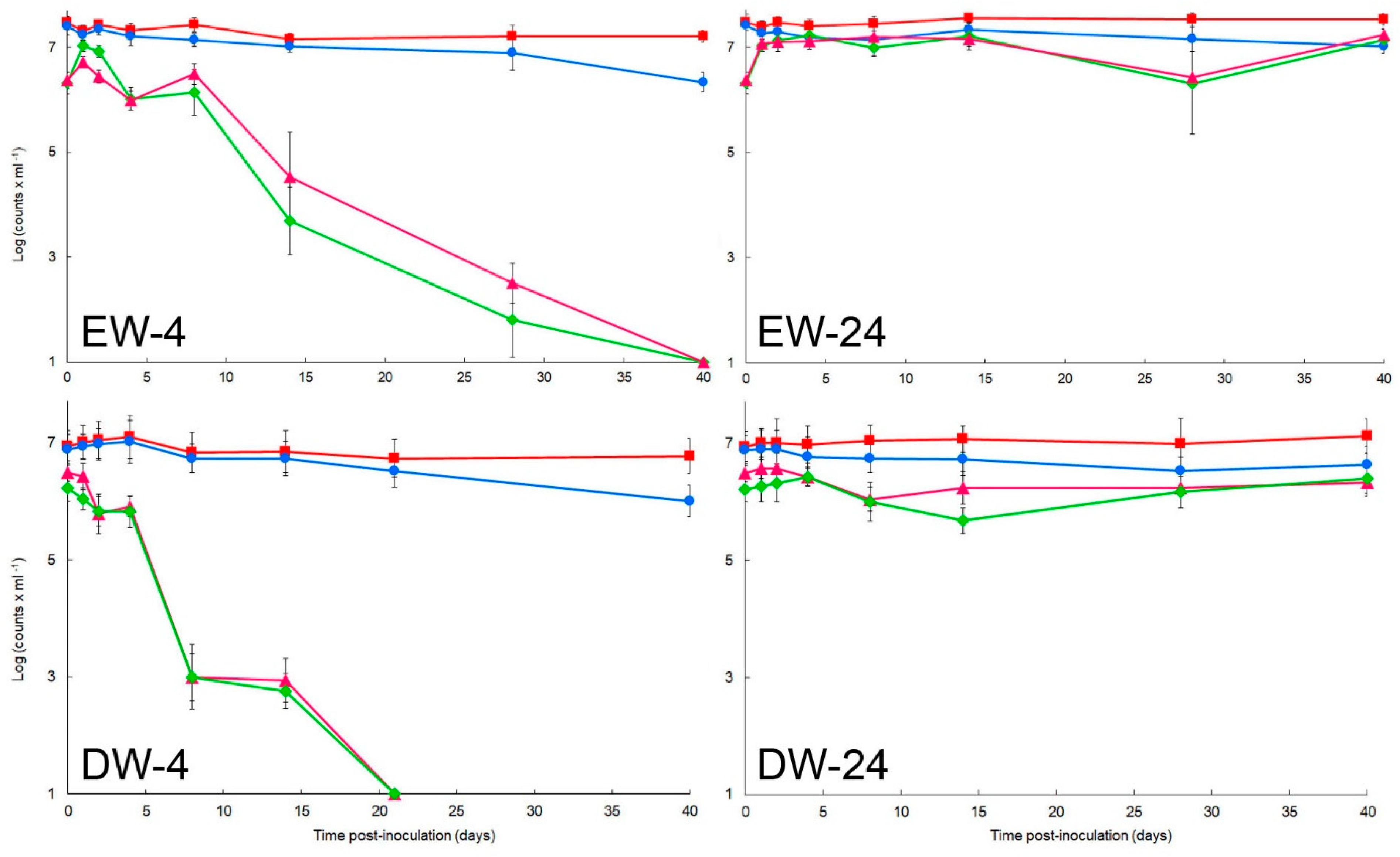
Figure 1. Effect of low temperature under nutrient-limiting conditions on survival of Ralstonia solanacearum strain IVIA-1602.1 during 40-day periods in water. Microcosms of: EW−4, environmental water at 4 °C; DW−4, distilled water at 4 °C; EW−24, environmental water at 24 °C, and DW−24, distilled water at 24 °C. Total (), viable (), and culturable cells on SMSA () and YPGA () media. Data from one representative environmental water sample have been plotted. Points are mean ± standard deviation of triplicate microcosms.
Microorganisms 10 02503 g001
Figure 2. Similarity in trends of culturable cell counts from Ralstonia solanacearum strain IVIA-1602.1-inoculated environmental water microcosms throughout 40-day periods at 4 °C and 24 °C. Inc stands for Increments, which were calculated with the differences between mean values of culturable data at both temperatures with respect to the values at time zero (Inc zero). (Left): comparison of culturable data on SMSA medium among environmental water (EW)—1 (), EW—2 (), EW—3 (), and EW—4 (). (Right): comparison between culturable data on SMSA (), and YPGA () media for the four environmental water samples.
Microorganisms 10 02503 g002
Figure 3. Proportions of coccoid cells appearing in Ralstonia solanacearum strain IVIA-1602.1 populations starved in the environmental water microcosms during 40-day periods at 24 °C, 14 °C, and 4 °C. (Left) Symbols for each temperature: 24 °C (), 14 °C (), and 4 °C (). Data from one representative environmental water sample have been plotted. Points are mean ± standard deviation of triplicate microcosms. (Right) Representative fluorescence microscopy images of proportions of coccoid cells (white arrows) of the bacterium at each temperature. Scale bars: 5 µm.
Microorganisms 10 02503 g003
Figure 4. Pathogenicity of Ralstonia solanacearum strain IVIA-1602.1 previously starved in environmental water microcosms during 40 days, in tomato plants cv. ‘Roma’. Viable (red bars) and culturable (pink bars) cells per plant, and percentage of wilted plants (). Only assays performed in at least weekly intervals from one representative environmental water (EW) at 4 °C (EW−4), 14 °C (EW−14) and 24 °C (EW−24) have been plotted. Points are mean ± standard deviation (SD) of triplicate microcosms. Absolute value for 100% wilting refers to 24 plants (6 × 4 sets). At the three temperatures, SD of wilting values for most of the points was zero, and ± 2.0% in some cases. Control plants inoculated with freshly grown R. solanacearum strain IVIA-1602.1 developed 100% wilting, while those inoculated with PBS were negative.
Microorganisms 10 02503 g004
Figure 5. Viability of Ralstonia solanacearum strain IVIA-1602.1 populations at 40 days post-inoculation in the environmental water microcosms. (A,B) starved cells at 24 °C, and (C,D) starved and cold-induced VBNC cells at 4 °C. Viability was similar between starved (A,B) and VBNC (C,D) R. solanacearum cells, although in (C,D) cells were no culturable on solid media. For VBNC cells, this time was considered time zero of VBNC induction of the R. solanacearum populations in the resuscitation assays. Viability was measured by the DVC method and subsequent staining with acridine orange [41,42], and estimated in 106 VBNC cells/mL. Scale bars: 5 µm.
Microorganisms 10 02503 g005
Table 1. Resuscitation of Ralstonia solanacearum strain IVIA-1602.1 previously induced to the VBNC state in environmental water microcosms at 4 °C.
Time in the VBNC State (Weeks)Culturable Cells (CFU/mL) Resuscitation Assays
In Vitro–In Enrichment Conditions
Direct−1−2−3−4−5−6−7−8
03Turbidity3/33/33/33/33/33/33/30/30/3
Culturability3/33/33/33/33/33/33/30/30/3
Pathogenicity3/33/33/33/33/33/33/30/30/3
10Turbidity3/33/33/33/33/33/30/30/3
Culturability3/33/33/33/33/33/30/30/3
Pathogenicity3/33/33/33/33/33/30/30/3
20Turbidity3/33/33/33/33/33/30/3
Culturability3/33/33/33/33/33/30/3
Pathogenicity3/33/33/33/33/33/30/3
30Turbidity3/33/33/33/33/32/30/3
Culturability3/33/33/33/33/32/30/3
Pathogenicity3/33/33/33/33/32/30/3
40Turbidity3/33/33/33/33/30/30/3
Culturability3/33/33/33/33/30/30/3
Pathogenicity3/33/33/33/33/30/30/3
In Vitro–In Environmental Water
Direct−1−2−3−4−5−6−7−8
03Culturability3/33/33/33/33/30/30/30/30/3
Pathogenicity3/33/33/33/33/30/30/30/30/3
20Culturability3/33/33/31/30/30/3
Pathogenicity3/33/33/31/30/30/3
40Culturability3/32/30/30/30/3
Pathogenicity3/32/30/30/30/3
In Planta
Direct−1−2−3−4−5
03Pathogenicity3/33/33/33/30/30/3
40Pathogenicity3/33/32/30/30/30/3
Data from one representative environmental water at 4 °C (EW−4) are summarized. Direct stands for direct aliquots, and the negative numbers stand for the serial ten-fold dilutions. Pathogenicity assays were considered positive when at least one of the two tomato plants cv. ‘Roma’ inoculated per microcosm showed bacterial wilt symptoms. R. solanacearum was re-isolated from wilted plants and PCR-identified. Control plants inoculated with R. solanacearum strain IVIA-1602.1 at each sampling time yielded 100% wilting in stem inoculation. Plants inoculated with PBS were negative. Data represent results from triplicate microcosms.
Publisher’s Note: MDPI stays neutral with regard to jurisdictional claims in published maps and institutional affiliations.