Review

The Historical Development of Cultivation Techniques for Methanogens and Other Strict Anaerobes and Their Application in Modern Microbiology

by
Nikola Hanišáková
1,
Monika Vítězová
1,* and
Simon K. -M. R. Rittmann
2,*
1
Laboratory of Anaerobic Microorganisms, Section of Microbiology, Department of Experimental Biology, Faculty of Science, Masaryk University, 62500 Brno, Czech Republic
2
Archaea Physiology & Biotechnology Group, Department of Functional and Evolutionary Ecology, Universität Wien, 1030 Wien, Austria
*
Authors to whom correspondence should be addressed.
Microorganisms 2022, 10(2), 412; https://doi.org/10.3390/microorganisms10020412
Submission received: 20 January 2022 / Revised: 3 February 2022 / Accepted: 9 February 2022 / Published: 10 February 2022
(This article belongs to the Section Microbial Biotechnology)

Abstract

:
The cultivation and investigation of strictly anaerobic microorganisms belong to the fields of anaerobic microbial physiology, microbiology, and biotechnology. Anaerobic cultivation methods differ from classic microbiological techniques in several aspects. The requirement for special instruments, which are designed to prevent the contact of the specimen with air/molecular oxygen by different means of manipulation, makes this field more challenging for general research compared to working with aerobic microorganisms. Anaerobic microbiological methods are required for many purposes, such as for the isolation and characterization of new species and their physiological examination, as well as for anaerobic biotechnological applications or medical indications. This review presents the historical development of methods for the cultivation of strictly anaerobic microorganisms focusing on methanogenic archaea, anaerobic cultivation methods that are still widely used today, novel methods for anaerobic cultivation, and almost forgotten, but still relevant, techniques.

1. Introduction

In the field of microbiology, anaerobic cultivation methods differ from the classic methods for aerobic cultivation. The main reason for the difference is that while manipulating strictly anaerobic microorganisms, it is necessary to prevent molecular oxygen (O2) exposure to the organisms. This is due to the fact that O2 is toxic to anaerobic microorganisms, to varying degrees and depending on the microbe [1], and on their oxidation-reduction potential (ORP), the optimal value of which differs among anaerobic species. As awareness of microorganisms that are not capable of growing while exposed to air was increasing towards the end of the 19th century, the need for special methods for their cultivation and manipulation was realized. The first efforts began more than a century ago and continued until the present variety of anaerobic cultivation methods and procedures was established [2,3,4,5]. For the most common anaerobic and aerotolerant microorganisms, cultivation techniques are often manageable under ordinary laboratory conditions, but more sophisticated cultivation methods are required for the study of strictly anaerobic microorganisms, such as methanogens, whose importance and biotechnological applications are now of utmost interest [6,7,8,9].
The relatively demanding procedures for handling strictly anaerobic microorganisms and technical requirements often prevent the cultivation of these organisms in common microbiological laboratories. Apart from this, there are laboratory limitations towards many different anaerobic cultivation setups with regard to experimental conditions. That means using high-throughput methods or studying requirements towards a particular gas composition and applied pressure. In addition, the toxicity and flammability of some microbial substrates and product gasses alone is an important factor, which might render cultivation difficult or impossible in standard microbiological laboratories.
It is of importance to know methods of anaerobic cultivation to perform such experiments, study microorganisms responsible for anaerobic substrate conversion, and measure their metabolic activity and degradation potential to enable us to fully understand their physiological processes. Mastering anaerobic cultivation techniques is necessary to describe new species, for anaerobic research and development, and to advance anaerobic microbiology and biotechnology.
The aim of this work is to present methods of anaerobic cultivation and their historical development. Although the primary focus of these techniques is to capture broad methods developed for cultivation of methanogenic archaea (methanogens), they are applicable to other strictly anaerobic microorganisms, for example clostridia, sulphate-reducing bacteria (SRB), or phototrophic bacteria. In addition, this review will introduce the methods of anaerobic microbiology that have been almost forgotten or are no longer in use, and show their potential applicability in modern microbiology and biotechnology. This insight into the principles of anaerobic cultivation might help scientists to choose among these methods to successfully isolate, cultivate and perform biotechnological experiments with anaerobic organisms.

2. Milestones in the Historical Development of Anaerobic Microbiology

2.1. The Origin of Anaerobic Cultivation

Methods for cultivation of microorganisms have been developed and adapted since their discovery. Initially, the studies were aimed at discovering the connection of microbes to infection, illnesses and to biotechnological applications [10,11]. Understanding the cause of the infections and diseases was crucial as it would lead to an understanding of their transmission, prevention and treatment.
Since the first discovery of ‘animalcules’ in 1680 in Leeuwenhoek’s specimen, the awareness of organisms smaller than the human eye can see brought a new perspective on how the world and its processes might be dependent on microbes. Even then, the anaerobic microorganisms noticed for the first time as some of the ‘animacules’ were growing in sealed glass tubes and producing gas [12]. The existence of organisms that could not be seen by eye was, of course, something that was never heard of before. However, the biggest development in microbial cultivation techniques occurred in the 20th century, when the importance of the natural processes caused by microorganisms increased [13,14,15,16].
It is understandable that the first cultivation techniques were aerobic and led to the discovery and description of bacteria. The greatest advance in the field of microbial cultivation was made by Robert Koch, who used prepared media for cultivation. The media were made from broth based on boiled meat or bovine serum. He also used media solidified by gelatine. However, working with gelatine was also unfruitful, as some bacterial species are capable of degrading gelatine, and at temperatures higher than 30 °C it is impossible to achieve gelatine solidification. Walter Hesse, with his wife’s suggestion, replaced the gelatine with agar. Gelatin-like agar is harvested from the red algae Gelidium sp. and Gracilaria sp. (division Rhodophyta). Agar is more stable, a dilute solution solidifies at 37 °C, does not affect the growth, and is not degraded by most of the microorganisms [17]. It was a breakthrough for the cultivation of plateable organisms.
Anaerobic microorganisms came to Pasteur’s attention during his research on the acidification of wine. The process was caused by contamination of bacteria that led to the production of acids. He described the fermentation process and suggested that the wine-making process was brought about by anaerobic microorganisms. Pasteur also described the effect that the substrate uptake rate increases under anaerobic conditions compared to aerobic conditions to produce the same amount of biomass, the so-called Pasteur effect. He cultivated Clostridium butyricum using meat extract broth as the medium, which was introduced to a vacuum. Under close examination of the microorganisms under the microscope, he found that the exposure of the cells to O2 resulted in a decrease in motility in some cases. He used the terms ‘aerobic’ and ‘anaerobic’ for the first time and also developed the first techniques of anoxic media preparation. These involved boiling or application of a vacuum [18,19].
The recognised significance of anaerobic microorganisms grew over the years, as they were discovered to be the cause of intoxications and infections leading to gas gangrenes [4,11,20,21]. Studies of the infections led to an increase in the number of anaerobes isolated and described. In order to cultivate these anaerobes, modifications of the basic microbiological methods needed to be carried out, and the number of reviews on that topic at that time also increased. In 1929, I. C. Hall extended his description of anaerobic bacteria isolation [2] and summarized the known methods of anaerobic cultivation in one of his reviews [13]. These methods later led to the isolation and cultivation of Clostridioides difficile. Thereafter, an interest in the study of C. difficile emerged, as the organism had been described as an anaerobic, pathogenic microorganism [22]. A typical method for anaerobic cultivation used deep tube techniques, with the medium in a semisolid or solid state (Table A1). For removing O2, boiling of the medium was recommended. The depth of the media column ensured that O2 reabsorption occurred only in a small surface volume and anaerobiosis persisted at the bottom of the tube. To reduce the contact with air, constricted tubes came to use, improved with a mechanical marble seal (Figure 1) [23]. Another option is to apply a layer of mineral oil that seals the surface of the liquid medium and eliminates contact with air. Since there are many types of oil, Hall suggests that paraffin oil is the most sufficient to serve as seal [13].
Another anaerobic cultivation method is Buchner’s method, in which pyrogallic acid is added onto the cotton plug inside the tube and the tube is then sealed with a rubber plug [5]. The method was later modified [24,25,26]. In addition, gas exhaustion and the use of inert gas were also described one hundred years ago as a form of anaerobic cultivation [4,21,27]. During experiments with fermentation and anaerobic cultivation of mixed samples, gas was produced, which aroused curiosity concerning the composition of this gas. The main components produced from selected anaerobic species were described as carbon dioxide (CO2), molecular hydrogen (H2) [28], and traces of hydrogen sulphide (H2S) [29]. Denitrification by bacteria was described as well as its inhibition by O2 [30,31].
In the mid-20th century, interest in anaerobic cultivation increased, as it was discovered that a large number of microorganisms possess strict anaerobic cultivation requirements in contrast to the majority of species that had been anaerobically cultivated until that time. Moreover, some of these anaerobic microorganisms were found to be responsible for cellulose degradation in the digestive tract of ruminants, or they were isolated from anaerobic infections in humans [10].
Efforts to cultivate these strictly O2-sensitive organisms resulted in the further modification of anaerobic culturing techniques and prepared the space for the cultivation of methanogens. The focus was on the cultivation of anaerobic bacteria from the rumen environment, such as cellulose and xylene-degrading bacteria [32,33]. A pioneer in the further development of anaerobic cultivation techniques was Hungate. His studies on cellulolytic bacteria led to the isolation of species such as Fibrobacter succinogenes (formerly Ruminobacter succinogenes) [33].

2.2. First Attempts of Methanogens Isolation and the Discovery of Archaea

In 1776, Alessandro Volta collected the gas formed bubbling in swamps [3,34]. He discovered that the gas was flammable. Almost a hundred years later, the gas was named methane by the German chemist August Wilhelm von Hofmann [35]. Further observations and gas analyses revealed that the gas formed in swamps consists of methane, CO2 and N2. Thereafter, the microbiological role in methane formation started to be studied in more detail.
Initially, methane formation and cellulose degradation were often associated with each other, and cellulose decomposition and gas formation were deeply studied [36,37]. The presence of methane in the formed gas led to studies based on two topics. The first was aimed at the isolation of methanogens [3,38], and second was the discovery of substrates used by these methanogens [39].
At the turn of the 20th century, Omelianski studied the fermentation of cellulose and noticed that some microorganisms produced H2 and some methane [36]. He also distinguished them based on temperature surveillance [3,36]. The methane bacilli were killed by pasteurisation while the spores of the H2-producing bacilli survived, and using the heating procedure, he obtained only the H2-producing consortium. In 1915, Mazé found spherical microorganisms in his enrichment cultures that produced methane. They fermented acetate to methane and formed aggregates [28,38]. In 1910, Söhngen discovered two types of methanogens. He described methanogenic rods and also observed and isolated acetate-utilizing methanogens that formed cell bundles and named them Sarcina (sarcina (lat.) meaning bundle) [3]. Söhngen’s experiments also proved that added H2 is rapidly consumed by the microorganisms and leads to methane production.
Based on his observations and calculations, he confirmed Equation (1) proposed by Omelianski:
CO2 + 4H2 → CH4 + 2H2O
His observations were published in his work with photographs of these microorganisms, coining the genera Methanobacterium and Methanosarcina [3].
The isolation of pure cultures of methanogens is complicated due to their strictly anaerobic nature. Agar plates could not be used, as the microorganisms did not grow on them because they were not incubated in a strictly anaerobic atmosphere. Agar shake tubes, however, proved efficient for isolating colonies.
The first methanogenic pure culture isolated was Methanobacterium omelianski by Barker in 1936 [38]. In the same work, he described the isolation and description of Söhngen’s methane-producing bacterium and named it Methanobacterium söhngenii (now Methanothrix söhngenii). In that work, he also described the isolation of Söhngen’s Sarcina. The isolated strain was able to form methane only from calcium acetate and was named Methanosarcina methanica. He also succeeded in isolating Mazé’s cocci and named them Methanococcus mazei (now Methanosarcina mazei). He emphasised that the latter three species were not strictly pure, but were only purified by repeated transfers and applications of techniques as a combination of agar shake tubes and special Hall’s tubes with marble seals.
Only later, the supposedly pure Methanobacterium omelianski, capable of oxidation of ethanol and further described in Barker’s next work [40], was discovered to consist of two different species. These were Methanobacterium bryantii and the “S” organism, which oxidized ethanol to acetic acid and produced H2 that served as a substrate for the syntrophically growing methanogen [41,42]. Other species of methanogens that were isolated were Methanococcus vannielii [43] and Methanobrevibacter ruminantium [44].
The breakthrough in anaerobic cultivation was achieved by Hungate. He innovated his techniques for cultivation of cellulolytic microorganisms, which led to new discoveries in the field of microbiology. In 1969, Hungate published “A Roll Tube Method for Cultivation of Strict Anaerobes”, which has become the most important source of techniques and procedures for anaerobic cultivation. Many of the principles described by Hungate or derivatives of these methods are widely used by many scientists today [14]. The application of these modifications led to the further description of numerous other species, expanding the entire group of methanogens [45,46,47].
In 1977, Woese and Fox published the seminal paper describing the Archaebacteria, now referred to as Archaea, which raised quite a commotion, as the third domain of life was established [48]. The study of the 16S rRNA structure was the key element for the distinction between Archaea and Bacteria. At first, the difference was found in methanogens, and later confirmed in halophilic archaea and extremely thermophilic archaea. More features of archaea were noticed to have different characteristics, e.g., cell wall, cytoplasmatic membrane, resistance to antibiotics and toxicity of the diphtheria toxin [49,50,51,52,53]. The existence of the third domain of life was supported by this evidence and led to further discoveries related to studies on the metabolism of these microorganisms, protein structure and function and their evolution. It also raised an interest in the cultivation and description of new species that might belong to the domain Archaea [54,55,56,57,58].

3. Anaerobic Cultivation Techniques

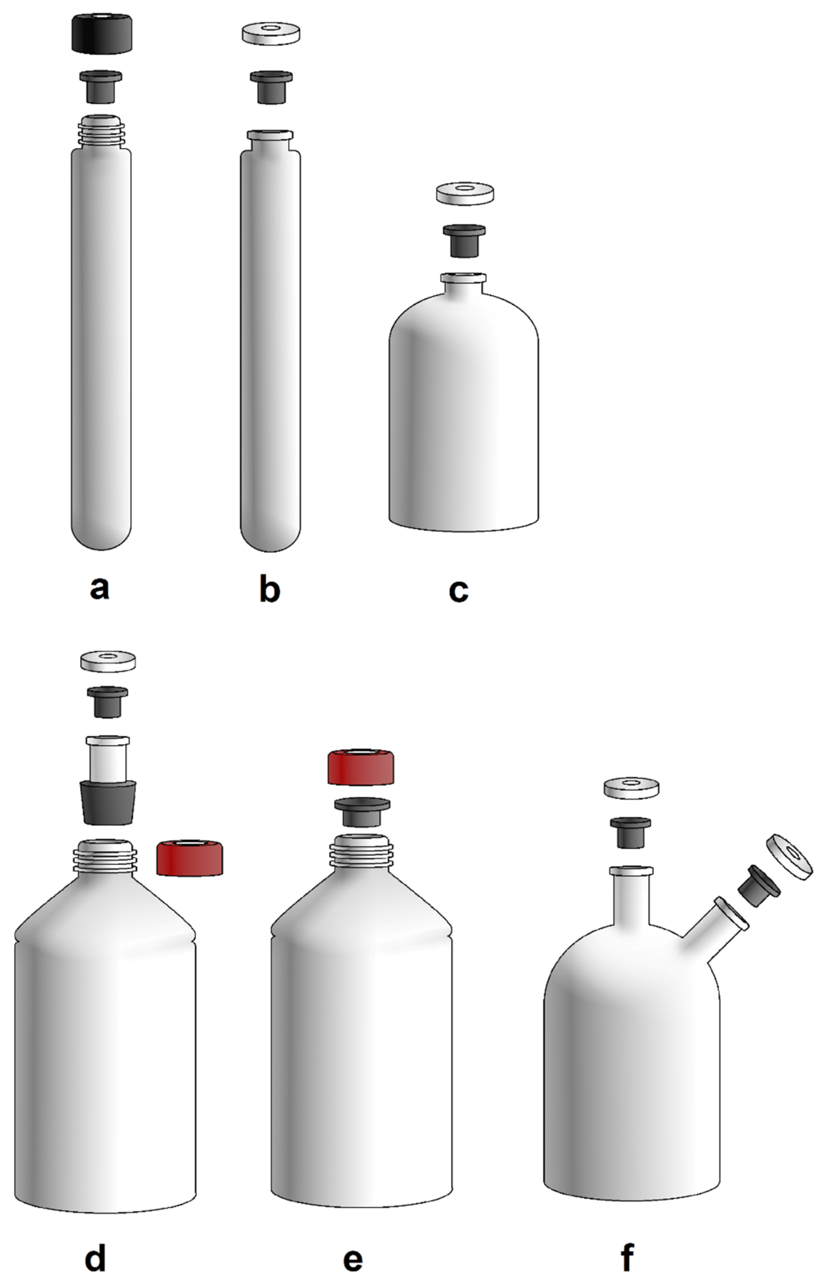
3.1. Laboratory Equipment for Cultivation of Methanogens

Anaerobic cultivation differs from aerobic cultivation in many aspects; one of these concerns the instruments with which the laboratory should be equipped. These instruments were established as novel methods of anaerobic cultivation to be introduced. The purpose of maintaining an anaerobic atmosphere and avoiding exposure to O2 led to a replacement of some classical instruments and cultivation vessels with the new ones.
The most commonly used cultivation vessels are Hungate or Balch tubes and serum bottles (Figure 2a–c). A Hungate tube is closed with a butyl stopper and a screw cap. Balch tubes and serum bottles are closed with butyl stoppers and sealed with aluminium seals by crimping. This allows the cultivation vessel to be pressurised with substrate gases [59]. However, it also allows the cultivation of gas-producing microbes. In the case of cultivation of, e.g., hydrogenotrophic, autotrophic methanogens, repeated gassing is still necessary because the gaseous substrate in the headspace is consumed according to the specific gas uptake rate and the biomass concentration of gas-utilizing microbes. In the case of thermophilic and hyperthermophilic methanogens, the headspace replenishment must be performed at higher frequency compared to mesophilic or psychrophilic methanogens. Cultivation in larger volumes can be performed using a modified bottle with a neck that can be sealed with a stopper and aluminium cap (Figure 2d,f). The bottle can consist of more than one opening for various purposes [16,60,61,62]. It is also possible to use a pressure-resistant Schott bottle for anaerobic cultivation, which is closed with a GL45 screw cap with an opening and the appropriate butyl rubber stopper (Figure 2e).
The use of anoxic gas or a gas mixture is necessary in anaerobic cultivation. To ensure that there is no O2 present in the gas, a copper column was used as another innovation to remove O2 from the used gas [14]. The tube filled with copper turnings or copper pellets is heated while the gas flows through the column. When the apparatus is constructed using copper turnings, the electric heating needs to exceed 350 °C. The copper pellets proved to be more effective, as a lower temperature of around 150 °C was sufficient to remove trace amounts of O2 [63]. Before introducing H2, the column must be absolutely free of O2, so it is gassed with N2 or argon beforehand. Today, when gasses of purity 5.0 (99.999%) are used in the laboratory, the removal of O2 by heated copper columns is no longer necessary.
For the purpose of quantitative gassing, the gassing manifold was invented [60,61,64]. The gassing manifold is necessary when a large number of media or cultures need to be gassed. This is because the gassing of cultures or anaerobization of media is a very time-consuming step. The gassing manifold divides the gas stream into several parallel streams, so that parallel gassing is possible [64]. The gassing manifold can be assembled from individual parts, or it is possible to buy prefabricated gassing manifold (Ochs Glasgerätebau, Bovenden, Germany). These manifolds are often operated manually. However, an electric gassing manifold is also available, e.g., Deoxidized Gas Pressure Injector; IP-8; Sanshin Industrial, Japan. The tubing exiting the gassing manifold can be terminated with a glass syringe containing sterile cotton or a Luer-Lock connector. In the latter case, a sterile filter is inserted between the tubing and the needles. Due to these settings, it is possible to connect the needle through the lock and manipulate closed vessels without exposing them to O2. Since all operations with cultures are also performed using syringes and needles, it is also recommended to flush the syringe with the N2 or CO2 gas before taking a sample of the culture or solutions [14,15].
Some assignments require a strictly anaerobic environment for manipulation, and it is complicated or even impossible to perform them without the use of the anaerobic glove box (Figure 3). It should be noted that the anaerobic glove box is an anoxic environment, but not a sterile one. Glove boxes with built-in HEPA filters are available (Labconco corporation, Kansas City, MO, USA), or a HEPA filter system could be added additionally (Coy Laboratory Products, Grass Lake, MI, USA).

3.2. Preparation of Anoxic Media for Methanogens

3.2.1. Composition of Medium for Methanogens

The preparation of media for methanogens can be a very time-consuming process, as many steps are required to make the media suitable for their growth. In addition, the media should reflect the growth conditions of each species in the natural environment, adapting it to the specific requirements of the species for laboratory experiments or for later biotechnological application.
The basis of the media is a mineral buffer solution. The salts in the media can be chlorides or sulphates, although sulphates increase the possibility of SRB growing in the media and competing with methanogens in enrichment cultures [65]. Another important component is the trace element solution, which contains important cofactors of metabolic enzymes. The most used solutions were described by Pfennig and often used by Widdel in his works with phototrophs and sulphate reducers, e.g., SL6 or SL10 [66,67,68,69] (Table 1), or Wolfe’s mineral solutions, made especially for methanogens [60]. Recently, 80 methanogens were grown in 22 chemically defined and/or complex media to assess their methane production and growth characteristics for the identification of high-performance methanogens. The analysis also included multivariate statistical analysis of the medium’s constituents [8].
Vitamin requirements differ among species. Many methanogens can be grown without the addition of vitamins [8,56,70,71], but previous studies have reported that the presence of vitamins supports their growth [72,73]. Other species might be fastidious and require a whole range of different vitamins, e.g., Methanimicrococcus blatticola [72,74,75]. Since vitamins are often thermolabile, their filter-sterilized stock solutions are added to media after the sterilization of a basic solution. Similarly to trace elements, complete solutions of vitamins are often prepared in advance and used in various media, e.g., Wolfe’s [60] or Widdel’s [69] vitamin solutions (Table 2).
The use of reducing agents in the media to keep the ORP within a specific range is required for the successful cultivation of anaerobic organisms, not only methanogens [76]. For methanogens, the ORP should not exceed -330 mV. Sodium sulphide (Na2S), sodium thioglycolate, dithiothreitol, L-cysteine or sodium dithionite could be used as a reducing agent, which should be added to the media prior to inoculation [76]. The most used reducing agent is Na2S. On the other hand, L-cysteine is also used as a source of sulphur for microorganisms instead of a pure reducing agent because an ORP of -340 mV cannot be achieved. The ORP is then set by the final addition of the reducing agent to the medium. If a redox indicator is present in the medium, the coloration of the medium will indicate whether the ORP is within the intended boundary. There are a number of different indicators with resazurin being the most commonly used [76]. However, when methanogens are frequently cultivated in the laboratory, redox indicators are not applied any more [8,64].
Since the organisms are adapted to live in a complex environment in the presence of various microorganisms, this may lead to the necessity of a component that is specific to particular environment and cannot be substituted in vitro. For the successful cultivation of these auxotrophic microorganisms, which initially appear unculturable, additional enhancement of the medium is required. The additive could be yeast extract, ruminal fluid, or sludge fluid [77,78,79]. Media containing carbonate buffer ensure that the pH remains stable. This is quite inconvenient when a wide pH range has to be measured and the optimal pH of the species determined. Different buffer systems may have to be tested.
This is the basic characterization of media for methanogens. Nowadays, there are different media depending on the species or environment of the microorganism of interest, which are freely available on the websites of microorganism collections such as the Deutsche Sammlung für Mikroorganismen und Zellkulturen (DSMZ) or the American Type Culture Collection (ATCC). Unfortunately, the recipes are updated without historical documentation of their former versions, which makes their use somewhat unreproducible. Moreover, the medium recipes are sometimes written in an unnecessarily complicated manner, especially when it comes to the recipe for the cultivation of methanogens. Therefore, it is recommended to assess up-to-date literature in order to select the appropriate medium. In addition, an assessment of growth characteristics of methanogens on different media was recently performed, which could help to select a medium for the cultivation of methanogens [8].

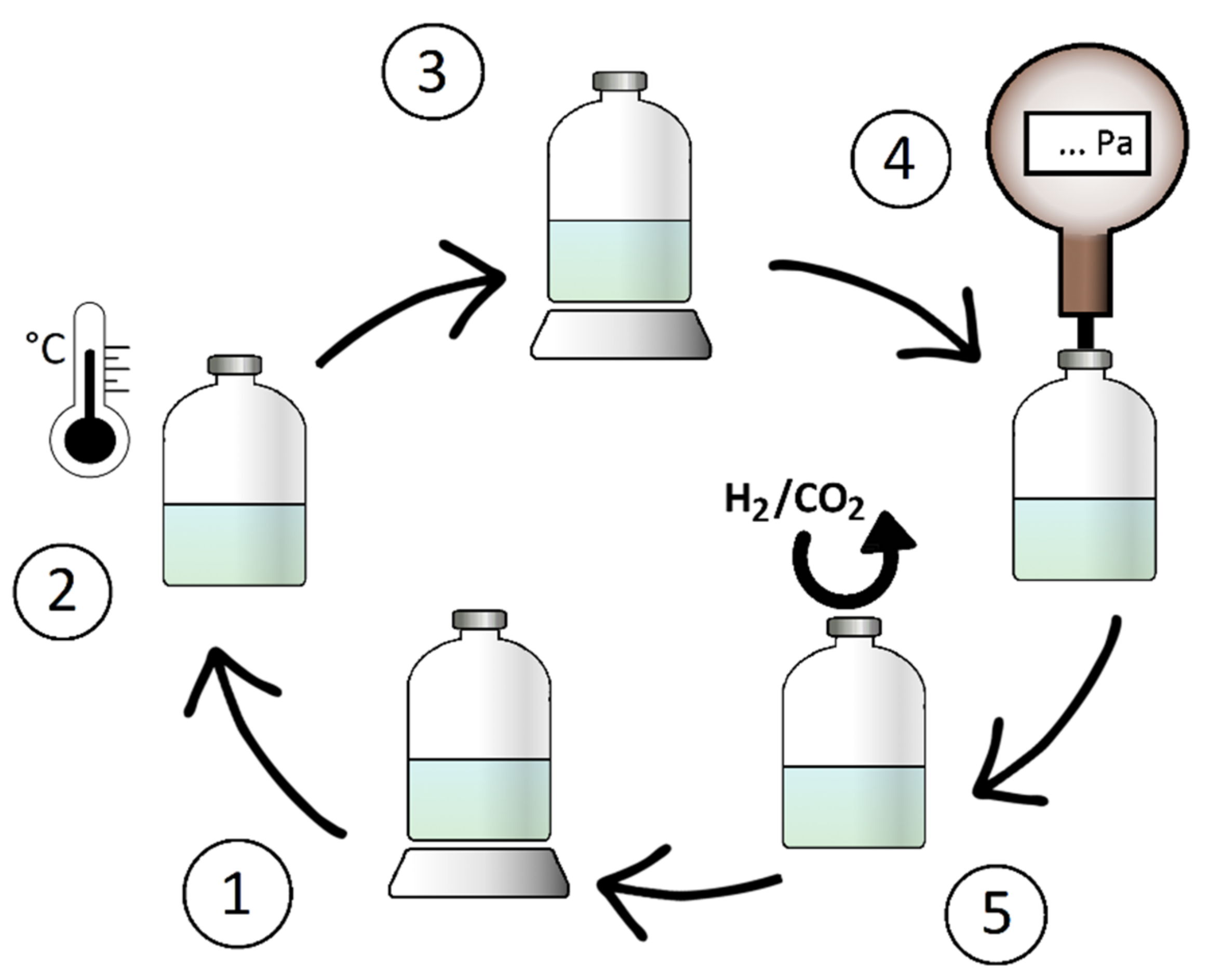
3.2.2. Process of Anaerobization of the Medium

Hungate’s technique (1969) for the preparation of O2-free media depends on the heat stability of the solution. Heat-stable media undergo a boiling process in which the dissolved O2 is expelled from the media. Heat-labile solutions should be purged with gas bubbles for about 30 min to one hour to remove residual O2.
Dispersal of the media can be performed prior to sterilization, which is recommended [14,64], or after sterilization, which carries the risk of contamination [80]. Previously, Hungate’s method of pipetting the media into the vessels while simultaneously gassing the media and the final vessel was quite difficult to manage, as one person must hold the pipette while orally pipetting the media and hold the Hungate tube at the same time (Figure 4a). The vessel is then closed with butyl stopper. A detailed description of this method of media preparation with slight modifications is also provided by Bryant [81]. Not only is the procedure complicated and difficult to perform, but oral pipetting is also prohibited nowadays. Later, apparatus to simplify the process of dispersing was invented, which can be used in the preparation of non-pressurised media (Figure 4b) [63].
Balch proposed to prepare the media in an anaerobic box, which simplified the whole pipetting process. The boiled medium is purged with a gas mixture of N2/CO2 (4:1 (v/v)), then the reducing agent is added, the medium is transferred into the anaerobic box and distributed into the cultivation vessels without being exposed to air. In the final step, the closed media are removed from the anaerobic box, the gas atmosphere is replaced, and the media are autoclaved [60]. In this way, the anoxic conditions are maintained and a better adjustment of pH is achieved, as it is carried out after the addition of Na2S.
Another modification as well as the first use of serum bottles in cultivation was devised by Miller and Wolin in 1974. The medium is prepared, then boiled and purged with O2-free gas while another stock solution is added. Afterwards, the medium is dispersed into serum bottles that are constantly purged with O2-free gas and closed with a butyl robber stopper while still being gassed. In this way, the O2 cannot enter the vessel. The media are sterilized by autoclaving.
When cultivating strictly anaerobic SRB and phototrophs, Widdel had also developed methodological variations and procedures. Since these microorganisms do not require a gas atmosphere, the apparatus was designed to fill the entire culturing vessel. The purpose of his designs is to autoclave the medium in the vessel simultaneously with the dispersing apparatus. After autoclaving, the medium is purged with gas while being stirred and dispersed into the culture vessels. The first model was a bottle with a butyl cap for gas purging and with an opening for dispersing the medium. Another model consisted of a conical vessel with a butyl cap, holes in the stopper for thermolabile solution addition, a gassing tube and a dispersing tube. During the process of dispersing, the vessel is turned upside down [69].
The final design is the Widdel flask (Figure 5). The reverse conical shape with a flat base ensures efficient stirring and the possibility of dispersing media from the whole volume of the flask. The individual openings for inserting and taking samples reduce the risk of contamination. At first, the vessel is filled with mineral solution and autoclaved. Then, it is continuously purged with the necessary gas or gas mixture while cooling, and the thermolabile components and reducing agents are added to the media. Due to the pressure created inside through gassing, the medium is poured through the dispensing apparatus into sterile vessels and closed. This method is suitable for media containing thermolabile components or for the media where the precipitation of salts during autoclaving occurs. The disadvantage of this method could be considered the contact with air during dispensing of the media or possible microbial contamination. If this method is used for cultivation of microorganisms that require a headspace, it is difficult to pour the exact volume of media and ensure anoxicity due to the filling mechanism, but it is not a problem to purge the culturing vessels with gas before and immediately after filling the media. Manipulation with this vessel is also described in detail elsewhere [82]. For the purpose of the cultivation of methanogens, this method of media preparation was used in the work on the isolation of Methanobacterium aarhusense [83].
In 2010, Wolfe published a method for the preparation of anoxic solutions and media in small volumes. The main advantage of this method is the speed that leads to anoxic solutions. Using the classic purging-with-gas method would require more time. The combination of vacuum–gas cycling and vortexing the entire system to increase the surface area of the liquid effectively expels O2 from the culturing vessel [84]. However, even without boiling or vortexing, vacuum–gas cycling prior to autoclaving has been proven efficient for media preparation and cultivation and is used as a proper method nowadays [85,86]. The number of cycles before autoclaving can range from three [85,86] to five [64]. The flushing of the aerobically dispersed media before autoclaving is also efficient [75].
To briefly summarize, the anaerobization of the media could be accomplished via different approaches depending on the objective of the media. The easiest and the most progressive, as well as the most suitable for manipulation and the least time-consuming, is the vacuum–gas cycling method combined with gassing manifold. This method does not require an anaerobic glove box, although a vacuum pump is required. Without a vacuum pump, flushing the media is also an efficient possibility, though slightly more time-consuming. For complicated media consisting of more solutions, the Widdel flask is still a present option that would save material such as needles, syringes and butyl stoppers, which could be destroyed by repeated piercing. These variants can substitute for an anaerobic glove box, which is not present in every laboratory; the manipulation is more difficult or which could be reserved for different tasks.

3.3. Cultivation and Pure Cultures Isolation Techniques

Using solid media for strictly anaerobic microorganisms and isolation of pure cultures may be seen as the most difficult part of anaerobic microbiology and biotechnology. The main obstacles are the slow growth rate of some methanogens and maintaining an anaerobic atmosphere. During cultivation, the agar could dry out quickly before the colonies become visible. Not every species is cultivable on solid media, and the higher temperature cannot be achieved with agar, so alternatives, e.g., Gellan Gum, have to be used [63]. Moreover, Petri dishes are not airtight. These are the reasons why anaerobes are mostly cultivated in liquid media.

3.3.1. Petri Dishes Cultivation in Anaerobic Jar

While cultivating anaerobic microorganisms on agar in a Petri dish, it is possible to incubate the plates in the anaerobic glove box. The atmosphere here is provided by the gas from the glove box, but it does not contain a sufficient amount of H2 if methanogens are to be cultivated, as the headspace gas contains up to 4.5 Vol.-% H2 in N2 (forming gas). The low partial pressure of H2 could slow down the generation time of methanogens. Anaerobic jar/pressure cylinders can be used to incubate inoculated Petri dishes inside (Figure 6). Some jars are equipped with gassing inlets to fill the headspace with substrate gas. The other option is to use a palladium catalyst inside the jar that uses H2 to reduce O2. It should be noted that palladium is deactivated by H2S, so the catalyst must be replaced before every experiment [87]. There are several commercially available anaerobic pack systems (e.g., AnaeroPouch-Anaero; Mitsubishi Gas Chemical Company, Japan) that have been used and described as support systems for anaerobic cultivation [88]. Some hydrogenotrophic, autotrophic methanogens could be cultivated on agar plates without a H2 atmosphere by using, for example, formate as a substrate [85].
When cultivating methanogens on agar, in addition to all the above-mentioned aspects, it is also essential to pay attention to the used agar, its type and brand, its concentration and the amount of Na2S [89]. The agar should be washed several times before use to remove all impurities. All factors could affect the morphology of the colonies and the specific growth rate. It is preferable to perform inoculation into deep agar or layered agar [90,91,92].
Special attention should also be paid to the material from which the anaerobic jar is constructed. For long-term cultivations, plastic materials do not have to be O2-tight. For this reason, even anaerobic jars with a set atmosphere should also be better kept in an anaerobic box if they are not made of metal [63]. The use of a modified canning jar instead of an anaerobic jar has also been described [90]. The agar plates have been used to isolate Methanocaldococcus jannaschii [93] or Methanothermococcus thermolithotrophicus [94].

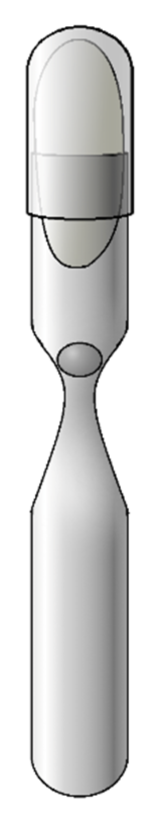
3.3.2. Hungate’s Roll Tube Technique

The most used technique for obtaining pure cultures of methanogens is Hungate’s roll tube technique [14,95,96,97]. This technique is based on the formation of a thin layer of inoculated agar, adhering to the walls of the Hungate tube. The gas phase is anaerobic. During growth, colonies become visible on the agar and as subsurface colonies. These are picked and further dispersed in the new dilution series or cultivated. Bryant preferred to inoculate the picked colonies into a slant medium rather than a liquid medium. The concentrated inoculum was found to be better adapted to the conditions in the new environment, as inoculation into liquid media is sometimes unsuccessful [32,81,98]. Applying this method, the microorganisms grow through the agar and form surface and subsurface colonies. Due to this effect, the morphology of the colonies could differ and that could be wrongly considered as a mixed culture [14]. It is also possible to apply thin layer of agar on the walls and inoculate the roll-tube after the solidification, as it was carried out during the isolation of Methanogenium marinum [99]. The purpose of the later inoculation was to avoid exposing the microorganisms to temperatures higher than 15 °C.
A variation of this technique was performed in 1973 by Miller and Wolin with the use of serum bottles instead of Hungate tubes. This method proved to be efficient for the isolation of anaerobic microorganisms such as Selenomonas ruminantium, Ruminococcus albus or Methanobrevibacter ruminantium [16].

3.3.3. Agar Shake Dilution Tube Method

Before the Hungate roll-tube method was developed, the agar shake dilution tube method was used for the cultivation of anaerobic microorganisms [4,27]. The latter method differs from Hungate’s method in the use of the column of semisolid agar in the tube, closed by a butyl stopper. During the growth of methanogens, visible colonies form inside the column. When the methanogens consume a liquid substrate (acetate or methylated compounds), or the technique is used for gas-producing anaerobes, such as Clostridia, they produce an excess volume of gas and the agar in the column is ruptured by the gas bubbles (Figure 7b) [100]. If there are facultatively anaerobic microorganisms present, they grow at the interface with the gas and consume the traces of O2, so the tube is completely anaerobic.
During the preparation of these agar shake tubes as well as the preparation of Hungate roll tubes, the temperature of agar must be optimal, as it should still be liquid but not too hot for microorganisms. For this method, modifications also have been introduced, changing the shape of the test tube slightly [101]. This method has been repeatedly used to isolate SRB [80,102] and has also been proven as a method for testing the antibiotic susceptibility of anaerobic microorganisms [103]. Even Hungate initially used the method [33] and it has also been repeatedly used for pure colony isolation in rather more recent publications [83,100], or in serum bottle modification [75].

3.3.4. Lee Tube Method

Another interesting method for the enumeration and cultivation of anaerobic microorganisms is the use of the Lee tube (Figure 8a) [104]. Colonies grow in the reduced agar present in the layer between the outer and inner tubes. The original Lee tube is made of a single piece of glass and sealed with a screw cap, which is more appropriate; a modified Lee tube consisting of two pieces and sealed with a cotton plug, which is not suitable for methanogens using a gaseous substrate, has also been described (Figure 8b) [105]. The Lee tube method has been used repeatedly to isolate and characterise anaerobic bacteria [106] and to maintain the cultures [107], but has not yet been used for methanogens.

3.3.5. Hermann’s Flat Flask Method

As mentioned above, cultivation on a Petri dish is not convenient due to evaporation of the water and drying of the agar. In 1986, Hermann published a method for anaerobic cultivation on poured agar in a flat flask used originally for phototrophic cultivation (Figure 8c) [108]. Later, in 1992, the method was modified by Olson. An additional opening was added that can be closed and sealed with aluminium. The second opening is used to flush the bottle with anoxic gas during manipulation and inoculation (Figure 8d). Both openings are then sealed and the flask is cultivated [109]. The advantage of this method is that the presence of an anaerobic box is not necessary, the manipulation could be carried out on a laboratory bench and the individual bottles can be checked separately, in comparison to the cultivation of Petri dishes in an anaerobic jar. This method was used, for example, for cultivation of Methanobrevibacter millerae and Methanobrevibacter olleyae [79].

3.3.6. Single Cell Isolation Methods

For the isolation of strict anaerobes, micromanipulation and single-cell cultivation is also a possible solution used in anaerobic cultivation. The purpose of this method is to take a single cell from the sample and inject it into the media. The isolation of the cell could be performed manually using the microinjectors BactoTip [110] or by laser microscope where the laser beam isolates the cell [111]. For some anaerobic microorganisms, this could be quite time consuming, as they are known for a longer generation time [112,113] and the cultivation of the extracted cells sometimes proves unsuccessful. The recovery rate has been studied and optimization of the technique for different common bacteria was recently accomplished [114]. This method proved successful not only for the isolation of the methanogen Methanobrevibacter sp. from termite gut [110], but also for extremophilic archaea such as Metallosphaera sedulla and Saccharolobus solfataricus [115] or hyperthermophilic archaea [116].

3.3.7. Dilution to Extinction Method

In case the microorganisms are fastidious and cannot be isolated by agar techniques, one option is dilution to extinction. The technique is based on serial dilutions in liquid media, ensuring the loss of most cells until the desired cells are the only present. This method is widely used, although it requires number of cultivation vessels to perform the method [71,117,118]. To remove contaminating microorganisms from an enrichment culture containing methanogens, the addition of appropriate antibiotics, such as ampicillin, vancomycin, clindamycin or kanamycin, could be used to simplify the process of purification [54,66,119]. To exclude fungal contamination, amphotericin B could also be added for the isolation of methanogens from the intestinal sphere [120,121].

3.4. Novel Insights in Cultivation Techniques

The anaerobic cultivation methods described by Hungate and their variants are still in use today and are widely applied in anaerobic microbiology and biotechnology. However, there are still many obstacles, and the cultivation of anaerobes is still considered to be very difficult and not always successful, even by following procedures step by step. In order to improve cultivation methods, reduce the difficulty level, increase experimental results and expand the application possibilities, the methods need to be further adapted to today’s requirements and novel devices need to be used for automation, although the improvement of the techniques has not been an issue in the last two decades.

3.4.1. The Six-Well Method

One of the novel methods for cultivation of strictly anaerobic microorganisms was published in 2011 by Nakamura et al. The six-well method using the AnaeroPack system was found to be efficient for the cultivation of a number of methanogens, SRB and syntrophic bacteria [88]. The AnaeroPack system is based on the use of special catalyst sachets without the presence of water or H2 production. In the work of Nakamura, inoculated plates in aerobic and anaerobic conditions were placed into a bag with the AnaeroPack system and the gas was recharged with H2/CO2 (4:1 (v/v)) mixture. Both variants proved to be successful, although anaerobic conditions for inoculation showed better results.

3.4.2. Growth in Syntrophic Communities

A completely different approach to anaerobic cultivation was published in 2009 by Sakai. There are a number of microorganisms that cannot be cultured or require specific conditions. Most methanogens are cultivated under an atmosphere of H2/CO2 (4:1 (v/v)) and an overpressure of 1 to 2 bar. The natural environment can provide even higher pressure and H2 concentrations, but there are also environments that have a low H2 concentration in which methanogens are also encountered. They often require a lower H2 concentration as they are accustomed to grow dependently on the H2-producing bacterial species, forming a syntrophic relationship between the two species. It is difficult to simulate the complex relationship in a laboratory. Co-cultivation is the key to successful cultivation and obtaining new species. Instead of adding direct substrate, pre-substrate such as ethanol, butyrate or propionate is consumed by bacteria and H2 is gradually released, which is then utilized by hydrogenotrophic, autotrophic methanogens. This method results in the capture of a wider range of methanogens, while the high H2 concentration tends to target fast-growing species [122]. The obstacle in the method is connected with the follow-up purification of the culture. Sometimes it is enough to apply the techniques mentioned above and cultivate the microorganism under conditions of higher H2 concentration using the liquid dilution method [123] or deep agar dilution tubes [124,125], but the purification is not always accomplished and the whole process is very time-consuming.

3.4.3. Microplate Reader Technique

One of the obstacles to anaerobic cultivation is the testing of chemical substances. In aerobic cultivation, it is possible to obtain results using microtiter plates for which different conditions can be set in one run. Several devices are designed for the continuous measurement of optical density during growth, e.g., Bioscreen C (Oy Growth Curves Ab Ltd., Helsinki, Finland) or Sunrise (Tecan, Männedorf, Switzerland) [126]. This method could be adapted to anaerobic conditions with the use of an oil layer to ensure a microaerobic environment [127], or some of the instruments may provide a Gas Control Module for changing the atmosphere composition. However, strictly anaerobic microorganisms are not able to grow using these methods because the oil layer is not absolutely gas-impermeable, or the gas composition does not favour the growth of methanogens. The third option is to place the entire device inside an anaerobic box to anaerobically measure the optical density continuously or just for a one-time measurement [128,129].
For quantitative experiments, a great amount of serum vials or Hungate tubes must be prepared, which is time-consuming and material-heavy. For this reason, various scientific groups have taken an interest in the microplate reading technique to enable large-scale experiments with methanogens. In 2012, Bang et al. tested antimicrobial peptides on methanogens. The microplate was prepared anaerobically in an anaerobic box, cultivated in an anaerobic jar under an atmosphere of H2/CO2 (4:1 (v/v)) and then read with a device placed in the anaerobic box to maintain anaerobic conditions [130]. This procedure resulted in the cultivation of hydrogenotrophs and the measurement of their susceptibility to various antimicrobial peptides. A similar experiment was performed to test chemical substances using a microtiter plate, but with the differences that the microtiter plate was cultivated directly in an anaerobic box and the microorganisms were growing on a substrate other than H2 [131].

3.4.4. Microfluidic Techniques

In recent years, microfluidics has achieved more recognition as an efficient and high-throughput method for cultivation and testing different conditions. It is obvious that its applications are broad, as it offers a variety of conditions in a small space. Applications of microfluidics in anaerobic conditions are focused primary on human gut microbiota and capturing species that are not yet culturable [132,133,134]. This technology was used to determine the optimal conditions for the growth of Methanosaeta concilii, using an anaerobic microbioreactor under a N2/CO2 atmosphere [135]. However, the aforementioned experiments were conducted in an anaerobic chamber, and hydrogenotrophic, autotrophic methanogenesis has not yet been investigated.
At the borderline between microfluidics and the classic microtiter plate method is a device designed for anaerobic cultivation in a 48-well plate (e.g., BioLector, m2p-labs GmbH, Baesweiler, Germany). It allows measurements of optical density, pH, biomass and fluorescence, as well as continuous feeding of the reactors. The instrument has been tested for fermentation of e.g., Clostridium sp. and Lactobacillus sp. [136], although the potential use for strict anaerobes such as methanogens has not yet been tested.

4. Quantification Techniques

During the cultivation of microorganisms, it is necessary to measure their growth, use these data to calculate growth parameters to define these microorganisms, and compare different growth conditions. Knowledge of the growth kinetics is also important for biotechnological applications, to relate the substrate conversion or product formation to biomass, or to directly calculate gas production using pure methanogenic cultures. Methods for quantification of growth of anaerobic microorganisms were recently reviewed by Mauerhofer et al. [76]. These quantification methods are non-growth-dependent.

4.1. Optical Density Measurement Technique

Optical density measurement is still a widely used and classic method to measure biomass and calculate specific growth rate (Equation (2)) [137]:
μ = ln OD 2 - ln OD 1 t 2 - t 1
where µ is the specific growth rate (h−1), OD1 and OD2 are the optical density values determined at the respective times t1 and t2 (h).
Optical density of methanogens is often measured at wavelength 578 nm [138,139,140] but it could be also measured at wavelength 600 or 660 [74,141]. The measured optical density has to be multiplied by a conversion factor, which is specific to every organism and must be experimentally determined, to obtain data for biomass or number of cells per volume (Equation (3)):
x V = a · OD
where x represents the number of cells in the sample, V is the volume of the sample (mL or L), OD is optical density and a is the conversion factor.
In some cases, this method cannot be used due to cell aggregation, the presence of precipitates or possible interference of resazurin in the medium. For the latter complication, it is recommended to add a reduction agent before measurement, which results in decolorization of the medium, or even better to omit resazurin addition into the media at all if OD measurement is considered.

4.2. ATP Determination Method

When optical density measurement is not an option for biomass quantification, there are other possible methods, such as the calculation based on direct cell counting [66,71] or adenosine triphosphate (ATP) determination. ATP determination could be also used to determine the growth of the culture [142,143]. Nowadays, there are many commercial products for ATP assay available, based on luminescence (luciferin/luciferase system) or colorimetric methods.

4.3. Methods Requiring the Cultivation of Anaerobic Microorganism

4.3.1. Methane Production Measurement Techniques

Methanogens have the advantage of not only measuring biomass, but also releasing methane as a metabolic end product. Methane, as the main component of biogas, carries the conserved energy and it is highly important to obtain the biggest energy yield from the different used substrates. The growth rate can be calculated from methane production with the use of Equation (4) [117,144,145]:
μ CH 4 = ln M 2 / M 1 t 2 - t 1
where µCH4 is the specific growth rate (h−1) calculated based on methane production. M1 and M2 represent the produced methane (mmol) measured at the respective times t1 and t2 (h).
When the culture’s growth slows down and enters the stationary phase, the optical density does not change rapidly. On the other hand, methane production still occurs at high rate. To capture the methane produced over time, methane evolution rate (MER) (Equation (5)) is used [64,146]:
MER = Δ n CH 4 ( t 2 - t 1 ) · V
where MER is methane evolution rate (mmol L−1 h−1), ΔnCH4 is the difference between the produced methane (mmol) measured at the respective culturing times t1 and t2 (h), and V is the volume of the media (L).
During hydrogenotrophic, autotrophic methanogenesis, the methane is produced according to the molar equation as mentioned above in Equation (1).
Based on the equation, four moles of H2 and one mole of CO2 is consumed as one mole of methane is produced. Since the substrates are both in the form of gas, it is possible to use the ideal gas law (IGL) (Equation (6)) for calculation the methane concentration in the headspace of the culture vessel:
n = p · V R · T
where n is amount of the substance of gas (mol), p is pressure (Pa), V is the volume of the headspace (m3), R is the universal gas constant (J K−1 mol−1), and T represents the temperature (K).
For the application of this law, two ways of measurement exist. The first is based on the change in pressure in the headspace, which is measured with a manometer. The pressure drop represents the four moles of consumed H2 (Equation (7)):
n CH 4 = ( p 0 - p ) · V R · T · 4
where nCH4 is the amount of produced methane in the headspace (mmol), p0 represents the initial pressure in the vessel (Pa), p represents the actual pressure in the vessel (Pa), and V is the volume of headspace (L).
The other option is to combine the actual pressure of the vessel to calculate absolute moles of gas in the headspace and the percentual concentration of methane measured by gas chromatography (GC) (Equation (8)):
n CH 4 = p · V R · T .   %   GC 100
where nCH4 is the amount of produced methane in the headspace (mmol), p represents the actual pressure in the vessel (Pa), V is the volume of headspace (L), and % GC represents percentual representation of methane in the headspace.
The first way is much faster but depends heavily on the pressurization of the vessels. It is also recommended to flush the headspace with the gas after every measurement to set the same conditions and pressure. This is because gas leaks may occur when measuring with a manometer and the results may be greatly affected during the second measurement. The use of GC is dependent on the actual pressure and the repeated flushing is not required if there is still a significant amount of substrates, respectively H2, present in the headspace [64]. The use of GC is also the only option to directly measure methane produced from formate or by methylotrophic or acetoclastic methanogens.

4.3.2. Manometric OxiTop Measurement

Apart from typical reactors, a simple device for measuring pressure in the vessel is the anaerobic OxiTop (OxiTop®-IDS, WTW GmbH, Germany). With two openings for manipulation through needles, the substrate can be added or the samples removed. Although it has not been yet directly used on pure cultures, potential application on cultures is possible. Continuous pressure measurement gives insight into gas formation kinetics.

4.3.3. Indirect Quantification of Produced Methane via Weight Gain

Another indirect method to determine produced methane uses the weight increase due to the formation of water during hydrogenotrophic methanogenesis [64]. This method is based on repeated measures of weight in regular intervals (Figure 9). The weight increment represents the two moles of water formed on the one mole of methane and is described as the water evolution rate (WER) (Equation (9)).
WER = Δ m H 2 O ( t 2 - t 1 ) . Mr H 2 O . V
where WER is the water evolution rate (mol L−1 h−1), ΔmH2O is the mass difference (g) measured in the respective culturing times t1 and t2 (h), V is the volume of the media (L), and MrH2O is the molar mass of water (g mol−1).
In combination with the pressure measurement, this approach presents the metabolic activity of hydrogenotrophic, autotrophic methanogens. It is applicable to fast-growing methanogens where the weight increase is easily measurable and noticeable. For slowly growing methanogens, it is recommended to set a longer interval of measurement. In addition, drops of the suspension may leak when handling the serum bottles, leading to inaccuracies in the measurement and a slow decrease in the weight in the control serum bottle [147].

5. Up-Scaling Process during Anaerobes Cultivation

Classic use of serum bottles is required for cultivation of species, capturing new species of microorganisms, and studying of material degradation in the primary steps of research. The setup is called closed-batch cultivation. However, the repeated gassing results in unbalanced growth and the maximal pressure of the headspace should not exceed 3 bar. Therefore, cultivation in an environment where the culture could achieve balanced growth according to a growth curve with enough substrate is necessary. Furthermore, in order to simulate processes in industrial reactors, up-scaling is needed to achieve more accurate results. It is also the way to obtain microbial cell mass for further analyses.

Batch, Fed-Batch and Continuous Cultivation

Bioreactors can be operated in batch, fed-batch, and continuous modes. This division is based on the experimental design and on the level of intervention in the reactor. Batch mode requires the least intervention. The medium, substrate and culture are prepared at the beginning and harvested at the end of the experiment. The fed-batch operation mode differs from the batch system as the carbon and energy source are added during the run of the reactor at a defined rate. This setup is limited by the volume of the reactor as there is no outflow of the excess liquid (in the case of a liquid feed). Continuous cultivation, as the name suggest, depends on the continuous inflow of substrate and nutrients and outflow of suspension and products.
Up-scaling the cultivation of methanogens encounters several different obstacles. Batch cultivation of pure cultures depends on the amount of substrate, which does not have to be sufficient to obtain the requested yield of biomass. The greatest advantage of this setup is the possibility of pressurisation of the reactor and setting conditions for observing the growth of the population. The more the reactor is pressurised, the more substrate is available to the culture and substrate conversion could be observed through pressure measurement [146,148]. In special occasions, the gas could be removed and the reactor repeatedly pressurised.
A fed-batch system ensures continuous substrate addition; in the case of hydrogenotrophic, autotrophic methanogens, this means a flow of H2/CO2 (4:1 (v/v)) mixture (Figure 10). Continuous cultivation also removes suspension and ensures a stable medium volume [76]. Reactors with a continuous supply of H2/CO2 gas mixture are hard to pressurise to increase the solubility of the gases. One way to solve the problem is to stir the suspension to increase the liquid-phase interface. Not every species of methanogen is able to withstand agitation and atmospheric pressure. There were some cases where the continuous reactor was pressurised to an absolute pressure of 1.22 bar [149] or 1.75 bar [150], but this does not reach the pressure achievable in closed batch cultivation, where a pressure of tens of bars could be easily set. There is one exception described of a constructed, pressurised fed-batch reactor, with a pressure 10 to 20 bars, which had been operated with M. marburgensis [151]. Due to water formation during hydrogenotrophic, autotrophic methanogenesis, it is also suggested to take a notice of increasing volume of medium during the planning of the experiment. If the expected production is greater, removal of the excess volume and addition of medium should be considered.
In 1968, Bryant et al. successfully cultivated Methanobacterium strain M.o.H. in a fed-batch setup with a reactor volume of 12 L and under continuous gassing [152]. To date, the experiments of fed-batch or continuous operation setups of different volumes have been successfully carried out with mainly thermophilic microorganisms, such as Methanothermobacter species [138,149,153,154,155,156], or microorganisms from the order Methanococcales [138,157]. The exceptions are mesophilic Methanococcus maripaludis [158] and the mentioned Methanobacterium strain M.o.H [152].
Acetoclastic and methylotrophic species are not dependent on pressure, and it is possible to cultivate them under a H2-free atmosphere. In the case of closed batch cultivation, the pressure would increase instead [145].

6. Conclusions

Cultivation techniques for anaerobic microorganisms, especially strictly anaerobic ones such as methanogens, vary greatly, from classic methods of aerobic cultivation to modern anaerobic high-throughput methods. Because of the difficulty of dealing with the fastidious anaerobic microorganisms and the required equipment for the cultivation, not many laboratories focus on anaerobic methods and anaerobic microorganisms. With this review, the historical development of culturing methods and the potential applications are presented in order to introduce anaerobic cultivation to the scientific community and to assist in reflecting on these methods to develop a new generation of anaerobic cultivation methods. This review also emphasises the importance of persevering in the knowledge and pitfalls of anaerobic cultivation techniques in anaerobic microbiology and biotechnology.

Author Contributions

Writing—original draft preparation, N.H.; figures design, N.H.; writing—review and editing, M.V. and S.K.-M.R.R. All authors have read and agreed to the published version of the manuscript.

Funding

Open Access Funding by the University of Vienna.

Acknowledgments

Open Access Funding by the University of Vienna.

Conflicts of Interest

The authors declare no conflict of interest.

Appendix A

Table A1. Milestones in historical development of cultivation of anaerobic microorganisms.
YearTechniqueDescriptionReference
1898Agar shake tubesAgar in test tube mixed with culture and cultivated after solidification of the agar[4]
1900Pyrogallic acid (Buchner’s method)The use of pyrogallic acid on the cotton plug together with alkaline solution to absorb oxygen in the tube[5]
1921Hall’s marble sealSpecial constricted tube with concave marble seal that is overpoured with sterile medium[23]
1929Paraffin oil sealLayer of paraffin oil on top of culture medium in test tube[13]
1969Roll tube methodLayer of agar-culture mixture on walls of Hungate tube made by rolling the tube with liquid mixture until solidification occurs[14]
1969Copper columnApplication of copper column for expelling O2 from used gas[14]
1969The use of reducing solutionsReducing the redox potential in the medium for creating a more suitable environment[14]
1972Syringes and needlesPreservation of anaerobic environment by application of needles and syringes without the oxygen exposure during manipulation[15]
1974Serum bottle modification of Hungate techniqueApplication of serum bottles for cultivation of anaerobic microorganisms in liquid and solidified medium[16]
1976Pressurization of culture vesselsIncreasing partial pressure in the cultivation vessel leads to lessening of the gassing frequency and an increase in the methanogenesis rate[59]
1979Gassing manifoldSpecial manifold for parallel gassing of culture vessels[60]
1979Lee’s tubeLayer of agar mixed with culture between two glass walls of special tube[104]
1981Widdel flaskSpecial conical flask for preparation media containing thermolabile solutions[69,80]
1986Modified Lee’s tubeLayer of agar mixed with culture between two walls of two tubes inserted in each other[105]
1986Hermann´s flat flask methodThe usage of closed flat flask for cultivation of anaerobic microorganisms on agar[108]
1992Modified Hermann´s flat flaskAddition of an opening for gassing the flask while picking up the colonies from medium[109]
1999Single cell isolation techniqueFirst application of a single-cell isolation technique which enables the picking up of single cells from sample on methanogens[110]
2007Microfluidics applicationThe first usage of microfluidics in anaerobic microbiology cultivating pure culture of methanogenic Methanosaeta concilii under N2/CO2 (4:1 (v/v)) conditions[135]
2009Vacuum-gas methodThe dispersed medium is put through cycles of gassing and gas exhaustion to set anaerobic conditions in the vessel[86]
2010Vacuum-vortex methodVortexing the dispersed medium in vessel while applying cycle gassing and gas exhaustion to set anaerobic conditions in the vessel[84]
2011Six-well methodAnaerobic cultivation and isolation using a six-well plate and supporting anaerobiosis generating system[88]
2012Application of microplate technique on methanogens cultivationThe first usage of microplate reader technique to cultivate methanogens under H2/CO2 (4:1 (v/v)) atmosphere and to measure their optical density[130]

References

  1. Liu, C.-T.; Miyaki, T.; Aono, T.; Oyaizu, H. Evaluation of Methanogenic Strains and Their Ability to Endure Aeration and Water Stress. Curr. Microbiol. 2008, 56, 214–218. [Google Scholar] [CrossRef]
  2. Hall, I.C. Practical Methods in the Purification of Obligate Anaerobes. J. Infect. Dis. 1920, 27, 576–590. [Google Scholar] [CrossRef]
  3. Söhngen, N.L. Sur Le Rôle Du Méthane Dans La Vie Organique. Recl. Trav. Chim. Pays-Bas Belg. 1910, 29, 238–274. [Google Scholar] [CrossRef]
  4. Veillon, A.; Zuber, A. Recherches Sur Quelques Microbes Strictement Anaérobies et Leur Rôle En Pathologie. Arch. Méd. Exp. Anat. Pathol. 1898, 10, 517–545. [Google Scholar]
  5. Wright, J.H. A Simple Method Of Cultivating Anaerobic Bacteria. J. Boston Soc. Med. Sci. 1900, 5, 114–115. [Google Scholar] [PubMed]
  6. Ergal, İ.; Fuchs, W.; Hasibar, B.; Thallinger, B.; Bochmann, G.; Rittmann, S.K.-M.R. The Physiology and Biotechnology of Dark Fermentative Biohydrogen Production. Biotechnol. Adv. 2018, 36, 2165–2186. [Google Scholar] [CrossRef] [PubMed]
  7. Kushkevych, I.; Bosáková, V.; Vítězová, M.; Rittmann, S.K.-M.R. Anoxygenic Photosynthesis in Photolithotrophic Sulfur Bacteria and Their Role in Detoxication of Hydrogen Sulfide. Antioxidants 2021, 10, 829. [Google Scholar] [CrossRef] [PubMed]
  8. Mauerhofer, L.-M.; Zwirtmayr, S.; Pappenreiter, P.; Bernacchi, S.; Seifert, A.H.; Reischl, B.; Schmider, T.; Taubner, R.-S.; Paulik, C.; Rittmann, S.K.-M.R. Hyperthermophilic Methanogenic Archaea Act as High-Pressure CH4 Cell Factories. Commun. Biol. 2021, 4, 289. [Google Scholar] [CrossRef]
  9. Rittmann, S.K.-M.R.; Lee, H.S.; Lim, J.K.; Kim, T.W.; Lee, J.-H.; Kang, S.G. One-Carbon Substrate-Based Biohydrogen Production: Microbes, Mechanism, and Productivity. Biotechnol. Adv. 2015, 33, 165–177. [Google Scholar] [CrossRef]
  10. Beigelman, P.M.; Rantz, L.A. Clinical Significance of Bacteroides. Arch. Intern. Med. (Chic). 1949, 84, 605–631. [Google Scholar] [CrossRef]
  11. Veillon, A.; Zuber, A. Sur Quelques Microbes Strictement Anaerobies et Leur Rôle Dans La Pathologie Humaine. Comptes Rendus Hebd. Séances Mém. Soc. Biol. 1897, 49, 253–255. [Google Scholar]
  12. Gest, H. The Discovery of Microorganisms by Robert Hooke and Antoni van Leeuwenhoek, Fellows of the Royal Society. Notes Rec. R. Soc. Lond. 2004, 58, 187–201. [Google Scholar] [CrossRef] [PubMed]
  13. Hall, I.C. A Review Of The Development And Application Of Physical And Chemical Principles In The Cultivation Of Obligately Anaerobic Bacteria. J. Bacteriol. 1929, 17, 255–301. [Google Scholar] [CrossRef] [PubMed] [Green Version]
  14. Hungate, R.E. Chapter IV A Roll Tube Method for Cultivation of Strict Anaerobes. In Methods in Microbiology; Norris, J.R., Ribbons, D.W., Eds.; Academic Press: Cambridge, MA, USA, 1969; Volume 3, pp. 117–132. [Google Scholar]
  15. Macy, J.M.; Snellen, J.E.; Hungate, R.E. Use of Syringe Methods for Anaerobiosis. Am. J. Clin. Nutr. 1972, 25, 1318–1323. [Google Scholar] [CrossRef] [PubMed]
  16. Miller, T.L.; Wolin, M.J. A Serum Bottle Modification of the Hungate Technique for Cultivating Obligate Anaerobes. Appl. Microbiol. 1974, 27, 985–987. [Google Scholar] [CrossRef] [PubMed]
  17. Hitchens, A.P.; Leikind, M.C. The Introduction of Agar-Agar into Bacteriology. J. Bacteriol. 1939, 37, 485–493. [Google Scholar] [CrossRef] [PubMed] [Green Version]
  18. Pasteur, L.; Faulkner, F. Studies on Fermentation: The Diseases of Beer, Their Causes, and the Means of Preventing Them; Macmillan & Company, London, UK, 1879.
  19. Sebald, M.; Hauser, D. Pasteur, Oxygen and the Anaerobes Revisited. Anaerobe 1995, 1, 11–16. [Google Scholar] [CrossRef]
  20. Finegold, S.M. A Century of Anaerobes: A Look Backward and a Call to Arms. Clin. Infect. Dis. Off. Publ. Infect. Dis. Soc. Am. 1993, 16 (Suppl. 4), S453–S457. [Google Scholar] [CrossRef] [PubMed]
  21. Pasteur, L. The Germ Theory And Its Applications To Medicine And Surgery. In Scientific Papers: Physiology, Medicine, Surgery, Geology: With Introductions, Notes and Illustrations; Harvard Classics; P.F. Collier & Son: New York, NY, USA, 1910; Volume 38, p. 440. ISBN 978-1-61640-122-1. [Google Scholar]
  22. Hall, I.C. Intestinal Flora in New-Born Infants. Am. J. Dis. Child. 1935, 49, 390–402. [Google Scholar] [CrossRef]
  23. Hall, I.C. A Constricted Tube with Mechanical Seal for Anaerobic Fermentation Tests. J. Infect. Dis. 1921, 29, 317–320. [Google Scholar] [CrossRef] [Green Version]
  24. Griffin, A.M. A Modification Of The Buchner Method Of Cultivating Anaerobic Bacteria. Science 1932, 75, 416–417. [Google Scholar] [CrossRef] [PubMed]
  25. Mellon, R.R. A Modification of the Wright-Buchner Anaerobic Tube. J. Bacteriol. 1919, 4, 295–297. [Google Scholar] [CrossRef] [PubMed] [Green Version]
  26. Rockwell, G.E. An Improved Method for Anaerobic Cultures. J. Infect. Dis. 1924, 35, 581–586. [Google Scholar] [CrossRef]
  27. Heller, H.H. Principles Concerning the Isolation of Anaerobes Studies in Pathogenic Anaerobes. Ii. J. Bacteriol. 1921, 6, 445–470. [Google Scholar] [CrossRef] [PubMed] [Green Version]
  28. Mazé, P. Ferment Formenique. Ferment Formenique de l’acetone. Procede de Culture Simple Du Ferment Formenique. C. R. Acad. Sci. 1915, 78, 398–405. [Google Scholar]
  29. Anderson, B.G. Gaseous Metabolism of Some Anaerobic Bacteria: XIX. Methods. J. Infect. Dis. 1924, 35, 213–243. [Google Scholar] [CrossRef]
  30. Lloyd, B.; Cranston, J.A. Studies in Gas Production by Bacteria: Denitrification and Bacterial Growth Phases. Biochem. J. 1930, 24, 529–548. [Google Scholar] [CrossRef] [PubMed]
  31. Sacks, L.E.; Barker, H.A. The Influence of Oxygen on Nitrate and Nitrite Reduction. J. Bacteriol. 1949, 58, 11–22. [Google Scholar] [CrossRef] [Green Version]
  32. Bryant, M.P.; Burkey, L.A. Cultural Methods and Some Characteristics of Some of the More Numerous Groups of Bacteria in the Bovine Rumen. J. Dairy Sci. 1953, 36, 205–217. [Google Scholar] [CrossRef]
  33. Hungate, R.E. The Anaerobic Mesophilic Cellulolytic Bacteria. Bacteriol. Rev. 1950, 14, 1–49. [Google Scholar] [CrossRef] [PubMed]
  34. Sowers, K.R. Methanogenesis. In Encyclopedia of Microbiology, 3rd ed.; Schaechter, M., Ed.; Academic Press: Oxford, UK, 2009; pp. 265–286. ISBN 978-0-12-373944-5. [Google Scholar]
  35. Hofmann, A.W.V.I. On the Action of Trichloride of Phosphorus on the Salts of the Aromatic Monamines. Proc. R. Soc. Lond. 1867, 15, 54–62. [Google Scholar] [CrossRef]
  36. Anaerobic Fermentations, 32nd ed.; Bulletin (Illinois State Water Survey); Buswell, A.M.; Hatfield, W.D. (Eds.) Department of Registration and Education, Division of the State Water Supply: Urbana, IL, USA, 1939.
  37. Omelianski, V.L. Sur La Fermentation Cellulosique. Comptes Rendus Hebd. Séances Mém. Soc. Biol. 1897, 125, 1131–1133. [Google Scholar]
  38. Barker, H.A. Studies upon the Methane-Producing Bacteria. Arch. Mikrobiol. 1936, 7, 420–438. [Google Scholar] [CrossRef]
  39. Beijer, W.H. Methane Fermentation in the Rumen of Cattle. Nature 1952, 170, 576–577. [Google Scholar] [CrossRef]
  40. Barker, H.A. Studies upon the Methane Fermentation. IV. The Isolation and Culture of Methanobacterium omelianskii. Antonie Leeuwenhoek 1939, 6, 201–220. [Google Scholar] [CrossRef]
  41. Bryant, M.P.; Wolin, E.A.; Wolin, M.J.; Wolfe, R.S. Methanobacillus omelianskii, a Symbiotic Association of Two Species of Bacteria. Arch. Mikrobiol. 1967, 59, 20–31. [Google Scholar] [CrossRef]
  42. Reddy, C.A.; Bryant, M.P.; Wolin, M.J. Characteristics of S Organism Isolated from Methanobacillus omelianskii. J. Bacteriol. 1972, 109, 539–545. [Google Scholar] [CrossRef] [PubMed] [Green Version]
  43. Stadtman, T.C.; Barker, H.A. Studies on the Methane Fermentation. X. A New Formate-Decomposing Bacterium, Methanococcus vannielii. J. Bacteriol. 1951, 62, 269–280. [Google Scholar] [CrossRef] [PubMed] [Green Version]
  44. Smith, P.H.; Hungate, R.E. Isolation And Characterization Of Methanobacterium ruminantion n. SP1. J. Bacteriol. 1958, 75, 713–718. [Google Scholar] [CrossRef] [Green Version]
  45. Ferry, J.G.; Smith, P.H.; Wolfe, R.S. Methanospirillum, a New Genus of Methanogenic Bacteria, and Characterization of Methanospirillum hungatii Sp. Nov. Int. J. Syst. Bacteriol. 1974, 24, 465–469. [Google Scholar] [CrossRef]
  46. Zeikus, J.G.; Henning, D.L. Methanobacterium arbophilicum Sp.Nov. An Obligate Anaerobe Isolated from Wetwood of Living Trees. Antonie Leeuwenhoek 1975, 41, 543–552. [Google Scholar] [CrossRef] [PubMed]
  47. Zeikus, J.G.; Wolfe, R.S. Methanobacterium thermoautotrophicus Sp. n., an Anaerobic, Autotrophic, Extreme Thermophile. J. Bacteriol. 1972, 109, 707–715. [Google Scholar] [CrossRef] [PubMed] [Green Version]
  48. Woese, C.R.; Fox, G.E. Phylogenetic Structure of the Prokaryotic Domain: The Primary Kingdoms. Proc. Natl. Acad. Sci. USA 1977, 74, 5088–5090. [Google Scholar] [CrossRef] [Green Version]
  49. Hammes, W.P.; Winter, J.; Kandler, O. The Sensitivity of the Pseudomurein-Containing Genus Methanobacterium to Inhibitors of Murein Synthesis. Arch. Microbiol. 1979, 123, 275–279. [Google Scholar] [CrossRef]
  50. Kandler, O.; König, H. Chemical Composition of the Peptidoglycan-Free Cell Walls of Methanogenic Bacteria. Arch. Microbiol. 1978, 118, 141–152. [Google Scholar] [CrossRef] [PubMed]
  51. Kates, M.; Yengoyan, L.S.; Sastry, P.S. A Diether Analog of Phosphatidyl Glycerophosphate in Halobacterium cutirubrum. Biochim. Biophys. Acta BBA—Lipids Lipid Metab. 1965, 98, 252–268. [Google Scholar] [CrossRef]
  52. Kessel, M.; Klink, F. Archaebacterial Elongation Factor Is ADP-Ribosylated by Diphtheria Toxin. Nature 1980, 287, 250–251. [Google Scholar] [CrossRef] [PubMed]
  53. Langworthy, T.A.; Smith, P.F.; Mayberry, W.R. Lipids of Thermoplasma acidophilum. J. Bacteriol. 1972, 112, 1193–1200. [Google Scholar] [CrossRef] [Green Version]
  54. Godsy, E.M. Isolation of Methanobacterium bryantii from a Deep Aquifer by Using a Novel Broth-Antibiotic Disk Method. Appl. Environ. Microbiol. 1980, 39, 1074–1075. [Google Scholar] [CrossRef] [Green Version]
  55. König, H. Isolation and Characterization of Methanobacterium uliginosum Sp. Nov. from a Marshy Soil. Can. J. Microbiol. 1984, 30, 1477–1481. [Google Scholar] [CrossRef]
  56. Schönheit, P.; Moll, J.; Thauer, R.K. Growth Parameters (K s, Μmax, Y s) of Methanobacterium thermoautotrophicum. Arch. Microbiol. 1980, 127, 59–65. [Google Scholar] [CrossRef]
  57. Stetter, K.O. Archaeoglobus Fulgidus Gen. Nov., Sp. Nov.: A New Taxon of Extremely Thermophilic Archaebacteria. Syst. Appl. Microbiol. 1988, 10, 172–173. [Google Scholar] [CrossRef]
  58. Zabel, H.P.; König, H.; Winter, J. Isolation and Characterization of a New Coccoid Methanogen, Methanogenium tatii Spec. Nov. from a Solfataric Field on Mount Tatio. Arch. Microbiol. 1984, 137, 308–315. [Google Scholar] [CrossRef]
  59. Balch, W.E.; Wolfe, R.S. New Approach to the Cultivation of Methanogenic Bacteria: 2-Mercaptoethanesulfonic Acid (HS-CoM)-Dependent Growth of Methanobacterium ruminantium in a Pressureized Atmosphere. Appl. Environ. Microbiol. 1976, 32, 781–791. [Google Scholar] [CrossRef] [PubMed] [Green Version]
  60. Balch, W.E.; Fox, G.E.; Magrum, L.J.; Woese, C.R.; Wolfe, R.S. Methanogens: Reevaluation of a Unique Biological Group. Microbiol. Rev. 1979, 43, 260–296. [Google Scholar] [CrossRef] [PubMed]
  61. Wolfe, R.S. Chapter One—Techniques for Cultivating Methanogens. In Methods in Enzymology; Methods in Methane Metabolism, Part A.; Rosenzweig, A.C., Ragsdale, S.W., Eds.; Academic Press: Cambridge, MA, USA, 2011; Volume 494, pp. 1–22. [Google Scholar]
  62. Ravichandran, M.; Munisamy, P.; Natarajan, S.D.; Varadharaju, C. Rare Detection And Identification of Methanogenic Bacteria from Diverse Ecological Niches in India for Carbon Balance and Management in Our Environment. Int. J. Adv. Res. 2016, 4, 1174–1186. [Google Scholar] [CrossRef] [Green Version]
  63. Sowers, K.R. Growth and Identification. In Archaea: A Laboratory Manual—Methanogens; Robb, F.T., Sowers, K.R., DasSarma, S., Place, A.R., Schreier, H.J., Fleischmann, E.M., Eds.; Cold Spring Harbor Laboratory Press: Plainview, NY, USA, 1995; pp. 15–59. [Google Scholar]
  64. Taubner, R.-S.; Rittmann, S.K.-M.R. Method for Indirect Quantification of CH4 Production via H2O Production Using Hydrogenotrophic Methanogens. Front. Microbiol. 2016, 7, 532. [Google Scholar] [CrossRef] [Green Version]
  65. Vítězová, M.; Kohoutová, A.; Vítěz, T.; Hanišáková, N.; Kushkevych, I. Methanogenic Microorganisms in Industrial Wastewater Anaerobic Treatment. Processes 2020, 8, 1546. [Google Scholar] [CrossRef]
  66. Kotelnikova, S.; Macario, A.J.L.; Pedersen, K. Methanobacterium subterraneum Sp. Nov., a New Alkaliphilic, Eurythermic and Halotolerant Methanogen Isolated from Deep Granitic Groundwater. Int. J. Syst. Evol. Microbiol. 1998, 48, 357–367. [Google Scholar] [CrossRef] [PubMed] [Green Version]
  67. Pfennig, N. Rhodopseudomonas globiformis, Sp. n., a New Species of the Rhodospirillaceae. Arch. Microbiol. 1974, 100, 197–206. [Google Scholar] [CrossRef]
  68. Widdel, F.; Kohring, G.-W.; Mayer, F. Studies on Dissimilatory Sulfate-Reducing Bacteria That Decompose Fatty Acids—III. Characterization of the Filamentous Gliding Desulfonema limicola Gen. Nov. Sp. Nov., and Desulfonema magnum Sp. Nov. Arch. Microbiol. 1983, 134, 286–294. [Google Scholar] [CrossRef]
  69. Widdel, F.; Bak, F. Gram-Negative Mesophilic Sulfate-Reducing Bacteria. In The Prokaryotes; Balows, A., Trüper, H.G., Dworkin, M., Harder, W., Schleifer, K.-H., Eds.; Springer: New York, NY, USA, 1992; pp. 3352–3378. ISBN 978-1-4757-2193-5. [Google Scholar]
  70. Cheng, L.; Dai, L.; Li, X.; Zhang, H.; Lu, Y. Isolation and Characterization of Methanothermobacter crinale Sp. Nov., a Novel Hydrogenotrophic Methanogen from the Shengli Oil Field. Appl. Environ. Microbiol. 2011, 77, 5212–5219. [Google Scholar] [CrossRef] [PubMed] [Green Version]
  71. Takai, K. Methanothermococcus okinawensis Sp. Nov., a Thermophilic, Methane-Producing Archaeon Isolated from a Western Pacific Deep-Sea Hydrothermal Vent System. Int. J. Syst. Evol. Microbiol. 2002, 52, 1089–1095. [Google Scholar] [CrossRef] [PubMed]
  72. Miller, T.L.; Wolin, M.J. Methanosphaera stadtmaniae Gen. Nov., Sp. Nov.: A Species That Forms Methane by Reducing Methanol with Hydrogen. Arch. Microbiol. 1985, 141, 116–122. [Google Scholar] [CrossRef] [PubMed]
  73. Whitman, W.B.; Ankwanda, E.; Wolfe, R.S. Nutrition and Carbon Metabolism of Methanococcus voltae. J. Bacteriol. 1982, 149, 852–863. [Google Scholar] [CrossRef] [PubMed] [Green Version]
  74. Morii, H.; Nishihara, M.; Koga, Y. Isolation, Characterization and Physiology of a New Formate-Assimilable Methanogenic Strain (A2) of Methanobrevibacter arboriphilus. Agric. Biol. Chem. 1983, 47, 2781–2789. [Google Scholar] [CrossRef] [Green Version]
  75. Sprenger, W.W.; van Belzen, M.C.; Rosenberg, J.; Hackstein, J.H.; Keltjens, J.T. Methanomicrococcus blatticola Gen. Nov., Sp. Nov., a Methanol- and Methylamine-Reducing Methanogen from the Hindgut of the Cockroach Periplaneta americana. Int. J. Syst. Evol. Microbiol. 2000, 50, 1989–1999. [Google Scholar] [CrossRef] [PubMed] [Green Version]
  76. Mauerhofer, L.-M.; Pappenreiter, P.; Paulik, C.; Seifert, A.H.; Bernacchi, S.; Rittmann, S.K.-M.R. Methods for Quantification of Growth and Productivity in Anaerobic Microbiology and Biotechnology. Folia Microbiol. 2019, 64, 321–360. [Google Scholar] [CrossRef] [PubMed] [Green Version]
  77. Miller, T.L.; Lin, C. Description of Methanobrevibacter gottschalkii Sp. Nov., Methanobrevibacter thaueri Sp. Nov., Methanobrevibacter woesei Sp. Nov. and Methanobrevibacter wolinii Sp. Nov. Int. J. Syst. Evol. Microbiol. 2002, 52, 819–822. [Google Scholar] [CrossRef]
  78. Paynter, M.J.B.; Hungate, R.E. Characterization of Methanobacterium mobilis, Sp. n., Isolated from the Bovine Rumen. J. Bacteriol. 1968, 95, 1943–1951. [Google Scholar] [CrossRef] [PubMed] [Green Version]
  79. Rea, S.; Bowman, J.P.; Popovski, S.; Pimm, C.; Wright, A.-D.G. Methanobrevibacter millerae Sp. Nov. and Methanobrevibacter olleyae Sp. Nov., Methanogens from the Ovine and Bovine Rumen That Can Utilize Formate for Growth. Int. J. Syst. Evol. Microbiol. 2007, 57, 450–456. [Google Scholar] [CrossRef] [PubMed]
  80. Widdel, F.; Pfennig, N. Studies on Dissimilatory Sulfate-Reducing Bacteria That Decompose Fatty Acids: I. Isolation of New Sulfate-Reducing Bacteria Enriched with Acetate from Saline Environments. Description of Desulfobacter postgatei Gen. Nov., Sp. Nov. Arch. Microbiol. 1981, 129, 395–400. [Google Scholar] [CrossRef]
  81. Bryant, M.P. Commentary on the Hungate Technique for Culture of Anaerobic Bacteria. Am. J. Clin. Nutr. 1972, 25, 1324–1328. [Google Scholar] [CrossRef]
  82. Laso-Pérez, R.; Krukenberg, V.; Musat, F.; Wegener, G. Establishing Anaerobic Hydrocarbon-Degrading Enrichment Cultures of Microorganisms under Strictly Anoxic Conditions. Nat. Protoc. 2018, 13, 1310–1330. [Google Scholar] [CrossRef] [Green Version]
  83. Shlimon, A.G. Methanobacterium aarhusense Sp. Nov., a Novel Methanogen Isolated from a Marine Sediment (Aarhus Bay, Denmark). Int. J. Syst. Evol. Microbiol. 2004, 54, 759–763. [Google Scholar] [CrossRef] [PubMed] [Green Version]
  84. Wolfe, R.S.; Metcalf, W.W. A Vacuum-Vortex Technique for Preparation of Anoxic Solutions or Liquid Culture Media in Small Volumes for Cultivating Methanogens or Other Strict Anaerobes. Anaerobe 2010, 16, 216–219. [Google Scholar] [CrossRef] [PubMed]
  85. Long, F.; Wang, L.; Lupa, B.; Whitman, W.B. A Flexible System for Cultivation of Methanococcus and Other Formate-Utilizing Methanogens. Archaea 2017, 2017, e7046026. [Google Scholar] [CrossRef] [PubMed] [Green Version]
  86. Stieglmeier, M.; Wirth, R.; Kminek, G.; Moissl-Eichinger, C. Cultivation of Anaerobic and Facultatively Anaerobic Bacteria from Spacecraft-Associated Clean Rooms. Appl. Environ. Microbiol. 2009, 75, 3484–3491. [Google Scholar] [CrossRef] [PubMed] [Green Version]
  87. Dowell, V.R.; Hawkins, T.M. Laboratory Methods in Anaerobic Bacteriology; CDC Laboratory Manual: Atlanta, GA, USA, 1974. [Google Scholar]
  88. Nakamura, K.; Tamaki, H.; Kang, M.S.; Mochimaru, H.; Lee, S.-T.; Nakamura, K.; Kamagata, Y. A Six-Well Plate Method: Less Laborious and Effective Method for Cultivation of Obligate Anaerobic Microorganisms. Microbes Environ. 2011, 26, 301–306. [Google Scholar] [CrossRef] [Green Version]
  89. Jones, W.J.; Whitman, W.B.; Fields, R.D.; Wolfe, R.S. Growth and Plating Efficiency of Methanococci on Agar Media. Appl. Environ. Microbiol. 1983, 46, 220–226. [Google Scholar] [CrossRef] [PubMed] [Green Version]
  90. Apolinario, E.A.; Sowers, K.R. Plate Colonization of Methanococcus maripaludis and Methanosarcina thermophila in a Modified Canning Jar. FEMS Microbiol. Lett. 1996, 145, 131–137. [Google Scholar] [CrossRef]
  91. Kiener, A.; Leisinger, T. Oxygen Sensitivity of Methanogenic Bacteria. Syst. Appl. Microbiol. 1983, 4, 305–312. [Google Scholar] [CrossRef]
  92. Sowers, K.R.; Boone, J.E.; Gunsalus, R.P. Disaggregation of Methanosarcina Spp. and Growth as Single Cells at Elevated Osmolarity. Appl. Environ. Microbiol. 1993, 59, 3832–3839. [Google Scholar] [CrossRef] [PubMed] [Green Version]
  93. Jones, W.J.; Leigh, J.A.; Mayer, F.; Woese, C.R.; Wolfe, R.S. Methanococcus jannaschii Sp. Nov., an Extremely Thermophilic Methanogen from a Submarine Hydrothermal Vent. Arch. Microbiol. 1983, 136, 254–261. [Google Scholar] [CrossRef]
  94. Huber, H.; Thomm, M.; König, H.; Thies, G.; Stetter, K.O. Methanococcus thermolithotrophicus, a Novel Thermophilic Lithotrophic Methanogen. Arch. Microbiol. 1982, 132, 47–50. [Google Scholar] [CrossRef] [Green Version]
  95. Battumur, U.; Yoon, Y.; Bae, G.S.; Kim, C.-H. Isolation and Characterization of New Methanosarcina mazei Strains KOR-3, -4, -5, and -6 from an Anaerobic Digester Using Pig Slurry. Asian-Australas. J. Anim. Sci. 2017, 30, 1198–1205. [Google Scholar] [CrossRef] [PubMed] [Green Version]
  96. Cuzin, N.; Labat, M.; Garcia, J.L.; Ouattara, A.S. Methanobacterium congolense Sp. Nov., from a Methanogenic Fermentation of Cassava Peel. Int. J. Syst. Evol. Microbiol. 2001, 51, 489–493. [Google Scholar] [CrossRef] [Green Version]
  97. Ma, K.; Liu, X.; Dong, X. Methanobacterium beijingense Sp. Nov., a Novel Methanogen Isolated from Anaerobic Digesters. Int. J. Syst. Evol. Microbiol. 2005, 55, 325–329. [Google Scholar] [CrossRef] [Green Version]
  98. Dehority, B.A. Characterization of Several Bovine Rumen Bacteria Isolated with a Xylan Medium. J. Bacteriol. 1966, 91, 1724–1729. [Google Scholar] [CrossRef] [Green Version]
  99. Chong, S.C.; Liu, Y.; Cummins, M.; Valentine, D.L.; Boone, D.R. Methanogenium marinum Sp. Nov., a H2-Using Methanogen from Skan Bay, Alaska, and Kinetics of H2 Utilization. Antonie Van Leeuwenhoek 2002, 81, 263–270. [Google Scholar] [CrossRef] [PubMed]
  100. Krüger, M.; Beckmann, S.; Engelen, B.; Thielemann, T.; Cramer, B.; Schippers, A.; Cypionka, H. Microbial Methane Formation from Hard Coal and Timber in an Abandoned Coal Mine. Geomicrobiol. J. 2008, 25, 315–321. [Google Scholar] [CrossRef]
  101. Miller, N.J.; Garrett, O.W.; Prickett, P.S. Anaerobic Technique–a Modified Deep Agar Shake. J. Food Sci. 1939, 4, 447–451. [Google Scholar] [CrossRef]
  102. Widdel, F.; Pfennig, N. A New Anaerobic, Sporing, Acetate-Oxidizing, Sulfate-Reducing Bacterium, Desulfotomaculum (Emend.) acetoxidans. Arch. Microbiol. 1977, 112, 119–122. [Google Scholar] [CrossRef] [PubMed]
  103. Evans, J.B.; Harrell, L.J. Agar Shake Tube Technique for Simultaneous Determination of Aerobic and Anaerobic Susceptibility to Antibioticst. Antimicrob. Agents Chemother. 1977, 12, 3. [Google Scholar] [CrossRef] [PubMed] [Green Version]
  104. Ogg, J.E.; Lee, S.Y.; Ogg, B.J. A Modified Tube Method for the Cultivation and Enumeration of Anaerobic Bacteria. Can. J. Microbiol. 1979, 25, 987–990. [Google Scholar] [CrossRef] [PubMed]
  105. Ababouch, L.; Busta, F.F. A Modified Lee Tube Technique for the Cultivation and Enumeration of Anaerobes. Int. J. Food Microbiol. 1986, 3, 211–216. [Google Scholar] [CrossRef]
  106. Lee, S.Y.; Moore, S.E.; Mabee, M.S. Selective-Differential Medium for Isolation and Differentiation of Pectinatus from Other Brewery Microorganisms. Appl. Environ. Microbiol. 1981, 41, 386–387. [Google Scholar] [CrossRef] [PubMed] [Green Version]
  107. Robertson, L.W.; Chandrasekaran, A.; Reuning, R.H.; Hui, J.; Rawal, B.D. Reduction of Digoxin to 20R-Dihydrodigoxin by Cultures of Eubacterium lentum. Appl. Environ. Microbiol. 1986, 51, 1300–1303. [Google Scholar] [CrossRef] [Green Version]
  108. Hermann, M.; Noll, K.M.; Wolfe, R.S. Improved Agar Bottle Plate for Isolation of Methanogens or Other Anaerobes in a Defined Gas Atmosphere. Appl. Environ. Microbiol. 1986, 51, 1124–1126. [Google Scholar] [CrossRef] [Green Version]
  109. Olson, K.D. Modified Bottle Plate for the Cultivation of Strict Anaerobes. J. Microbiol. Methods 1992, 14, 267–269. [Google Scholar] [CrossRef]
  110. Fröhlich, J.; König, H. Rapid Isolation of Single Microbial Cells from Mixed Natural and Laboratory Populations with the Aid of a Micromanipulator. Syst. Appl. Microbiol. 1999, 22, 249–257. [Google Scholar] [CrossRef]
  111. Huber, R.; Huber, H.; Stetter, K.O. Towards the Ecology of Hyperthermophiles: Biotopes, New Isolation Strategies and Novel Metabolic Properties. FEMS Microbiol. Rev. 2000, 24, 615–623. [Google Scholar] [CrossRef]
  112. Huser, B.A.; Wuhrmann, K.; Zehnder, A.J.B. Methanothrix soehngenii Gen. Nov. Sp. Nov., a New Acetotrophic Non-Hydrogen-Oxidizing Methane Bacterium. Arch. Microbiol. 1982, 132, 1–9. [Google Scholar] [CrossRef]
  113. Mochimaru, H.; Tamaki, H.; Katayama, T.; Imachi, H.; Sakata, S.; Kamagata, Y. Methanomicrobium antiquum Sp. Nov., a Hydrogenotrophic Methanogen Isolated from Deep Sedimentary Aquifers in a Natural Gas Field. Int. J. Syst. Evol. Microbiol. 2016, 66, 4873–4877. [Google Scholar] [CrossRef] [PubMed]
  114. Hohnadel, M.; Maumy, M.; Chollet, R. Development of a Micromanipulation Method for Single Cell Isolation of Prokaryotes and Its Application in Food Safety. PLoS ONE 2018, 13, e0198208. [Google Scholar] [CrossRef] [PubMed]
  115. Ishøy, T.; Kvist, T.; Westermann, P.; Ahring, B.K. An Improved Method for Single Cell Isolation of Prokaryotes from Meso-, Thermo- and Hyperthermophilic Environments Using Micromanipulation. Appl. Microbiol. Biotechnol. 2006, 69, 510–514. [Google Scholar] [CrossRef]
  116. Huber, R.; Burggraf, S.; Mayer, T.; Barns, S.M.; Rossnagel, P.; Stetter, K.O. Isolation of a Hyperthermophilic Archaeum Predicted by in Situ RNA Analysis. Nature 1995, 376, 57–58. [Google Scholar] [CrossRef]
  117. Kita, A.; Suehira, K.; Miura, T.; Okamura, Y.; Aki, T.; Matsumura, Y.; Tajima, T.; Nishio, N.; Nakashimada, Y. Characterization of a Halotolerant Acetoclastic Methanogen Highly Enriched from Marine Sediment and Its Application in Removal of Acetate. J. Biosci. Bioeng. 2016, 121, 196–202. [Google Scholar] [CrossRef] [Green Version]
  118. Mori, K.; Iino, T.; Suzuki, K.-I.; Yamaguchi, K.; Kamagata, Y. Aceticlastic and NaCl-Requiring Methanogen “Methanosaeta Pelagica” Sp. Nov., Isolated from Marine Tidal Flat Sediment. Appl. Environ. Microbiol. 2012, 78, 3416–3423. [Google Scholar] [CrossRef] [PubMed] [Green Version]
  119. Sowers, K.R.; Ferry, J.G. Isolation and Characterization of a Methylotrophic Marine Methanogen, Methanococcoides methylutens Gen. Nov., Sp. Nov. Appl. Environ. Microbiol. 1983, 45, 684–690. [Google Scholar] [CrossRef] [Green Version]
  120. Dridi, B.; Fardeau, M.-L.; Ollivier, B.; Raoult, D.; Drancourt, M. The Antimicrobial Resistance Pattern of Cultured Human Methanogens Reflects the Unique Phylogenetic Position of Archaea. J. Antimicrob. Chemother. 2011, 66, 2038–2044. [Google Scholar] [CrossRef]
  121. Khelaifia, S.; Drancourt, M. Susceptibility of Archaea to Antimicrobial Agents: Applications to Clinical Microbiology. Clin. Microbiol. Infect. 2012, 18, 841–848. [Google Scholar] [CrossRef] [PubMed] [Green Version]
  122. Sakai, S.; Imachi, H.; Sekiguchi, Y.; Tseng, I.-C.; Ohashi, A.; Harada, H.; Kamagata, Y. Cultivation of Methanogens under Low-Hydrogen Conditions by Using the Coculture Method. Appl. Environ. Microbiol. 2009, 75, 4892–4896. [Google Scholar] [CrossRef] [Green Version]
  123. Imachi, H.; Sakai, S.; Sekiguchi, Y.; Hanada, S.; Kamagata, Y.; Ohashi, A.; Harada, H. Methanolinea tarda Gen. Nov., Sp. Nov., a Methane-Producing Archaeon Isolated from a Methanogenic Digester Sludge. Int. J. Syst. Evol. Microbiol. 2008, 58, 294–301. [Google Scholar] [CrossRef] [PubMed] [Green Version]
  124. Sakai, S.; Imachi, H.; Sekiguchi, Y.; Ohashi, A.; Harada, H.; Kamagata, Y. Isolation of Key Methanogens for Global Methane Emission from Rice Paddy Fields: A Novel Isolate Affiliated with the Clone Cluster Rice Cluster I. Appl. Environ. Microbiol. 2007, 73, 4326–4331. [Google Scholar] [CrossRef] [PubMed] [Green Version]
  125. Sakai, S.; Imachi, H.; Hanada, S.; Ohashi, A.; Harada, H.; Kamagata, Y. Methanocella paludicola Gen. Nov., Sp. Nov., a Methane-Producing Archaeon, the First Isolate of the Lineage “Rice Cluster I”, and Proposal of the New Archaeal Order Methanocellales Ord. Nov. Int. J. Syst. Evol. Microbiol. 2008, 58, 929–936. [Google Scholar] [CrossRef]
  126. Mytilinaios, I.; Salih, M.; Schofield, H.K.; Lambert, R.J.W. Growth Curve Prediction from Optical Density Data. Int. J. Food Microbiol. 2012, 154, 169–176. [Google Scholar] [CrossRef] [Green Version]
  127. Ahn, S.-J.; Ahn, S.-J.; Browngardt, C.M.; Burne, R.A. Changes in Biochemical and Phenotypic Properties of Streptococcus Mutans during Growth with Aeration. Appl. Environ. Microbiol. 2009, 75, 2517–2527. [Google Scholar] [CrossRef] [Green Version]
  128. Candry, P.; Van Daele, T.; Denis, K.; Amerlinck, Y.; Andersen, S.J.; Ganigué, R.; Arends, J.B.A.; Nopens, I.; Rabaey, K. A Novel High-Throughput Method for Kinetic Characterisation of Anaerobic Bioproduction Strains, Applied to Clostridium kluyveri. Sci. Rep. 2018, 8, 9724. [Google Scholar] [CrossRef]
  129. Stringer, S.C.; Webb, M.D.; George, S.M.; Pin, C.; Peck, M.W. Heterogeneity of Times Required for Germination and Outgrowth from Single Spores of Nonproteolytic Clostridium botulinum. Appl. Environ. Microbiol. 2005, 71, 4998–5003. [Google Scholar] [CrossRef] [Green Version]
  130. Bang, C.; Schilhabel, A.; Weidenbach, K.; Kopp, A.; Goldmann, T.; Gutsmann, T.; Schmitz, R.A. Effects of Antimicrobial Peptides on Methanogenic Archaea. Antimicrob. Agents Chemother. 2012, 56, 4123–4130. [Google Scholar] [CrossRef] [PubMed] [Green Version]
  131. Weimar, M.R.; Cheung, J.; Dey, D.; McSweeney, C.; Morrison, M.; Kobayashi, Y.; Whitman, W.B.; Carbone, V.; Schofield, L.R.; Ronimus, R.S.; et al. Development of Multiwell-Plate Methods Using Pure Cultures of Methanogens To Identify New Inhibitors for Suppressing Ruminant Methane Emissions. Appl. Environ. Microbiol. 2017, 83. [Google Scholar] [CrossRef] [PubMed] [Green Version]
  132. Ma, L.; Kim, J.; Hatzenpichler, R.; Karymov, M.A.; Hubert, N.; Hanan, I.M.; Chang, E.B.; Ismagilov, R.F. Gene-Targeted Microfluidic Cultivation Validated by Isolation of a Gut Bacterium Listed in Human Microbiome Project’s Most Wanted Taxa. Proc. Natl. Acad. Sci. USA 2014, 111, 9768–9773. [Google Scholar] [CrossRef] [Green Version]
  133. Villa, M.M.; Bloom, R.J.; Silverman, J.D.; Durand, H.K.; Jiang, S.; Wu, A.; Dallow, E.P.; Huang, S.; You, L.; David, L.A. Interindividual Variation in Dietary Carbohydrate Metabolism by Gut Bacteria Revealed with Droplet Microfluidic Culture. mSystems 2020, 5. [Google Scholar] [CrossRef] [PubMed]
  134. Watterson, W.J.; Tanyeri, M.; Watson, A.R.; Cham, C.M.; Shan, Y.; Chang, E.B.; Eren, A.M.; Tay, S. Droplet-Based High-Throughput Cultivation for Accurate Screening of Antibiotic Resistant Gut Microbes. eLife 2020, 9, e56998. [Google Scholar] [CrossRef]
  135. Steinhaus, B.; Garcia, M.L.; Shen, A.Q.; Angenent, L.T. A Portable Anaerobic Microbioreactor Reveals Optimum Growth Conditions for the Methanogen Methanosaeta concilii. Appl. Environ. Microbiol. 2007, 73, 1653–1658. [Google Scholar] [CrossRef] [PubMed] [Green Version]
  136. Birgen, C.; Degnes, K.F.; Markussen, S.; Wentzel, A.; Sletta, H. Butanol Production from Lignocellulosic Sugars by Clostridium beijerinckii in Microbioreactors. Biotechnol. Bioeng. 2020, 14, 1–12. [Google Scholar] [CrossRef]
  137. Widdel, F. Theory and Measurement of Bacterial Growth. In Grundpraktikum Mikrobiologie, 4. sem. (B. Sc); Universität Bremen: Bremen, Germany, 2007; p. 11. [Google Scholar]
  138. Azim, A.A.; Pruckner, C.; Kolar, P.; Taubner, R.-S.; Fino, D.; Saracco, G.; Sousa, F.L.; Rittmann, S.K.-M.R. The Physiology of Trace Elements in Biological Methane Production. Bioresour. Technol. 2017, 241, 775–786. [Google Scholar] [CrossRef] [PubMed]
  139. Smith, G.C.; Floodgate, G.D. A Chemical Method for Estimating Methanogenic Biomass. Cont. Shelf Res. 1992, 12, 1187–1196. [Google Scholar] [CrossRef]
  140. Widdel, F.; Wolfe, R.S. Expression of Secondary Alcohol Dehydrogenase in Methanogenic Bacteria and Purification of the F420-Specific Enzyme from Methanogenium thermophilum Strain TCI. Arch. Microbiol. 1989, 152, 322–328. [Google Scholar] [CrossRef]
  141. Shimizu, S.; Ueno, A.; Tamamura, S.; Naganuma, T.; Kaneko, K. Methanoculleus horonobensis Sp. Nov., a Methanogenic Archaeon Isolated from a Deep Diatomaceous Shale Formation. Int. J. Syst. Evol. Microbiol. 2013, 63, 4320–4323. [Google Scholar] [CrossRef] [PubMed]
  142. Kaesler, B.; Schönheit, P. Methanogenesis and ATP Synthesis in Methanogenic Bacteria at Low Electrochemical Proton Potentials. Eur. J. Biochem. 1988, 174, 189–197. [Google Scholar] [CrossRef] [PubMed]
  143. Schönheit, P.; Beimborn, D.B. ATP Synthesis in Methanobacterium thermoautotrophicum Coupled to CH4 Formation from H2 and CO2 in the Apparent Absence of an Electrochemical Proton Potential across the Cytoplasmic Membrane. Eur. J. Biochem. 1985, 148, 545–550. [Google Scholar] [CrossRef] [PubMed]
  144. Fukuzaki, S.; Nishio, N.; Nagai, S. Kinetics of the Methanogenic Fermentation of Acetate. Appl. Environ. Microbiol. 1990, 56, 3158–3163. [Google Scholar] [CrossRef] [Green Version]
  145. Yang, S.T.; Okos, M.R. Kinetic Study and Mathematical Modeling of Methanogenesis of Acetate Using Pure Cultures of Methanogens. Biotechnol. Bioeng. 1987, 30, 661–667. [Google Scholar] [CrossRef] [PubMed]
  146. Pappenreiter, P.A.; Zwirtmayr, S.; Mauerhofer, L.-M.; Rittmann, S.K.-M.R.; Paulik, C. Development of a Simultaneous Bioreactor System for Characterization of Gas Production Kinetics of Methanogenic Archaea at High Pressure. Eng. Life Sci. 2019, 19, 537–544. [Google Scholar] [CrossRef] [Green Version]
  147. Hanišáková, N. Methanogenic Archaea in Environmental Samples. Master’s Thesis, Masarykova Universita, Brno, Czechia, 2020. [Google Scholar]
  148. Taubner, R.-S.; Pappenreiter, P.; Zwicker, J.; Smrzka, D.; Pruckner, C.; Kolar, P.; Bernacchi, S.; Seifert, A.; Krajete, A.; Bach, W.; et al. Biological Methane Production under Putative Enceladus-like Conditions. Nat. Commun. 2018, 9, 748. [Google Scholar] [CrossRef] [PubMed] [Green Version]
  149. Martin, M.R.; Fornero, J.J.; Stark, R.; Mets, L.; Angenent, L.T. A Single-Culture Bioprocess of Methanothermobacter thermautotrophicus to Upgrade Digester Biogas by CO2-to-CH4 Conversion with H2. Archaea 2013, 2013, 157529. [Google Scholar] [CrossRef] [Green Version]
  150. Seifert, A.H.; Rittmann, S.; Herwig, C. Analysis of Process Related Factors to Increase Volumetric Productivity and Quality of Biomethane with Methanothermobacter marburgensis. Appl. Energy 2014, 132, 155–162. [Google Scholar] [CrossRef]
  151. Pappenreiter, P.A. Development of a Pressurised Biomethanation Reactor System Using CO2-Type Methanogenic Strains. Ph.D. Thesis, Johannes Kepler University Linz, Linz, Austria, 2020. [Google Scholar]
  152. Bryant, M.P.; McBride, B.C.; Wolfe, R.S. Hydrogen-Oxidizing Methane Bacteria I. Cultivation and Methanogenesis. J. Bacteriol. 1968, 95, 1118–1123. [Google Scholar] [CrossRef] [Green Version]
  153. Hoffarth, M.; Broeker, T.; Schneider, J. Effect of N2 on Biological Methanation in a Continuous Stirred-Tank Reactor with Methanothermobacter Marburgensis. Fermentation 2019, 5, 56. [Google Scholar] [CrossRef] [Green Version]
  154. Mauerhofer, L.-M.; Reischl, B.; Schmider, T.; Schupp, B.; Nagy, K.; Pappenreiter, P.; Zwirtmayr, S.; Schuster, B.; Bernacchi, S.; Seifert, A.H.; et al. Physiology and Methane Productivity of Methanobacterium thermaggregans. Appl. Microbiol. Biotechnol. 2018, 102, 7643–7656. [Google Scholar] [CrossRef] [PubMed] [Green Version]
  155. Rittmann, S.; Seifert, A.; Herwig, C. Quantitative Analysis of Media Dilution Rate Effects on Methanothermobacter marburgensis Grown in Continuous Culture on H2 and CO2. Biomass Bioenergy 2012, 36, 293–301. [Google Scholar] [CrossRef]
  156. Schill, N.; van Gulik, W.M.; Voisard, D.; von Stockar, U. Continuous Cultures Limited by a Gaseous Substrate: Development of a Simple, Unstructured Mathematical Model and Experimental Verification with Methanobacterium thermoautotrophicum. Biotechnol. Bioeng. 1996, 51, 645–658. [Google Scholar] [CrossRef]
  157. Mukhopadhyay, B.; Johnson, E.F.; Wolfe, R.S. Reactor-Scale Cultivation of the Hyperthermophilic Methanarchaeon Methanococcus jannaschii to High Cell Densities. Appl. Environ. Microbiol. 1999, 65, 5059–5065. [Google Scholar] [CrossRef] [PubMed] [Green Version]
  158. Shieh, J.; Whitman, W.B. Autotrophic Acetyl Coenzyme A Biosynthesis in Methanococcus maripaludis. J. Bacteriol. 1988, 170, 3072–3079. [Google Scholar] [CrossRef] [PubMed] [Green Version]
Figure 1. Hall’s constricted tube with marble seal (Hanišáková).
Microorganisms 10 00412 g001
Figure 2. The most used cultivation vessels: (a) Hungate tube; (b) Balch tube; (c) serum bottle; and culturing vessels for greater volume: (d) modified bottle with neck (according to Balch, 1979); (e) pressure bottle with butyl stopper and GL45 opening; (f) modification of bottle with more openings (Hanišáková, according to Miller and Wolin, 1974).
Microorganisms 10 00412 g002
Figure 3. Anaerobic box (Coy Laboratory Products, USA) (Photo: Laboratory).
Microorganisms 10 00412 g003
Figure 4. (a) Media pipetting described by Hungate and Bryant. (b) Dispersing media illustrated according to Sowers (Hanišáková).
Microorganisms 10 00412 g004
Figure 5. Widdel’s flask. ① Openings for input/output of media samples. ② Opening for gas entry. ③ Central opening for media filling. ④ Clamp for media filling. ⑤ Filling funnel for media. ⑥ Magnetic stirrer (Hanišáková).
Microorganisms 10 00412 g005
Figure 6. Illustration of anaerobic jar for anaerobic Petri dish cultivation (Hanišáková).
Microorganisms 10 00412 g006
Figure 7. Test tube for agar shake. (a) Test tube with butyl stopper. (b) Photo of gas bubbles inside the agar (Hanišáková, photo: laboratory).
Microorganisms 10 00412 g007
Figure 8. Cultivation vessels for pure colony isolation. (a) Lee tube; (b) modified Lee tube; (c) flat flask — original used by Hermann; (d) flat flask modification with additional opening (Hanišáková).
Microorganisms 10 00412 g008
Figure 9. The process of quantification of methane via produced water during hydrogenotrophic methanogenesis. ① The weight of inoculated serum bottle is measured. ② Incubation of the serum bottle. ③ The weight of the serum bottle after incubation is measured. ④ The pressure in the headspace is determined. ⑤ The serum bottle is flushed with gas. (1) The weight of the serum bottle is again measured, presenting the weight difference and gained mass of water (Taubner & Rittmann, 2016, redrawn by Hanišáková).
Microorganisms 10 00412 g009
Figure 10. Model of fed-batch bioreactor. ① Gas mixture inflow. ② Gas outflow. ③ Acid-base and sulphide solution input. ④ Stirring mechanism. ⑤ Opening for inoculum input. ⑥ Sample output. ⑦ Temperature sensor and pH electrode (Hanišáková).
Microorganisms 10 00412 g010
Table 1. Composition of the most common trace element solution used in methanogenic media preparation.
Wolfe’s Solution 1
(g/L)
SL10 2
(g/L)
SL6
(g/L)
Nitrilotriacetic acid (NTA)1.5--
MgSO4.7H2O3--
MnSO4.H2O0.5--
MnCl2.4H2O-0.10.003
NaCl1--
NiCl2.6H2O-0.0240.002
FeSO4.7H2O0.1--
FeCl2.4H2O-1.5-
CoCl2.6H2O0.10.190.02
CaCl20.1--
ZnSO4.7H2O0.1-0.01
ZnCl2-0.07-
CuSO4.5H2O0.01--
CuCl2.2H2O-0.0020.001
AlK(SO)4.12H2O0.01--
H3BO30.010.0060.03
Na2MoO4.2H2O0.010.0360.003
Reference[60][68][67]
1 dissolve NTA in 500 ml water and adjust pH to 6.5 with KOH, then add the rest of the compounds. 2 dissolve FeCl2.4H2O in 10 ml 25% HCl, add deionized water and dissolve the rest of the salts. Fill to volume of 1000 mL.
Table 2. The most common vitamin solution used in methanogenic media preparation.
Wolfe’s Solution
(mg/L)
Widdel’s 5 Vitamin Solution
(mg/L)
Pyridoxine-HCl1015
Thiamine-HCl5-
Riboflavin5-
Nicotinic acid510
Calcium pantothenate55
p-Aminobenzoic acid54
α-Lipoic acid5-
Biotin21
Folic acid2-
Cyanocobalamin0.1-
Reference[60][69]
Publisher’s Note: MDPI stays neutral with regard to jurisdictional claims in published maps and institutional affiliations.

