Article

Revising the Freshwater Thelohania to Astathelohania gen. et comb. nov., and Description of Two New Species

by
Cheyenne E. Stratton
1,
Lindsey S. Reisinger
1,
Donald C. Behringer
1,2 and
Jamie Bojko
3,4,*
1
Fisheries and Aquatic Sciences, University of Florida, Gainesville, FL 32653, USA
2
Emerging Pathogens Institute, University of Florida, Gainesville, FL 32611, USA
3
School of Health and Life Sciences, Teesside University, Middlesbrough TS1 3BA, UK
4
National Horizons Centre, Teesside University, Darlington DL1 1HG, UK
*
Author to whom correspondence should be addressed.
Microorganisms 2022, 10(3), 636; https://doi.org/10.3390/microorganisms10030636
Submission received: 16 February 2022 / Revised: 14 March 2022 / Accepted: 15 March 2022 / Published: 17 March 2022
(This article belongs to the Special Issue Advances in Microsporidiolog)

Abstract

:
Crayfish are common hosts of microsporidian parasites, prominently from the genus Thelohania. Thelohania is a polyphyletic genus, with multiple genetically distinct lineages found from freshwater and marine environments. Researchers have been calling for a revision of this group for over a decade. We provide evidence that crayfish-infecting freshwater Thelohania are genetically and phylogenetically distinct from the marine Thelohania (Clade V/Glugeida), whilst also describing two new species that give further support to the taxonomic revision. We propose that the freshwater Thelohania should be transferred to their own genus, Astathelohania gen. et comb. nov., in a new family (Astathelohaniidae n. fam.). This results in the revision of Thelohania contejeani (Astathelohania contejeani), Thelohania montirivulorum (Astathelohania montirivulorum), and Thelohania parastaci (Astathelohania parastaci). We also describe two novel muscle-infecting Astathelohania species, A. virili n. sp. and A. rusti n. sp., from North American crayfishes (Faxonius sp.). We used histological, molecular, and ultrastructural data to formally describe the novel isolates. Our data suggest that the Astathelohania are genetically distinct from other known microsporidian genera, outside any described family, and that their SSU rRNA gene sequence diversity follows their host species and native geographic location. The range of this genus currently includes North America, Europe, and Australia.

1. Introduction

Microsporidia are intracellular, spore-forming parasites that commonly infect animals in freshwater environments [1]. In one common group of freshwater arthropods, crayfish, microsporidiosis is often referred to as “cotton-tail” or “porcelain disease”, since the muscle tissue of infected individuals often turns opaque white [2]. Infections are usually chronic and result in muscle function loss and ultimately death [2]. Several microsporidian genera have been identified from crayfish globally, including: Cambaraspora, Nosema, Ovipleistophora, Pleistophora, Thelohania, and Vavraia [3,4,5,6]. Four of these genera (Cambaraspora, Ovipleistophora, Pleistophora, Thelohania) infect North American crayfish species; however, the presence of only two genera (Ovipleistophora and Cambaraspora) has been confirmed using molecular tools [5,6]. One genus, the Thelohania (Thelohaniidae; Clade V), is polyphyletic and genetically distinct between marine and freshwater environments—only freshwater Thelohania infect crayfish [7].
The Thelohania are one major group of crayfish pathogens. To date, three species have been formally identified (Thelohania contejeani, Thelohania montirivulorum, Thelohania parastaci) [8,9,10]. Thelohania contejeani was the first crayfish-infecting Thelohania species to be described and has three known hosts in Europe [11,12,13,14,15]. Two Thelohania species have been described from Australia, T. montirivulorum and T. parastaci, which were identified from common yabby (Cherax destructor) [9,10]. All three of these species share a similar development with a dimorphic pattern of sporogony, and free binucleate spores and uninucleate spores contained within sporophorous vesicles (SPVs). In North America, there have been two suspected T. contejeani infections in crayfish. Thelohania contejeani was reported in 1979 in virile crayfish (Faxonius virilis) in Ontario, Canada, using morphology (spore measurements) [16,17]. The same parasite was also reported in a signal crayfish (Pacifastacus leniusculus) from California in 1983, based on spore morphology [18]. Pacifastacus leniusculus has been diagnosed with T. contejeani in its invasive range in Europe, confirmed using molecular diagnostics [14]. An unofficial Thelohania, T. cambari, was described from Appalachian brook crayfish (Cambarus bartonii) in Georgia and South Carolina based on morphology [19]. Thelohania cambari has not been reported since its initial description in 1950 and there are no molecular or ultrastructural data available. In recent years, a high diversity of crayfish-infecting microsporidia has been reported from North America with supporting DNA sequence data, but the taxonomy surrounding historic, morphology-based observations is unreliable, and it is unknown whether any were truly Thelohania species [5,6,20].
Historically, the genus Thelohania (Thelohaniidae) was considered to house over 80 described species from terrestrial, freshwater, and marine environments. The genus had a broad geographical and host range that included vertebrates, crustaceans, and terrestrial insects [21]. There are no genetic or ultrastructural data available for the Thelohania type species T. giardia, a parasite of Crangon crangon (marine decapod shrimp) [8,21]. The description of the genus was broad, leading to many microsporidia being incorrectly classified into the Thelohania [8,22]. The first ‘true’ Thelohania with gene sequence data available, T. butleri, was identified from Canadian pink shrimp (Pandalus jordani) off the coast of British Columbia, Canada [21]. Thelohania butleri is considered a ‘true’ Thelohania because it infects a marine decapod host and has a similar development to T. giardia, phylogenetically grouping within the Thelohaniidae family and Clade V of the Microsporidia [8,21]. The availability of genetic data for a ‘true’ Thelohania has already led to the taxonomic revision of two terrestrial Thelohania species [23,24]. The genetic data provided by this ‘true’ Thelohania member suggest the placement of freshwater, crayfish-infecting microsporidia in this genus is phylogenetically inaccurate, despite possible morphological similarities. Genomic data for T. contejeani also support that it is not a Clade V (Glugeida) or Thelohaniidae member [25]. Genetically, the crayfish-infecting, freshwater Thelohania currently reside within an ‘orphan’ lineage (also termed Clade VI), including Hamiltosporidium, Neoflabelliforma, and Areospora [26,27,28,29,30]. Therefore, the genetically distinct freshwater Thelohania genus requires taxonomic revision at both the genus, family, and possibly higher taxonomic levels.
Here we propose a taxonomic revision, removing the freshwater Thelohania from this genus and associated family (Thelohaniidae), and erecting a new genus and family Astathelohania n. gen. (Astathelohaniidae n. fam.) to represent the phylogenetically-grouping, freshwater, crayfish-infecting, microsporidia that share high levels of genetic similarity to one another, but not the marine Thelohania. We describe two new species of Astathelohania n. gen., Astathelohania virili n. sp. and Astathelohania rusti n. sp., which infect F. virilis and rusty crayfish (Faxonius rusticus), respectively. These novel isolates are the first confirmed cases of freshwater Thelohania (now Astathelohania) infections in North America based on a combination of histological, molecular, and ultrastructural data.

2. Materials and Methods

2.1. Crayfish Locality and Collection

Four F. virilis adults, some presenting white muscle tissue, were collected from two lakes in Wisconsin, USA (Table 1). Animals were stored in lake water and immediately brought back to Trout Lake Station where they were dissected. In addition, two F. rusticus presenting white muscle tissue were collected from their native range in Ohio, USA (Table 1). These individuals were shipped overnight to the Fisheries and Aquatic Sciences laboratory at the University of Florida where they were dissected for histopathological analysis.

2.2. Histopathology

For histopathological screening, crayfish were dissected to obtain antennal gland, eye, gill, gonad, gut, heart, hepatopancreas, muscle, and nerve tissue. These tissues were preserved in Davidson’s Freshwater Fixative (35.5% tap water, 31% 95%-ethanol, 22% formaldehyde, 11.5% glacial acetic acid) for 24–48 h and then moved to 70% ethanol. The tissues were wax-embedded, sectioned (3–4 μm), mounted on glass slides, and stained with hematoxylin and alcoholic eosin as specified in Bojko et al. [5]. Histology slides were screened using a Leica DM500 microscope. Biopsies of the antennal gland, gill, hepatopancreas, and muscle tissue were also fixed in 96% molecular grade ethanol for molecular diagnostics and a third biopsy of the same tissues placed into 2.5% glutaraldehyde in a 0.1% sodium cacodylate buffer for transmission electron microscopy (TEM).

2.3. Transmission Electron Microscopy

Microsporidia-infected muscle tissue was transferred from 2.5% glutaraldehyde in a 0.1% sodium cacodylate buffer to 4% paraformaldehyde with 2.5% glutaraldehyde in 0.1 M sodium cacodylate (pH 7.24). A Pelco BioWave Pro laboratory microwave (Ted Pella, Redding, CA, USA) aided with processing of fixed tissues. Samples were washed in 0.1 M sodium cacodylate (pH 7.24) then postfixed in 2% osmium tetroxide followed by two water washes. Samples were dehydrated in a graded ethanol series (25% to 100% in 5–10% increments) followed by 100% acetone. The samples were resin infiltrated using a ARALDITE/Embed epoxy resin and Z6040 embedding primer (Electron Microscopy Services (EMS), Hatfield, PA, USA) in increments of 3:1, 1:1, 1:3 anhydrous acetone:ARALDITE/Embed followed by 100% ARALDITE/Embed.
Resin infiltrated samples were cured for 72 h at 60 °C before semi-thick sections (500 nm) were stained with toluidine blue. Ultra-thin sections were collected on carbon coated Formvar 100 mesh grid (EMS, Hatfield, PA, USA). Sections were stained with 2% aqueous uranyl acetate and lead citrate (EMS, Hatfield, PA, USA). Sections were viewed with an FEI Teenai G2 Spirit Twin TEM (FEI Corp., Hillsboro, OR, USA) and digital images were captured with a Gatan UltraScan 2k × 2k camera and Digital Micrograph software (Gatan Inc., Pleasanton, CA, USA). All morphology measurements were acquired from TEM images and ImageJ software [31].

2.4. Molecular Diagnostics

Microsporidia-infected muscle tissue underwent DNA extraction using Qiagen’s DNeasy Blood and Tissue kit (Qiagen, Hilden, Germany) following the manufacturer’s protocol. Extracted DNA was used in a Promega ‘Flexi-Tag’ PCR (4Promega, Madison, WI, USA) consisting of 2.5 mM MgCl2, 1 mM dNTPs, 0.25 μL Promega Taq polymerase, 10 μL buffer, 1 μM forward primer V1F (5′-CACCAGGTTGATTCTGCCTGAC-3′), 1 μM reverse primer MC3r (5′-GATAACGACGGGCGGTGTGTACAA-3′) in a 50 μL reaction volume [32]. The thermocycler conditions for the reaction consisted of an initial denature at 94 °C for five minutes followed by 35 cycles of 94 °C–55 °C–72 °C, with each temperature held for one minute, and a final extension period at 72 °C for seven minutes. The resulting amplicons were visualized using gel electrophoresis on a 1.5% agarose gel. The microsporidia-specific amplicon size was ~1100 bp. The bands were excised from the gel and extracted using Qiagen’s gel extraction kit (Qiagen, Hilden, Germany). The amplicons were sent for sequencing using Eurofins Genomics (eurofinsgenomics.com; accessed on 20 January 2022) for both forward and reverse orientation.

2.5. Phylogenetics and Genetic Comparisons

A maximum-likelihood (ML) phylogenetic tree was constructed for representative species from across the Microsporidia (n = 150), including all available Thelohania isolates and those sequenced in this study. Sequences were downloaded from NCBI, or provided by authors, and aligned using MAFFT in CIPRES [33], resulting in 2519 bp comparable columns (including gaps). The alignment was uploaded to the IQtree server [34] for ML tree construction, resulting in a tree inferred from 1000 bootstrap replicates and based on the evolutionary model: GTR+F+I+G4, according to Bayesian information criterion (BIC). The resulting tree was annotated in FigTree v.1.4.4. (tree.bio.ed.ac.uk/software/figtree/; accessed on 22 January 2022) and rooted to a Metchnikovella isolate.
The sequence demarcation tool v.1.2. [35] was used to compare the genetic similarity of the rRNA (SSU) gene for all available freshwater Thelohania isolates, along with T. butleri (marine; Glugeida), other genera in the ‘orphan lineage’ (Hamiltosporidium, Neoflabelliforma, Areospora), and the new isolates sequenced in this study.
Additional phylogenetic comparison was conducted for crayfish, using a 742 bp fragment of the mitochondrial cytochrome oxidase 1 gene, representing four families: Cambaridae (n = 23), Cambaroididae (n = 3), Astacidae (n = 5), Parastacidae (n = 23). The ML phylogenetic analysis was conducted in IQtree [34], after alignment in CLC genomics workbench v.22 (MUSCLE), using 1000 bootstraps and evolutionary model TIM2+F+I+G4 (according to BIC).

3. Results

3.1. Pathology, Ultrastructure, and Development for Microsporidiosis in Faxonius virilis

One of the four F. virilis specimens exhibited signs of microsporidiosis in the form of white muscle tissue, visible through the ventral cuticle of the abdomen (Figure 1A–C). This individual had a loss of righting response and subsequent decline in physiological condition in captivity. The remaining three F. virilis individuals did not exhibit clear signs of gross pathology. Histological screening of all individuals revealed microsporidian spores developing within sporophorous vesicles (SPV) within the sarcolemma of host skeletal and heart muscle fibers (Figure 1D–I). Multiple developmental stages were observed during histological screening.
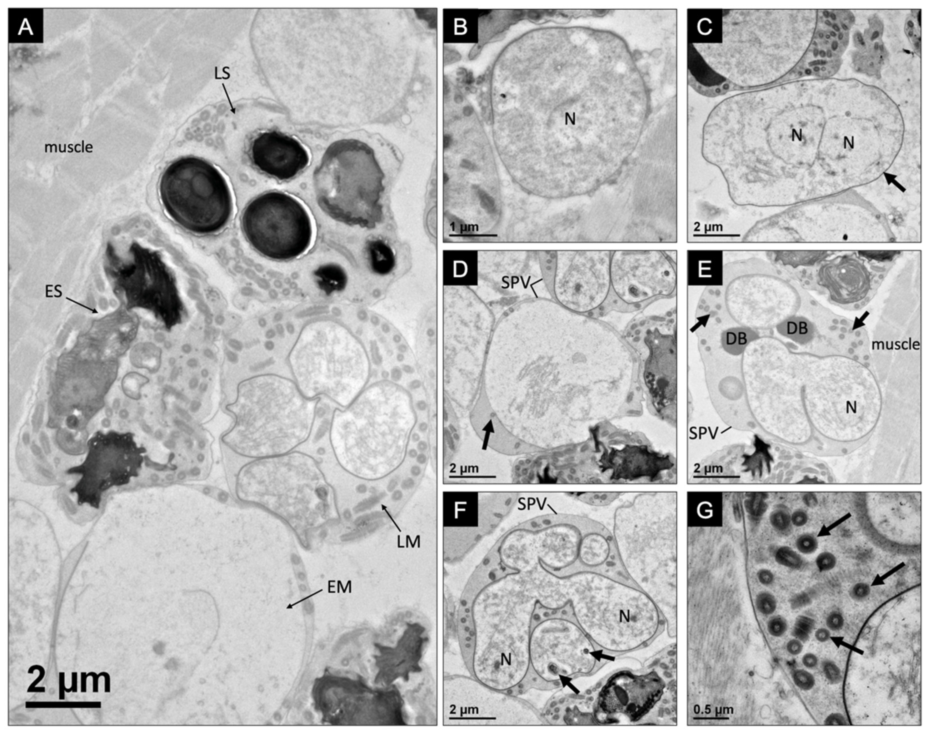
The developmental pattern for the microsporidium-infecting F. virilis occurred within the sarcolemma of the muscle fibers, with various stages of spore development occurring within close proximity to one another (Figure 2A). The development began with a binucleate meront in direct contact with the host cytoplasm and often proximally associated with host muscle fibers (Figure 2B,C). SPVs (8.1 ± 0.7 μm in diameter; n = 10, SD) were observed to house developing meronts, which divided into up to eight early sporonts (Figure 2D). During sporogony, a sporogonial plasmodium, which is presumably formed from the merging of binucleate counterparts and subsequent meiosis (not observed), divides into up to eight uninucleate sporoblasts via rosette-like division (Figure 2E,F). Dense bodies created by aggregations of granules were observed within the SPVs in the early stages of sporogony prior to the formation of individual sporoblasts (Figure 2E). As the sporonts developed into sporoblasts, electron-dense organelles began to develop (Figure 2F). Microtubular-like (73 ± 10 nm in diameter; n = 10, SD) and tubular-like (241 ± 26 nm in diameter; n = 10, SD) structures were observable within the episporontal space, which became more numerous as the development of the sporoblasts progressed (Figure 2G and Figure 3A). Sporoblasts were characterized by a thick electron-dense plasmalemma and the early development of the organelles, including the polar filament and anchoring disc (Figure 3B–D).
All mature spores observed were uninucleate. Uninucleate mature spores were contained within SPVs and were oval in shape, with a wider posterior end (Figure 3E). Mature spores were 3.4 ± 0.1 μm (n = 7, SD) in length and 2.0 ± 0.3 μm (n = 10, SD) in width, with 16–17 coils of the polar filament (118 ± 3 nm in diameter; n = 10, SD) arranged in two or three layers (Figure 3F). The mature spore ultrastructure included an anchoring disc, bilaminar polarplast, a coiled polar filament, and a posterior vacuole (Figure 3G). The spore wall was composed of an electron-lucent endospore (82 ± 12 nm; n = 10, SD) and an electron-dense exospore (25 ± 3 nm; n = 10, SD), which thinned at the apex of the spore above the anchoring disc (Figure 3H).

3.2. Pathology, Ultrastructure, and Development for Microsporidiosis in Faxonius rusticus

Two F. rusticus specimens exhibited signs of microsporidiosis, with white muscle tissue visible through the ventral cuticle of the abdomen. Upon dissection, white musculature was seen throughout the body cavity of the specimen (Figure 1A–C). Histological screening of the infected F. rusticus individuals revealed microsporidian spores developing within SPVs within the sarcolemma of the hosts’ skeletal and heart muscle fibers (Figure 1D–I). Multiple developmental stages were observed during our histological screening, which were observed in greater detail using TEM.
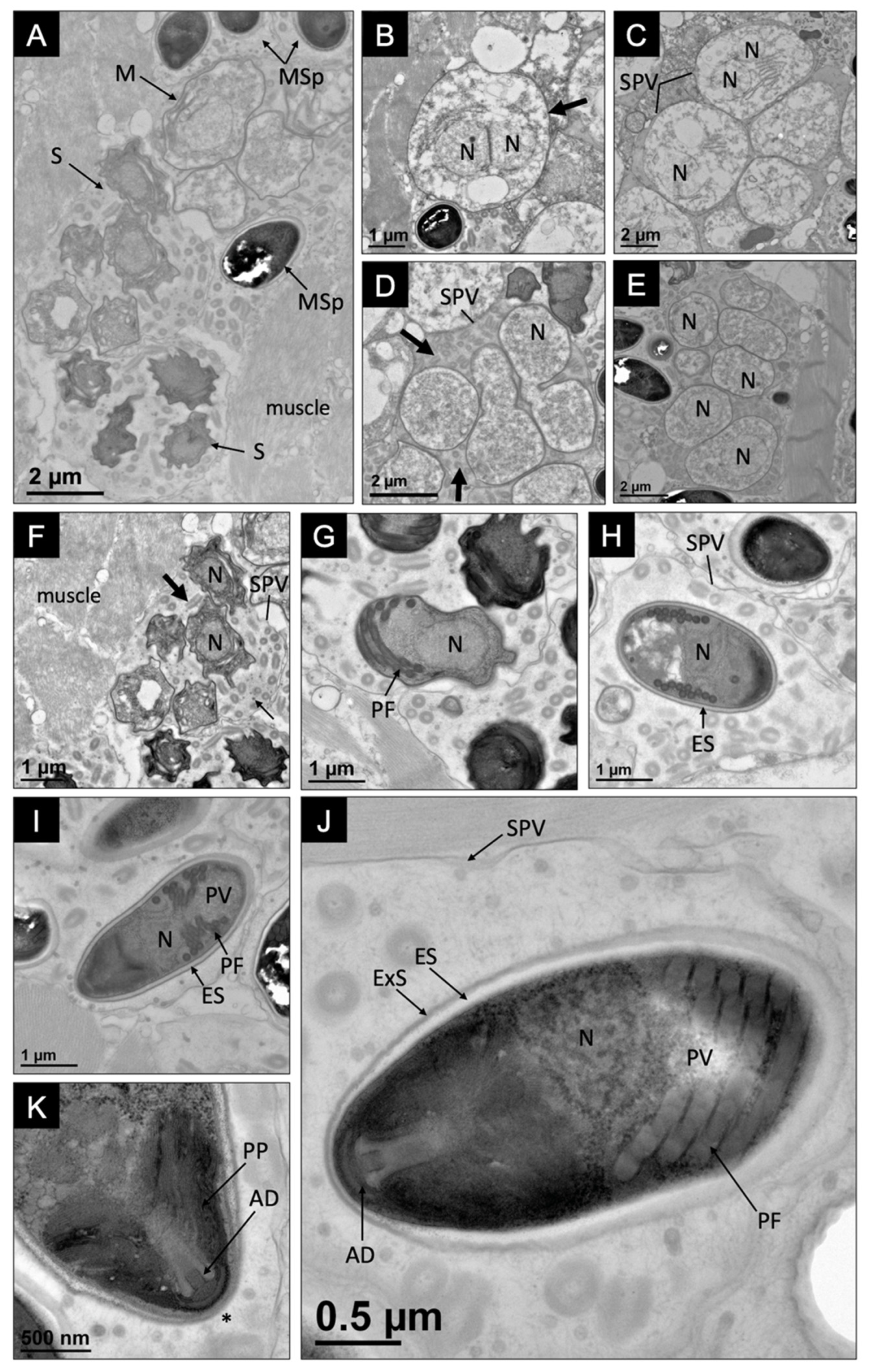
The development of the novel microsporidium occurred within the sarcolemma of the host muscle fibers and various developmental stages were visible in close proximity to one another within individual SPVs (Figure 4A). Mature spores were not found to be dimorphic, and all observed spores were uninucleate. The development of this microsporidium began with large binucleate meronts developing in direct contact with host cytoplasm (Figure 4B). Meronts were not contained within an SPV and had a simple plasmalemma.
Merogony included the development of an SPV (5.2 ± 0.6 μm in diameter; n = 10, SD), which appeared to develop from the plasmalemma (Figure 4C). The binucleate meront progressed into a rosette-shaped plasmodium, which divided to form eight uninucleate sporoblasts (Figure 4D,E). Microtubular-like (70 ± 9 nm in diameter; n = 10, SD) and tubular-like structures (244 ± 32 nm in diameter; n = 10, SD) were abundant within the episporontal space at this stage of development. As the sporoblasts continued to develop, their plasmalemma thickened and became more electron dense. They developed organelles, beginning with the polar filament (Figure 4F,G). As the sporoblast progressed into a mature spore, a thick, electron-lucent endospore became apparent (Figure 4H).
The ultrastructure of a mature spore included an anchoring disc, a bilaminar polarplast, a posterior vacuole, and a polar filament, which coiled 13–14 times (141 ± 14 nm in diameter; n = 10, SD) (Figure 4I–K). The mature spores were uninucleate and oval, with a wider posterior end. The spores were 3.2 ± 0.5 um (n = 10, SD) in length and 1.7 ± 0.3 um (n = 10, SD) in width with a spore wall composed of an electron-lucent endospore (57 ± 18 nm; n = 10, SD) and electron-dense exospore (25 ± 6 nm; n = 10, SD), which thinned at the apex of the spore above the anchoring disc (Figure 4J,K).
Table 2 provides morphological information for the two new species, and provides a comparison to other related species, following a table provided by Moodie et al. [9].

3.3. Genetic Similarity and Phylogenetic Placement of the Novel Microsporidians

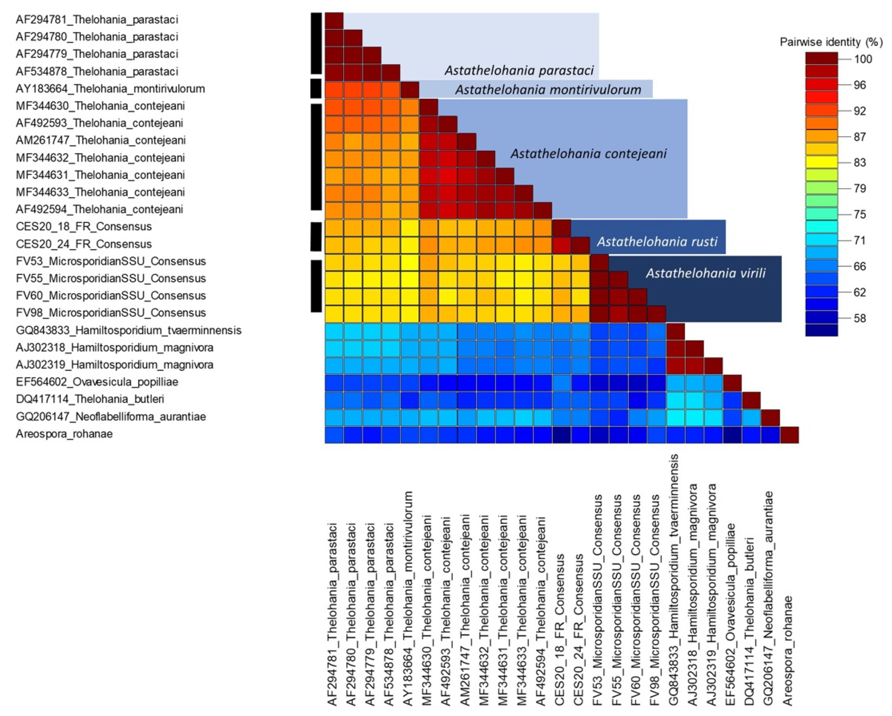
The four microsporidian SSU sequence isolates from F. virilis were identical to one another, as were the two isolates from F. rusticus (Figure 5); however, the novel isolates from each host were genetically distinct (98% coverage; 83.71% similarity; e-value: 0.0). A 775 bp sequence from the novel microsporidium-infecting F. virilis (OM630068) showed 84.79% similarity to a T. contejeani isolate (MF344630: 97% coverage; e-value: 0.0) from Austropotamobius pallipes in Italy. Similarly, a 735 bp sequence from the novel microsporidium-infecting F. rusticus (OM630067) was 87.36% similar to the same T. contejeani isolate (MF344630: 96% coverage; e-value: 0.0). Our sequence demarcation plot highlights the genetically distinct freshwater Thelohania species based on the geographic location from which the isolates were found (Figure 5).
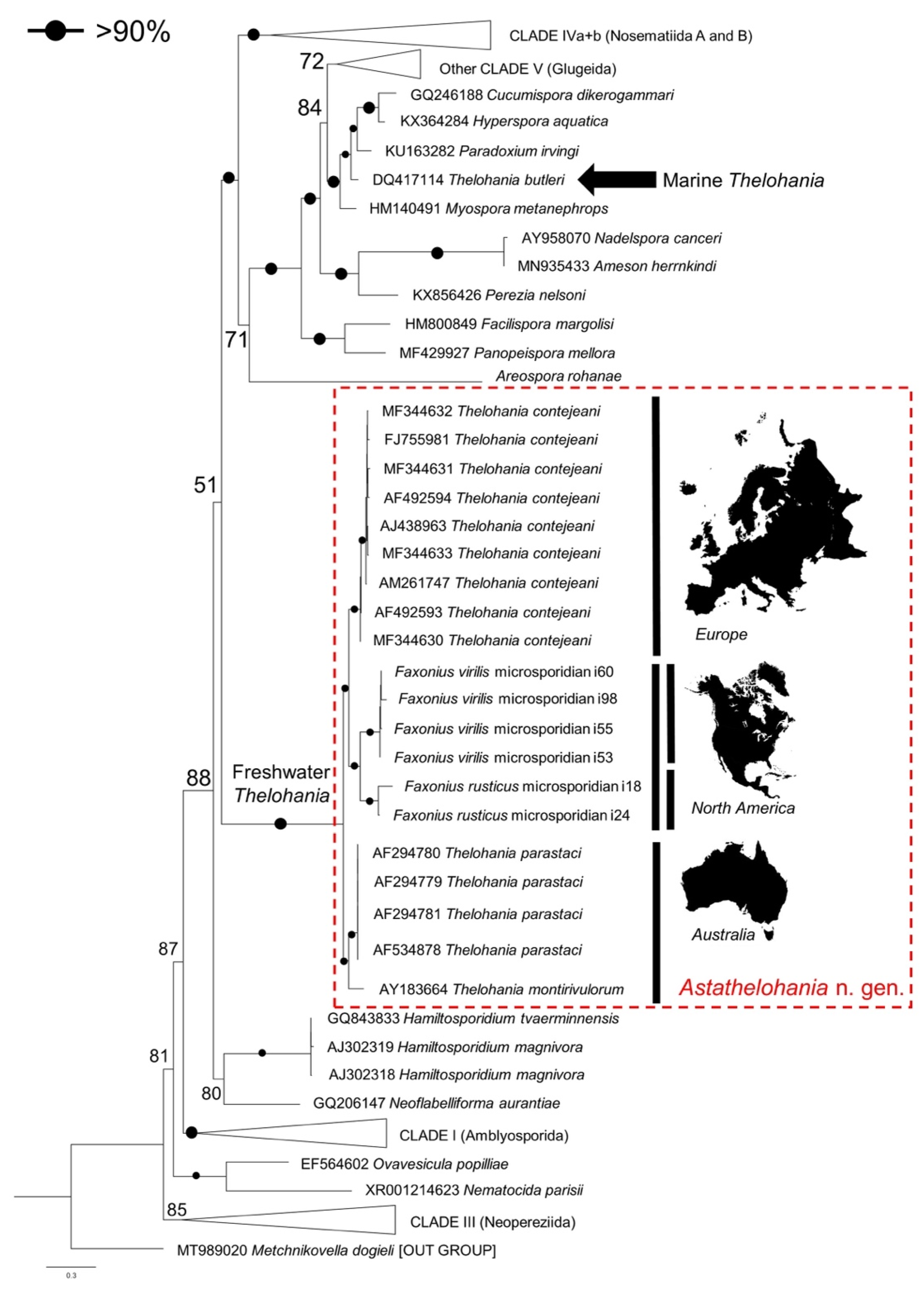
Phylogenetic analysis revealed that our novel microsporidia grouped in an ‘orphan’ lineage at the base of Clades IV and V, along with the other freshwater Thelohania isolates from Europe and Australia (bootstrap: 100%), revealing a genetic similarity between species from specific continental ranges (Figure 6 and Figure 7). Grouping below our microsporidia and the existing freshwater Thelohania are the genera Hamiltosporidium and Neoflabelliforma (Figure 6). The phylogenetic analysis also revealed that freshwater Thelohania and marine (‘true’) Thelohania spp. are genetically distinct, with T. butleri branching separately in Clade V (Figure 6). A sequence demarcation plot of the SSU rRNA gene of all isolates found in the ‘orphan’ lineage, and also comparing T. butleri, emphasizes the genetic dissimilarity between freshwater Thelohania and marine (‘true’) Thelohania with <75% similarity (Figure 5). Therefore, we propose the freshwater members of the genus Thelohania be relocated to a new genus, Astathelohania gen. et comb. nov., based on genetic and phylogenetic dissimilarity of the 18S rRNA sequences. The novel microsporidia described here are named Astathelohania virili n. sp. and Astathelohania rusti n. sp., and the species T. contejeani, T. montirivulorum, and T. parastaci, are revised to become members of this genus.

4. Taxonomic Summary

4.1. Higher Taxonomy

Superphylum: Opisthosporidia (Karpov et al. [36])
Phylum: Rozellomycota (Tedersoo et al. [37]), including the Microsporidia (Balbiani [38]; Wijayawardene et al. [39])
Class: ‘Orphan lineage’ or Clade VI (Dubuffet et al. [29])
Order: Undetermined
Family: Astathelohaniidae Stratton, Reisinger, Behringer, Bojko 2022
Family description: Binucleate, uninucleate, and potentially dimorphic microsporidian parasites that develop within sporophorous vesicles in the muscle tissue of freshwater crustacean hosts. Spores are ellipsoidal, oval, or pear-shaped. Species considered to be members of this family should phylogenetically group with other members of this family using DNA, RNA, or amino acid sequence data, and clade with the type genus and species (Astathelohania virili).
Type genus and species: Astathelohania virili n. sp. Stratton, Reisinger, Behringer, Bojko 2022
Genus: Thelohania (freshwater) replaced by Astathelohania Stratton, Reisinger, Behringer, Bojko 2022
Astathelohania genus description: This genus should accommodate uninucleate or binucleate species that undergo merogony and sporogony in a sporophorous vesicle. Members of this genus infect freshwater Astacoidea Latreille, 1802 hosts (crayfish), which are globally present. Gene sequence data should be considered when determining the placement of a species into this genus and that data should be used to infer a phylogenetic analysis, showing clustering with other Astathelohania species, accounting for possible geographic sequence diversity observed in this study.
Type species: Astathelohania virili n. sp. Stratton, Reisinger, Behringer, Bojko 2022

4.2. Astathelohania virili n. sp. Stratton, Reisinger, Behringer, Bojko 2022

Species description: The microsporidian parasite infects the muscle and heart tissue of F. virilis and undergoes merogony and sporogony in a sporophorous vesicle. The spores are uninucleate and include 16–17 coils of the polar filament. The spores are oval in shape with a wider posterior end and measure 3.4 ± 0.1 µm (SD) in length and 2.0 ± 0.3 µm (SD) in width. To be a candidate for this species, sequence similarity must be shared by comparison to available SSU sequence data for this isolate. Phylogenetically, the parasite must clade with the original sequence provided in this manuscript for Astathelohania virili.
Type host: Faxonius virilis (Hagen, 1870)
Type locality: South Turtle Lake (46.217698, –89.891143), Vilas County, WI, USA.
Site of infection: This species infects the muscle and heart tissue of the host.
Etymology: The species ‘virili’ is named for the host species (Faxonius virilis) in which this novel species was found to infect.
Type material: Histology slides, resin blocks, ethanol-fixed tissue, and glutaraldehyde-fixed tissue are stored at the University of Florida, Reisinger Laboratory. SSU sequence data are deposited in NCBI, under the accession OM630068.

4.3. Astathelohania rusti n. sp. Stratton, Reisinger, Behringer, Bojko 2022

Species description: The microsporidian parasite infects the muscle and heart tissue of F. rusticus and undergoes merogony and sporogony in a sporophorous vesicle. The spores are uninucleate and include 13–14 coils of the polar filament. The spores are oval in shape with a wider posterior end and measure 3.2 ± 0.5 m (SD) in length and 1.7 ± 0.3 µm (SD) in width. To be a candidate for this species, sequence similarity must be shared by comparison to available SSU sequence data for this isolate. Phylogenetically, the parasite must clade with the original sequence provided in this manuscript for Astathelohania rusti.
Type host: Faxonius rusticus (Girard, 1852)
Type locality: Darby Creek (40.013388, –83.383180), Madison County, OH, USA.
Site of infection: This species infects the muscle and heart tissue of the host.
Etymology: The species of this parasite ‘rusti’ is named for the host species (Faxonius rusticus) in which this novel species was first identified.
Type material: Histology slides, resin blocks, ethanol-fixed tissue, and glutaraldehyde-fixed tissue are stored at the University of Florida, Reisinger Laboratory. SSU sequence data are deposited in NCBI, under the accession OM630067.

4.4. Novel and Redescribed Astathelohania Species

Astathelohania rusti n. sp. (Stratton, Reisinger, Behringer, Bojko 2022)
Astathelohania virili n. sp. (Stratton, Reisinger, Behringer, Bojko 2022)
Thelohania contejeani (Henneguy and Thélohan [8]), gen. et comb. nov., Astathelohania contejeani
Thelohania montirivulorum (Moodie et al. [9]), gen. et comb. nov., Astathelohania montirivulorum
Thelohania parastaci (Moodie et al. [10]), gen. et comb. nov., Astathelohania parastaci

5. Discussion

Crayfish can harbor a diverse suite of pathogens, and the freshwater Thelohania are a major group of crayfish-infecting microsporidia [4,5]. In this study, we present a taxonomic revision for freshwater Thelohania based on SSU rRNA sequence data and phylogenetics, proposing that crayfish-infecting, freshwater members of Thelohania, a polyphyletic genus, be transferred to the Astathelohania gen. et comb. nov., housed in the family Astahelohaniidae n. fam., making a clear distinction from the Clade V family, Thelohaniidae, which now houses marine and terrestrial Thelohania spp. In addition, we describe two new species of Astathelohania, Astathelohania virili n. sp. and Astathelohania rusti n. sp., from two crayfish hosts in North America, using histopathology, ultrastructure, intracellular development, and SSU phylogenetics.

5.1. Renaming the Freshwater Thelohania to Astathelohania n. gen.

As genetic data become increasingly available for microsporidia, it has become clearer that traditional data (e.g., phenotypic, ecological, developmental) alone are unable to delineate accurate phylogenies—a combination of these data are required to currently identify species and their taxonomy, evident by several recent species revisions [40,41]. For some of the first microsporidian genera described, such as the Nosema, it has proven vital to incorporate genetic data as part of a revision [41]. Several studies have called for a taxonomic revision of the polyphyletic genus Thelohania since it has become increasingly apparent that the marine Thelohania and the freshwater Thelohania are not closely related genetically and are in fact clades apart [7,15,21]. Other studies have begun to revise the polyphyletic genus by placing terrestrial Thelohania species into more appropriate genera based on genetic, phylogenetic, developmental, and ecological data [23,24]. Our study provides further evidence to support taxonomic revision through the discovery of two new species in this ‘orphan lineage’.
Based on our phylogenetic analysis, freshwater Thelohania branch outside of both Clades IV and V, and importantly branch together in a well-supported group separate from the marine T. butleri (Clade V), the only ‘true’ Thelohania species with genetic data available (Figure 6). Our taxonomic revision is further supported by several recent studies [15,29]. The sequence demarcation plot we provide illustrates the dissimilarity between marine and freshwater Thelohania, based on the SSU rRNA gene (Figure 5).
Further, the family Thelohaniidae remains polyphyletic and also requires taxonomic revision [42]. Many of the genera and species assigned to this family have undergone recent revision on the basis of genetic dissimilarity [23,24,43,44,45]. Our phylogenetic tree further highlights the need for the novel Astathelohania genus to be placed into a new family (Astathelohaniidae n. fam.) considering that all crayfish-infecting, freshwater Thelohania do not fall into the same clade as any genetically validated members of the family Thelohaniidae (Figure 6) [39].
Therefore, we propose a revision in which the crayfish-infecting, freshwater members of the Thelohania are distinguished and relocated to the Astathelohania n. gen. and Astathelohaniidae n. fam. This new genus and family are named for the Thelohania, maintaining their important historic connotations, but additionally represent the freshwater crayfish hosts of this genetically distinct lineage, helping to maintain the historic genus and family names that once represented these species for over a century of published literature.

5.2. Two Novel Crayfish Parasites in the USA

To date, seven microsporidia have been formally described from crayfish hosts, but none of these are known from the crayfish genus Faxonius [5]. The genus Faxonius is the third most species-rich genus of crayfish in the world behind Procambarus and Cambarus, yet little is known about the pathogens this group harbors [4,46]. Astathelohania virili n. sp. and A. rusti n. sp. are the first formally described microsporidia found to infect members of the genus Faxonius. Both crayfish hosts, F. virilis and F. rusticus, have invasive ranges throughout North America, but these novel parasites were found in the native range of each host. Further research should examine whether these novel parasites are found in the invaded ranges of the crayfish hosts.
There have been reports of two suspected T. contejeani infection within North America and an unofficial T. cambari species reported [17,18,19]. These reports were all based on the observation of octosporous development and spore size. However, the size range of spores for the suspected T. contejeani infections overlap with both our spore size ranges and the range described for A. contejeani and A. parastaci (Table 2) [10,15,17,18]. We also now know of many microsporidian groups that undergo octosporous development within an SPV outside of Thelohania [15,47]. Therefore, until these infections are rediscovered, and genetic data become available, we cannot say whether these reports are accurate. Similarly, the unofficial species T. cambari was placed in the genus based on spore size and observation of octosporous development [19]. The spores were much larger in size than our Astathelohania species but do overlap with the size range reported for binucleate spores of A. montirivulorum and A. parastaci (Table 2) [9,10,19]. Genetic and ultrastructural data must become available before T. cambari can be formally recognized.

5.3. Host–Parasite Co-Evolution

The discovery of these novel parasites allowed us to examine the possibility of host–parasite co-evolution of crayfish hosts and Astathelohania microsporidia. Phylogenetic studies of the superfamily Astacoidea illustrate that the divergence of families and genera are geographically affiliated [46,48]. Families in the Northern (Cambaroididae, Astacidae, and Cambaridae) and Southern (Parastacidae) hemispheres diverged over 265 mya [49]. The family Cambaridae is the youngest yet most diverse crayfish lineage, undergoing diversification and radiation approximately 90 mya [50].
The diversity observed within the Astathelohania genus may also represent a geographic split. The microsporidia A. montirivulorum and A. parastaci are only known from the Australian crayfish C. destructor in the family Parastacidae [9,10]. Astathelohania contejeani has been found to infect three members of the family Astacidae which include A. pallipes, Astacus astacus, and P. leniusculus, and all isolates were discovered in Europe [13,14,15]. Finally, A. virili and A. rusti infect two members of the North American family Cambaridae. Our sequence demarcation plot highlights that isolates discovered in the oldest host family (Parastacidae) are least similar to isolates from the youngest family (Cambaridae) (Figure 5, Figure 6 and Figure 7). There is little genetic variation between European isolates since they are all the same microsporidian species; however, two strains of A. contejeani have been described and are evident in the phylogenetic tree (Figure 5 and Figure 6) [13]. In Australia, the Astathelohania (A. montirivulorum and A. parastaci) infect the same host species and are 93% similar to one another [9,10]. In North America, A. virili and A. rusti show considerable genetic variation which may be because North American crayfishes are a significantly more diverse group compared to crayfishes in the families Astacidae and Parastacidae. Therefore, if there is a host–parasite co-evolution it would make sense that their parasites would also be more genetically diverse.

6. Conclusions

It is a vital taxonomic step to separate the crayfish-infecting, freshwater Thelohania into their own distinct genus, avoiding polyphyly in ongoing taxonomic studies concerning the ‘true’ marine Thelohania. Here, we have provided a description of the Astathelohania n. gen., in the family Astathelohaniidae n. fam., to provide valuable systematic distinction for this lineage. This has resulted in three species of Thelohania being revised and the addition of two new species. The two new species we describe provide a North American perspective of Astathelohania diversity, which is now viewed as a globally diverse genus. We see well-supported groups in our phylogeny, which combine all suggested Astathelohania species with 100% bootstrap support, as well as splitting the various genera based on geography and host diversity.

Author Contributions

Conceptualization, C.E.S., L.S.R., D.C.B. and J.B.; methodology, C.E.S., L.S.R., D.C.B. and J.B.; validation, C.E.S. and J.B.; formal analysis, C.E.S. and J.B.; investigation, C.E.S., L.S.R., D.C.B. and J.B.; resources, L.S.R., D.C.B. and J.B.; data curation, C.E.S. and J.B.; writing—original draft preparation, C.E.S. and J.B.; writing—review and editing, C.E.S., L.S.R., D.C.B. and J.B.; visualization, C.E.S. and J.B.; supervision, L.S.R., D.C.B. and J.B.; project administration, C.E.S. and L.S.R.; funding acquisition, L.S.R., D.C.B. and J.B. All authors have read and agreed to the published version of the manuscript.

Funding

This research was funded by the Wisconsin Department of Natural Resources, grant number AIRD11519.

Institutional Review Board Statement

Not applicable.

Informed Consent Statement

Not applicable.

Data Availability Statement

Histology slides, resin blocks, ethanol-fixed tissue, and glutaraldehyde-fixed tissue are stored at the University of Florida, Reisinger Laboratory. SSU sequence data are deposited in NCBI, under the accession numbers OM630066–OM630071.

Acknowledgments

Thanks to Emily An, Bana Kabalan, Lauren Pintor, and Natalie Stephens for assistance with the collection of crayfish and to Nicole Machi at the Electron Microscopy Core (University of Florida, Interdisciplinary Center for Biotechnology Research, RRID:SCR_019146) for their work on these pathogens.

Conflicts of Interest

The authors declare no conflict of interest.

References

  1. Stentiford, G.D.; Feist, S.W.; Stone, D.M.; Bateman, K.S.; Dunn, A.M. Microsporidia: Diverse, Dynamic, and Emergent Pathogens in Aquatic Systems. Trends Parasitol. 2013, 29, 567–578. [Google Scholar] [CrossRef]
  2. Freeman, M.A.; Turnbill, J.F.; Yeomans, W.E.; Bean, C.W. Prospects for Management Strategies of Invasive Crayfish Populations with an Emphasis on Biological Control. Aquat. Conserv. Mar. Freshw. Ecosyst. 2010, 20, 211–223. [Google Scholar] [CrossRef]
  3. Edgerton, B.F.; Evans, L.H.; Stephens, F.J.; Overstreet, R.M. Synopsis of Freshwater Crayfish Diseases and Commensal Organisms. Aquaculture 2002, 206, 57–135. [Google Scholar] [CrossRef] [Green Version]
  4. Longshaw, M. Diseases of Crayfish: A Review. J. Invertebr. Pathol. 2011, 106, 54–70. [Google Scholar] [CrossRef] [PubMed]
  5. Bojko, J.; Behringer, D.C.; Moler, P.; Stratton, C.E.L.; Reisinger, L. A New Lineage of Crayfish-Infecting Microsporidia: The Cambaraspora floridanus n. gen. n. sp. (Glugeida: Glugeidae) Complex from Floridian Freshwaters (USA). J. Invertebr. Pathol. 2020, 171, 107345. [Google Scholar] [CrossRef] [PubMed]
  6. Bojko, J.; Behringer, D.C.; Moler, P.; Reisinger, L. Ovipleistophora diplostomuri, a Parasite of Fish and Their Trematodes, also Infects the Crayfish Procambarus bivittatus. J. Invertebr. Pathol. 2020, 169, 107306. [Google Scholar] [CrossRef] [PubMed]
  7. Stentiford, G.D.; Ross, S.H.; Kerr, R.; Bass, D.; Bateman, K.S. Paradoxium irvingi n. gen. n. sp. (Microsporidia) Infecting the Musculature of European Pink Shrimp Pandalus montagui. J. Invertebr. Pathol. 2015, 130, 1–8. [Google Scholar] [CrossRef]
  8. Henneguy, G.; Thélohan, P. Myxosporides Parasites Des Muscles Chez Quelques Crustaces Decapodes. Ann Microgr. 1892, 4, 617–641. [Google Scholar]
  9. Moodie, E.G.; Le Jambre, L.F.; Katz, M.E. Thelohania montirivulorum sp. nov. (Microspora: Thelohaniidae), a Parasite of the Australian Freshwater Crayfish, Cherax destructor (Decapoda: Parastacidae): Fine Ultrastructure, Molecular Characteristics and Phylogenetic Relationships. Parasitol. Res. 2003, 91, 215–228. [Google Scholar] [CrossRef]
  10. Moodie, E.G.; Le Jambre, L.F.; Katz, M.E. Thelohania parastaci sp. nov. (Microspora: Thelohaniidae), a Parasite of the Australian Freshwater Crayfish, Cherax destructor (Decapoda: Parastacidae). Parasitol. Res. 2003, 91, 151–165. [Google Scholar] [CrossRef]
  11. Cossins, A.R.; Bowler, K. A Histological and Ultrastructural Study of Thelohania contejeani Henneguy, 1892 (Nosematidae), Microsporidian Parasite of Austropotamobius pallipes Lereboullet. Parasitology 1974, 68, 81–91. [Google Scholar] [CrossRef]
  12. Vivares, C.P. Etude Comparative Faite En Microscopies Photonique et Electronique de Trois Especes de Microsporidies Appartenant Au Genre Thelohania Henneguy, 1892, Parasites de Crustaces Decapodes Marins. Ann. Sci. Nat. 1975, 17, 141–178. [Google Scholar]
  13. Lom, J.; Nilsen, F.; Dyková, I. Thelohania contejeani Henneguy, 1892: Dimorphic Life Cycle and Taxonomic Affinities, as Indicated by Ultrastructural and Molecular Study. Parasitol. Res. 2001, 87, 860–872. [Google Scholar] [CrossRef] [PubMed]
  14. Dunn, J.C.; McClymont, H.E.; Christmas, M.; Dunn, A.M. Competition and Parasitism in the Native White Clawed Crayfish Austropotamobius pallipes and the Invasive Signal Crayfish Pacifastacus leniusculus in the UK. Biol. Invasions 2009, 11, 315–324. [Google Scholar] [CrossRef] [Green Version]
  15. Pretto, T.; Montesi, F.; Ghia, D.; Berton, V.; Abbadi, M.; Gastaldelli, M.; Manfrin, A.; Fea, G. Ultrastructural and Molecular Characterization of Vairimorpha austropotamobii sp. nov. (Microsporidia: Burenellidae) and Thelohania contejeani (Microsporidia: Thelohaniidae), Two Parasites of the White-Clawed Crayfish, Austropotamobius pallipes Complex (Decapoda: Astacidae). J. Invertebr. Pathol. 2018, 151, 59–75. [Google Scholar] [CrossRef] [PubMed]
  16. France, R.; Graham, L. Increased Microsporidian Parasitism of the Crayfish Orconectes virilis in an Experimentally Acidified Lake. Water Air Soil Pollut. 1985, 26, 129–1336. [Google Scholar] [CrossRef]
  17. Graham, L.; France, R. Attempts To Transmit Experimentally the Microsporidian Thelohania contejeani in Freshwater Crayfish (Orconectes virilis). Crustaceana 1986, 51, 208–211. [Google Scholar] [CrossRef]
  18. McGriff, D.; Modin, J. Thelohania contejeani Parasitism of the Crayfish, Pacifastacus leniusculus, in California. Calif. Fish Game 1983, 69, 178–183. [Google Scholar]
  19. Sprague, V. Thelohania cambarii n. sp., a Microsporidian Parasite of North American Crayfish. J. Parasitol. 1950, 36, 46. [Google Scholar]
  20. Stentiford, G.D.; Bateman, K.S.; Feist, S.W.; Chambers, E.; Stone, D.M. Plastic Parasites: Extreme Dimorphism Creates a Taxonomic Conundrum in the Phylum Microsporidia. Int. J. Parasitol. 2013, 43, 339–352. [Google Scholar] [CrossRef] [PubMed]
  21. Brown, A.M.V.; Adamson, M.L. Phylogenetic Distance of Thelohania butleri (Microsporidia; Thelohaniidae), a Parasite of the Smooth Pink Shrimp Pandalus jordani, from Its Congeners Suggests Need for Major Revision of the Genus Thelohania Henneguy, 1892. J. Eukaryot. Microbiol. 2006, 53, 445–455. [Google Scholar] [CrossRef] [PubMed]
  22. Sprague, V. Annotated List of Species of Microsporidia. In Comparative Pathology, 2nd ed.; Bulla, L.A., Cheng, T.C., Eds.; Plenum Press: New York, NY, USA, 1977; pp. 1–333. [Google Scholar]
  23. Vavra, J.; Hylis, M.; Vossbrinck, C.R.; Pilarska, D.K.; Linde, A.; Weiser, J.; Mcmanus, M.L.; Hoch, G.; Solter, L.F. Vairimorpha disparis n. comb. (Microsporidia: Burenellidae): A Redescription and Taxonomic Revision of Thelohania disparis Timofejeva 1956, a Microsporidian Parasite of the Gypsy Moth Lymantria dispar (L.) (Lepidoptera: Lymantriidae). J. Eukaryot. Microbiol. 2006, 53, 292–304. [Google Scholar] [CrossRef] [PubMed]
  24. Sokolova, Y.Y.; Fuxa, J.R. Biology and Life-cycle of the Microsporidium Kneallhazia solenopsae Knell Allan Hazard 1977 gen. n., comb. n., from the Fire Ant Solenopsis invicta. Parasitology 2008, 135, 903–929. [Google Scholar] [CrossRef]
  25. Cormier, A.; Wattier, R.; Giraud, I.; Teixeira, M.; Grandjean, F.; Rigaud, T.; Cordaux, R. Draft Genome Sequences of Thelohania contejeani and Cucumispora dikerogammari, Pathogenic Microsporidia of Freshwater Crustaceans. Microbiol. Resour. Announc. 2021, 10, e01346-20. [Google Scholar] [CrossRef] [PubMed]
  26. Morris, D.J.; Freeman, M.A. Hyperparasitism has Wide-Ranging Implications for Studies on the Invertebrate Phase of Myxosporean (Myxozoa) Life Cycles. Int. J. Parasitol. 2010, 40, 357–369. [Google Scholar] [CrossRef] [PubMed]
  27. Haag, K.L.; Larsson, J.I.R.; Refardt, D.; Ebert, D. Cytological and Molecular Description of Hamiltosporidium tvaerminnensis gen. et sp. nov., a Microsporidian Parasite of Daphnia magna, and Establishment of Hamiltosporidium magnivora comb. nov. Parasitology 2011, 138, 447–462. [Google Scholar] [CrossRef] [PubMed] [Green Version]
  28. Stentiford, G.D.; Bateman, K.S.; Feist, S.W.; Oyarzún, S.; Uribe, J.C.; Palacios, M.; Stone, D.M. Areospora rohanae n. gen. n. sp. (Microsporidia; Areosporiidae n. fam.) Elicits Multi-Nucleate Giant-Cell Formation in Southern King Crab (Lithodes santolla). J. Invertebr. Pathol. 2014, 118, 1–11. [Google Scholar] [CrossRef] [PubMed]
  29. Dubuffet, A.; Chauvet, M.; Moné, A.; Debroas, D.; Lepère, C. A Phylogenetic Framework to Investigate the Microsporidian Communities through Metabarcoding and Its Application to Lake Ecosystems. Environ. Microbiol. 2021, 23, 4344–4359. [Google Scholar] [CrossRef] [PubMed]
  30. Bojko, J.; Reinke, A.W.; Stentiford, G.D.; Williams, B.; Rogers, M.S.J.; Bass, D. Microsporidia: A Taxonomic, Evolutionary, and Ecological Synthesis. Trends Parasitol. 2022, in press. [Google Scholar]
  31. Schneider, C.A.; Rasband, W.S.; Eliceiri, K.W. NIH Image to ImageJ: 25 Years of Image Analysis. Nat. Methods 2012, 9, 671–675. [Google Scholar] [CrossRef] [PubMed]
  32. Ovcharenko, M.O.; Bacela, K.; Wilkinson, T.; Ironside, J.E.; Rigaud, T.; Wattier, R.A. Cucumispora dikerogammari n. gen. (Fungi: Microsporidia) Infecting the Invasive Amphipod Dikerogammarus villosus: A Potential Emerging Disease in European Rivers. Parasitology 2010, 137, 191–204. [Google Scholar] [CrossRef] [PubMed] [Green Version]
  33. Miller, M.A.; Pfeiffer, W.; Schwartz, T. The CIPRES Science Gateway: A Community Resource for Phylogenetic Analyses. In Proceedings of the 2011 TeraGrid Conference on Extreme Digital Discovery—TG’11, Salt Lake City, UT, USA, 18–21 July 2011; ACM Press: Salt Lake City, UT, USA, 2011; p. 1. [Google Scholar]
  34. Nguyen, L.-T.; Schmidt, H.A.; von Haeseler, A.; Minh, B.Q. IQ-TREE: A Fast and Effective Stochastic Algorithm for Estimating Maximum-Likelihood Phylogenies. Mol. Biol. Evol. 2015, 32, 268–274. [Google Scholar] [CrossRef]
  35. Muhire, B.M.; Varsani, A.; Martin, D.P. SDT: A Virus Classification Tool Based on Pairwise Sequence Alignment and Identity Calculation. PLoS ONE 2014, 9, e108277. [Google Scholar] [CrossRef] [PubMed]
  36. Karpov, S.A.; Mamkaeva, M.A.; Aleoshin, V.V.; Nassonova, E.; Lilje, O.; Gleason, F.H. Morphology, Phylogeny, and Ecology of the Aphelids (Aphelidea, Opisthokonta) and Proposal for the New Superphylum Opisthosporidia. Front. Microbiol. 2014, 5, 112. [Google Scholar] [CrossRef] [PubMed] [Green Version]
  37. Tedersoo, L.; Sánchez-Ramírez, S.; Kõljalg, U.; Bahram, M.; Döring, M.; Schigel, D.; May, T.; Ryberg, M.; Abarenkov, K. High-Level Classification of the Fungi and a Tool for Evolutionary Ecological Analyses. Fungal Divers. 2018, 90, 135–159. [Google Scholar] [CrossRef] [Green Version]
  38. Balbiani, E. Sur Les Microsporidies Ou Psorospermies Des Articules. Compt. Rend. Acad. Sco. Paris 1882, 95, 1168–1171. [Google Scholar]
  39. Wijayawardene, N.N.; Hyde, K.D.; Dai, D.Q.; Sánchez-García, M.; Goto, B.T.; Saxena, R.K.; Erdoğdu, M.; Selçuk, F.; Rajeshkumar, K.C.; Aptroot, A.; et al. Outline of Fungi and fungus-like taxa—2021. Mycosphere 2022, 13, 53–453. [Google Scholar] [CrossRef]
  40. Vossbrinck, C.R.; Debrunner-Vossbrinck, B.A. Molecular Phylogeny of the Microsporidia: Ecological, Ultrastructural and Taxonomic Considerations. Folia Parasitol. 2005, 52, 131–142. [Google Scholar] [CrossRef] [PubMed] [Green Version]
  41. Tokarev, Y.S.; Huang, W.-F.; Solter, L.F.; Malysh, J.M.; Becnel, J.J.; Vossbrinck, C.R. A Formal Redefinition of the Genera Nosema and Vairimorpha (Microsporidia: Nosematidae) and Reassignment of Species Based on Molecular Phylogenetics. J. Invertebr. Pathol. 2020, 169, 107279. [Google Scholar] [CrossRef] [PubMed]
  42. Hazard, E.I.; Oldacre, S.W. Revision of Microsporidia (Protozoa) Close to Thelohania, with Descriptions of One New Family, Eight New Genera, and Thirteen New Species; US Department of Agriculture, Agricultural Research Service: Washington, DC, USA, 1976. [CrossRef]
  43. Weiser, J. Contribution to the Classification of Microsporidia. Vestn. Cesk. Spol. Zool. 1977, 41, 308–320. [Google Scholar]
  44. Baker, M.D.; Vossbrinck, C.R.; Becnel, J.J.; Andreadis, T.G. Phylogeny of Amblyospora (Microsporida: Amblyosporidae) and Related Genera Based on Small Subunit Ribosomal DNA Data: A Possible Example of Host Parasite Cospeciation. J. Invertebr. Pathol. 1998, 71, 199–206. [Google Scholar] [CrossRef] [PubMed] [Green Version]
  45. Sokolova, Y.; Pelin, A.; Hawke, J.; Corradi, N. Morphology and Phylogeny of Agmasoma penaei (Microsporidia) from the Type Host, Litopenaeus setiferus, and the Type Locality, Louisiana, USA. Int. J. Parasitol. 2015, 45, 1–16. [Google Scholar] [CrossRef] [PubMed]
  46. Crandall, K.A.; De Grave, S. An Updated Classification of the Freshwater Crayfishes (Decapoda: Astacidea) of the World, with a Complete Species List. J. Crustac. Biol. 2017, 37, 615–653. [Google Scholar] [CrossRef] [Green Version]
  47. Moodie, E.G.; Le Jambre, L.F.; Katz, M.E. Ultrastructural Characteristics and Small Subunit Ribosomal DNA Sequence of Vairimorpha cheracis sp. nov., (Microspora: Burenellidae), a Parasite of the Australian Yabby, Cherax destructor (Decapoda: Parastacidae). J. Invertebr. Pathol. 2003, 84, 198–213. [Google Scholar] [CrossRef] [PubMed]
  48. Stern, D.B.; Breinholt, J.; Pedraza-Lara, C.; López-Mejía, M.; Owen, C.L.; Bracken-Grissom, H.; Fetzner, J.W.; Crandall, K.A. Phylogenetic Evidence from Freshwater Crayfishes That Cave Adaptation Is Not an Evolutionary Dead-end. Evolution 2017, 71, 2522–2532. [Google Scholar] [CrossRef] [PubMed] [Green Version]
  49. Bracken-Grissom, H.D.; Ahyong, S.T.; Wilkinson, R.D.; Feldmann, R.M.; Schweitzer, C.E.; Breinholt, J.W.; Bendall, M.; Palero, F.; Chan, T.-Y.; Felder, D.L.; et al. The Emergence of Lobsters: Phylogenetic Relationships, Morphological Evolution and Divergence Time Comparisons of an Ancient Group (Decapoda: Achelata, Astacidea, Glypheidea, Polychelida). Syst. Biol. 2014, 63, 457–479. [Google Scholar] [CrossRef] [Green Version]
  50. Reynolds, J.D.; Souty-Grosset, C. Management of Freshwater Biodiversity: Crayfish as Bioindicators; Cambridge University Press: Cambridge, UK; New York, NY, USA, 2012; ISBN 978-0-521-51400-2. [Google Scholar]
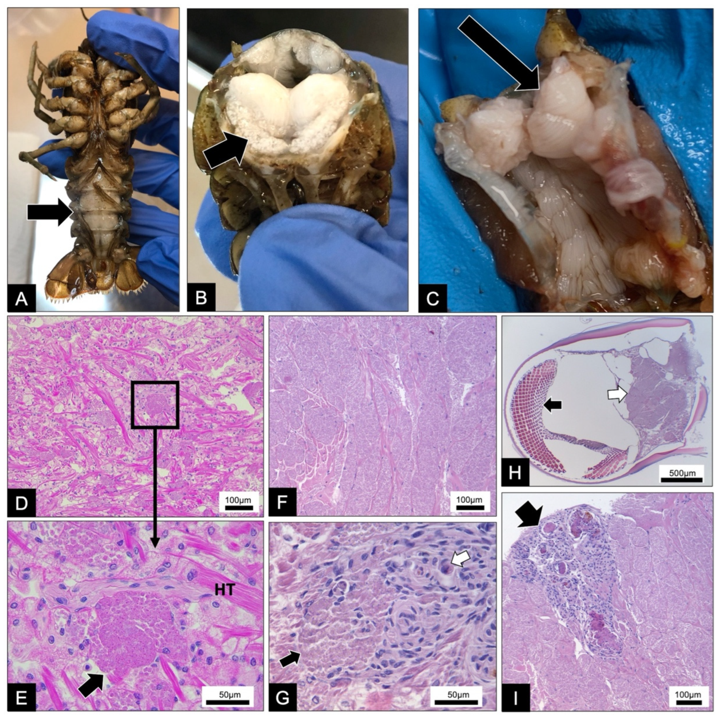
Figure 1. Gross pathology and histopathology of microsporidian infections in Faxonius virilis and Faxonius rusticus: (A) muscle tissue of infected crayfish is white and visible through the ventral cuticle of the abdomen (black arrow); (B) a transverse section of the abdomen reveals white muscle tissue (black arrow) presumably due to infection; (C) during dissection, white muscle tissue throughout the body cavity was white (black arrow) from the infection; (D) heart tissue with groups of developing spores; (E) a higher magnification of (D) of one cluster of developing spores in the heart tissue (HT) and the evident sporophorous vesicles containing the spores (black arrow); (F) abdominal muscle tissue exhibiting a heavy microsporidian infection; (G) high magnification image of a cluster of spores (black arrow) developing within the heart tissue and the production of granulomas (white arrow); (H) microsporidian spores (white arrow) developing within the muscle stalk of the eye (black arrow); (I) an immune response to the microsporidian infection in the abdominal muscle resulting in the production of several granulomas (black arrow).
Microorganisms 10 00636 g001
Figure 2. Merogony of Astathelohania virili n. sp. (A) This image identifies an early merogonal stage (EM), a late merogonal stage (LM), an early sporoblast stage (ES), and a late sporoblast stage (LS). Each stage is developing within their own sporophorous vesicle in close proximity to one another and near host muscle tissue; (B) a binucleate meront (N = nucleus) with a thin cell wall; (C) a binucleate meront (N = nuclei) with a thickening cell wall (arrow); (D) sporophorous vesicle (SPV) developing from meront with tubular-like structures present (arrow); (E) division of sporont into sporoblasts within SPV with one visible nucleus (N). SPV contains dense bodies (DB) and tubular-like structures (arrows); (F) another dividing sporont within an SPV. Electron-dense organelles beginning to develop within developing sporonts (arrows); (G) high magnification image of tubular-like structures (arrows) found within late merogony SPVs.
Microorganisms 10 00636 g002
Figure 3. Sporogony and spore ultrastructure of Astathelohania virili n. sp. (A) Sporoblasts developing within a single sporophorous vesicle (SPV) that contains microtubular-like (small arrow) and tubular-like structures (large arrow); (B) sporoblasts beginning to develop electron-dense organelles including the polar filament (PF); (C) sporoblasts continuing to develop within SPV containing tubular-like structures (arrows) in close association with host muscle tissue; (D) a uninucleate (N) sporoblast with developing organelles including the anchoring disc (AD) and polar filament (PF); (E) near mature spores within an SPV; (F) spore with thickening endospore (ES); (G) uninucleate spore with a well-developed anchoring disc (AD) and bilaminar polarplast (PP). Inset shows fine details of polar filament and the two-layer arrangement; (H) a uninucleate spore with posterior vacuole and spore wall consisting of thickening electron-lucent endospore (ES) and electron-dense exospore (ExoS).
Microorganisms 10 00636 g003
Figure 4. Intracellular developmental cycle of Astathelohania rusti n. sp. within the muscle tissue of Faxonius rusticus. (A) This image identifies a merogony stage (M), a sporogony (S), and mature spores (MSp) developing within their own sporophorous vesicles (SPV) in close proximity to host muscle tissue; (B) a binucleate meront (N = nuclei) with a thickening cell wall (arrow); (C) nuclei dividing within meronts and SPVs developing around each meront; (D) division of sporont within SPV. SPV contains tubular-like structures (arrows); (E) early uninucleate (N) sporoblasts maturing within SPVs; (F) uninucleate sporoblast developing within SPV with both microtubular-like (small arrow) and tubular-like structures (large arrow) present; (G) uninucleate sporoblast developing organelles including the polar filament (PF); (H) a near mature uninucleate spore developing within an SPV with a thickening electron-lucent endospore (ES); (I) a near mature uninucleate spore with a thicker endospore and well-developed polar filament (PF) and posterior vacuole (PV); (J) the ultrastructure of a mature uninucleate spore developing within an SPV includes a posterior vacuole (PV), polar filament (PF), anchoring disc (AD), and a spore wall with a thick electron-lucent endospore (ES) and electron-dense exospore (ExoS); (K) shows the fine details of the bilaminar polarplast (PP) and anchoring disc (AD) with the spore wall thinning above the anchoring disc (*).
Microorganisms 10 00636 g004
Figure 5. A similarity matrix reflecting the percent similarity between different Astathelohania (=Thelohania) rRNA (SSU) gene isolates. The key provides a color scheme that reflects the similarity between isolates (blue/low to red/high). The figure was designed using the sequence demarcation Tool v1.2 [35].
Microorganisms 10 00636 g005
Figure 6. A maximum-likelihood phylogenetic tree of all crayfish-infecting, freshwater Thelohania isolates as well as wide-scale Microsporidia representation of each existing clade. The annotated maps demonstrate the distinct crayfish-infecting Thelohania species present per continent. The isolates sequenced in this study are denoted on the tree using the host (Faxonius sp.) and the microsporidian isolate number. Two isolates are present for a novel microsporidian species from F. rusticus (i18 and i24), and four isolates were sequenced from F. virilis (i60, i98, i55, i53). The tree was constructed using MAFFT aligned rRNA (SSU) gene sequences followed by IQtree [34]. The tree was annotated in FigTree v.1.4.4.
Microorganisms 10 00636 g006
Figure 7. Representative phylogenetic-inferred cladograms of native crayfish species (“Crayfish”) from the families Cambaridae (native geography: North America), Astacidae (native geography: Europe and North America), Cambaroididae (native geography: China and Japan), and Parastacidae (native geography: South America, Madagascar, Australia, New Zealand), compared with microsporidian isolates from the freshwater Thelohania (now revised to Astathelohania) (“Parasites”). The accession numbers for the isolates are listed by the name of the species on each tree. The microsporidian cladogram was developed from the tree presented in Figure 6. For the “Crayfish” tree, cytochrome oxidase 1 DNA sequence data were aligned using MAFFT and constructed using IQtree [34]. The trees were drawn and annotated in FigTree v.1.4.4.
Microorganisms 10 00636 g007
Table 1. Sampling detail of each individual crayfish collected with indication of what microsporidian species each crayfish was infected with and the data available for each crayfish.
Host
Species
SiteCoordinatesCollection DateSexCarapace Length (mm)Microsporidian SpeciesSSUHistologyElectron
Microscopy
Accession Number
F. rusticusDarby Creek, OH40.013388, −83.38318030 June 2021MII27A. rusti n. sp.OM630066
F. rusticusDarby Creek, OH40.013388, −83.38318030 June 2021MI32A. rusti n. sp.OM630067
F. virilisSouth Turtle Lake, WI46.217698, −89.89114309 July 2019MII51A. virili n. sp.OM630068
F. virilisSouth Turtle Lake, WI46.217698, −89.89114309 July 2019MII50A. virili n. sp.OM630069
F. virilisSouth Turtle Lake, WI46.217698, −89.89114309 July 2019MII43A. virili n. sp.OM630070
F. virilisCrab Lake, WI46.203368, −89.72925519 July 2019MII40A. rusti n. sp.OM630071
Table 2. Comparison of morphological features of all described Astathelohania (previously Thelohania) species. In addition, the morphological features of two suspected and an unofficial Thelohania in North America are included. The table was adapted from Moodie et al. [9], n/a indicates data were not available.
Morphological featureA. rusti n. sp.A. virili n. sp.A. montirivulorumA. parastaciA. contejeaniA. contejeaniT. contejeaniT. contejeaniT. cambari
Moodie et al. [9]Moodie et al. [10]Lom et al. [13]Pretto et al. [15]Graham and France [17]McGriff and Modin [18]Sprague [19]
Shore shapeOval, wider
posterior end
Oval, wider
posterior end
Lozenge, round endsLozenge, round endsOval, wider posterior endOval, wider posterior endOvalOvalOval, wider posterior end
Uninucleate spore length (μm)3.2 ± 0.5 1n = 103.4 ± 0.1 1n = 7n/an/a4.2 23.6 ± 0.4 2n = 503.3 (2.8–3.6)n = 503.0–3.84.6
Uninucleate spore width (μm)1.7 ± 0.31n = 102.0 ± 0.3 1n = 10n/an/a2.1 22.3 ± 0.3 2n = 502.2 (2.0–2.6)n = 501.8–2.42.2
Binucleate spore length (μm)n/a n/a 5.9 (4.9–7.2) 23.9 (3.2–4.9) 23.8 23.3 ± 0.5 2n = 50n/a n/an/a
Binucleate spore width (μm)n/a n/a 2.6 (2.0–3.1) 22.0 (1.5–2.7) 21.8 21.7 ± 0.2 2n = 50n/a n/an/a
Uninucleate—no. coils in polar filament13–14 16–17 20–2211–209–109–12 n/a n/an/a
Uninucleate—polar filament diameter (nm)141 ± 14n = 10118 ± 3n = 1098 (82–111)59 (53–74)120–180 377n = 10n/a n/an/a
Binucleate—no. coils in polar filamentn/a n/a 20–226–85–75–6 n/a n/an/a
Binucleate—polar filament diameter (nm)n/a n/a 107 (90–140)83 (65–102)n/a108n = 10n/a n/an/a
SPV diameter (μm)5.2 ± 0.6 1n = 108.1 ± 0.7 1n = 108.4 (7.0–9.6) 28.8 (7.4–10.5) 28–9 39.4 ± 0.6 2n = 207.9 (6.4–8.1)n = 10n/an/a
SPV tubular-like structure diameter (nm)244 ± 32n = 10241 ± 26n = 10171 (130–249)249 (205–307)220155–185n = 20n/a n/an/a
SPV microtubular-like structure diameter (nm)70 ± 9n = 1073 ± 10n = 1085 (63–117)73 (50–99)80–10075–85n = 20n/a n/an/a
Lateral exospore thickness of
uninucleate spores (nm)
25 ± 6n = 1025 ± 3n = 1031 (30–40)24 (20–40)24–30 428n = 15n/a n/an/a
Lateral endospore thickness of
uninucleate spores (nm)
57 ± 18n = 1082 ± 12n = 10108 (80–130)73 (56–110)60–90 478n = 15n/a n/an/a
Lateral exospore thickness of
binucleate spores (nm)
n/a n/a 22 (17–30)34 (30–40)n/a32n = 8n/a n/an/a
Lateral endospore thickness of
binucleate spores (nm)
n/a n/a 65 (40–80)58 (50–60)n/a55n = 8n/a n/an/a
Dimorphic sporogony?No No YesYesYesYes n/a n/an/a
1 Resin infiltrated. 2 Light microscopy. 3 Cossins and Bowler [11]. 4 Vivares [12].
Publisher’s Note: MDPI stays neutral with regard to jurisdictional claims in published maps and institutional affiliations.