Article

Structural and Functional Characterization of the Holliday Junction Resolvase RuvC from Deinococcus radiodurans

MOE Key Laboratory of Biosystems Homeostasis and Protection, Institute of Biophysics, College of Life Sciences, Zhejiang University, Hangzhou 310058, China
*
Authors to whom correspondence should be addressed.
Microorganisms 2022, 10(6), 1160; https://doi.org/10.3390/microorganisms10061160
Submission received: 21 April 2022 / Revised: 24 May 2022 / Accepted: 2 June 2022 / Published: 6 June 2022
(This article belongs to the Section Environmental Microbiology)

Abstract

:
Holliday junctions (HJs) are four-way DNA structures, which are an important intermediate in the process of homologous recombination. In most bacteria, HJs are cleaved by specific nucleases called RuvC resolvases at the end of homologous recombination. Deinococcus radiodurans is an extraordinary radiation-resistant bacterium and is known as an ideal model organism for elucidating DNA repair processes. Here, we described the biochemical properties and the crystal structure of RuvC from D. radiodurans (DrRuvC). DrRuvC exhibited an RNase H fold that belonged to the retroviral integrase family. Among many DNA substrates, DrRuvC specifically bound to HJ DNA and cleaved it. In particular, Mn2+ was the preferred bivalent metal co-factor for HJ cleavage, whereas high concentrations of Mg2+ inhibited the binding of DrRuvC to HJ. In addition, DrRuvC was crystallized and the crystals diffracted to 1.6 Å. The crystal structure of DrRuvC revealed essential amino acid sites for cleavage and binding activities, indicating that DrRuvC was a typical resolvase with a characteristic choice for metal co-factor.

1. Introduction

Homologous recombination (HR) enables the cell to access and copy intact DNA sequence information in trans, which is an essential process in life [1]. In somatic cells, HR plays a key role in conserving genetic information by facilitating DNA repair. In higher organisms, HR is also involved in the meiosis that generates genetic diversity by reshuffling genes [2]. At the end of HR, after homologous pairing and strand exchange, two DNA double strands will form a four-way structure of DNA intermediates, called Holliday junctions (HJs), which must be removed by specific nucleases called resolvases at the end of the recombination process [3]. Resolvases from different biological kingdoms of life have significant diversity and belong to different classes of nucleases [4,5,6,7]. In eukaryotes, HJs are removed primarily by “dissolution”, a pathway involving the combined activities of a DNA helicase and a type IA topoisomerase, which catalyze branch migration and decatenation of the double HJ into non-crossover products [8,9]. In most bacterial cells, HJs are mainly processed by the RuvC resolvasome. The dimeric endonuclease RuvC symmetrically introduces two nicks at the junction, resulting in two separate recombinant DNA duplexes that can be directly repaired by DNA ligases [10,11,12]. In addition to RuvC, functional RuvA and RuvB are also needed for efficient Holliday junction resolution [13,14,15]. These proteins were initially identified by mutations that cause genetic defects in ultraviolet-induced DNA damage repair [16,17]. It is generally believed that these three proteins form a complex molecular machine called RuvABC lysosome, which coordinates the two main events of late recombination: (1) Two homologous duplex arms pass through the RuvA octamer to exchange their pairing partners [18]. (2) The RuvB complex functions as a pump to relocate the junction point to any cleavable sequences by pulling DNA duplex arms [11,12]. (3) RuvC catalyzes the decomposition of HJs via a pair of symmetrical incisions across the junction point [19].
Deinococcus radiodurans belongs to the Deinococcus-Thermus phylum and is highly resistant to various extreme environments and agents, including desiccation, ionizing radiation (IR), ultraviolet (UV) radiation and oxidative stress, thanks to its strong ability to repair DNA damage [20,21]. Deinococcus seldom invoke translesion synthesis and non-homologous end joining, but rather adopt homologous recombination to guarantee the fidelity of DNA repair [22]. Therefore, the efficient DNA damage repair ability of D. radiodurans may benefit from its powerful HR repair system. Despite the importance of RuvC in DNA recombination and repair across bacteria, there is essentially no biochemical and crystal study reported for RuvC from extremophiles. In this present study, we presented biochemical and structural analyses of RuvC from D. radiodurans (DrRuvC) and demonstrated that Mn2+ rather than Mg2+ played key roles in the process of DNA binding and cleavage activities. This study will provide useful information for elucidating the efficiency of HR repair in D. radiodurans.

2. Materials and Methods

2.1. Oligonucleotides

All oligonucleotides and single nucleotide maker were purchased from Sangon (Shanghai, China). For DNA imaging, one strand of the double-helical DNAs was fluorescently labeled with 6-carboxyfluorescein (FAM). The sequences are listed in the Supplementary Table S2 [23]. DNA annealing was carried out by mixing an equal molar amount of strands with complementary sequences in the annealing buffer (20 mM Tris-HCl pH 8.0, 50 mM NaCl), wherein the equal molar amount of strands with complementary sequences was mixed for annealing [24]. The mixture is heated at 98 °C for 5 min and then decreases by one degree per minute to room temperature.

2.2. Protein Expression and Purification

The gene encoding DrRuvC was amplified by polymerase chain reaction and cloned into the expression vector pET28a between NdeI and BamHI sites. The encoded protein carries a 6 × His-tag sequence at the N-terminal. The constructed recombinant plasmid was transformed into BL21 (DE3) competent cells and cultured on LB plate containing 40 mg/L kanamycin. All the expressed strains were grown in LB medium containing 40 mg/L kanamycin at 37 °C to an optical density at 600 nm (OD600) of 0.6–0.8. Protein expression was induced at 30 °C for 5 h by adding 0.2 mM isopropyl β-D-1-thiogalactopyranoside (IPTG) [25].
After harvesting, cells were re-suspended in buffer A (20 mM Tris-HCl (pH 7.5), 500 mM NaCl, 10% (v/v) glycerol, and 1 mM β-mercaptoethanol), disrupted by sonication, and insoluble material was removed by centrifugation at 15,000 rpm for 35 min. All subsequent steps were performed at 4 °C. Furthermore, the supernatants were successively loaded onto nickel, desalting, ion exchange (Heparin, GE Healthcare, Pittsburgh, PA, USA), and size exclusion (Superdex™ 75/Superdex™ 200, GE Healthcare, Pittsburgh, PA, USA) columns using AKTA pure 25 (GE Healthcare, Pittsburgh, PA, USA) [26]. The columns and buffers used for each protein purification process were detailed in Supplementary Table S1. Finally, fractions were concentrated, aliquoted and stored at −80 °C in 50% glycerol. The purified protein was verified by SDS-PAGE (Figure 1A). The DrRuvC mutants were expressed and purified similarly.

2.3. DNA Binding Assay

First, 50 nM of 5′-FAM labeled substrates were mixed with various amounts of DrRuvC proteins to obtain a final volume of 20 μL, and the final solution conditions were 50 mM Tris-HCl, pH 7.0, 100 mM NaCl, 1 mM EDTA, and 1% glycerol. Binding reactions were incubated on ice for 30 min, then samples were electrophoresed through a 5% native polyacrylamide gel for 30 min at 200 V on ice. Gels were scanned by Typhoon FLA 9500 apparatus (GE Healthcare) [27].

2.4. DNA Cleavage Assay

For DrRuvC enzyme nuclease activity assay, 100 nM DrRuvC protein were mixed with 50 nM HJs in a 10 μL reaction mixture containing 50 mM Tris (pH 7.0), 100 mM NaCl, 1 mM MnCl2 and 0.1 mg/mL bovine serum albumin (BSA) at 37 °C for 5 min [27]. The reactions were terminated by the stopped solution containing protease K, SDS and EDTA. For divalent metal ion preferences of the nuclease activity assay, DrRuvC and substrates were incubated with various concentration of MgCl2, MnCl2, CaCl2 or ZnCl2. The reaction mixture was separated on 5–10% polyacrylamide native gels, imaged and analyzed with Typhoon FLA 9500 apparatus (GE Healthcare). To ensure reproducibility, all assays were repeated at least three times independently.

2.5. Crystallization, Data Collection and Structure Determination

Purified DrRuvC protein containing N-terminal 6 × His-Tag was concentrated to 3 mg/mL, and crystals were grown by the sitting-drop vapor diffusion method, using Index™-HR-144 Scoring Sheet (Hampton Research, Aliso Viejo, CA, USA) for initial screening at 293 K. DrRuvC crystals were optimized and grown in the reservoir solution containing 0.2 M ammonium sulfate, 0.1 M BIS-TRIS pH 6.5, 25% w/v Polyethylene glycol 3350. Cryocooling was done by soaking the crystals in the reservoir solution supplemented with 20% (v/v) glycerol as cryoprotectant and fast-frozen in liquid nitrogen. Diffraction intensities were recorded on beam-line BL17U at the Shanghai Synchrotron Radiation Facility (Shanghai, China) and were integrated and scaled using the XDS suite [28,29]. The structure was determined by molecular replacement using a published EcRuvC structure (PDB ID: 1HJR) as the search model. Structures were refined using PHENIX [30,31] and interspersed with manual model building using COOT [32]. Data collection and refinement statistics are summarized in Table 1. The coordinates and structure factors have been deposited to Protein Data Bank with accession codes 7XHJ. All structural figures in this study were generated with the PyMOL program. All figures were generated using the program PyMOL.

2.6. Circular Dichroism Spectroscopy

Circular dichroism spectra were obtained using a JASCO J-1500 spectrometer, equipped with a N2 purge and a Peltier system (PTC-4235) to control the temperature. The spectra were recorded between 200 nm and 260 nm. Measurements were carried out at 100 nm min−1 scan speed with a response time of 1 s and bandwidth of 1 nm. All the samples were measured in a precision cell made of Quartz Suprasil (Hellma) with a path-length of 1 mm. The intensities of the CD spectra were normalized as follows:
θ = θ measured · M e n z y m e 10 · C RuvC · l
where θmeasured is the measured spectrum in mdeg, l is cuvette path-length, CRuvC is the concentration of DrRuvC in the measured sample and Menzyme is the molecular weight of DrRuvC.

3. Results

3.1. DrRuvC Is a Typical Holliday Junction Resolvase

Bioinformatics analysis showed that DR_0440 from D. radiodurans is a RuvC homolog (we designate it DrRuvC) with a longer C-terminal tail (residues 163–179) compared with some homologous proteins (Supplementary Figure S1). Both the full-length and the C-terminal tail truncated mutant DrRuvC were overexpressed in the E. coli cells and purified. The full-length DrRuvC protein was eluted as homodimer on the Superdex 75 size exclusion column (Figure 1A).
Previous studies have shown that Holliday junction resolvase recognizes substrates in a structure-specific manner. Sequence-dependent HJ resolution has been reported for RuvC (5′-A/TTT↓G/C-3′), Cce1 (5′-ACT↓A-3′), Ydc2 (5′-C/TT↓−3′), and MOC1 (5′-C↓C-3′) except the homologous proteins in viruses [23,33,34,35]. The DNA binding activities of DrRuvC to the synthetic DNA substrates with various structures including duplex, splayed duplex, bulge, flap, Y-junction, HJ-0X and HJ-12X (Supplementary Figure S2) were measured. It was demonstrated that HJs are the preferred substrate for DrRuvC binding, and possesses the highest binding affinity, forming a stable DNA-protein complex band (Figure 1B). No stable shifted-bands were observed with the increase of DrRuvC concentration in the presence of other DNA structures (Figure 1C). Consistently, DrRuvC specifically cleaved the HJs, but not other DNA substrates (Figure 1D). To verify the sequence-specific cleavage ability of DrRuvC for two kinds of Holliday junctions with different core sequences, HJ-0X and HJ-12X, were tested to verify the sequence specificity of DrRuvC. HJ-12X has 12 bp homologous regions so that it can be migrated within a certain range, whereas HJ-0X is completely fixed and cannot be migrated (Supplementary Figure S3). Native PAGE analysis showed that, DrRuvC can effectively cleaved HJ-12X, but has no activity for HJ-0X (Figure 1D), which is similar to other RuvC homologues. However, DrRuvC showed no significant difference in the binding of the two kinds of substrates (Figure 1B), suggesting that the homologous core can only affect the digestion ability of DrRuvC, but not its binding ability. These data show that DrRuvC functions as a typical HJ-DNA resolvase with strict specificity for the cleavage of branched DNA forms.

3.2. Mn2+ Is Essential for the Resolvase Activity of DrRuvC

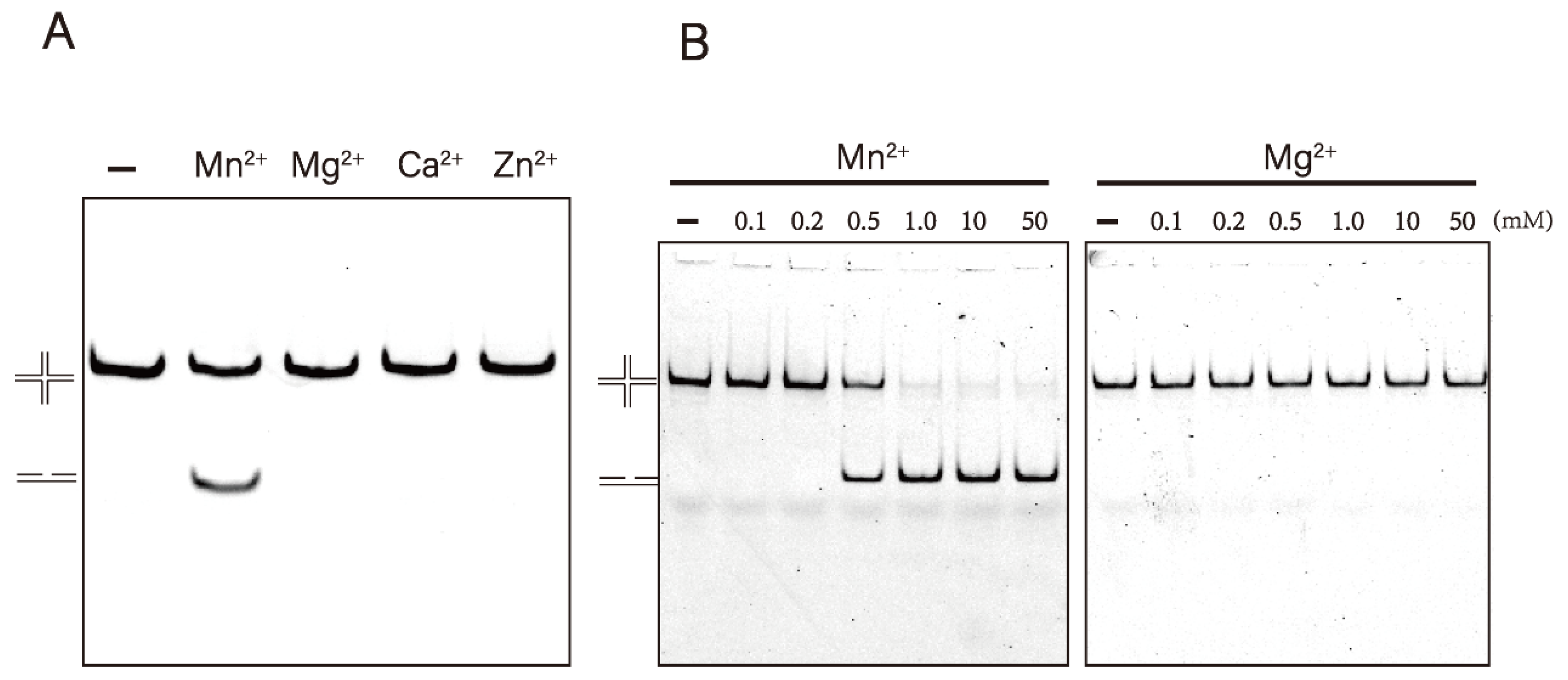
Resolvase usually prefers Mg2+ as a co-factor for catalytic reactions, however, Mn2+ can act as a substitute [19,36,37,38]. To study the metal ion dependence of DrRuvC, we used an HJ-12X substrate with a 12 bp homology region to react under different metal ion conditions. To create a contrast, 10 mM EDTA was included in the reaction buffer to sequester divalent metal ions. Interestingly, Mn2+ rather than Mg2+ is the only catalytic co-factor of DrRuvC resolvase. DrRuvC cleaved HJ-12X efficiently in the presence of Mn2+. However, no activity was detected in the presence of Ca2+, Zn2+, or Mg2+ (Figure 2A). Concentration-dependent experiments showed that no activity was detected with Mg2+ even up to 50 mM (Figure 2B). On the contrary, the DNA substrate was completely cleaved with Mn2+ at a concentration of 1 mM. We also found that 50 mM Mn2+ did not display any inhibitory effect on the activity of DrRuvC (Supplementary Figure S5), although studies have shown that high concentrations (>50 mM) of metal ions can inhibit the resolvase activity of other RuvC [10,37]. Since Mn2+ can enhance the digestion ability of DrRuvC, we tried to perform HJ-0X cleavage assay in the presenc of Mn2+. Unfortunately, even a high concentration of Mn2+ cannot catalyze DrRuvC to cleavage HJ-0X (Supplementary Figure S4), suggesting that the homologous core is strictly needed in the enzyme digestion reaction.

3.3. High Mg2+ Concentration Inhibits the Binding Activity of DrRuvC

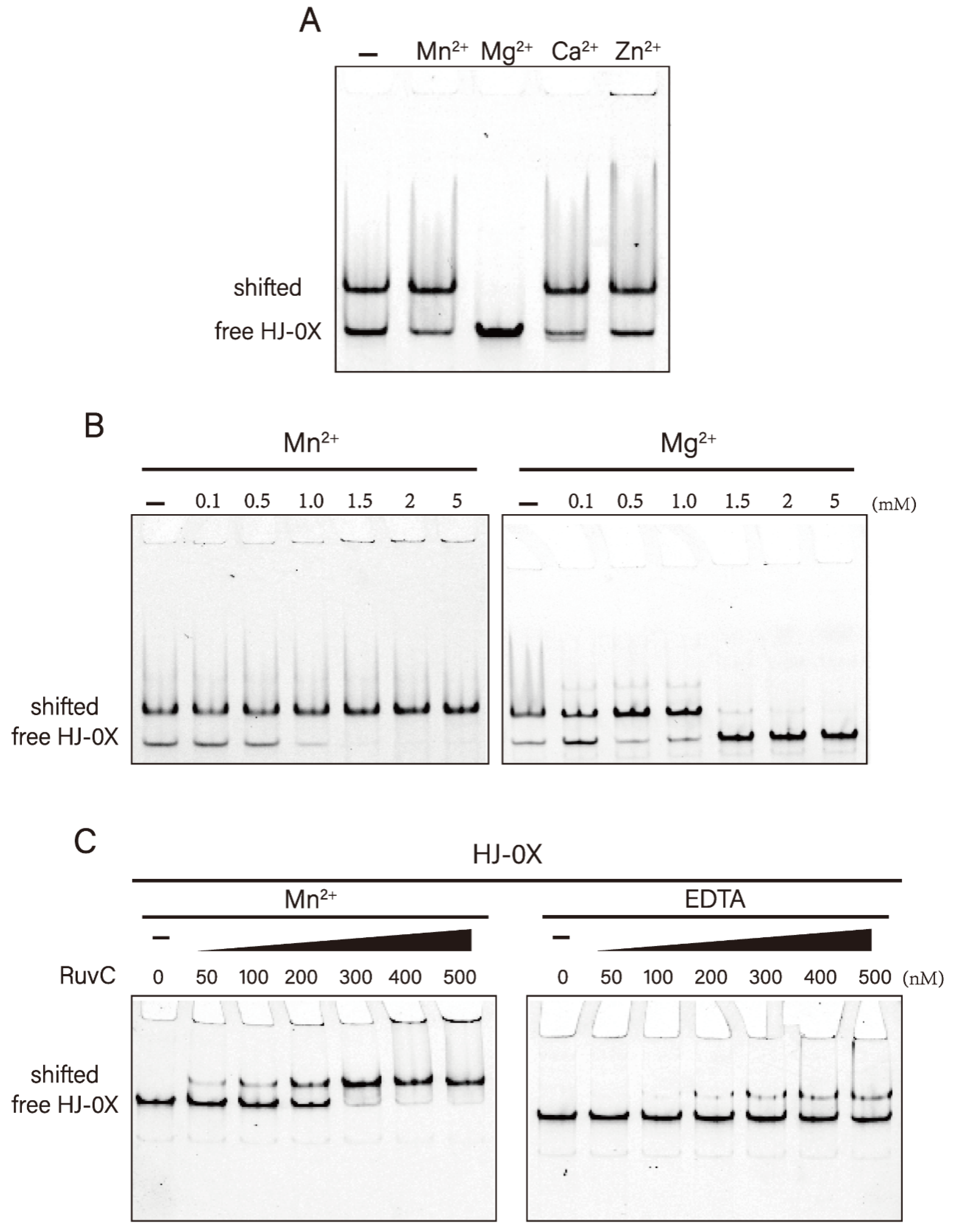
It is well-known that divalent metal ions are essential for the activity of resolvase, but not for DNA binding. We thus performed band-shift DNA binding analysis under different divalent metal ion conditions. To prevent DNA cleavage, we used HJ-0X without a homologous region to react. Similarly, we included 10 mM EDTA in the binding reaction buffer as a control. Native PAGE analysis showed that stable complexes were formed between DrRuvC and HJ either in the absence of metal ions or the presence of Mn2+, Ca2+, and Zn2+. However, no shifted DNA appeared in the presence of Mg2+ (Figure 3A).
We further set a concentration gradient to detect the effect of Mg2+ and Mn2+ on DNA binding. The results showed that when the concentration of Mg2+ was higher than 1.5 mM, the shifted DNA band disappeared completely. However, all HJs were combined with DrRuvC to form stable complexes with an increase in Mn2+ (Figure 3B). We also prepared a protein–DNA complex by adding different amounts of protein and a fixed concentration of DNA. We found that the binding ability of DrRuvC to HJs in the presence of Mn2+ is much higher than that in the absence of metal ions (Figure 3C). Our results show that Mg2+ inhibits the binding of DrDuvC to HJs, whereas Mn2+ facilitates the formation of the complex. This helps to explain the inhibitory effect of Mg2+ on the activity of DrRuvC resolvase.

3.4. Crystal Structure of DrRuvC Reveals Its Essential Amino Acid Sites

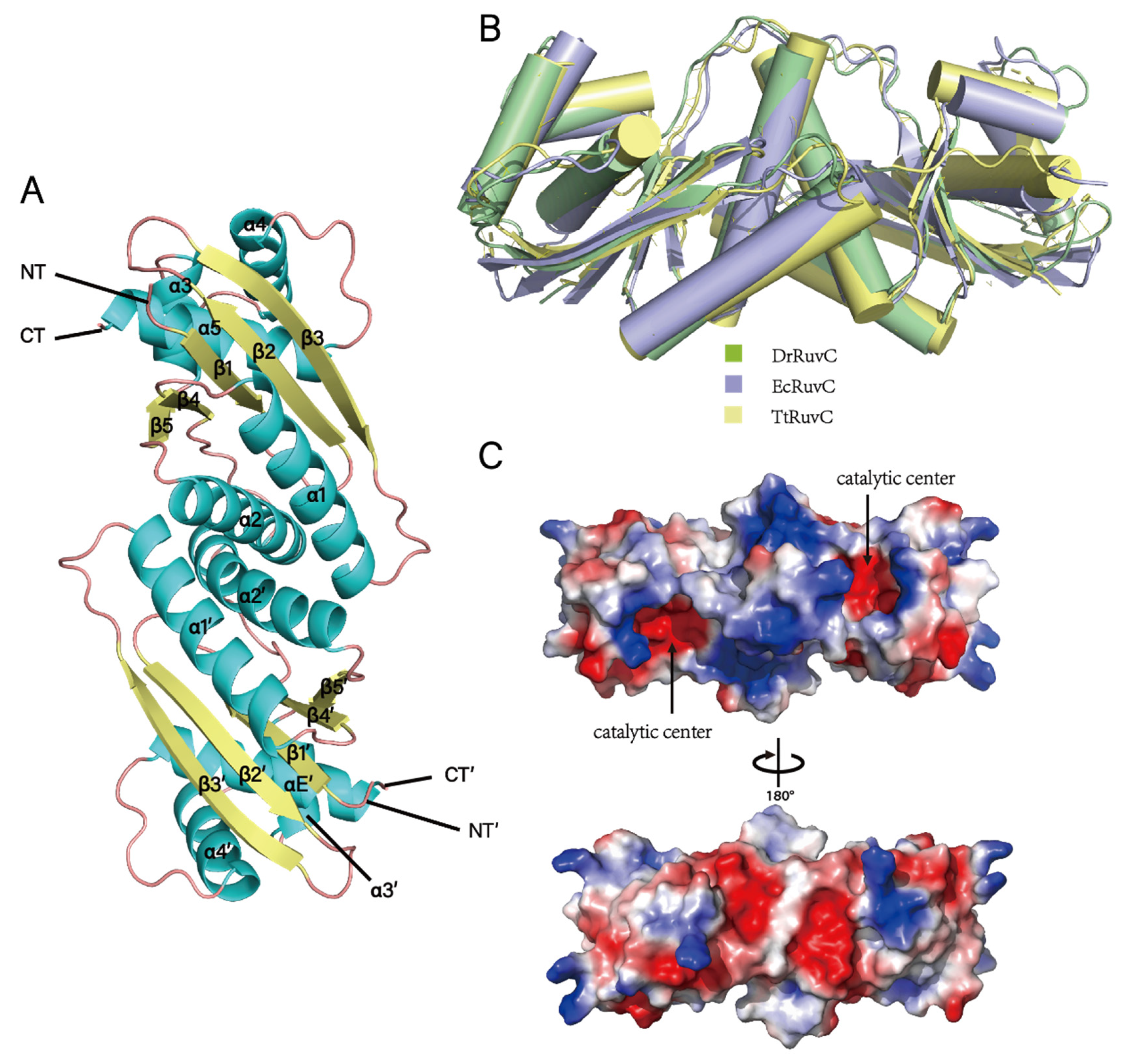
The structure of EcRuvC (PDB ID: 1HJR) was used as a search model, and the crystal structure of DrRuvC was determined at 1.6 Å using the molecular replacement method. It contains two monomers in an asymmetric unit, and each monomer comprises 1–154 residues with an elongated overall shape. For each protomer, the protein folds of DrRuvC consist of three antiparallel (β1β2β3) and two parallel strands (β4β5), sandwiched between five helices (α1–5), which is characteristic of enzymes from the retrovirus integrase superfamily (Figure 4A). Structural superimposition of DrRuvC with EcRuvC and TtRuvC showed similar protomers (with an RMSD value of 1.656 Å) and dimeric architectures (Figure 4B), as expected from the high amino acid sequence identity between the two proteins. The structural deviations are mainly reflected in the secondary structural elements and the disordered region of the C-terminal.
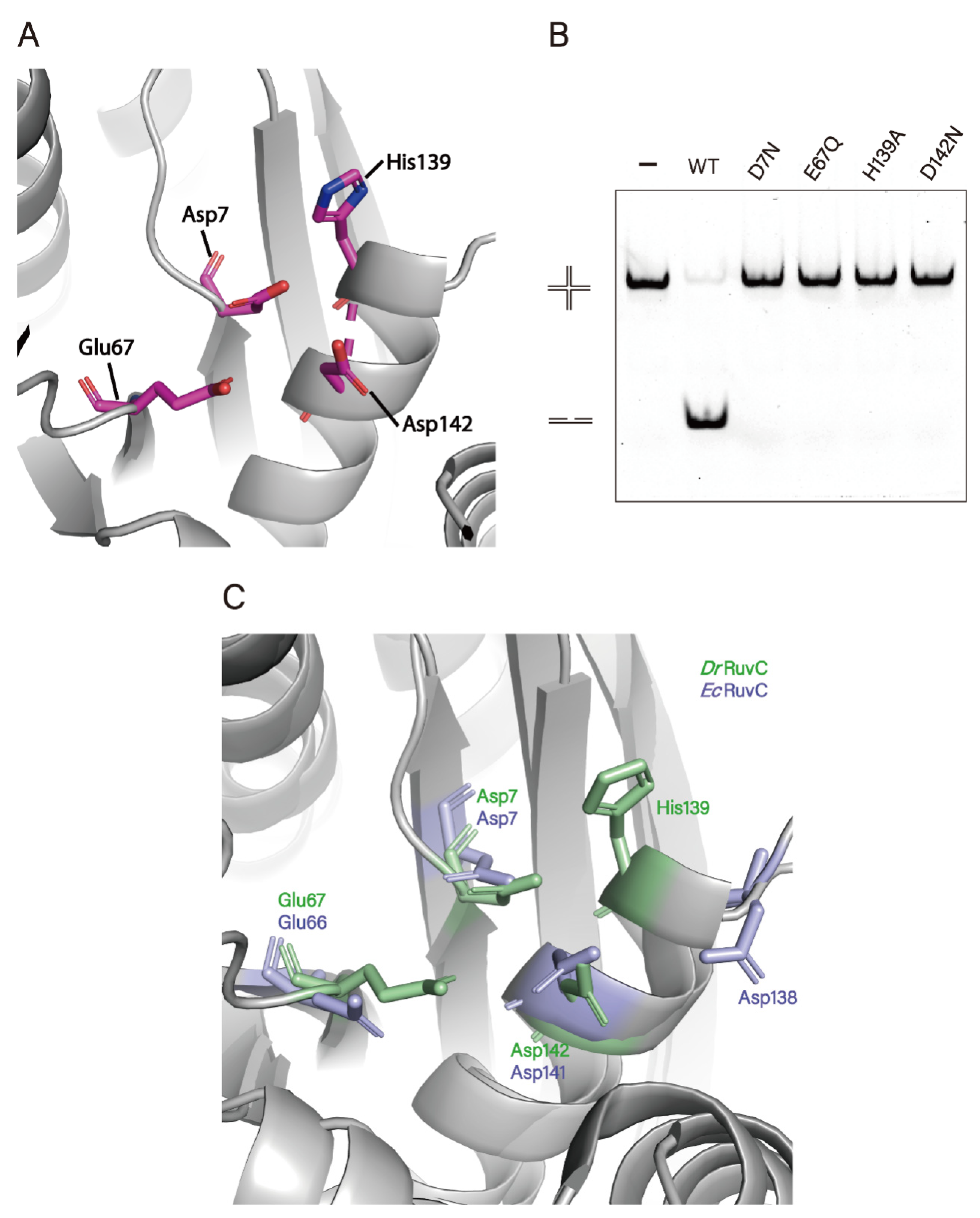
The distribution of the electrostatic surface potential of DrRuvC revealed that the catalytic center with a negatively charged groove is surrounded by a positively charged surface (Figure 4C), which corresponds to divalent metal ion and DNA binding sites. As expected for other known Holliday junction resolvases [4,18,36,39], the two catalytic centers in the DrRuvC dimer are positioned on the same side composed of Asp7, Glu67, His139 and Asp142 (Figure 5A). The results of amino acids site mutation showed that the substitution of these amino acid residues can eliminate the activity of DrRuvC completely (Figure 5B). Although these catalytic residues are conserved in most homologous proteins, the third metal-chelating residue of DrDuvC is His143, corresponding to Asp138 of EcRuvC (Figure 5C). Structurally, the pocket of the catalytic center is tighter in DrRuvC than in TtRuvC, which may be because Mn2+ is much smaller than Mg2+. It is worth noting that EcRuvC does not choose Mg2+ as the only catalytic co-factor, although its third metal-chelating residue is His143.
Previous studies have revealed that the base-specific recognition loop (BR-loop, corresponding to E67-A75 of DrRuvC) of MOC1 mediates its sequence-specific interaction with HJ [24]. As RuvC and MOC1 are evolutionarily conserved, we compared the BR-loop between RuvC proteins [40]. The results showed that the amino acid residues of DrRuvC in the BR-loop were significantly different from that of other homologous proteins (Supplementary Figure S1). Generally, aromatic residues such as tyrosine or phenylalanine are present within the loop. Following the aromatic residue (Tyr or Phe), there are charged (Asp, Lys, Arg) or polar (Asn, Gln, Ser) residues, which might mediate specific base recognition [24]. However, the BR-loop of DrRuvC does not contain any amino acids with aromatic bases. In particular, the residue F69 in EcRuvC is replaced by I70, which can intercalate into the bases one nucleotide before the cleavage site, forming stacking interactions with the flanking nucleotide bases [39,41,42]. The substitution of amino acids at these key sites may result in a distinctive mechanism of HJ cleavage by DrRuvC.

4. Discussion

HR plays critical roles in repairing DNA damage including double-strand breaks [43]. RuvC proteins have been implicated as a key that mediates the decomposition of HJs at the end of HR [44]. As an important model organism, D. radiodurans is of great value in the study of DNA repair [45]. In this study, besides confirming the structure specificity of DrRuvC, we explored the unique metal ion dependence of DrRuvC. To gain insights into the cleavage mechanisms of RuvC, we determined the crystal structures of DrRuvC and performed crystallographic analyses.
A panel of different branched DNA substrates was used to analyze the DNA binding and cleavage activities of DrRuvC. Our data revealed the enzyme had the highest binding affinity for HJ DNA, consistent with the result that HJs, but no other substrates, were cleaved, suggesting that the recognition of DNA branches by the formation of the DNA–protein complex is the basis for determining the catalytic specificity of DNA substrates. Sequence-dependent cleavage by RuvC resolvases is another important property that needed to be addressed. Bacteriophage enzymes can cleave a broad range of branched DNA structures, including the HJ, with little sequence-specificity [33,35,46]. In contrast, sequence-dependent HJ resolution was reported in various cellular resolvases [19]. Two kinds of HJ substrates were used in our experiment: (1) HJ-12X with a 12 bp homologous region core and (2) HJ-0X with a non-homologous region core. The results suggested that the homologous core of the HJ is the basis for determining cleavage activity, but it does not affect the binding ability. The specific preference in the sequences of cleavage sites remains to be accurately analyzed.
Our results seem to show that DrRuvC has a strict requirement for manganese as its metal co-factor. At present, all known RuvC proteins require divalent cations as catalytic co-factors for cleavage activity, among which Mg2+ is the most suitable [36,37,39], and Mn2+ could substitute for Mg2+ as a catalytic co-factor with weaker catalytic ability. However, it seems that DrRuvC prefers Mn2+ as the only catalytic co-factor, whereas high Mg2+ concentration inhibits the binding between DrRuvC and HJ. In addition, Mn2+ facilitates the formation of the DrRuvC-HJ complex, although it is generally believed that this topology-specific binding does not require divalent cations. In fact, the similar phenomenon regarding the selection of Mn2+ and Mg2+ is widespread in D. radiodurans. Compared with E. coli homologues that mainly use Mg2+ as cofactors, metalloenzymes in D. radiodurans exhibited a strong preference for Mn2+ rather than Mg2+. For example, Mn2+ are found in the structures of DrXth [26], RecJ [47], RNase J [48], MazG [49], MntH [50], and SodA [51] in D. radiodurans. The circular dichroism (CD) results showed that the overall configuration of DrRuvC protein did not change significantly under different metal ionic conditions (Supplementary Figure S6). In the presence of metal ions, the helicity of protein decreased, which may be beneficial to the catalytic reaction of the protein [52]. In particular, Mg2+ makes DrRuvC more relaxed than Mn2+, while less amount of α-helix conformation may hinder the enzyme digestion. D. radiodurans is known to accumulate very high intracellular manganese and low iron level compared to radiosensitive bacteria [53,54,55]. The high manganese concentration was suggested to be essential for relieving oxidative stress and protecting proteins from damage caused by reactive oxygen species [55,56]. The abundance of Mn2+ in the D. radiodurans cells may be used to explain why DrRuvC has adopted Mn2+ as its metal co-factor.
The crystal structure of DrRuvC shows an overall protein fold similar to that of EcRuvC, but DrRuvC has a tighter catalytic pocket by replacing the Asp138 of the catalytic active site with His139. In the base-specific recognition loop, the aromatic amino acids of DrRuvC are replaced by leucine and isoleucine, indicating that DrRuvC may have a specific cleavage mechanism. To determine the complex structure, a series of HJ with varying arm lengths were used to co-crystallize with DrRuvC, but without success. The detailed structural mechanisms of metal ion preference and HJ resolution require further investigation. Notably, it has been recently proposed that the TtRuvC resolvase could catalyze DNA cleavage through a general mechanism that is shared with CRISPR-Cas9 [57]. The DDE motif in Cas9 matches the Mn2+-coordinating ligands in DrRuvC, and the catalytic residue H139 is located at H983 in Cas9. Although the metal-dependent mechanisms of the two enzyme digestion systems are different, it is worthy to probe the histidine-activated mechanism of DrRuvC in recombination and genome editing.

Supplementary Materials

The following supporting information can be downloaded at: https://www.mdpi.com/article/10.3390/microorganisms10061160/s1, Table S1. The columns and buffers used for protein purification process; Table S2. The columns and buffers used for protein purification process; Figure S1. Multiple sequence alignment of RuvC homologs; Figure S2. Annealing of DNA substrates used in this study; Figure S3. Two Holliday junctions with different cores; Figure S4. HJ-0X cleavage assays; Figure S5. Time-resolved cleavage assay; Figure S6. The CD results of DrRuvC in different ion-containing buffer.

Author Contributions

Y.H. conceived the project. C.Q., L.W. and Y.H. designed the experiments and drafted the manuscript. C.Q. carried out biochemical experiments. W.H. and Y.X. performed the crystallization. Y.Z. determined the crystal structures. L.W., Y.Z., H.X. and B.T. gave technical support and conceptual advice. All authors have read and agreed to the published version of the manuscript.

Funding

This work was supported by the National Key Research and Development Program of China (2017YFA0503900) to Y.H., National Natural Science Foundation of China (31870051) to Y.H., and the Public Project of Zhejiang Province (LGN22C010002) to L.W.

Institutional Review Board Statement

Not applicable.

Informed Consent Statement

Not applicable.

Data Availability Statement

The coordinates and structure factors have been deposited to Protein Data Bank with accession codes 7XHJ.

Acknowledgments

We would like to thank Cancer Research Institute of Zhejiang University and the staff at the Shanghai Synchrotron Radiation Facility (SSRF in China) for assistance in the data collection.

Conflicts of Interest

The authors declare no conflict of interest.

References

  1. Wright, W.D.; Shah, S.S.; Heyer, W.-D. Homologous recombination and the repair of DNA double-strand breaks. J. Biol. Chem. 2018, 293, 10524–10535. [Google Scholar] [CrossRef] [Green Version]
  2. Mehta, A.; Haber, J.E. Sources of DNA Double-Strand Breaks and Models of Recombinational DNA Repair. Cold Spring Harb. Perspect. Biol. 2014, 6, a016428. [Google Scholar] [CrossRef] [Green Version]
  3. Holliday, R. A mechanism for gene conversion in fungi (Reprinted). Genet. Res. 2007, 89, 285–307. [Google Scholar] [CrossRef] [PubMed] [Green Version]
  4. Bond, C.S.; Kvaratskhelia, M.; Richard, D.; White, M.F.; Hunter, W.N. Structure of Hjc, a Holliday junction resolvase, from Sulfolobus solfataricus. Proc. Natl. Acad. Sci. USA 2001, 98, 5509–5514. [Google Scholar] [CrossRef] [Green Version]
  5. Hadden, J.M.; Declais, A.-C.; Carr, S.B.; Lilley, D.M.J.; Phillips, S.E.V. The structural basis of Holliday junction resolution by T7 endonuclease. Nature 2007, 449, 621-U15. [Google Scholar] [CrossRef]
  6. Kim, J.S.; Chang, J.H.; Kim, J.J.; Cho, Y. Crystal structure of the Mus81-Eme1 complex. Acta Crystallogr.-Found. Adv. 2008, 64, C301. [Google Scholar] [CrossRef] [Green Version]
  7. Nishino, T.; Komori, K.; Tsuchiya, D.; Ishino, Y.; Morikawa, K. Crystal structure of the archaeal Holliday Junction resolvase Hjc and implications for DNA recognition. Structure 2001, 9, 197–204. [Google Scholar] [CrossRef] [Green Version]
  8. Bizard, A.H.; Hickson, I.D. The Dissolution of Double Holliday Junctions. Cold Spring Harb. Perspect. Biol. 2014, 6, a016477. [Google Scholar] [CrossRef]
  9. Manthei, K.A.; Keck, J.L. The BLM dissolvasome in DNA replication and repair. Cell. Mol. Life Sci. 2013, 70, 4067–4084. [Google Scholar] [CrossRef] [Green Version]
  10. Bennett, R.; Dunderdale, H.; West, S. Resolution of Holliday Junctions by Ruvc Resolvase—Cleavage Specificity and Dna Distortion. Cell 1993, 74, 1021–1031. [Google Scholar] [CrossRef]
  11. Dunderdale, H.; Benson, F.; Parsons, C.; Sharples, G.; Lloyd, R.; West, S. Formation and Resolution of Recombination Intermediates by Escherichia-Coli Reca and Ruvc Proteins. Nature 1991, 354, 506–510. [Google Scholar] [CrossRef]
  12. Iwasaki, H.; Takahagi, M.; Shiba, T.; Nakata, A.; Shinagawa, H. Escherichia-Coli Ruvc Protein Is an Endonuclease That Resolves the Holliday Structure. Embo J. 1991, 10, 4381–4389. [Google Scholar] [CrossRef] [PubMed]
  13. Eggleston, A.K.; Mitchell, A.H.; West, S.C. In vitro reconstitution of the late steps of genetic recombination in E. coli. Cell 1997, 89, 607–617. [Google Scholar] [CrossRef] [Green Version]
  14. van Gool, A.J.; Shah, R.; Mézard, C.; West, S.C. Functional interactions between the holliday junction resolvase and the branch migration motor of Escherichia coli. EMBO J. 1998, 17, 1838–1845. [Google Scholar] [CrossRef] [Green Version]
  15. Mandal, T.N.; Mahdi, A.A.; Sharples, G.J.; Lloyd, R.G. Resolution of Holliday intermediates in recombination and DNA repair: Indirect suppression of ruvA, ruvB, and ruvC mutations. J. Bacteriol. 1993, 175, 4325–4334. [Google Scholar] [CrossRef] [Green Version]
  16. Sharples, G.J.; Ingleston, S.M.; Lloyd, R.G. Holliday junction processing in bacteria: Insights from the evolutionary conservation of RuVABC, RecG, and RusA. J. Bacteriol. 1999, 181, 5543–5550. [Google Scholar] [CrossRef] [PubMed] [Green Version]
  17. Shinagawa, H.; Iwasaki, H. Processing the Holliday junction in homologous recombination. Trends Biochem. Sci. 1996, 21, 107–111. [Google Scholar] [CrossRef]
  18. Parsons, C.; Stasiak, A.; Bennett, R.; West, S. Structure of a Multisubunit Complex That Promotes Dna Branch Migration. Nature 1995, 374, 375–378. [Google Scholar] [CrossRef] [PubMed]
  19. Wyatt, H.D.M.; West, S.C. Holliday Junction Resolvases. Cold Spring Harb. Perspect. Biol. 2014, 6, a023192. [Google Scholar] [CrossRef] [Green Version]
  20. Battista, J.R. Against all odds: The survival strategies of Deinococcus radiodurans. Annu. Rev. Microbiol. 1997, 51, 203–224. [Google Scholar] [CrossRef] [PubMed]
  21. Munteanu, A.-C.; Uivarosi, V.; Andries, A. Recent progress in understanding the molecular mechanisms of radioresistance in Deinococcus bacteria. Extremophiles 2015, 19, 707–719. [Google Scholar] [CrossRef]
  22. Slade, D.; Radman, M. Oxidative Stress Resistance in Deinococcus radiodurans. Microbiol. Mol. Biol. Rev. 2011, 75, 133–191. [Google Scholar] [CrossRef] [Green Version]
  23. Kobayashi, Y.; Misumi, O.; Odahara, M.; Ishibashi, K.; Hirono, M.; Hidaka, K.; Endo, M.; Sugiyama, H.; Iwasaki, H.; Kuroiwa, T.; et al. Holliday junction resolvases mediate chloroplast nucleoid segregation. Science 2017, 356, 631–634. [Google Scholar] [CrossRef] [PubMed]
  24. Yan, J.; Hong, S.; Guan, Z.; He, W.; Zhang, D.; Yin, P. Structural insights into sequence-dependent Holliday junction resolution by the chloroplast resolvase MOC1. Nat. Commun. 2020, 11, 1417. [Google Scholar] [CrossRef]
  25. Cai, J.; Pan, C.; Zhao, Y.; Xu, H.; Tian, B.; Wang, L.; Hua, Y. DRJAMM Is Involved in the Oxidative Resistance in Deinococcus radiodurans. Front. Microbiol. 2022, 12, 756867. [Google Scholar] [CrossRef]
  26. He, Y.; Wang, Y.; Qin, C.; Xu, Y.; Cheng, K.; Xu, H.; Tian, B.; Zhao, Y.; Wang, L.; Hua, Y. Structural and Functional Characterization of a Unique AP Endonuclease From Deinococcus radiodurans. Front. Microbiol. 2020, 11, 1178. [Google Scholar] [CrossRef] [PubMed]
  27. Zhou, X.; Chen, X.; An, Y.; Lu, H.; Wang, L.; Xu, H.; Tian, B.; Zhao, Y.; Hua, Y. Biochemical characterization of a unique DNA polymerase A from the extreme radioresistant organism Deinococcus radiodurans. Biochimie 2021, 185, 22–32. [Google Scholar] [CrossRef]
  28. Minor, W.; Cymborowski, M.; Otwinowski, Z.; Chruszcz, M. HKL-3000: The integration of data reduction and structure solution —From diffraction images to an initial model in minutes. Acta Crystallogr. Sect.-Struct. Biol. 2006, 62, 859–866. [Google Scholar] [CrossRef] [Green Version]
  29. Otwinowski, Z.; Minor, W. Processing of X-ray diffraction data collected in oscillation mode. Macromol. Crystallogr. Pt. A 1997, 276, 307–326. [Google Scholar] [CrossRef]
  30. Adams, P.D.; Grosse-Kunstleve, R.W.; Hung, L.W.; Ioerger, T.R.; McCoy, A.J.; Moriarty, N.W.; Read, R.J.; Sacchettini, J.C.; Sauter, N.K.; Terwilliger, T.C. PHENIX: Building new software for automated crystallographic structure determination. Acta Crystallogr. Sect.-Struct. Biol. 2002, 58, 1948–1954. [Google Scholar] [CrossRef] [Green Version]
  31. McCoy, A.J.; Grosse-Kunstleve, R.W.; Adams, P.D.; Winn, M.D.; Storoni, L.C.; Read, R.J. Phaser crystallographic software. J. Appl. Crystallogr. 2007, 40, 658–674. [Google Scholar] [CrossRef] [PubMed] [Green Version]
  32. Emsley, P.; Cowtan, K. Coot: Model-building tools for molecular graphics. Acta Crystallogr. Sect.-Struct. Biol. 2004, 60, 2126–2132. [Google Scholar] [CrossRef] [Green Version]
  33. Shah, R. Genetic recombination in E. coli: RuvC protein cleaves Holliday junctions at resolution hotspots in vitro. Cell 1994, 79, 853–864. [Google Scholar] [CrossRef]
  34. White, M.F.; Lilley, D.M.J. Characterization of a Holliday junction-resolving enzyme from Schizosaccharomyces pombe. Mol. Cell. Biol. 1997, 17, 6465–6471. [Google Scholar] [CrossRef] [PubMed] [Green Version]
  35. White, M.F.; Lilley, D.M.J. The Structure-selectivity and Sequence-preference of the Junction-resolving Enzyme CCE1 of Saccharomyces cerevisiae. J. Mol. Biol. 1996, 257, 330–341. [Google Scholar] [CrossRef]
  36. Hu, Y.; He, Y.; Lin, Z. Biochemical and structural characterization of the Holliday junction resolvase RuvC from Pseudomonas aeruginosa. Biochem. Biophys. Res. Commun. 2020, 525, 265–271. [Google Scholar] [CrossRef] [PubMed]
  37. Komori, K.; Sakae, S.; Fujikane, R.; Morikawa, K.; Shinagawa, H.; Ishino, Y. Biochemical characterization of the Hjc Holliday junction resolvase of Pyrococcus furiosus. Nucleic Acids Res. 2000, 28, 4544–4551. [Google Scholar] [CrossRef] [PubMed] [Green Version]
  38. Takahagi, M.; Iwasaki, H.; Shinagawa, H. Structural requirements of substrate DNA for binding to and cleavage by RuvC, a Holliday junction resolvase. J. Biol. Chem. 1994, 269, 15132–15139. [Google Scholar] [CrossRef]
  39. Chen, L.; Shi, K.; Yin, Z.; Aihara, H. Structural asymmetry in the Thermus thermophilus RuvC dimer suggests a basis for sequential strand cleavages during Holliday junction resolution. Nucleic Acids Res. 2013, 41, 648–656. [Google Scholar] [CrossRef] [Green Version]
  40. Lin, H.; Zhang, D.; Zuo, K.; Yuan, C.; Li, J.; Huang, M.; Lin, Z. Structural basis of sequence-specific Holliday junction cleavage by MOC1. Nat. Chem. Biol. 2019, 15, 1241–1248. [Google Scholar] [CrossRef] [PubMed]
  41. Górecka, K.M.; Komorowska, W.; Nowotny, M. Crystal structure of RuvC resolvase in complex with Holliday junction substrate. Nucleic Acids Res. 2013, 41, 9945–9955. [Google Scholar] [CrossRef]
  42. Yoshikawa, M.; Iwasaki, H.; Shinagawa, H. Evidence that phenylalanine 69 in Escherichia coli RuvC resolvase forms a stacking interaction during binding and destabilization of a holliday junction DNA substrate. J. Biol. Chem. 2001, 276, 10432–10436. [Google Scholar] [CrossRef] [Green Version]
  43. West, S.C. Molecular views of recombination proteins and their control. Nat. Rev. Mol. Cell Biol. 2003, 4, 435–445. [Google Scholar] [CrossRef]
  44. Holliday, R. Mechanism for Gene Conversion in Fungi. Genet. Res. 1964, 5, 282–304. [Google Scholar] [CrossRef] [Green Version]
  45. Lu, H.; Hua, Y. PprI: The Key Protein in Response to DNA Damage in Deinococcus. Front. Cell Dev. Biol. 2021, 8, 609714. [Google Scholar] [CrossRef]
  46. Shah, R. The RuvC protein dimer resolves Holliday junctions by a dual incision mechanism that involves base-specific contacts. EMBO J. 1997, 16, 1464–1472. [Google Scholar] [CrossRef] [PubMed] [Green Version]
  47. Cheng, K.; Xu, H.; Chen, X.; Wang, L.; Tian, B.; Zhao, Y.; Hua, Y. Structural basis for DNA 5′-end resection by RecJ. eLife 2016, 5, e14294. [Google Scholar] [CrossRef]
  48. Zhao, Y.; Lu, M.; Zhang, H.; Hu, J.; Zhou, C.; Xu, Q.; Ul Hussain Shah, A.M.; Xu, H.; Wang, L.; Hua, Y. Structural insights into catalysis and dimerization enhanced exonuclease activity of RNase J. Nucleic Acids Res. 2015, 43, 5550–5559. [Google Scholar] [CrossRef] [Green Version]
  49. Gonçalves, A.M.D.; de Sanctis, D.; McSweeney, S.M. Structural and Functional Insights into DR2231 Protein, the MazG-like Nucleoside Triphosphate Pyrophosphohydrolase from Deinococcus radiodurans. J. Biol. Chem. 2011, 286, 30691–30705. [Google Scholar] [CrossRef] [PubMed] [Green Version]
  50. Bozzi, A.T.; Zimanyi, C.M.; Nicoludis, J.M.; Lee, B.K.; Zhang, C.H.; Gaudet, R. Structures in multiple conformations reveal distinct transition metal and proton pathways in an Nramp transporter. eLife 2019, 8, e41124. [Google Scholar] [CrossRef]
  51. Dennis, R.J.; Micossi, E.; McCarthy, J.; Moe, E.; Gordon, E.J.; Kozielski-Stuhrmann, S.; Leonard, G.A.; McSweeney, S. Structure of the manganese superoxide dismutase from Deinococcus radiodurans in two crystal forms. Acta Crystallograph. Sect. F Struct. Biol. Cryst. Commun. 2006, 62, 325–329. [Google Scholar] [CrossRef] [Green Version]
  52. Subinya, M.; Steudle, A.K.; Jurkowski, T.P.; Stubenrauch, C. Conformation and activity of lipase B from Candida antarctica in bicontinuous microemulsions. Colloids Surf. B-Biointerfaces 2015, 131, 108–114. [Google Scholar] [CrossRef] [PubMed]
  53. Daly, M.J.; Gaidamakova, E.K.; Matrosova, V.Y.; Vasilenko, A.; Zhai, M.; Venkateswaran, A.; Hess, M.; Omelchenko, M.V.; Kostandarithes, H.M.; Makarova, K.S.; et al. Accumulation of Mn(II) in, Deinococcus radiodurans facilitates gamma-radiation resistance. Science 2004, 306, 1025–1028. [Google Scholar] [CrossRef] [PubMed] [Green Version]
  54. Sun, H.; Xu, G.; Zhan, H.; Chen, H.; Sun, Z.; Tian, B.; Hua, Y. Identification and evaluation of the role of the manganese efflux protein in Deinococcus radiodurans. Bmc Microbiol. 2010, 10, 319. [Google Scholar] [CrossRef] [PubMed] [Green Version]
  55. Tabares, L.C.; Un, S. In Situ Determination of Manganese(II) Speciation in Deinococcus radiodurans by High Magnetic Field EPR Detection of High Levels of Mn(II) Bound to Proteins. J. Biol. Chem. 2013, 288, 5050–5055. [Google Scholar] [CrossRef] [PubMed] [Green Version]
  56. Sharma, A.; Gaidamakova, E.K.; Matrosova, V.Y.; Bennett, B.; Daly, M.J.; Hoffman, B.M. Responses of Mn2+ speciation in Deinococcus radiodurans and Escherichia coli to gamma-radiation by advanced paramagnetic resonance methods. Proc. Natl. Acad. Sci. USA 2013, 110, 5945–5950. [Google Scholar] [CrossRef] [Green Version]
  57. Casalino, L.; Nierzwicki, Ł.; Jinek, M.; Palermo, G. Catalytic Mechanism of Non-Target DNA Cleavage in CRISPR-Cas9 Revealed by Ab Initio Molecular Dynamics. ACS Catal. 2020, 10, 13596–13605. [Google Scholar] [CrossRef] [PubMed]
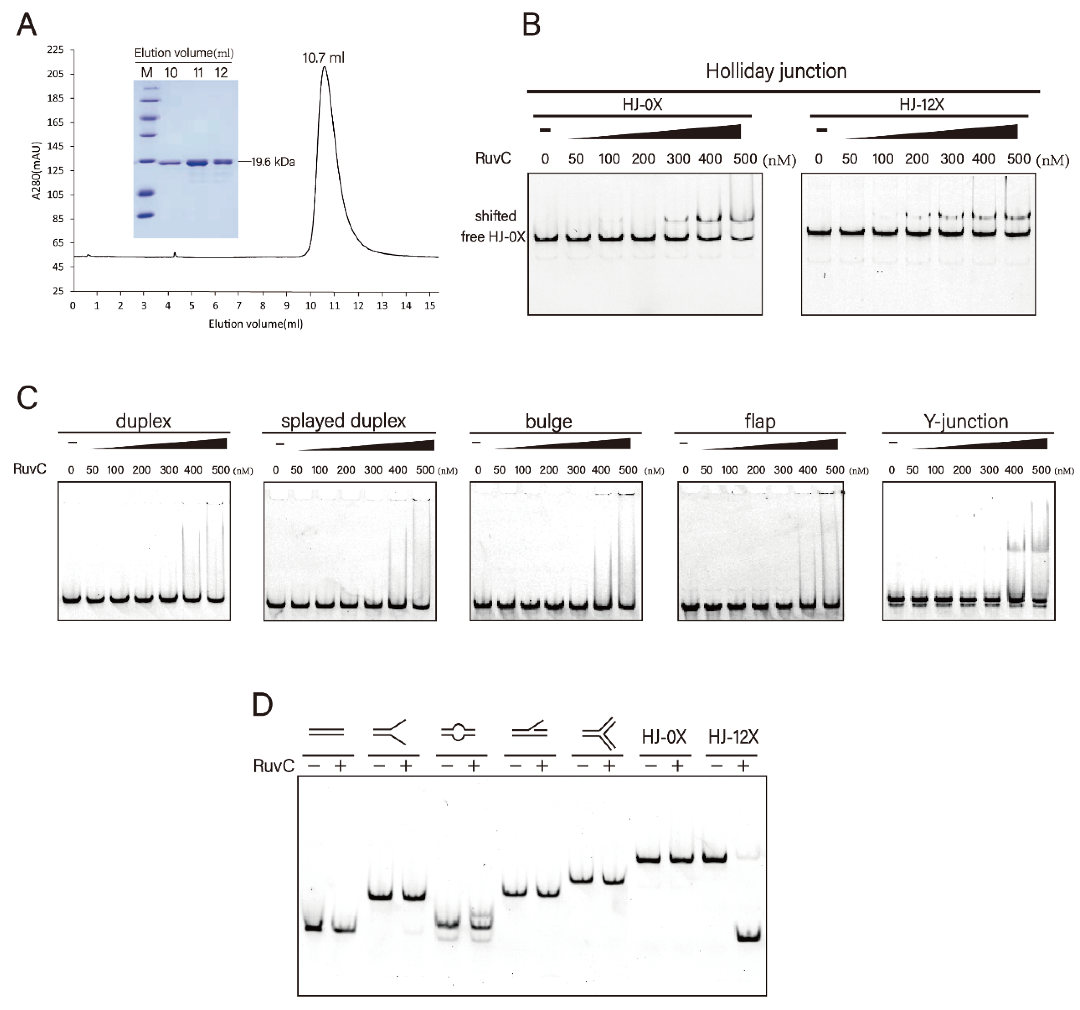
Figure 1. Protein purification and substrate specificities of DrRuvC. (A) Size exclusion chromatogram (Superdex 75 10/300 GL column) of recombinant DrRuvC protein. SDS-PAGE of representative eluted fractions is shown in inset. (B) Bandshift analysis of Holliday junctions (HJ-0X and HJ-12X). (C) Bandshift analysis of the indicated DNA structures. From left to right, the binding reactions contained 0, 50, 100, 200, 300, 400, 500 nM of DrRuvC protein. (D) Native PAGE analysis of DrRuvC cleavage of the indicated DNA structures.
Microorganisms 10 01160 g001
Figure 2. Effect of metal ions on the resolvase activity of DrRuvC. (A) The Holliday junction cleavage assays were performed in the absence or presence of indicated metal ions. Substrate uses HJ-12X with 12 bp homologous core. 500 μM of each metal ion was used. (B) Metal ion titration experiment of DrRuvC cleavage activity.
Microorganisms 10 01160 g002
Figure 3. Effect of metal ions on the HJ binding of DrRuvC. (A) The Holliday junction binding abilities of DrRuvC (250 nM) in the absence or presence of indicated metal ions (2 mM). The substrate HJ-0X with non-homologous core was used. (B) Mn2+ and Mg2+ titration experiments of HJ binding. (C) The binding abilities of DrRuvC to HJ-0X in the presence of 1 mM Mn2+ or in the absence of bivalent metal ions.
Microorganisms 10 01160 g003
Figure 4. Crystal structure of DrRuvC. (A) Cartoon diagram of the overall structure of DrRuvC. The secondary structural elements are labeled. The N- and C-termini are indicated. (B) Structural superimposition of DrRuvC with EcRuvC and TtRuvC. (C) Electrostatic surface of the DrRuvC dimer colored according to the electrostatic surface potential (red: −1 kT/e to blue: +1 kT/e), shown in two orientations rotated by 180°. The location of the active sites is indicated by arrows.
Microorganisms 10 01160 g004
Figure 5. The active sites of DrRuvC. (A) The active site of DrRuvC. DrRuvC is shown in grey and the catalytic residues are highlighted in magenta sticks. (B) Holliday junction cleavage assay of DrRuvC wild type (WT) or various mutants. (C) Superposition of the catalytic residues from DrRuvC (green) and EcRuvC (blue).
Microorganisms 10 01160 g005
Table 1. Data collection, phasing and refinement statistics.
DrRuvC
Data collection
Space groupP212121
Cell dimensions
a, b, c (Å)
α, β, ɣ (°)
40.02, 72.60, 113.90
90, 90, 90
Wavelength (Å)0.9793
Resolution (Å)30.0–1.60
Rsym (%)5.7 (41.1)
II16.5 (4.3)
Completeness (%)94.8(98.6)
Redundancy3.7 (3.8)
Refinement
Resolution (Å)30.0–1.60 (1.64–1.60)
No. reflections42,358
Rwork/Rfree20.5/22.4
No. atoms
Protein2270
Water151
B-factors
Protein30.7
Water42.5
R.m.s deviations
Bond lengths (Å) 0.005
Bond angles (°)0.770
Ramachandran statistics
Favored (%)98.7
Allowed (%)1.3
Ourlier (%)0
Publisher’s Note: MDPI stays neutral with regard to jurisdictional claims in published maps and institutional affiliations.