Abstract
The human gut microbiome plays a key role in regulating host physiology. In a stable state, both the microbiota and the gut work synergistically. The overall homeostasis of the intestinal flora can be affected by multiple factors, including disease states and the treatments given for those diseases. In this review, we examine the relatively well-characterised abnormalities that develop in the microbiome in idiopathic inflammatory bowel disease, and compare and contrast them to those that are found in radiation enteropathy. We discuss how these changes may exert their effects at a molecular level, and the possible role of manipulating the microbiome through the use of a variety of therapies to reduce the severity of the underlying condition.
1. Role of the Human Gut Microbiome
The human gut microbiome is made up of a collection of microorganisms, including bacteria, viruses, archaea, and protists [1]. It is a collection of over 1014 microorganisms, and plays a key role in regulating host physiology [2,3].
They assist in metabolising indigestible polysaccharides, producing vitamins that are required for the growth and maturation of intestinal epithelium and immune system; defend against attack by opportunistic pathogens [2]; and are instrumental in sustaining tissue homeostasis. In a “stable” state, both the microbiota and the gut work synergistically. Dysbiosis is a term that is used to describe an alteration that results in the gut microbiota composition being changed to one less harmonious, which can occur for a variety of reasons, including a change in diet, enteric infections, the use of antibiotics, or abdominal surgery [4].
Inflammatory bowel disease (IBD), which includes Crohn’s disease (CD) and ulcerative colitis (UC), is a significant burden to health. It is more prevalent in the Western world, especially in the United States and Europe [5]. However, recent analysis shows that it is now becoming a global health problem, with increasing incidence and prevalence in different parts of the world [5]. It may present in a number of ways, varying from presenting with a relatively mild disease phenotype to a very severe phenotype. It is not currently possible to predict the onset of IBD or the course of the disease in humans. This is because the aetiology is unknown, as are many of the underlying pathological processes [6]. However, IBD is thought to be multifactorial, where diet and environment trigger a dysregulated intestinal mucosal response to intestinal microbiota in genetically susceptible individuals [6].
In contrast, radiation enteropathy is a potentially more valuable human model of gastrointestinal (GI) inflammation and fibrosis. First, patients treated with radiotherapy (RT) for cancer often develop progressive changes that are initiated by the RT, and these changes can be followed over time. Secondly, patients receiving RT for pelvic tumours frequently complain of GI-related symptoms similar to those suffered by patients with IBD—including ulceration and bleeding, diarrhoea, steatorrhoea, haemorrhoids, nausea, and abdominal or anal pain [7]—which significantly affect their quality of life (QoL). These problems caused by RT are sometimes called “pelvic radiation disease” (PRD).
2. The Oxygen Hypothesis
There is a decrease in gut oxygen levels from the duodenum to the large intestine [8]. This is a consequence of the host being able to maintain a high oxygen concentration in the proximal small intestine [8], whilst also delivering nitrate to the distal end [9]. The microbial community composition of the small intestine is therefore dominated by facultatively anaerobic bacteria [10], whereas in the large intestine, there is predominantly obligately anaerobic bacteria as a consequence of limited oxygen and nitrate [11].
An imbalance between these obligate and facultative anaerobes is what characterises IBD dysbiosis [12]. The key factors influencing this imbalance are oxygen and reactive oxygen species (ROS) [13]. The “oxygen hypothesis” suggests there is an increased release of oxyhaemoglobin and ROS into the intestinal lumen due to chronic inflammation of the intestinal walls [14]. Radiation-induced dysbiosis has similarities to IBD, also exhibiting increased levels of ROS [15]. These changes create a microenvironment that favours facultative anaerobes [16,17]. As a consequence, the proportions of obligate anaerobes, such as F. prausnitzii, that release anti-inflammatory compounds are reduced, which leads to increased inflammation [18], creating a positive feedback loop that enhances the disease process [19].
3. IBD and the Gut Microbiome
Early studies on animal models have revealed that immune cells are unable to cause inflammation without the presence of intestinal bacteria, thereby indicating a role for the intestinal microbiome in the induction and/or maintenance of local inflammation and disease [20]. This is further supported by the finding that intestinal inflammation is most severe in parts of the bowel with larger bacterial populations [21]. Other studies have shown that specific patterns in microbiotic changes may be linked to the risk of IBD [21].
The changes that have been described in the literature are shown in Table 1. The most consistent alterations detected in the gut microbiota of patients with IBD are a reduction in diversity, particularly of Firmicutes, compared to healthy individuals [22,23,24]. Increases in some species may promote inflammation, while reductions in others may do the same. These changes lead to a reduction in mucosal integrity either directly or by affecting colonic butyrate production, an important fuel for colonic epithelium, or by influencing cytokine production. The mechanisms via which these changes occur are further explained in Figure 1.

Table 1.
Summarising the changes in bacterial population in IBD and the likely effect of the change.
Table 1.
Summarising the changes in bacterial population in IBD and the likely effect of the change.
| Bacterial Species | Nature of Change (Increase/Decrease) | Change in UC or Crohn’s Disease | Likely Effect of the Change | |
|---|---|---|---|---|
| Proteobacteria | Escherichia Salmonella Legionellales | Varies | Both | Pro-inflammatory if increased [25,26], anti-inflammatory if decreased [23,27]. In CD patients, intestinal permeability is increased due to adhesion-invasive E. coli, which leads to inflammation [28,29]. |
| Bacteroidetes | Varies | Both | Pro-inflammatory if increased [25,26], anti-inflammatory if decreased [23,27]. | |
| Firmicutes | Faecalibacterium prausnitzii | Decreased | Both | Pro-inflammatory [30,31] and increased risk of post-operative occurrence in CD [32] due to reduction in short-chain fatty acids, especially butyrate [33]; this has an anti-inflammatory effect, provides energy for colonic epithelial cells, may strengthen epithelial barrier integrity, and plays a role in GI immune responses [34]. Recovery of population associated with maintenance of clinical remission in UC [32] due to production of interleukin (IL)-10 and inhibition of inflammatory cytokines, such as IL-12 and interferon-γ [35]. |
| Roseburia inulinvorans | Decreased | CD | Pro-inflammatory [30,31], higher genetic risk of IBD in healthy individuals with decreased levels [36]. | |
| Ruminococcus torques | Decreased | CD | Pro-inflammatory [30,31] | |
| Blautia faecis | Decreased | CD | Pro-inflammatory [30,31] | |
| Clostridium lavalense | Decreased | CD | Pro-inflammatory [30,31] | |
| Erysipelotrichales Clostridiales | Decreased | CD | Pro-inflammatory due to reduction in butyrate production [28,29,36,37]. | |
| Veillonellaceae | Increased | CD | Pro-inflammatory [36] | |
| Enterobacteriaceae Pasteurellaceae Fusobacteriaceae | Increased | CD | Pro-inflammatory [36] | |

Figure 1.
Altered gut bacteria implication in the pathogenesis of IBD. (Taken from Pavel et al., 2021 [38]).
4. Studies Assessing the Microbiome of Twins with IBD
A further insight into the role of the gut microbiome in the pathogenesis of IBD is through studies looking at the development of IBD in twins. Previous studies exploring this have reported differences in the gut microbiome composition in IBD-affected twins compared with their healthy co-twins [39,40,41,42,43] (see Table 2).
Table 2.
Summarising the changes in bacterial population in twin studies.
However, these studies were performed in small numbers, no more than 10, of IBD-discordant or concordant twin pairs [40,41,42,43], and either did not include an unrelated matched healthy control group [39,43], or only included a small non-matched control group [40,41,42]. In addition, they were based on 16S rRNA sequencing [39,41,42,43], which does not assess microbial functional pathways.
A more recent study by Brand et al. [44] showed no significant differences in the relative abundance of species and pathways between healthy co-twins and their IBD-twins. However, they found an overlap in species, between healthy co-twins and IBD-twins and healthy co-twins and unrelated patients with IBD, respectively. Many of these shared species have previously been associated with IBD, such as Escherichia unclassified. The gut microbiome may therefore display IBD-like signatures that precede the onset of IBD [44]. However, longitudinal follow-up studies are needed to infer a causal relationship [44].
5. The Effects of Radiotherapy on the Gut Microbiome
Both curative and palliative cancer patients may receive RT, in combination with chemotherapy, as part of their cancer care [45]. Approximately 50% of all cancer patients receive RT [46], with 90% of those receiving pelvic RT developing a permanent change in their bowel habits [47]. Despite the well-established benefits of RT in oncology, RT-induced toxicities may detract from this, significantly impairing the QoL of patients. Radiation enteritis can be either acute or chronic. The chronic form, more correctly called radiation enteropathy, usually develops between 3 months to many decades after treatment [47], and occurs in approximately 5% to 55% of patients treated with pelvic RT [48]. Severe symptoms arising from chronic radiation enteropathy not only affect cancer patient’s QoL, but also add to the cost of medical treatment by increasing the use of medication for symptom relief, prolonging hospital stays, and temporarily or permanently stopping cancer treatment [49,50].
There is a close relationship between gut microbiota dysbiosis and intestinal injury after RT. This is summarised in Table 3.
Table 3.
Detailing the observed changes in microbiota following exposure to radiation.
6. The Human Gut Virome
The human gut virome contains eukaryotic viruses, prokaryotic viruses, and phages, known as bacteriophages and bacterial viruses (the phageome) [68]. According to the Global Virome Database, phages make up 97.7% of the gut virome, with 2.1% being eukaryotic viruses and 0.1% being prokaryotic viruses [69]. Cross-kingdom interactions between phages and bacteria, and between viruses/phages and the host immune system, underlie the function of the human gut virome in health and disease [70]. The virome population can affect its host in the following ways [70]:
- Eukaryotic viruses that infect human cells trigger immune responses, which can then lead to disease.
- Phages can affect the host indirectly via modulation of bacterial composition and bacterial fitness.
Potential activators of chronic inflammation can be released when enteric eukaryotic viruses and bacteriophages kill host cells [71]. The resulting dysbiosis of the enteral system is one of the key factors in the pathogenesis of I BD [72]. Several studies have delineated the events that can occur in inflammatory bowel disease (Table 4). There are no studies yet in patients treated with radiotherapy. As a result of significant improvements in sequencing technology in recent times, the diversity of the enteric human virome is being increasingly revealed, leading to new ways of targeting the gut microbiota to prevent or treat disease [71].
Table 4.
Changes in the Hunan Gut Virome of Patients with IBD.
7. How the Microbiome Acts as the Guardian of the Gut from Radiation: Parallels with IBD
It has been established that toll-like receptors (TLRs) act as the centre of immune responses to microbes in the gut [83]. TLRs are a group of proteins that are expressed by a number of immune cells, including macrophages, neutrophils, dendritic cells, and epithelial cells [4]. They recognise pathogen-associated molecular patterns (PAMPs), which are highly conserved structures of microbes [84]. Upon activation, TLRs induce a number of inflammatory cytokines by mediating the phosphorylation of IκB to activate NF-κB [83]. It also regulates the maturation of dendritic cells (DCs), and the proliferation and differentiation of Th1 and Th2 T cells [83].
A proliferation-inducing ligand (APRIL) and thymic stromal lymphopoietin (TSLP) are cytokines that are expressed following stimulation of intestinal epithelial cells (IECs), which promote class switch recombination (CSR) of IgM and IgA1 to protease-resistant IgA2 [85]. IgA2 stops invasion by bacteria by binding them to the apical surface of IECs [85]. The production of trefoil factor 3 (TFF3) is also increased following the activation of TLR2, which promotes the repair of gaps in the epithelial monolayer [85]. Microbicidal peptides and lectins, such as α-defensins and regenerating islet-derived protein 3γ (REG3γ), are released by Paneth cells due to TLR stimulation [85].
TLR signalling facilitates the optimal functioning of the immune mechanisms within a healthy host by protecting barrier integrity and maintaining commensal composition and tolerance. However, dysfunctional TLR signalling in susceptible individuals may impair commensal–mucosal homeostasis, thus leading to a worsening of tissue injury and ultimately to chronic inflammation in IBD [86] (see Table 5).
Table 5.
Role of TLRs in IBD. (Adapted from Lu et al., 2018 [83]).
Similarly, as in IBD, the gut microbiome may prevent injury induced by radiation through the activation of TLRs [4]. Entolimod, a TLR5 ligand, has previously been shown to decrease the rate of apoptosis of intestinal crypt cells, as well as cells within the lamina propria in mice and primates, when given as a pre-irradiation injection [102]. In another study in mice, pre-treatment with a TLR9 ligand reduced small bowel radiation injury through a MyD88-dependent signalling pathway [103]. Bacterial flagellin and CpG (cytidine–phosphate–guanosine) DNA, which are TLR5 and TLR9 ligands, are found in bacteria and viruses, respectively [4].
Additionally, the use of lipopolysaccharide, a membrane component of Gram-negative bacteria, before radiation provides protection to intestinal crypts via the induction of cyclooxygenase-2 and the production of prostaglandins [104]. The release of tumour necrosis factor (TNF)-α, which occurs due to the stimulation of TLR4-expressing cells by lipopolysaccharide, also results in increased production of prostaglandins and decreased radiation-induced apoptosis of epithelial stem cells [105]. TLR may also exert its protective effects against radiation through the activation of nuclear factor-kappa B (NF-κB) signalling [106], which is required for defending the gut against radiation-induced apoptosis. NF-κB activation also moderates the radioprotective effects of lipopolysaccharide [107], suggesting that TLRs have an effect on the intestinal response to radiation-induced epithelial damage through the NF-κB pathway [108].
In contrast, radiation damage of the bowel can be worsened through the activation of TLR3, and possibly TLR4. Injection with the TLR3 ligand Poly I:C, found naturally in viruses, resulted in more severe GI symptoms after whole body irradiation [4]. In addition, TLR3 knockout mice appeared more radioresistant by having less apoptotic intestinal epithelial cells, and also a larger proportion of radiation surviving crypts. Another study using knockout mice revealed that pre-treatment with the TLR4 antagonist, C34, reduced radiation-induced cell damage and death [109].
These compounds may provide the bowel with radioprotection through their effect on the systemic immune system. Furthermore, it is has been established that germ-free mice are more able to withstand radiation-induced bowel injury than conventional mice colonised with the microbiome [110,111]. These findings suggest that a key factor in the development of radiation enteropathy could be the gut microbiota, thus allowing the possibility to prevent or treat radiation enteropathy by manipulating its composition.
8. Treatment in IBD
There are several ways of regulating the gut microbiota during therapy. One example is the use of mitochondria-associated membrane (MAM) proteins when there is a reduction in anti-inflammatory bacteria; these are anti-inflammatory molecules produced by F. prausnitzii [112]. In cases such as this, probiotics, prebiotics, synbiotics, and antibiotics can be utilised to replenish anti-inflammatory bacteria and their substrates. Another way is to target the inflammatory bacteria with antibiotics or phage therapy. Faecal microbiota transplantation (FMT) can also be used to reset the whole microbiome. Research has shown the therapeutic benefits of the microbiota; for example, altered organisms, whose purposes are to release anti-inflammatory cytokines or other molecules, can be delivered straight to the area of inflammation [113].
9. Probiotics
Probiotics are microorganisms that are able to withstand the acidic environment of the stomach. A number of ways in which probiotics act have been proposed [114,115,116,117]:
- Triggering a rise in anti-inflammatory cytokines (IL-10, transforming growth factor beta (TGF β)).
- Release of antimicrobial products and halting of bacterial development.
- Stimulating the immune response.
- Enhancing epithelial barrier function.
- Stopping T-cell generation.
In order for the microorganisms to be classed as probiotic agents, they must have the following criteria [38]:
- To be able to withstand the acid secretions of the stomach, gallbladder, and pancreas, thus remaining viable when they reach the small and large intestines.
- To remain functionable during transfer and storing.
- Not to have any adverse effect on normal tissue structures.
- To benefit the host.
- To stick to intestinal epithelial cells.
- To stabilise the intestinal microbiota.
- To secrete antimicrobial products.
The evidence that probiotic treatment is effective in IBD is outlined in Table 6. Effects may differ slightly in UC and CD, and different strains of probiotics have been trialled under the two conditions. Concomitant use of multiple strains in patients seems to have better outcomes than the use of a single microorganism. The optimal doses have not been determined yet, and a number of studies use doses above what is recommended, while other studies do not state the dose given [38].
Table 6.
Probiotics used in IBD.
10. Prebiotics
Prebiotics are indigestible carbohydrates that are broken down by select bacteria in the intestine, resulting in their growth and providing benefit to their host [133].
Prebiotics are comprised of inulin, fructo-oligosaccharide (FOS), galacto-oligosaccharide (GOS), and lactulose, which occur in higher levels in healthy populations of commensal Lactobacillus and/or Bifidobacterium spp. [134]. Prebiotic use in IBD works in a variety of ways, including the selective growth of native bacteria within the intestinal microbiota, and the enhanced production of SCFAs (i.e., acetate, butyrate, and propionate) [135].
There have been several studies in animal colitis models and IBD patients demonstrating the potential benefits of prebiotic use [136,137,138,139,140]. Both inulin and FOS reduced prolonged intestinal inflammation in HLA-B27 transgenic rats through regulation of the gut microbiota, and by increasing the availability of probiotics Bacteroides–Prevotella–Porphyromonas and Bifidobacteria [141]. Moreover, it was found, in IBD models, that these agents play a significant role in reducing 2, 4, 6-trinitrobenzene sulfonic acid (TNBS)-induced colitis by increasing the abundance of probiotics (Lactobacillus and Bifidobacterium) and the production of SCFAs [142]. Although the effects of prebiotics are promising, studies of their use in IBD remain limited and controversial [136]. In short, it cannot be definitively concluded that they improve IBD symptoms [143], and so further research is required to confirm their potential benefits.
11. Synbiotics
Synbiotics are a mixture of probiotics and prebiotics [144,145], and require the prebiotic compound to selectively favour the probiotic organism. They were developed to improve the survival of probiotics when passing through the upper GI tract. The purpose of a synbiotic is, therefore, to facilitate the delivery of a probiotic to the colon and to augment the growth of probiotic strains [146]. Furrie et al. [147] found that there were reduced microscopic inflammatory lesions of the rectal mucosa, and also lower levels of pro-inflammatory cytokines, such as TNF-α and IL-1β, following use of a synbiotic consisting of B. longum and oligofructose-enriched inulin in UC patients. However, a study in children with IBD by Hansen et al. [148] did not confirm this finding. Chermesh et al. [149] also found that there was no benefit to post-operative recurrence in 30 patients with CD that were treated with a mixture of four probiotic species and four prebiotics. Nonetheless, there may well be a beneficial effect of using a variety of selected synbiotics on the intestinal mucosa in IBD, but further investigation into their use is required [144].
12. Faecal Microbial Transplantation
FMT is a therapy in which faecal matter from a healthy donor is placed into the GI tract of a patient with a chronic condition so that it can be treated by restoring the normal intestinal microbiome [150]. Currently, FMT is frequently used in the management of recurrent Clostridioides difficile infections [151]. It has also been shown to be useful in the treatment of patients with IBD, with one meta-analysis showing an effectiveness of 21% in UC and 30% in CD patients [152]. In another study, FMT use was found to be beneficial in 20.9% of patients with mild to moderate IBD, and 32.3% of those with moderate to severe IBD [153]. This suggests that FMT may be more efficacious in those with moderate to severe IBD, and could be considered as an alternative rescue therapy for refractory disease. However, there are substantial differences between recently conducted studies due to variances in transplantation methods and routes, as well as through the use of fresh or frozen faeces and the selection criteria of donors.
The mechanisms through which FMT benefits patients are thought to be associated with the alteration of the intestinal microbiota, with an increase in diversity and the composition shifting towards that of the donor profile [154]. Paramsothy et al. [153] also revealed a rise in the diversity of intestinal flora and an alteration in its composition following FMT [155]. In patients who benefitted from FMT, their faecal and colon samples contained higher levels of Eubacterium hallii, and clinical remission positively correlated with donor stools that contained Bacteroides species [156]. However, there was no response to FMT if the donor stool contained Streptococcus species [157]. Further studies are therefore required in order to ascertain the best way of selecting donors, so that FMT can exert its most beneficial effect on the microbiome changes that occur in IBD patients.
13. Antibiotics
Antibiotics can affect the clinical course of IBD by decreasing bacterial concentrations in the lumen, and also by changing the intestinal microbiota composition to a more advantageous one [117,158]. Table 7 and Table 8 detail the studies that have been performed assessing the use of antibiotics in both active CD and active UC. It is important to note that there is less data concerning the treatment of UC with antibiotics, which consists of studies that contain a small number of patients and a lack of well-designed, placebo-controlled trials [159].
Table 7.
Detailing studies assessing the efficacy of antibiotics in active Crohn’s disease.
Table 8.
Detailing studies assessing the efficacy of antibiotics in active ulcerative colitis.
14. Diet
Fats, proteins, carbohydrates, and fibres can all have an impact on the onset of IBD, with a Western diet associated with an increase risk [33]. However, there are no studies assessing similar uses in patients post RT. Therefore, we will only focus on the benefits of following specific diets in terms of the microbiome in IBD (Table 9).
Table 9.
The benefits of specific diets in IBD.
15. Faecal Virome Transplantation
Faecal virome transplantation (FVT) is a refined method of FMT that removes faecal bacteria, thereby decreasing the risk of bacterial infection associated with FMT [190]. Several studies in non-IBD patients have shown much promise for its use (see Table 10).
Table 10.
Studies in non-IBD patients showing the benefit of FVT.
Although the efficacy of FVT has been established in non-IBD patients, we need to remain cautious as pathogenic eukaryotic viruses can be co-transferred along with phages, and thus pose potential health concerns [195]. This is particularly important in IBD patients and those treated for cancer, who may be immuno-compromised. Nonetheless, FVT presents a very useful method for treating microbiome-dysbiosis-related disease, and so further investigation into its efficacy and safety in this patient group should be performed.
16. Phage Therapy
Phage therapy is a process whereby virulent phages are given directly to the patient with the purpose of lysing the bacterial pathogen [196], and so represents a method of restoring intestinal eubiosis.
Some advantages linked to phage therapy include [197]:
- An ability to increase their number where their host is present.
- Being highly specific and infecting only a few bacterial strains.
- Remaining in an environment only when their hosts are present.
- Able to modify themselves in relation to evolving bacteria, allowing them to remain capable of infecting and lysing the bacteria.
There is currently limited research into the use of phage therapy in the management of IBD, and none with regards to treating radiation enteropathy. Currently, the use of phage therapy in IBD has been mainly targeted at adherent invasive Escherichia coli (AIEC), which has been shown to be more prevalent in CD patients [198], and in maintaining intestinal inflammation in IBD [199,200,201]. Studies have shown the potential efficacy of phage therapy against AIEC (see Table 11).
Table 11.
Details of studies looking into the efficacy of phage therapy against Escherichia coli.
Although these recent studies on phage therapy are promising, further work is needed to learn more about their use and safety.
17. Therapeutic Options for Radiation-Induced Intestinal Injury
Radiation damage to the GI tract can be a severe complication that can contribute to multiple organ failure [206]. It has also been shown in many experimental models that multiple organ failure may be the result of excessive inflammatory responses following intestinal injury [207]. Therefore, targeting early intestinal changes that occur after radiation exposure may help to prevent or reduce radiation syndrome [208]. As used in IBD, the gut microbiota and its metabolites can be effective treatments for radiation-induced intestinal injury.
18. Probiotics
Probiotics were found, as early as 1988, to be useful treatments for GI symptoms occurring post RT (see Table 12).
Table 12.
Probiotics used in post-radiotherapy patients.
These findings share parallels with the use of probiotics in IBD. Therefore, their use as mitigators against radiation toxicity is potentially very exciting, and so it is necessary that further research is conducted with regards to how best to improve the formulation, administration and absorption of these products.
19. Prebiotics
Prebiotics have also been shown to play an important role in immune regulation and have anti-tumour properties. In a randomised trial, Garcia-Peris et al. [215] found that SCFA-producing bacteria, such as Roseburia, were increased in pelvic RT patients who were given a mixture containing inulin and that this reduced the severity of diarrhoea [208]. However, studies assessing the use of prebiotics in patients receiving RT are limited and further work needs to be conducted to explore their potential benefit in reducing the risk of radiation enteritis.
20. Faecal Microbial Transplantation
It has been reported that FMT is safe and effective in patients with radiation enteropathy, improving intestinal symptoms and mucosal injury for a certain amount of time [216]. It can also be used to improve the prognosis of cancer patients after RT by ameliorating the toxicity that is caused as a result of radiation damage [217]. Recently, it has also been shown to be a successful treatment in immunotherapy-induced colitis [218]. Potential adverse effects that can occur as a result of transmission from the donor’s faeces may be avoided through careful selection of healthy donors. The standardisation of this therapy and the normalisation of its use are, therefore, two key factors in the usage of this treatment that are required to meet the needs of patients [219]. Additionally, similar to IBD, further studies are required to understand how best to target the microbiome in order to implement the changes needed to alleviate symptoms.
21. Antibiotics
Antibiotics have been shown to be beneficial in the restoration of gut microbes in irradiated mice. It has been reported that an antibiotic cocktail and metronidazole pre-treatment results in less severe intestinal inflammation, which occurs due to a reduced level of lipopolysaccharide (LPS) in the ileum and inhibition of TLR4/MyD88/NF-κB signalling [26]. In addition, antibiotic pre-treatment regulates macrophage polarisation in the ileum, and downregulates the expression of TGF-β1, phosphorylated Smad-3, and α-SMA, leading to reduced intestinal fibrosis [26,217]. These results provide evidence that antibiotic pre-treatment can be an effective means of easing gut microbial dysbiosis and intestinal injury caused by RT. It will therefore be important to further our understanding of the pathogenesis of radiation enteritis in humans, and the role antibiotics play in alleviating it.
22. Conclusions and Future
A number of processes can disturb the intestinal flora, as well as conditions, such as IBD and cancer, which alter the health status of the host and, thus, affect the overall homeostasis of intestinal flora. It is crucial that future studies are carried out that use healthy people as a control group, and that assess bacterial function as well as numbers.
This will allow for comparison and may lead to the revelation of bacterial genera that are altered in radiation enteritis. With regards to treatment, the microbiome of those with intestinal damage from radiation can be targeted so that it can be changed to a more healthy composition [220]. Our review has shown the many ways in which this is already being conducted in patients with IBD, and how it can possibly be performed on patients with radiation enteritis. The microbiota are the guardians of our gut, and we should use them to our benefit; this will require the development of considerable collaboration across medical and scientific disciplines, which currently rarely meet.
Author Contributions
Conceptualization, D.F., J.A.; writing—original draft preparation, D.F., J.A.; writing—review and editing, D.F., J.A. All authors have read and agreed to the published version of the manuscript.
Funding
This research received no external funding.
Institutional Review Board Statement
Not applicable.
Informed Consent Statement
Not applicable.
Conflicts of Interest
The authors declare no conflict of interest.
References
- Fernandes, A.; Oliveira, A.; Soares, R.; Barata, P. The Effects of Ionizing Radiation on Gut Microbiota, a Systematic Review. Nutrients 2021, 13, 3025. [Google Scholar] [CrossRef] [PubMed]
- Gill Steven, R.; Pop, M.; DeBoy Robert, T.; Eckburg Paul, B.; Turnbaugh Peter, J.; Samuel Buck, S.; Gordon Jeffrey, I.; Relman David, A.; Fraser–Liggett Claire, M.; Nelson Karen, E. Metagenomic analysis of the human distal gut microbiome. Science 2006, 312, 1355–1359. [Google Scholar] [CrossRef] [PubMed] [Green Version]
- Turnbaugh, P.J.; Ley, R.E.; Mahowald, M.A.; Magrini, V.; Mardis, E.R.; Gordon, J.I. An Obesity-Associated Gut Microbiome with Increased Capacity for Energy Harvest. Nature 2006, 444, 1027–1031. [Google Scholar] [CrossRef] [PubMed]
- Kumagai, T.; Rahman, F.; Smith, A.M. The Microbiome and Radiation Induced-Bowel Injury: Evidence for Potential Mechanistic Role in Disease Pathogenesis. Nutrients 2018, 10, 1405. [Google Scholar] [CrossRef] [PubMed] [Green Version]
- Alatab, S.; Sepanlou, S.G.; Ikuta, K.; Vahedi, H.; Bisignano, C.; Safiri, S.; Sadeghi, A.; Nixon, M.R.; Abdoli, A.; Abolhassani, H.; et al. The global, regional, and national burden of inflammatory bowel disease in 195 countries and territories, 1990–2017: A systematic analysis for the Global Burden of Disease Study 2017. Lancet Gastroenterol. Hepatol. 2020, 5, 17–30. [Google Scholar] [CrossRef] [Green Version]
- DeWitt, T.; Hegazi, R. Nutrition in pelvic radiation disease and inflammatory bowel disease: Similarities and differences. BioMed Res. Int. 2014, 2014, 716579. [Google Scholar] [CrossRef]
- Andreyev, H.J.; Benton, B.E.; Lalji, A.; Norton, C.; Mohammed, K.; Gage, H.; Pennert, K.; Lindsay, J.O. Algorithm-based management of patients with gastrointestinal symptoms in patients after pelvic radiation treatment (ORBIT): A randomised controlled trial. Lancet 2013, 382, 2084–2092. [Google Scholar] [CrossRef]
- Friedman, E.S.; Bittinger, K.; Esipova, T.V.; Hou, L.; Chau, L.; Jiang, J.; Mesaros, C.; Lund, P.J.; Liang, X.; FitzGerald, G.A.; et al. Microbes vs. chemistry in the origin of the anaerobic gut lumen. Proc. Natl. Acad. Sci. USA 2018, 115, 4170–4175. [Google Scholar] [CrossRef] [Green Version]
- Rivera-Chavez, F.; Lopez, C.A.; Zhang, L.F.; Garcia-Pastor, L.; Chavez-Arroyo, A.; Lokken, K.L.; Tsolis, R.M.; Winter, S.E.; Baumler, A.J. Energy Taxis toward Host-Derived Nitrate Supports a Salmonella Pathogenicity Island 1-Independent Mechanism of Invasion. mBio 2016, 7, e00960-16. [Google Scholar] [CrossRef] [Green Version]
- Sundin, O.H.; Mendoza-Ladd, A.; Zeng, M.; Diaz-Arevalo, D.; Morales, E.; Fagan, B.M.; Ordonez, J.; Velez, P.; Antony, N.; McCallum, R.W. The human jejunum has an endogenous microbiota that differs from those in the oral cavity and colon. BMC Microbiol. 2017, 17, 160. [Google Scholar] [CrossRef] [Green Version]
- Byndloss, M.X.; Olsan, E.E.; Rivera-Chavez, F.; Tiffany, C.R.; Cevallos, S.A.; Lokken, K.L.; Torres, T.P.; Byndloss, A.J.; Faber, F.; Gao, Y.; et al. Microbiota-activated PPAR-gamma signaling inhibits dysbiotic Enterobacteriaceae expansion. Science 2017, 357, 570–575. [Google Scholar] [CrossRef] [PubMed]
- Henson, M.A.; Phalak, P. Microbiota dysbiosis in inflammatory bowel diseases: In silico investigation of the oxygen hypothesis. BMC Syst. Biol. 2017, 11, 145. [Google Scholar] [CrossRef] [PubMed]
- Rigottier-Gois, L. Dysbiosis in inflammatory bowel diseases: The oxygen hypothesis. ISME J. 2013, 7, 1256–1261. [Google Scholar] [CrossRef] [PubMed]
- Zhu, H.; Li, Y.R. Oxidative stress and redox signaling mechanisms of inflammatory bowel disease: Updated experimental and clinical evidence. Exp. Biol. Med. 2012, 237, 474–480. [Google Scholar] [CrossRef] [PubMed]
- Sokol, H.; Adolph, T.E. The microbiota: An underestimated actor in radiation-induced lesions? Gut 2018, 67, 1–2. [Google Scholar] [CrossRef] [Green Version]
- Albenberg, L.; Esipova, T.V.; Judge, C.P.; Bittinger, K.; Chen, J.; Laughlin, A.; Grunberg, S.; Baldassano, R.N.; Lewis, J.D.; Li, H.; et al. Correlation between intraluminal oxygen gradient and radial partitioning of intestinal microbiota. Gastroenterology 2014, 147, 1055–1063.e8. [Google Scholar] [CrossRef] [Green Version]
- Hartman, A.L.; Lough, D.M.; Barupal, D.K.; Fiehn, O.; Fishbein, T.; Zasloff, M.; Eisen, J.A. Human gut microbiome adopts an alternative state following small bowel transplantation. Proc. Natl. Acad. Sci. USA 2009, 106, 17187–17192. [Google Scholar] [CrossRef] [Green Version]
- Kelly, C.J.; Zheng, L.; Campbell, E.L.; Saeedi, B.; Scholz, C.C.; Bayless, A.J.; Wilson, K.E.; Glover, L.E.; Kominsky, D.J.; Magnuson, A.; et al. Crosstalk between Microbiota-Derived Short-Chain Fatty Acids and Intestinal Epithelial HIF Augments Tissue Barrier Function. Cell Host Microbe 2015, 17, 662–671. [Google Scholar] [CrossRef] [Green Version]
- Miquel, S.; Leclerc, M.; Martin, R.; Chain, F.; Lenoir, M.; Raguideau, S.; Hudault, S.; Bridonneau, C.; Northen, T.; Bowen, B.; et al. Identification of metabolic signatures linked to anti-inflammatory effects of Faecalibacterium prausnitzii. mBio 2015, 6, e00300-15. [Google Scholar] [CrossRef] [Green Version]
- Veltkamp, C.; Tonkonogy, S.L.; De Jong, Y.P.; Albright, C.; Grenther, W.B.; Balish, E.; Terhorst, C.; Sartor, R.B. Continuous stimulation by normal luminal bacteria is essential for the development and perpetuation of colitis in Tg(epsilon26) mice. Gastroenterology 2001, 120, 900–913. [Google Scholar] [CrossRef]
- Glassner, K.L.; Abraham, B.P.; Quigley, E.M.M. The microbiome and inflammatory bowel disease. J. Allergy Clin. Immunol. 2020, 145, 16–27. [Google Scholar] [CrossRef] [PubMed] [Green Version]
- Belkaid, Y.; Hand, T.W. Role of the microbiota in immunity and inflammation. Cell 2014, 157, 121–141. [Google Scholar] [CrossRef] [PubMed] [Green Version]
- Kamada, N.; Seo, S.U.; Chen, G.Y.; Nunez, G. Role of the gut microbiota in immunity and inflammatory disease. Nat. Rev. Immunol. 2013, 13, 321–335. [Google Scholar] [CrossRef] [PubMed]
- Manichanh, C.; Borruel, N.; Casellas, F.; Guarner, F. The gut microbiota in IBD. Nat. Rev. Gastroenterol. Hepatol. 2012, 9, 599–608. [Google Scholar] [CrossRef]
- Wang, M.; Dong, Y.; Wu, J.; Li, H.; Zhang, Y.; Fan, S.; Li, D. Baicalein ameliorates ionizing radiation-induced injuries by rebalancing gut microbiota and inhibiting apoptosis. Life Sci. 2020, 261, 118463. [Google Scholar] [CrossRef]
- Zhao, Z.; Cheng, W.; Qu, W.; Shao, G.; Liu, S. Antibiotic Alleviates Radiation-Induced Intestinal Injury by Remodeling Microbiota, Reducing Inflammation, and Inhibiting Fibrosis. ACS Omega 2020, 5, 2967–2977. [Google Scholar] [CrossRef]
- Matsuoka, K.; Kanai, T. The gut microbiota and inflammatory bowel disease. Semin. Immunopathol. 2015, 37, 47–55. [Google Scholar] [CrossRef] [Green Version]
- Cui, B.; Feng, Q.; Wang, H.; Wang, M.; Peng, Z.; Li, P.; Huang, G.; Liu, Z.; Wu, P.; Fan, Z.; et al. Fecal microbiota transplantation through mid-gut for refractory Crohn’s disease: Safety, feasibility, and efficacy trial results. J. Gastroenterol. Hepatol. 2015, 30, 51–58. [Google Scholar] [CrossRef]
- Ahmed, I.; Roy, B.C.; Khan, S.A.; Septer, S.; Umar, S. Microbiome, Metabolome and Inflammatory Bowel Disease. Microorganisms 2016, 4, 20. [Google Scholar] [CrossRef] [Green Version]
- Fujimoto, T.; Imaeda, H.; Takahashi, K.; Kasumi, E.; Bamba, S.; Fujiyama, Y.; Andoh, A. Decreased abundance of Faecalibacterium prausnitzii in the gut microbiota of Crohn’s disease. J. Gastroenterol. Hepatol. 2013, 28, 613–619. [Google Scholar] [CrossRef]
- Takahashi, K.; Nishida, A.; Fujimoto, T.; Fujii, M.; Shioya, M.; Imaeda, H.; Inatomi, O.; Bamba, S.; Sugimoto, M.; Andoh, A. Reduced Abundance of Butyrate-Producing Bacteria Species in the Fecal Microbial Community in Crohn’s Disease. Digestion 2016, 93, 59–65. [Google Scholar] [CrossRef] [PubMed]
- Varela, E.; Manichanh, C.; Gallart, M.; Torrejon, A.; Borruel, N.; Casellas, F.; Guarner, F.; Antolin, M. Colonisation by Faecalibacterium prausnitzii and maintenance of clinical remission in patients with ulcerative colitis. Aliment. Pharmacol. Ther. 2013, 38, 151–161. [Google Scholar] [CrossRef] [PubMed]
- Mentella, M.C.; Scaldaferri, F.; Pizzoferrato, M.; Gasbarrini, A.; Miggiano, G.A.D. Nutrition, IBD and Gut Microbiota: A Review. Nutrients 2020, 12, 944. [Google Scholar] [CrossRef] [PubMed] [Green Version]
- Frank, D.N.; St Amand, A.L.; Feldman, R.A.; Boedeker, E.C.; Harpaz, N.; Pace, N.R. Molecular-phylogenetic characterization of microbial community imbalances in human inflammatory bowel diseases. Proc. Natl. Acad. Sci. USA 2007, 104, 13780–13785. [Google Scholar] [CrossRef] [Green Version]
- Sokol, H.; Pigneur, B.; Watterlot, L.; Lakhdari, O.; Bermudez-Humaran, L.G.; Gratadoux, J.J.; Blugeon, S.; Bridonneau, C.; Furet, J.P.; Corthier, G.; et al. Faecalibacterium prausnitzii is an anti-inflammatory commensal bacterium identified by gut microbiota analysis of Crohn disease patients. Proc. Natl. Acad. Sci. USA 2008, 105, 16731–16736. [Google Scholar] [CrossRef] [Green Version]
- Imhann, F.; Vich Vila, A.; Bonder, M.J.; Fu, J.; Gevers, D.; Visschedijk, M.C.; Spekhorst, L.M.; Alberts, R.; Franke, L.; van Dullemen, H.M.; et al. Interplay of host genetics and gut microbiota underlying the onset and clinical presentation of inflammatory bowel disease. Gut 2018, 67, 108–119. [Google Scholar] [CrossRef]
- Ding, X.; Bin, P.; Wu, W.; Chang, Y.; Zhu, G. Tryptophan Metabolism, Regulatory T Cells, and Inflammatory Bowel Disease: A Mini Review. Mediat. Inflamm. 2020, 2020, 9706140. [Google Scholar] [CrossRef]
- Pavel, F.M.; Vesa, C.M.; Gheorghe, G.; Diaconu, C.C.; Stoicescu, M.; Munteanu, M.A.; Babes, E.E.; Tit, D.M.; Toma, M.M.; Bungau, S. Highlighting the Relevance of Gut Microbiota Manipulation in Inflammatory Bowel Disease. Diagnostics 2021, 11, 1090. [Google Scholar] [CrossRef]
- Willing, B.P.; Dicksved, J.; Halfvarson, J.; Andersson, A.F.; Lucio, M.; Zheng, Z.; Jarnerot, G.; Tysk, C.; Jansson, J.K.; Engstrand, L. A pyrosequencing study in twins shows that gastrointestinal microbial profiles vary with inflammatory bowel disease phenotypes. Gastroenterology 2010, 139, 1844–1854.e1. [Google Scholar] [CrossRef]
- Erickson, A.R.; Cantarel, B.L.; Lamendella, R.; Darzi, Y.; Mongodin, E.F.; Pan, C.; Shah, M.; Halfvarson, J.; Tysk, C.; Henrissat, B.; et al. Integrated metagenomics/metaproteomics reveals human host-microbiota signatures of Crohn’s disease. PLoS ONE 2012, 7, e49138. [Google Scholar] [CrossRef] [Green Version]
- Dicksved, J.; Halfvarson, J.; Rosenquist, M.; Jarnerot, G.; Tysk, C.; Apajalahti, J.; Engstrand, L.; Jansson, J.K. Molecular analysis of the gut microbiota of identical twins with Crohn’s disease. ISME J. 2008, 2, 716–727. [Google Scholar] [CrossRef] [PubMed]
- Lepage, P.; Hasler, R.; Spehlmann, M.E.; Rehman, A.; Zvirbliene, A.; Begun, A.; Ott, S.; Kupcinskas, L.; Dore, J.; Raedler, A.; et al. Twin study indicates loss of interaction between microbiota and mucosa of patients with ulcerative colitis. Gastroenterology 2011, 141, 227–236. [Google Scholar] [CrossRef] [PubMed]
- Willing, B.; Halfvarson, J.; Dicksved, J.; Rosenquist, M.; Jarnerot, G.; Engstrand, L.; Tysk, C.; Jansson, J.K. Twin studies reveal specific imbalances in the mucosa-associated microbiota of patients with ileal Crohn’s disease. Inflamm. Bowel Dis. 2009, 15, 653–660. [Google Scholar] [CrossRef]
- Brand, E.C.; Klaassen, M.A.Y.; Gacesa, R.; Vich Vila, A.; Ghosh, H.; De Zoete, M.R.; Boomsma, D.I.; Hoentjen, F.; Horjus Talabur Horje, C.S.; Van De Meeberg, P.C.; et al. Healthy Cotwins Share Gut Microbiome Signatures with Their Inflammatory Bowel Disease Twins and Unrelated Patients. Gastroenterology 2021, 160, 1970–1985. [Google Scholar] [CrossRef] [PubMed]
- Allison, R.R.; Patel, R.M.; McLawhorn, R.A. Radiation oncology: Physics advances that minimize morbidity. Future Oncol. 2014, 10, 2329–2344. [Google Scholar] [CrossRef] [PubMed]
- Barton, M.B.; Jacob, S.; Shafiq, J.; Wong, K.; Thompson, S.R.; Hanna, T.P.; Delaney, G.P. Estimating the demand for radiotherapy from the evidence: A review of changes from 2003 to 2012. Radiother. Oncol. 2014, 112, 140–144. [Google Scholar] [CrossRef]
- Stacey, R.; Green, J.T. Radiation-induced small bowel disease: Latest developments and clinical guidance. Ther. Adv. Chronic Dis. 2014, 5, 15–29. [Google Scholar] [CrossRef] [Green Version]
- Lefevre, J.H.; Amiot, A.; Joly, F.; Bretagnol, F.; Panis, Y. Risk of recurrence after surgery for chronic radiation enteritis. Br. J. Surg. 2011, 98, 1792–1797. [Google Scholar] [CrossRef]
- Stokman, M.A.; Spijkervet, F.K.; Boezen, H.M.; Schouten, J.P.; Roodenburg, J.L.; de Vries, E.G. Preventive intervention possibilities in radiotherapy- and chemotherapy-induced oral mucositis: Results of meta-analyses. J. Dent. Res. 2006, 85, 690–700. [Google Scholar] [CrossRef]
- Villa, A.; Sonis, S.T. Mucositis: Pathobiology and management. Curr. Opin. Oncol. 2015, 27, 159–164. [Google Scholar] [CrossRef]
- Reis Ferreira, M.; Andreyev, H.J.N.; Mohammed, K.; Truelove, L.; Gowan, S.M.; Li, J.; Gulliford, S.L.; Marchesi, J.R.; Dearnaley, D.P. Microbiota- and Radiotherapy-Induced Gastrointestinal Side-Effects (MARS) Study: A Large Pilot Study of the Microbiome in Acute and Late-Radiation Enteropathy. Clin. Cancer Res. 2019, 25, 6487–6500. [Google Scholar] [CrossRef] [PubMed] [Green Version]
- Wang, Z.; Wang, Q.; Wang, X.; Zhu, L.; Chen, J.; Zhang, B.; Chen, Y.; Yuan, Z. Gut microbial dysbiosis is associated with development and progression of radiation enteritis during pelvic radiotherapy. J. Cell Mol. Med. 2019, 23, 3747–3756. [Google Scholar] [CrossRef] [PubMed]
- Gerassy-Vainberg, S.; Blatt, A.; Danin-Poleg, Y.; Gershovich, K.; Sabo, E.; Nevelsky, A.; Daniel, S.; Dahan, A.; Ziv, O.; Dheer, R.; et al. Radiation induces proinflammatory dysbiosis: Transmission of inflammatory susceptibility by host cytokine induction. Gut 2018, 67, 97–107. [Google Scholar] [CrossRef] [PubMed] [Green Version]
- Bennett, K.W.; Eley, A. Fusobacteria: New taxonomy and related diseases. J. Med. Microbiol. 1993, 39, 246–254. [Google Scholar] [CrossRef]
- Cuzzolin, L.; Zambreri, D.; Donini, M.; Griso, C.; Benoni, G. Influence of radiotherapy on intestinal microflora in cancer patients. J. Chemother. 1992, 4, 176–179. [Google Scholar] [CrossRef]
- Sheikh Sajjadieh, M.R.; Kuznetsova, L.V.; Bojenko, V.B. Dysbiosis in ukrainian children with irritable bowel syndrome affected by natural radiation. Iran. J. Pediatr. 2012, 22, 364–368. [Google Scholar]
- Garcia-Peris, P.; Velasco, C.; Lozano, M.A.; Moreno, Y.; Paron, L.; de la Cuerda, C.; Breton, I.; Camblor, M.; Garcia-Hernandez, J.; Guarner, F.; et al. Effect of a mixture of inulin and fructo-oligosaccharide on Lactobacillus and Bifidobacterium intestinal microbiota of patients receiving radiotherapy: A randomised, double-blind, placebo-controlled trial. Nutr. Hosp. 2012, 27, 1908–1915. [Google Scholar] [CrossRef]
- Yamanouchi, K.; Tsujiguchi, T.; Sakamoto, Y.; Ito, K. Short-term follow-up of intestinal flora in radiation-exposed mice. J. Radiat. Res. 2019, 60, 328–332. [Google Scholar] [CrossRef] [Green Version]
- Yi, Y.; Shen, L.; Shi, W.; Xia, F.; Zhang, H.; Wang, Y.; Zhang, J.; Wang, Y.; Sun, X.; Zhang, Z.; et al. Gut Microbiome Components Predict Response to Neoadjuvant Chemoradiotherapy in Patients with Locally Advanced Rectal Cancer: A Prospective, Longitudinal Study. Clin. Cancer Res. 2021, 27, 1329–1340. [Google Scholar] [CrossRef]
- Wang, A.; Ling, Z.; Yang, Z.; Kiela, P.R.; Wang, T.; Wang, C.; Cao, L.; Geng, F.; Shen, M.; Ran, X.; et al. Gut microbial dysbiosis may predict diarrhea and fatigue in patients undergoing pelvic cancer radiotherapy: A pilot study. PLoS ONE 2015, 10, e0126312. [Google Scholar] [CrossRef] [Green Version]
- Sahly, N.; Moustafa, A.; Zaghloul, M.; Salem, T.Z. Effect of radiotherapy on the gut microbiome in pediatric cancer patients: A pilot study. PeerJ 2019, 7, e7683. [Google Scholar] [CrossRef] [PubMed]
- Yoshida, N.; Yamashita, T.; Kishino, S.; Watanabe, H.; Sasaki, K.; Sasaki, D.; Tabata, T.; Sugiyama, Y.; Kitamura, N.; Saito, Y.; et al. A possible beneficial effect of Bacteroides on faecal lipopolysaccharide activity and cardiovascular diseases. Sci. Rep. 2020, 10, 13009. [Google Scholar] [CrossRef] [PubMed]
- Goudarzi, M.; Mak, T.D.; Jacobs, J.P.; Moon, B.H.; Strawn, S.J.; Braun, J.; Brenner, D.J.; Fornace, A.J., Jr.; Li, H.H. An Integrated Multi-Omic Approach to Assess Radiation Injury on the Host-Microbiome Axis. Radiat. Res. 2016, 186, 219–234. [Google Scholar] [CrossRef] [PubMed] [Green Version]
- Salyers, A.A. Bacteroides of the human lower intestinal tract. Annu. Rev. Microbiol. 1984, 38, 293–313. [Google Scholar] [CrossRef]
- Wexler, H.M. Bacteroides: The good, the bad, and the nitty-gritty. Clin. Microbiol. Rev. 2007, 20, 593–621. [Google Scholar] [CrossRef] [Green Version]
- Iizumi, T.; Battaglia, T.; Ruiz, V.; Perez Perez, G.I. Gut Microbiome and Antibiotics. Arch. Med. Res. 2017, 48, 727–734. [Google Scholar] [CrossRef]
- Villeger, R.; Lopes, A.; Carrier, G.; Veziant, J.; Billard, E.; Barnich, N.; Gagniere, J.; Vazeille, E.; Bonnet, M. Intestinal Microbiota: A Novel Target to Improve Anti-Tumor Treatment? Int. J. Mol. Sci. 2019, 20, 4584. [Google Scholar] [CrossRef] [Green Version]
- Shkoporov, A.N.; Hill, C. Bacteriophages of the Human Gut: The “Known Unknown” of the Microbiome. Cell Host Microbe 2019, 25, 195–209. [Google Scholar] [CrossRef] [Green Version]
- Gregory, A.C.; Zablocki, O.; Zayed, A.A.; Howell, A.; Bolduc, B.; Sullivan, M.B. The Gut Virome Database Reveals Age-Dependent Patterns of Virome Diversity in the Human Gut. Cell Host Microbe 2020, 28, 724–740.e8. [Google Scholar] [CrossRef]
- Liang, G.; Bushman, F.D. The human virome: Assembly, composition and host interactions. Nat. Rev. Microbiol. 2021, 19, 514–527. [Google Scholar] [CrossRef]
- Mukhopadhya, I.; Segal, J.P.; Carding, S.R.; Hart, A.L.; Hold, G.L. The gut virome: The ‘missing link’ between gut bacteria and host immunity? Therap. Adv. Gastroenterol. 2019, 12, 1756284819836620. [Google Scholar] [CrossRef] [PubMed]
- Lopetuso, L.R.; Ianiro, G.; Scaldaferri, F.; Cammarota, G.; Gasbarrini, A. Gut Virome and Inflammatory Bowel Disease. Inflamm. Bowel Dis. 2016, 22, 1708–1712. [Google Scholar] [CrossRef] [PubMed]
- Lepage, P.; Colombet, J.; Marteau, P.; Sime-Ngando, T.; Dore, J.; Leclerc, M. Dysbiosis in inflammatory bowel disease: A role for bacteriophages? Gut 2008, 57, 424–425. [Google Scholar] [CrossRef] [PubMed] [Green Version]
- Wagner, J.; Maksimovic, J.; Farries, G.; Sim, W.H.; Bishop, R.F.; Cameron, D.J.; Catto-Smith, A.G.; Kirkwood, C.D. Bacteriophages in gut samples from pediatric Crohn’s disease patients: Metagenomic analysis using 454 pyrosequencing. Inflamm. Bowel Dis. 2013, 19, 1598–1608. [Google Scholar] [CrossRef]
- Perez-Brocal, V.; Garcia-Lopez, R.; Nos, P.; Beltran, B.; Moret, I.; Moya, A. Metagenomic Analysis of Crohn’s Disease Patients Identifies Changes in the Virome and Microbiome Related to Disease Status and Therapy, and Detects Potential Interactions and Biomarkers. Inflamm. Bowel Dis. 2015, 21, 2515–2532. [Google Scholar] [CrossRef]
- Wang, W.; Jovel, J.; Halloran, B.; Wine, E.; Patterson, J.; Ford, G.; O’Keefe, S.; Meng, B.; Song, D.; Zhang, Y.; et al. Metagenomic analysis of microbiome in colon tissue from subjects with inflammatory bowel diseases reveals interplay of viruses and bacteria. Inflamm. Bowel Dis. 2015, 21, 1419–1427. [Google Scholar] [CrossRef]
- Norman, J.M.; Handley, S.A.; Baldridge, M.T.; Droit, L.; Liu, C.Y.; Keller, B.C.; Kambal, A.; Monaco, C.L.; Zhao, G.; Fleshner, P.; et al. Disease-specific alterations in the enteric virome in inflammatory bowel disease. Cell 2015, 160, 447–460. [Google Scholar] [CrossRef] [Green Version]
- Zuo, T.; Lu, X.J.; Zhang, Y.; Cheung, C.P.; Lam, S.; Zhang, F.; Tang, W.; Ching, J.Y.L.; Zhao, R.; Chan, P.K.S.; et al. Gut mucosal virome alterations in ulcerative colitis. Gut 2019, 68, 1169–1179. [Google Scholar] [CrossRef] [Green Version]
- Clooney, A.G.; Sutton, T.D.S.; Shkoporov, A.N.; Holohan, R.K.; Daly, K.M.; O’Regan, O.; Ryan, F.J.; Draper, L.A.; Plevy, S.E.; Ross, R.P.; et al. Whole-Virome Analysis Sheds Light on Viral Dark Matter in Inflammatory Bowel Disease. Cell Host Microbe 2019, 26, 764–778.e5. [Google Scholar] [CrossRef]
- Fernandes, M.A.; Verstraete, S.G.; Phan, T.G.; Deng, X.; Stekol, E.; LaMere, B.; Lynch, S.V.; Heyman, M.B.; Delwart, E. Enteric Virome and Bacterial Microbiota in Children with Ulcerative Colitis and Crohn Disease. J. Pediatr. Gastroenterol. Nutr. 2019, 68, 30–36. [Google Scholar] [CrossRef]
- Yan, A.; Butcher, J.; Mack, D.; Stintzi, A. Virome Sequencing of the Human Intestinal Mucosal-Luminal Interface. Front. Cell. Infect. Microbiol. 2020, 10, 582187. [Google Scholar] [CrossRef] [PubMed]
- Liang, G.; Conrad, M.A.; Kelsen, J.R.; Kessler, L.R.; Breton, J.; Albenberg, L.G.; Marakos, S.; Galgano, A.; Devas, N.; Erlichman, J.; et al. Dynamics of the Stool Virome in Very Early-Onset Inflammatory Bowel Disease. J. Crohn’s Colitis 2020, 14, 1600–1610. [Google Scholar] [CrossRef] [PubMed]
- Lu, Y.; Li, X.; Liu, S.; Zhang, Y.; Zhang, D. Toll-like Receptors and Inflammatory Bowel Disease. Front. Immunol. 2018, 9, 72. [Google Scholar] [CrossRef] [PubMed] [Green Version]
- Lim, K.H.; Staudt, L.M. Toll-like receptor signaling. Cold Spring Harb. Perspect. Biol. 2013, 5, a011247. [Google Scholar] [CrossRef] [Green Version]
- Abreu, M.T. Toll-like receptor signalling in the intestinal epithelium: How bacterial recognition shapes intestinal function. Nat. Rev. Immunol. 2010, 10, 131–144. [Google Scholar] [CrossRef]
- Cario, E. Toll-like receptors in inflammatory bowel diseases: A decade later. Inflamm. Bowel Dis. 2010, 16, 1583–1597. [Google Scholar] [CrossRef]
- Kamdar, K.; Khakpour, S.; Chen, J.; Leone, V.; Brulc, J.; Mangatu, T.; Antonopoulos, D.A.; Chang, E.B.; Kahn, S.A.; Kirschner, B.S.; et al. Genetic and Metabolic Signals during Acute Enteric Bacterial Infection Alter the Microbiota and Drive Progression to Chronic Inflammatory Disease. Cell Host Microbe 2016, 19, 21–31. [Google Scholar] [CrossRef] [Green Version]
- Toiyama, Y.; Araki, T.; Yoshiyama, S.; Hiro, J.; Miki, C.; Kusunoki, M. The expression patterns of Toll-like receptors in the ileal pouch mucosa of postoperative ulcerative colitis patients. Surg. Today 2006, 36, 287–290. [Google Scholar] [CrossRef]
- Berger, M.; Hsieh, C.Y.; Bakele, M.; Marcos, V.; Rieber, N.; Kormann, M.; Mays, L.; Hofer, L.; Neth, O.; Vitkov, L.; et al. Neutrophils express distinct RNA receptors in a non-canonical way. J. Biol. Chem. 2012, 287, 19409–19417. [Google Scholar] [CrossRef] [Green Version]
- Depaolo, R.W.; Tang, F.; Kim, I.; Han, M.; Levin, N.; Ciletti, N.; Lin, A.; Anderson, D.; Schneewind, O.; Jabri, B. Toll-like receptor 6 drives differentiation of tolerogenic dendritic cells and contributes to LcrV-mediated plague pathogenesis. Cell Host Microbe 2008, 4, 350–361. [Google Scholar] [CrossRef] [Green Version]
- Sugiura, Y.; Kamdar, K.; Khakpour, S.; Young, G.; Karpus, W.J.; DePaolo, R.W. TLR1-induced chemokine production is critical for mucosal immunity against Yersinia enterocolitica. Mucosal Immunol. 2013, 6, 1101–1109. [Google Scholar] [CrossRef] [PubMed]
- Sainathan, S.K.; Bishnupuri, K.S.; Aden, K.; Luo, Q.; Houchen, C.W.; Anant, S.; Dieckgraefe, B.K. Toll-like receptor-7 ligand Imiquimod induces type I interferon and antimicrobial peptides to ameliorate dextran sodium sulfate-induced acute colitis. Inflamm. Bowel Dis. 2012, 18, 955–967. [Google Scholar] [CrossRef] [PubMed] [Green Version]
- McKernan, D.P.; Finn, D.P. An apPEAling new therapeutic for ulcerative colitis? Gut 2014, 63, 1207–1208. [Google Scholar] [CrossRef] [PubMed]
- Junker, Y.; Zeissig, S.; Kim, S.J.; Barisani, D.; Wieser, H.; Leffler, D.A.; Zevallos, V.; Libermann, T.A.; Dillon, S.; Freitag, T.L.; et al. Wheat amylase trypsin inhibitors drive intestinal inflammation via activation of toll-like receptor 4. J. Exp. Med. 2012, 209, 2395–2408. [Google Scholar] [CrossRef]
- Zevallos, V.F.; Raker, V.; Tenzer, S.; Jimenez-Calvente, C.; Ashfaq-Khan, M.; Russel, N.; Pickert, G.; Schild, H.; Steinbrink, K.; Schuppan, D. Nutritional Wheat Amylase-Trypsin Inhibitors Promote Intestinal Inflammation via Activation of Myeloid Cells. Gastroenterology 2017, 152, 1100–1113.e2. [Google Scholar] [CrossRef] [PubMed] [Green Version]
- Gibson, D.L.; Montero, M.; Ropeleski, M.J.; Bergstrom, K.S.; Ma, C.; Ghosh, S.; Merkens, H.; Huang, J.; Mansson, L.E.; Sham, H.P.; et al. Interleukin-11 reduces TLR4-induced colitis in TLR2-deficient mice and restores intestinal STAT3 signaling. Gastroenterology 2010, 139, 1277–1288. [Google Scholar] [CrossRef]
- Chassaing, B.; Ley, R.E.; Gewirtz, A.T. Intestinal epithelial cell toll-like receptor 5 regulates the intestinal microbiota to prevent low-grade inflammation and metabolic syndrome in mice. Gastroenterology 2014, 147, 1363–1377.e7. [Google Scholar] [CrossRef] [Green Version]
- Saruta, M.; Targan, S.R.; Mei, L.; Ippoliti, A.F.; Taylor, K.D.; Rotter, J.I. High-frequency haplotypes in the X chromosome locus TLR8 are associated with both CD and UC in females. Inflamm. Bowel Dis. 2009, 15, 321–327. [Google Scholar] [CrossRef]
- Obermeier, F.; Dunger, N.; Strauch, U.G.; Hofmann, C.; Bleich, A.; Grunwald, N.; Hedrich, H.J.; Aschenbrenner, E.; Schlegelberger, B.; Rogler, G.; et al. CpG motifs of bacterial DNA essentially contribute to the perpetuation of chronic intestinal inflammation. Gastroenterology 2005, 129, 913–927. [Google Scholar] [CrossRef]
- Weber, A.; Wasiliew, P.; Kracht, M. Interleukin-1 (IL-1) pathway. Sci. Signal. 2010, 3, cm1. [Google Scholar] [CrossRef]
- Atreya, R.; Bloom, S.; Scaldaferri, F.; Gerardi, V.; Admyre, C.; Karlsson, A.; Knittel, T.; Kowalski, J.; Lukas, M.; Lofberg, R.; et al. Clinical Effects of a Topically Applied Toll-like Receptor 9 Agonist in Active Moderate-to-Severe Ulcerative Colitis. J. Crohn’s Colitis 2016, 10, 1294–1302. [Google Scholar] [CrossRef] [PubMed] [Green Version]
- Burdelya, L.G.; Krivokrysenko, V.I.; Tallant, T.C.; Strom, E.; Gleiberman, A.S.; Gupta, D.; Kurnasov, O.V.; Fort, F.L.; Osterman, A.L.; Didonato, J.A.; et al. An agonist of toll-like receptor 5 has radioprotective activity in mouse and primate models. Science 2008, 320, 226–230. [Google Scholar] [CrossRef] [PubMed] [Green Version]
- Saha, S.; Bhanja, P.; Liu, L.; Alfieri, A.A.; Yu, D.; Kandimalla, E.R.; Agrawal, S.; Guha, C. TLR9 agonist protects mice from radiation-induced gastrointestinal syndrome. PLoS ONE 2012, 7, e29357. [Google Scholar] [CrossRef] [PubMed]
- Riehl, T.; Cohn, S.; Tessner, T.; Schloemann, S.; Stenson, W.F. Lipopolysaccharide is radioprotective in the mouse intestine through a prostaglandin-mediated mechanism. Gastroenterology 2000, 118, 1106–1116. [Google Scholar] [CrossRef]
- Riehl, T.E.; Newberry, R.D.; Lorenz, R.G.; Stenson, W.F. TNFR1 mediates the radioprotective effects of lipopolysaccharide in the mouse intestine. Am. J. Physiol. Gastrointest. Liver Physiol. 2004, 286, G166–G173. [Google Scholar] [CrossRef]
- van Vliet, M.J.; Harmsen, H.J.; de Bont, E.S.; Tissing, W.J. The role of intestinal microbiota in the development and severity of chemotherapy-induced mucositis. PLoS Pathog. 2010, 6, e1000879. [Google Scholar] [CrossRef] [Green Version]
- Egan, L.J.; Eckmann, L.; Greten, F.R.; Chae, S.; Li, Z.W.; Myhre, G.M.; Robine, S.; Karin, M.; Kagnoff, M.F. IkappaB-kinasebeta-dependent NF-kappaB activation provides radioprotection to the intestinal epithelium. Proc. Natl. Acad. Sci. USA 2004, 101, 2452–2457. [Google Scholar] [CrossRef] [Green Version]
- Liu, J.; Liu, C.; Yue, J. Radiotherapy and the gut microbiome: Facts and fiction. Radiat. Oncol. 2021, 16, 9. [Google Scholar] [CrossRef]
- Banerjee, S.; Fu, Q.; Shah, S.K.; Ponnappan, U.; Melnyk, S.B.; Hauer-Jensen, M.; Pawar, S.A. Role of TLR4 in the Pathogenesis of Radiation-Induced Ibtestinal Injury in C/EBP Delta-Knockout Mice. Shock 2017, 47, 84. [Google Scholar]
- McLaughlin, M.M.; Dacquisto, M.P.; Jacobus, D.P.; Horowitz, R.E. Effects of the Germfree State on Responses of Mice to Whole-Body Irradiation. Radiat. Res. 1964, 23, 333–349. [Google Scholar] [CrossRef]
- Crawford, P.A.; Gordon, J.I. Microbial regulation of intestinal radiosensitivity. Proc. Natl. Acad. Sci. USA 2005, 102, 13254–13259. [Google Scholar] [CrossRef] [PubMed] [Green Version]
- Regueiro, M.; Velayos, F.; Greer, J.B.; Bougatsos, C.; Chou, R.; Sultan, S.; Singh, S. American Gastroenterological Association Institute Technical Review on the Management of Crohn’s Disease after Surgical Resection. Gastroenterology 2017, 152, 277–295.e3. [Google Scholar] [CrossRef] [PubMed] [Green Version]
- Santavirta, J.; Mattila, J.; Kokki, M.; Matikainen, M. Mucosal morphology and faecal bacteriology after ileoanal anastomosis. Int. J. Colorectal Dis. 1991, 6, 38–41. [Google Scholar] [CrossRef] [PubMed]
- Nerstedt, A.; Nilsson, E.C.; Ohlson, K.; Hakansson, J.; Thomas Svensson, L.; Lowenadler, B.; Svensson, U.K.; Mahlapuu, M. Administration of Lactobacillus evokes coordinated changes in the intestinal expression profile of genes regulating energy homeostasis and immune phenotype in mice. Br. J. Nutr. 2007, 97, 1117–1127. [Google Scholar] [CrossRef] [Green Version]
- Tomasik, P.J.; Tomasik, P. Probiotics and Prebiotics. Cereal Chem. 2003, 80, 113–117. [Google Scholar] [CrossRef]
- Delzenne, N.M. Oligosaccharides: State of the art. Proc. Nutr. Soc. 2003, 62, 177–182. [Google Scholar] [CrossRef]
- Sartor, R.B. Therapeutic manipulation of the enteric microflora in inflammatory bowel diseases: Antibiotics, probiotics, and prebiotics. Gastroenterology 2004, 126, 1620–1633. [Google Scholar] [CrossRef] [Green Version]
- Guslandi, M.; Mezzi, G.; Sorghi, M.; Testoni, P.A. Saccharomyces boulardii in maintenance treatment of Crohn’s disease. Dig. Dis. Sci. 2000, 45, 1462–1464. [Google Scholar] [CrossRef]
- Generoso, S.V.; Viana, M.L.; Santos, R.G.; Arantes, R.M.; Martins, F.S.; Nicoli, J.R.; Machado, J.A.; Correia, M.I.; Cardoso, V.N. Protection against increased intestinal permeability and bacterial translocation induced by intestinal obstruction in mice treated with viable and heat-killed Saccharomyces boulardii. Eur. J. Nutr. 2011, 50, 261–269. [Google Scholar] [CrossRef]
- Steed, H.; Macfarlane, G.T.; Blackett, K.L.; Bahrami, B.; Reynolds, N.; Walsh, S.V.; Cummings, J.H.; Macfarlane, S. Clinical trial: The microbiological and immunological effects of synbiotic consumption—A randomized double-blind placebo-controlled study in active Crohn’s disease. Aliment. Pharmacol. Ther. 2010, 32, 872–883. [Google Scholar] [CrossRef]
- Fedorak, R.N.; Feagan, B.G.; Hotte, N.; Leddin, D.; Dieleman, L.A.; Petrunia, D.M.; Enns, R.; Bitton, A.; Chiba, N.; Pare, P.; et al. The probiotic VSL#3 has anti-inflammatory effects and could reduce endoscopic recurrence after surgery for Crohn’s disease. Clin. Gastroenterol. Hepatol. 2015, 13, 928–935.e2. [Google Scholar] [CrossRef] [PubMed]
- Mallon, P.; McKay, D.; Kirk, S.; Gardiner, K. Probiotics for induction of remission in ulcerative colitis. Cochrane Database Syst. Rev. 2007, 3, CD005573. [Google Scholar] [CrossRef]
- Sood, A.; Midha, V.; Makharia, G.K.; Ahuja, V.; Singal, D.; Goswami, P.; Tandon, R.K. The probiotic preparation, VSL#3 induces remission in patients with mild-to-moderately active ulcerative colitis. Clin. Gastroenterol. Hepatol. 2009, 7, 1202–1209.e1. [Google Scholar] [CrossRef] [PubMed]
- Miele, E.; Pascarella, F.; Giannetti, E.; Quaglietta, L.; Baldassano, R.N.; Staiano, A. Effect of a probiotic preparation (VSL#3) on induction and maintenance of remission in children with ulcerative colitis. Am. J. Gastroenterol. 2009, 104, 437–443. [Google Scholar] [CrossRef] [PubMed]
- Huynh, H.Q.; deBruyn, J.; Guan, L.; Diaz, H.; Li, M.; Girgis, S.; Turner, J.; Fedorak, R.; Madsen, K. Probiotic preparation VSL#3 induces remission in children with mild to moderate acute ulcerative colitis: A pilot study. Inflamm. Bowel Dis. 2009, 15, 760–768. [Google Scholar] [CrossRef]
- Kato, K.; Mizuno, S.; Umesaki, Y.; Ishii, Y.; Sugitani, M.; Imaoka, A.; Otsuka, M.; Hasunuma, O.; Kurihara, R.; Iwasaki, A.; et al. Randomized placebo-controlled trial assessing the effect of bifidobacteria-fermented milk on active ulcerative colitis. Aliment. Pharmacol. Ther. 2004, 20, 1133–1141. [Google Scholar] [CrossRef]
- Shanahan, F.; Collins, S.M. Pharmabiotic manipulation of the microbiota in gastrointestinal disorders, from rationale to reality. Gastroenterol. Clin. N. Am. 2010, 39, 721–726. [Google Scholar] [CrossRef]
- Kruis, W.; Schutz, E.; Fric, P.; Fixa, B.; Judmaier, G.; Stolte, M. Double-blind comparison of an oral Escherichia coli preparation and mesalazine in maintaining remission of ulcerative colitis. Aliment. Pharmacol. Ther. 1997, 11, 853–858. [Google Scholar] [CrossRef]
- Rembacken, B.J.; Snelling, A.M.; Hawkey, P.M.; Chalmers, D.M.; Axon, A.T. Non-pathogenic Escherichia coli versus mesalazine for the treatment of ulcerative colitis: A randomised trial. Lancet 1999, 354, 635–639. [Google Scholar] [CrossRef]
- Kruis, W.; Fric, P.; Pokrotnieks, J.; Lukas, M.; Fixa, B.; Kascak, M.; Kamm, M.A.; Weismueller, J.; Beglinger, C.; Stolte, M.; et al. Maintaining remission of ulcerative colitis with the probiotic Escherichia coli Nissle 1917 is as effective as with standard mesalazine. Gut 2004, 53, 1617–1623. [Google Scholar] [CrossRef]
- Guslandi, M. Saccharomyces boulardii plus rifaximin in mesalamine-intolerant ulcerative colitis. J. Clin. Gastroenterol. 2010, 44, 385. [Google Scholar] [CrossRef] [PubMed]
- Colombel, J.F.; D’Haens, G.; Lee, W.J.; Petersson, J.; Panaccione, R. Outcomes and Strategies to Support a Treat-to-target Approach in Inflammatory Bowel Disease: A Systematic Review. J. Crohn’s Colitis 2020, 14, 254–266. [Google Scholar] [CrossRef] [PubMed] [Green Version]
- Gibson, G.R.; Roberfroid, M.B. Dietary modulation of the human colonic microbiota: Introducing the concept of prebiotics. J. Nutr. 1995, 125, 1401–1412. [Google Scholar] [CrossRef] [PubMed]
- Gibson, G.R.; Hutkins, R.; Sanders, M.E.; Prescott, S.L.; Reimer, R.A.; Salminen, S.J.; Scott, K.; Stanton, C.; Swanson, K.S.; Cani, P.D.; et al. Expert consensus document: The International Scientific Association for Probiotics and Prebiotics (ISAPP) consensus statement on the definition and scope of prebiotics. Nat. Rev. Gastroenterol. Hepatol. 2017, 14, 491–502. [Google Scholar] [CrossRef] [Green Version]
- Aggeletopoulou, I.; Konstantakis, C.; Assimakopoulos, S.F.; Triantos, C. The role of the gut microbiota in the treatment of inflammatory bowel diseases. Microb. Pathog. 2019, 137, 103774. [Google Scholar] [CrossRef]
- Laurell, A.; Sjoberg, K. Prebiotics and synbiotics in ulcerative colitis. Scand. J. Gastroenterol. 2017, 52, 477–485. [Google Scholar] [CrossRef]
- Valcheva, R.; Koleva, P.; Martinez, I.; Walter, J.; Ganzle, M.G.; Dieleman, L.A. Inulin-type fructans improve active ulcerative colitis associated with microbiota changes and increased short-chain fatty acids levels. Gut Microbes 2019, 10, 334–357. [Google Scholar] [CrossRef]
- Cai, Y.; Liu, W.; Lin, Y.; Zhang, S.; Zou, B.; Xiao, D.; Lin, L.; Zhong, Y.; Zheng, H.; Liao, Q.; et al. Compound polysaccharides ameliorate experimental colitis by modulating gut microbiota composition and function. J. Gastroenterol. Hepatol. 2019, 34, 1554–1562. [Google Scholar] [CrossRef]
- Ferenczi, S.; Szegi, K.; Winkler, Z.; Barna, T.; Kovacs, K.J. Oligomannan Prebiotic Attenuates Immunological, Clinical and Behavioral Symptoms in Mouse Model of Inflammatory Bowel Disease. Sci. Rep. 2016, 6, 34132. [Google Scholar] [CrossRef]
- Hedin, C.R.; Mullard, M.; Sharratt, E.; Jansen, C.; Sanderson, J.D.; Shirlaw, P.; Howe, L.C.; Djemal, S.; Stagg, A.J.; Lindsay, J.O.; et al. Probiotic and prebiotic use in patients with inflammatory bowel disease: A case-control study. Inflamm. Bowel Dis. 2010, 16, 2099–2108. [Google Scholar] [CrossRef]
- Koleva, P.T.; Valcheva, R.S.; Sun, X.; Ganzle, M.G.; Dieleman, L.A. Inulin and fructo-oligosaccharides have divergent effects on colitis and commensal microbiota in HLA-B27 transgenic rats. Br. J. Nutr. 2012, 108, 1633–1643. [Google Scholar] [CrossRef] [PubMed] [Green Version]
- Rufino, M.N.; Aleixo, G.F.P.; Trombine-Batista, I.E.; Giuffrida, R.; Keller, R.; Bremer-Neto, H. Systematic review and meta-analysis of preclinical trials demonstrate robust beneficial effects of prebiotics in induced inflammatory bowel disease. J. Nutr. Biochem. 2018, 62, 1–8. [Google Scholar] [CrossRef] [PubMed]
- Shanahan, F.; Quigley, E.M. Manipulation of the microbiota for treatment of IBS and IBD-challenges and controversies. Gastroenterology 2014, 146, 1554–1563. [Google Scholar] [CrossRef] [PubMed]
- Damaskos, D.; Kolios, G. Probiotics and prebiotics in inflammatory bowel disease: Microflora ‘on the scope’. Br. J. Clin. Pharmacol. 2008, 65, 453–467. [Google Scholar] [CrossRef] [PubMed] [Green Version]
- Olveira, G.; Gonzalez-Molero, I. An update on probiotics, prebiotics and symbiotics in clinical nutrition. Endocrinol. Nutr. 2016, 63, 482–494. [Google Scholar] [CrossRef]
- Pandey, K.R.; Naik, S.R.; Vakil, B.V. Probiotics, prebiotics and synbiotics—A review. J. Food Sci. Technol. 2015, 52, 7577–7587. [Google Scholar] [CrossRef]
- Furrie, E.; Macfarlane, S.; Kennedy, A.; Cummings, J.H.; Walsh, S.V.; O’Neil D, A.; Macfarlane, G.T. Synbiotic therapy (Bifidobacterium longum/Synergy 1) initiates resolution of inflammation in patients with active ulcerative colitis: A randomised controlled pilot trial. Gut 2005, 54, 242–249. [Google Scholar] [CrossRef]
- Hansen, R.; Mahdi, G.; McIntyre, K.; Macfarlane, G.T.; Macfarlane, S.; Wilson, D.C. Synbiotics for inflammatory bowel disease: Useful in adults but problematic in paediatrics. Arch. Dis. Child. 2011, 96, A18–A19. [Google Scholar] [CrossRef] [Green Version]
- Chermesh, I.; Tamir, A.; Reshef, R.; Chowers, Y.; Suissa, A.; Katz, D.; Gelber, M.; Halpern, Z.; Bengmark, S.; Eliakim, R. Failure of Synbiotic 2000 to prevent postoperative recurrence of Crohn’s disease. Dig. Dis. Sci. 2007, 52, 385–389. [Google Scholar] [CrossRef]
- Akutko, K.; Stawarski, A. Probiotics, Prebiotics and Synbiotics in Inflammatory Bowel Diseases. J. Clin. Med. 2021, 10, 2466. [Google Scholar] [CrossRef]
- Borody, T.J.; Clancy, A. Fecal microbiota transplantation for ulcerative colitis-where to from here? Transl. Gastroenterol. Hepatol. 2019, 4, 48. [Google Scholar] [CrossRef]
- Moayyedi, P.; Surette, M.G.; Kim, P.T.; Libertucci, J.; Wolfe, M.; Onischi, C.; Armstrong, D.; Marshall, J.K.; Kassam, Z.; Reinisch, W.; et al. Fecal Microbiota Transplantation Induces Remission in Patients with Active Ulcerative Colitis in a Randomized Controlled Trial. Gastroenterology 2015, 149, 102–109.e6. [Google Scholar] [CrossRef] [Green Version]
- Paramsothy, S.; Kamm, M.A.; Kaakoush, N.O.; Walsh, A.J.; van den Bogaerde, J.; Samuel, D.; Leong, R.W.L.; Connor, S.; Ng, W.; Paramsothy, R.; et al. Multidonor intensive faecal microbiota transplantation for active ulcerative colitis: A randomised placebo-controlled trial. Lancet 2017, 389, 1218–1228. [Google Scholar] [CrossRef]
- Sood, A.; Singh, A.; Midha, V.; Mahajan, R.; Kao, D.; Rubin, D.T.; Bernstein, C.N. Fecal Microbiota Transplantation for Ulcerative Colitis: An Evolving Therapy. Crohn’s Colitis 360 2020, 2, otaa067. [Google Scholar] [CrossRef]
- Lai, C.Y.; Sung, J.; Cheng, F.; Tang, W.; Wong, S.H.; Chan, P.K.S.; Kamm, M.A.; Sung, J.J.Y.; Kaplan, G.; Chan, F.K.L.; et al. Systematic review with meta-analysis: Review of donor features, procedures and outcomes in 168 clinical studies of faecal microbiota transplantation. Aliment. Pharmacol. Ther. 2019, 49, 354–363. [Google Scholar] [CrossRef]
- Fang, H.; Fu, L.; Wang, J. Protocol for Fecal Microbiota Transplantation in Inflammatory Bowel Disease: A Systematic Review and Meta-Analysis. Biomed Res. Int. 2018, 2018, 8941340. [Google Scholar] [CrossRef] [Green Version]
- Qazi, T.; Amaratunga, T.; Barnes, E.L.; Fischer, M.; Kassam, Z.; Allegretti, J.R. The risk of inflammatory bowel disease flares after fecal microbiota transplantation: Systematic review and meta-analysis. Gut Microbes 2017, 8, 574–588. [Google Scholar] [CrossRef] [Green Version]
- Scott, K.P.; Antoine, J.M.; Midtvedt, T.; van Hemert, S. Manipulating the gut microbiota to maintain health and treat disease. Microb. Ecol. Health Dis. 2015, 26, 25877. [Google Scholar] [CrossRef]
- Nitzan, O.; Elias, M.; Peretz, A.; Saliba, W. Role of antibiotics for treatment of inflammatory bowel disease. World J. Gastroenterol. 2016, 22, 1078–1087. [Google Scholar] [CrossRef]
- Khan, K.J.; Ullman, T.A.; Ford, A.C.; Abreu, M.T.; Abadir, A.; Marshall, J.K.; Talley, N.J.; Moayyedi, P. Antibiotic therapy in inflammatory bowel disease: A systematic review and meta-analysis. Am. J. Gastroenterol. 2011, 106, 661–673. [Google Scholar] [CrossRef]
- Wang, S.L.; Wang, Z.R.; Yang, C.Q. Meta-analysis of broad-spectrum antibiotic therapy in patients with active inflammatory bowel disease. Exp. Ther. Med. 2012, 4, 1051–1056. [Google Scholar] [CrossRef] [Green Version]
- Su, J.W.; Ma, J.J.; Zhang, H.J. Use of antibiotics in patients with Crohn’s disease: A systematic review and meta-analysis. J. Dig. Dis. 2015, 16, 58–66. [Google Scholar] [CrossRef]
- Arnold, G.L.; Beaves, M.R.; Pryjdun, V.O.; Mook, W.J. Preliminary study of ciprofloxacin in active Crohn’s disease. Inflamm. Bowel Dis. 2002, 8, 10–15. [Google Scholar] [CrossRef]
- Steinhart, A.H.; Feagan, B.G.; Wong, C.J.; Vandervoort, M.; Mikolainis, S.; Croitoru, K.; Seidman, E.; Leddin, D.J.; Bitton, A.; Drouin, E.; et al. Combined budesonide and antibiotic therapy for active Crohn’s disease: A randomized controlled trial. Gastroenterology 2002, 123, 33–40. [Google Scholar] [CrossRef]
- Sutherland, L.; Singleton, J.; Sessions, J.; Hanauer, S.; Krawitt, E.; Rankin, G.; Summers, R.; Mekhjian, H.; Greenberger, N.; Kelly, M.; et al. Double blind, placebo controlled trial of metronidazole in Crohn’s disease. Gut 1991, 32, 1071–1075. [Google Scholar] [CrossRef] [Green Version]
- Prantera, C.; Zannoni, F.; Scribano, M.L.; Berto, E.; Andreoli, A.; Kohn, A.; Luzi, C. An antibiotic regimen for the treatment of active Crohn’s disease: A randomized, controlled clinical trial of metronidazole plus ciprofloxacin. Am. J. Gastroenterol. 1996, 91, 328–332. [Google Scholar]
- Jigaranu, A.O.; Nedelciuc, O.; Blaj, A.; Badea, M.; Mihai, C.; Diculescu, M.; Cijevschi-Prelipcean, C. Is rifaximin effective in maintaining remission in Crohn’s disease? Dig. Dis. 2014, 32, 378–383. [Google Scholar] [CrossRef]
- Chapman, R.W.; Selby, W.S.; Jewell, D.P. Controlled trial of intravenous metronidazole as an adjunct to corticosteroids in severe ulcerative colitis. Gut 1986, 27, 1210–1212. [Google Scholar] [CrossRef] [Green Version]
- Gilat, T.; Suissa, A.; Leichtman, G.; Delpre, G.; Pavlotzky, M.; Grossman, A.; Fireman, Z. A comparative study of metronidazole and sulfasalazine in active, not severe, ulcerative colitis. An Israeli multicenter trial. J. Clin. Gastroenterol. 1987, 9, 415–417. [Google Scholar] [CrossRef]
- Mantzaris, G.J.; Archavlis, E.; Christoforidis, P.; Kourtessas, D.; Amberiadis, P.; Florakis, N.; Petraki, K.; Spiliadi, C.; Triantafyllou, G. A prospective randomized controlled trial of oral ciprofloxacin in acute ulcerative colitis. Am. J. Gastroenterol. 1997, 92, 454–456. [Google Scholar]
- Husserl, E. Logical Investigations, Volumes One and Two; Findlay, J.N., Ed.; With translation corrections and with a new Introduction by Dermot Moran; With a new Preface by Michael Dummett; A new and revised edition of the original English translation by J. N. Findlay; Routledge & Kegan Paul: London, UK, 1970. [Google Scholar]
- Burke, D.A.; Axon, A.T.; Clayden, S.A.; Dixon, M.F.; Johnston, D.; Lacey, R.W. The efficacy of tobramycin in the treatment of ulcerative colitis. Aliment. Pharmacol. Ther. 1990, 4, 123–129. [Google Scholar] [CrossRef]
- Mantzaris, G.J.; Hatzis, A.; Kontogiannis, P.; Triadaphyllou, G. Intravenous tobramycin and metronidazole as an adjunct to corticosteroids in acute, severe ulcerative colitis. Am. J. Gastroenterol. 1994, 89, 43–46. [Google Scholar] [PubMed]
- Turunen, U.M.; Farkkila, M.A.; Hakala, K.; Seppala, K.; Sivonen, A.; Ogren, M.; Vuoristo, M.; Valtonen, V.V.; Miettinen, T.A. Long-term treatment of ulcerative colitis with ciprofloxacin: A prospective, double-blind, placebo-controlled study. Gastroenterology 1998, 115, 1072–1078. [Google Scholar] [CrossRef]
- Petersen, A.M.; Mirsepasi, H.; Halkjaer, S.I.; Mortensen, E.M.; Nordgaard-Lassen, I.; Krogfelt, K.A. Ciprofloxacin and probiotic Escherichia coli Nissle add-on treatment in active ulcerative colitis: A double-blind randomized placebo controlled clinical trial. J. Crohn’s Colitis 2014, 8, 1498–1505. [Google Scholar] [CrossRef]
- Gionchetti, P.; Rizzello, F.; Ferrieri, A.; Venturi, A.; Brignola, C.; Ferretti, M.; Peruzzo, S.; Miglioli, M.; Campieri, M. Rifaximin in patients with moderate or severe ulcerative colitis refractory to steroid-treatment: A double-blind, placebo-controlled trial. Dig. Dis. Sci. 1999, 44, 1220–1221. [Google Scholar] [CrossRef]
- Limketkai, B.N.; Wolf, A.; Parian, A.M. Nutritional Interventions in the Patient with Inflammatory Bowel Disease. Gastroenterol Clin. N. Am. 2018, 47, 155–177. [Google Scholar] [CrossRef]
- Knight-Sepulveda, K.; Kais, S.; Santaolalla, R.; Abreu, M.T. Diet and Inflammatory Bowel Disease. Gastroenterol. Hepatol. 2015, 11, 511–520. [Google Scholar]
- Weber, A.T.; Shah, N.D.; Sauk, J.; Limketkai, B.N. Popular Diet Trends for Inflammatory Bowel Diseases: Claims and Evidence. Curr. Treat. Options Gastroenterol. 2019, 17, 564–576. [Google Scholar] [CrossRef]
- Kakodkar, S.; Farooqui, A.J.; Mikolaitis, S.L.; Mutlu, E.A. The Specific Carbohydrate Diet for Inflammatory Bowel Disease: A Case Series. J. Acad. Nutr. Diet. 2015, 115, 1226–1232. [Google Scholar] [CrossRef] [Green Version]
- Suskind, D.L.; Wahbeh, G.; Cohen, S.A.; Damman, C.J.; Klein, J.; Braly, K.; Shaffer, M.; Lee, D. Patients Perceive Clinical Benefit with the Specific Carbohydrate Diet for Inflammatory Bowel Disease. Dig. Dis. Sci. 2016, 61, 3255–3260. [Google Scholar] [CrossRef]
- Kamal, A.; Pimentel, M. Influence of Dietary Restriction on Irritable Bowel Syndrome. Am. J. Gastroenterol. 2019, 114, 212–220. [Google Scholar] [CrossRef] [PubMed]
- Pedersen, N.; Ankersen, D.V.; Felding, M.; Wachmann, H.; Vegh, Z.; Molzen, L.; Burisch, J.; Andersen, J.R.; Munkholm, P. Low-FODMAP diet reduces irritable bowel symptoms in patients with inflammatory bowel disease. World J. Gastroenterol. 2017, 23, 3356–3366. [Google Scholar] [CrossRef] [PubMed]
- Bodini, G.; Zanella, C.; Crespi, M.; Lo Pumo, S.; Demarzo, M.G.; Savarino, E.; Savarino, V.; Giannini, E.G. A randomized, 6-wk trial of a low FODMAP diet in patients with inflammatory bowel disease. Nutrition 2019, 67–68, 110542. [Google Scholar] [CrossRef]
- Herfarth, H.H.; Martin, C.F.; Sandler, R.S.; Kappelman, M.D.; Long, M.D. Prevalence of a gluten-free diet and improvement of clinical symptoms in patients with inflammatory bowel diseases. Inflamm. Bowel Dis. 2014, 20, 1194–1197. [Google Scholar] [CrossRef] [PubMed]
- Schreiner, P.; Yilmaz, B.; Rossel, J.B.; Franc, Y.; Misselwitz, B.; Scharl, M.; Zeitz, J.; Frei, P.; Greuter, T.; Vavricka, S.R.; et al. Vegetarian or gluten-free diets in patients with inflammatory bowel disease are associated with lower psychological well-being and a different gut microbiota, but no beneficial effects on the course of the disease. United Eur. Gastroenterol. J. 2019, 7, 767–781. [Google Scholar] [CrossRef] [Green Version]
- Olendzki, B.C.; Silverstein, T.D.; Persuitte, G.M.; Ma, Y.; Baldwin, K.R.; Cave, D. An anti-inflammatory diet as treatment for inflammatory bowel disease: A case series report. Nutr. J. 2014, 13, 5. [Google Scholar] [CrossRef] [Green Version]
- De Filippis, F.; Pellegrini, N.; Vannini, L.; Jeffery, I.B.; La Storia, A.; Laghi, L.; Serrazanetti, D.I.; Di Cagno, R.; Ferrocino, I.; Lazzi, C.; et al. High-level adherence to a Mediterranean diet beneficially impacts the gut microbiota and associated metabolome. Gut 2016, 65, 1812–1821. [Google Scholar] [CrossRef]
- Marlow, G.; Ellett, S.; Ferguson, I.R.; Zhu, S.; Karunasinghe, N.; Jesuthasan, A.C.; Han, D.Y.; Fraser, A.G.; Ferguson, L.R. Transcriptomics to study the effect of a Mediterranean-inspired diet on inflammation in Crohn’s disease patients. Hum. Genomics 2013, 7, 24. [Google Scholar] [CrossRef] [Green Version]
- Rasmussen, T.S.; Koefoed, A.K.; Jakobsen, R.R.; Deng, L.; Castro-Mejia, J.L.; Brunse, A.; Neve, H.; Vogensen, F.K.; Nielsen, D.S. Bacteriophage-mediated manipulation of the gut microbiome—Promises and presents limitations. FEMS Microbiol. Rev. 2020, 44, 507–521. [Google Scholar] [CrossRef]
- Ott, S.J.; Waetzig, G.H.; Rehman, A.; Moltzau-Anderson, J.; Bharti, R.; Grasis, J.A.; Cassidy, L.; Tholey, A.; Fickenscher, H.; Seegert, D.; et al. Efficacy of Sterile Fecal Filtrate Transfer for Treating Patients with Clostridium difficile Infection. Gastroenterology 2017, 152, 799–811.e7. [Google Scholar] [CrossRef] [Green Version]
- Kao, D.H.; Roach, B.; Walter, J.; Lobenberg, R.; Wong, K. A51 Effect of lyophilized sterile fecal filtrate vs lyophilized donor stool on recurrent clostridium difficile infection (RCDI): Preliminary results from a randomized, double-blind pilot study. J. Can. Assoc. Gastroenterol. 2019, 2, 101–102. [Google Scholar] [CrossRef]
- Draper, L.A.; Ryan, F.J.; Dalmasso, M.; Casey, P.G.; McCann, A.; Velayudhan, V.; Ross, R.P.; Hill, C. Autochthonous faecal viral transfer (FVT) impacts the murine microbiome after antibiotic perturbation. BMC Biol. 2020, 18, 173. [Google Scholar] [CrossRef] [PubMed]
- Brunse, A.; Deng, L.; Pan, X.; Hui, Y.; Castro-Mejia, J.L.; Kot, W.; Nguyen, D.N.; Secher, J.B.; Nielsen, D.S.; Thymann, T. Fecal filtrate transplantation protects against necrotizing enterocolitis. ISME J. 2022, 16, 686–694. [Google Scholar] [CrossRef] [PubMed]
- Cao, Z.; Sugimura, N.; Burgermeister, E.; Ebert, M.P.; Zuo, T.; Lan, P. The gut virome: A new microbiome component in health and disease. EBioMedicine 2022, 81, 104113. [Google Scholar] [CrossRef] [PubMed]
- Gordillo Altamirano, F.L.; Barr, J.J. Phage Therapy in the Postantibiotic Era. Clin. Microbiol. Rev. 2019, 32, e00066-18. [Google Scholar] [CrossRef] [Green Version]
- Rehman, S.; Ali, Z.; Khan, M.; Bostan, N.; Naseem, S. The dawn of phage therapy. Rev. Med. Virol. 2019, 29, e2041. [Google Scholar] [CrossRef]
- Lamps, L.W.; Madhusudhan, K.T.; Havens, J.M.; Greenson, J.K.; Bronner, M.P.; Chiles, M.C.; Dean, P.J.; Scott, M.A. Pathogenic Yersinia DNA is detected in bowel and mesenteric lymph nodes from patients with Crohn’s disease. Am. J. Surg. Pathol. 2003, 27, 220–227. [Google Scholar] [CrossRef]
- Sartor, R.B.; Wu, G.D. Roles for Intestinal Bacteria, Viruses, and Fungi in Pathogenesis of Inflammatory Bowel Diseases and Therapeutic Approaches. Gastroenterology 2017, 152, 327–339.e4. [Google Scholar] [CrossRef] [Green Version]
- Palmela, C.; Chevarin, C.; Xu, Z.; Torres, J.; Sevrin, G.; Hirten, R.; Barnich, N.; Ng, S.C.; Colombel, J.F. Adherent-invasive Escherichia coli in inflammatory bowel disease. Gut 2018, 67, 574–587. [Google Scholar] [CrossRef]
- Chervy, M.; Barnich, N.; Denizot, J. Adherent-Invasive E. coli: Update on the Lifestyle of a Troublemaker in Crohn’s Disease. Int. J. Mol. Sci. 2020, 21, 3734. [Google Scholar] [CrossRef]
- Galtier, M.; De Sordi, L.; Sivignon, A.; de Vallee, A.; Maura, D.; Neut, C.; Rahmouni, O.; Wannerberger, K.; Darfeuille-Michaud, A.; Desreumaux, P.; et al. Bacteriophages Targeting Adherent Invasive Escherichia coli Strains as a Promising New Treatment for Crohn’s Disease. J. Crohn’s Colitis 2017, 11, 840–847. [Google Scholar] [CrossRef] [PubMed] [Green Version]
- Vahedi, A.; Soltan Dallal, M.M.; Douraghi, M.; Nikkhahi, F.; Rajabi, Z.; Yousefi, M.; Mousavi, M. Isolation and identification of specific bacteriophage against enteropathogenic Escherichia coli (EPEC) and in vitro and in vivo characterization of bacteriophage. FEMS Microbiol. Lett. 2018, 365, fny136. [Google Scholar] [CrossRef] [PubMed] [Green Version]
- Yu, L.; Wang, S.; Guo, Z.; Liu, H.; Sun, D.; Yan, G.; Hu, D.; Du, C.; Feng, X.; Han, W.; et al. A guard-killer phage cocktail effectively lyses the host and inhibits the development of phage-resistant strains of Escherichia coli. Appl. Microbiol. Biotechnol. 2018, 102, 971–983. [Google Scholar] [CrossRef] [PubMed]
- Febvre, H.P.; Rao, S.; Gindin, M.; Goodwin, N.D.M.; Finer, E.; Vivanco, J.S.; Lu, S.; Manter, D.K.; Wallace, T.C.; Weir, T.L. PHAGE Study: Effects of Supplemental Bacteriophage Intake on Inflammation and Gut Microbiota in Healthy Adults. Nutrients 2019, 11, 666. [Google Scholar] [CrossRef] [Green Version]
- Francois, A.; Milliat, F.; Guipaud, O.; Benderitter, M. Inflammation and immunity in radiation damage to the gut mucosa. BioMed Res. Int. 2013, 2013, 123241. [Google Scholar] [CrossRef] [Green Version]
- Li, Y.; Zhang, Y.; Wei, K.; He, J.; Ding, N.; Hua, J.; Zhou, T.; Niu, F.; Zhou, G.; Shi, T.; et al. Review: Effect of Gut Microbiota and Its Metabolite SCFAs on Radiation-Induced Intestinal Injury. Front. Cell. Infect. Microbiol. 2021, 11, 577236. [Google Scholar] [CrossRef]
- Kim, S.H.; Lee, H.J.; Kim, J.S.; Moon, C.; Kim, J.C.; Park, H.R.; Jung, U.; Jang, J.S.; Jo, S.K. Protective effect of an herbal preparation (HemoHIM) on radiation-induced intestinal injury in mice. J. Med. Food 2009, 12, 1353–1358. [Google Scholar] [CrossRef]
- Ciorba, M.A.; Hallemeier, C.L.; Stenson, W.F.; Parikh, P.J. Probiotics to prevent gastrointestinal toxicity from cancer therapy: An interpretive review and call to action. Curr. Opin. Support Palliat. Care 2015, 9, 157–162. [Google Scholar] [CrossRef] [Green Version]
- Salminen, E.; Elomaa, I.; Minkkinen, J.; Vapaatalo, H.; Salminen, S. Preservation of intestinal integrity during radiotherapy using live Lactobacillus acidophilus cultures. Clin. Radiol. 1988, 39, 435–437. [Google Scholar] [CrossRef]
- Segers, C.; Verslegers, M.; Baatout, S.; Leys, N.; Lebeer, S.; Mastroleo, F. Food Supplements to Mitigate Detrimental Effects of Pelvic Radiotherapy. Microorganisms 2019, 7, 97. [Google Scholar] [CrossRef] [Green Version]
- Ki, Y.; Kim, W.; Cho, H.; Ahn, K.; Choi, Y.; Kim, D. The effect of probiotics for preventing radiation-induced morphological changes in intestinal mucosa of rats. J. Korean Med. Sci. 2014, 29, 1372–1378. [Google Scholar] [CrossRef] [PubMed] [Green Version]
- Badgeley, A.; Anwar, H.; Modi, K.; Murphy, P.; Lakshmikuttyamma, A. Effect of probiotics and gut microbiota on anti-cancer drugs: Mechanistic perspectives. Biochim. Biophys. Acta Rev. Cancer 2021, 1875, 188494. [Google Scholar] [CrossRef] [PubMed]
- Demers, M.; Dagnault, A.; Desjardins, J. A randomized double-blind controlled trial: Impact of probiotics on diarrhea in patients treated with pelvic radiation. Clin. Nutr. 2014, 33, 761–767. [Google Scholar] [CrossRef]
- Garcia-Peris, P.; Velasco, C.; Hernandez, M.; Lozano, M.A.; Paron, L.; de la Cuerda, C.; Breton, I.; Camblor, M.; Guarner, F. Effect of inulin and fructo-oligosaccharide on the prevention of acute radiation enteritis in patients with gynecological cancer and impact on quality-of-life: A randomized, double-blind, placebo-controlled trial. Eur. J. Clin. Nutr. 2016, 70, 170–174. [Google Scholar] [CrossRef]
- Ding, X.; Li, Q.; Li, P.; Chen, X.; Xiang, L.; Bi, L.; Zhu, J.; Huang, X.; Cui, B.; Zhang, F. Fecal microbiota transplantation: A promising treatment for radiation enteritis? Radiother. Oncol. 2020, 143, 12–18. [Google Scholar] [CrossRef] [PubMed]
- Cui, M.; Xiao, H.; Li, Y.; Zhou, L.; Zhao, S.; Luo, D.; Zheng, Q.; Dong, J.; Zhao, Y.; Zhang, X.; et al. Faecal microbiota transplantation protects against radiation-induced toxicity. EMBO Mol. Med. 2017, 9, 448–461. [Google Scholar] [CrossRef] [PubMed]
- Wang, Y.; Wiesnoski, D.H.; Helmink, B.A.; Gopalakrishnan, V.; Choi, K.; DuPont, H.L.; Jiang, Z.D.; Abu-Sbeih, H.; Sanchez, C.A.; Chang, C.C.; et al. Fecal microbiota transplantation for refractory immune checkpoint inhibitor-associated colitis. Nat. Med. 2018, 24, 1804–1808. [Google Scholar] [CrossRef]
- Ren, R.R.; Sun, G.; Yang, Y.S.; Peng, L.H.; Wang, S.F.; Shi, X.H.; Zhao, J.Q.; Ban, Y.L.; Pan, F.; Wang, X.H.; et al. Chinese physicians’ perceptions of fecal microbiota transplantation. World J. Gastroenterol. 2016, 22, 4757–4765. [Google Scholar] [CrossRef]
- Jian, Y.; Zhang, D.; Liu, M.; Wang, Y.; Xu, Z.X. The Impact of Gut Microbiota on Radiation-Induced Enteritis. Front. Cell. Infect. Microbiol. 2021, 11, 586392. [Google Scholar] [CrossRef]
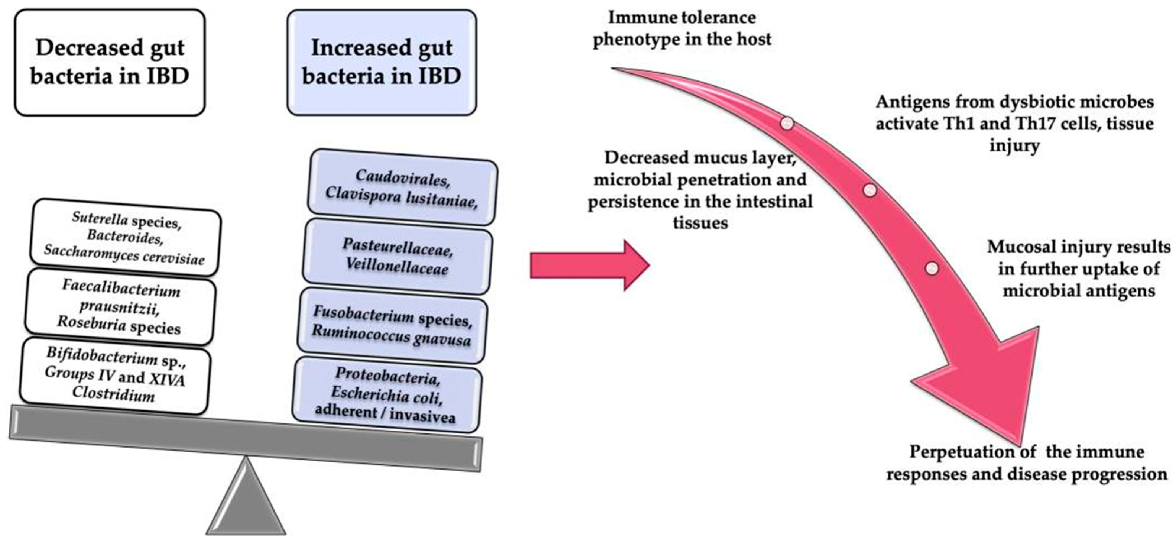
Publisher’s Note: MDPI stays neutral with regard to jurisdictional claims in published maps and institutional affiliations. |
© 2022 by the authors. Licensee MDPI, Basel, Switzerland. This article is an open access article distributed under the terms and conditions of the Creative Commons Attribution (CC BY) license (https://creativecommons.org/licenses/by/4.0/).