Article

Identification of a Phylogenetically Divergent Vanillate O-Demethylase from Rhodococcus ruber R1 Supporting Growth on Meta-Methoxylated Aromatic Acids

by
Raúl A. Donoso
1,2,
Ricardo Corbinaud
1,2,
Carla Gárate-Castro
1,
Sandra Galaz
1,2 and
Danilo Pérez-Pantoja
1,*
1
Programa Institucional de Fomento a la Investigación, Desarrollo e Innovación (PIDi), Universidad Tecnológica Metropolitana, Santiago 8940577, Chile
2
Center of Applied Ecology and Sustainability (CAPES), Santiago 6513677, Chile
*
Author to whom correspondence should be addressed.
Microorganisms 2023, 11(1), 78; https://doi.org/10.3390/microorganisms11010078
Submission received: 7 December 2022 / Revised: 22 December 2022 / Accepted: 23 December 2022 / Published: 27 December 2022

Abstract

:
Rieske-type two-component vanillate O-demethylases (VanODs) catalyze conversion of the lignin-derived monomer vanillate into protocatechuate in several bacterial species. Currently, VanODs have received attention because of the demand of effective lignin valorization technologies, since these enzymes own the potential to catalyze methoxy group demethylation of distinct lignin monomers. In this work, we identified a phylogenetically divergent VanOD from Rhodococcus ruber R1, only distantly related to previously described homologues and whose presence, along with a 3-hydroxybenzoate/gentisate pathway, correlated with the ability to grow on other meta-methoxylated aromatics, such as 3-methoxybenzoate and 5-methoxysalicylate. The complementation of catabolic abilities by heterologous expression in a host strain unable to grow on vanillate, and subsequent resting cell assays, suggest that the vanAB genes of R1 strain encode a proficient VanOD acting on different vanillate-like substrates; and also revealed that a methoxy group in the meta position and a carboxylic acid moiety in the aromatic ring are key for substrate recognition. Phylogenetic analysis of the oxygenase subunit of bacterial VanODs revealed three divergent groups constituted by homologues found in Proteobacteria (Type I), Actinobacteria (Type II), or Proteobacteria/Actinobacteria (Type III) in which the R1 VanOD is placed. These results suggest that VanOD from R1 strain, and its type III homologues, expand the range of methoxylated aromatics used as substrates by bacteria.

1. Introduction

The Rhodococcus genus is characterized by displaying a diverse range of metabolic capabilities, comprising degradation of short-chain, long-chain, and halogenated hydrocarbons; and numerous aromatic compounds, including substituted aromatics, heteroaromatics, and polycyclic aromatic hydrocarbons [1,2,3,4]. Accordingly, Rhodococcus spp. are considered as promising degraders of persistent contaminants, offering a multitude of novel enzymes able to perform challenging reactions [5,6,7].
Recently, we isolated a bacterial strain belonging to the Rhodococcus genus from a pulp mill wastewater treatment plant identified as Rhodococcus ruber strain R1, whose genome sequence consisted of one chromosome (~5.3 Mbp) and two plasmids (~179 and ~33 kbp), revealing an extensive catabolic potential [8]. Strain R1 has the ability to grow on various lignin-derived phenolic monomers, including p-coumarate and 4-hydroxybenzoate [8], which are typically catabolized via β-ketoadipate through the ring-cleavage intermediate protocatechuate (PCA) in Rhodococcus species [9,10]. Accordingly, we have currently confirmed that strain R1 is capable to grow on vanillate (VA), an additional lignin-derived product generated from the metabolism of ferulate and vanillin [10,11], as a sole carbon and energy source. The VA catabolism also proceeds via the PCA intermediate in several bacterial species, such as Acinetobacter, Comamonas, Corynebacterium, Pseudomonas, Streptomyces, and Rhodococcus as well, and is mediated by a two-component enzyme called vanillate O-demethylase (VanOD). This enzyme comprises a Rieske domain-containing oxygenase subunit encoded by vanA, and a reductase subunit that encompasses FMN, NADPH, and [2Fe-2S] cluster binding domains encoded by vanB, and providing electron equivalents to enable the enzymatic conversion [11,12,13,14,15,16,17,18,19]. It was shown that the VanOD encoded by vanAB genes from different bacteria were able to catalyze two types of reaction: methoxy group demethylation at the meta position of VA and analogs such as 3-methoxybenzoate (3-MB), veratrate, or syringate, with concomitant release of formaldehyde; or methyl group hydroxylation in m-toluate, 4-hydroxy-3-methylbenzoate, or 4-hydroxy-3,5-dimethylbenzoate; although, with the exception of veratrate and syringate, none of the analogs have been reported to support cell growth employing this enzyme [16,18,19,20,21]. Alternatively, it has been reported that distinct tetrahydrofolate (H4folate)-dependent O-demethylases, analogous to aromatic O-demethylases from anaerobic bacteria, are responsible for VA O-demethylation in Sphingobium sp. SYK-6 [22,23,24,25,26,27]. Remarkably, VanOD has gained increased interest since biocatalytic dealkylation of aryl methyl ethers have become attractive reactions for valorization of lignin-derived components towards fine chemicals and polymer precursors [28], highlighting the relevance of substrate-range studies for different VanOD enzymes.
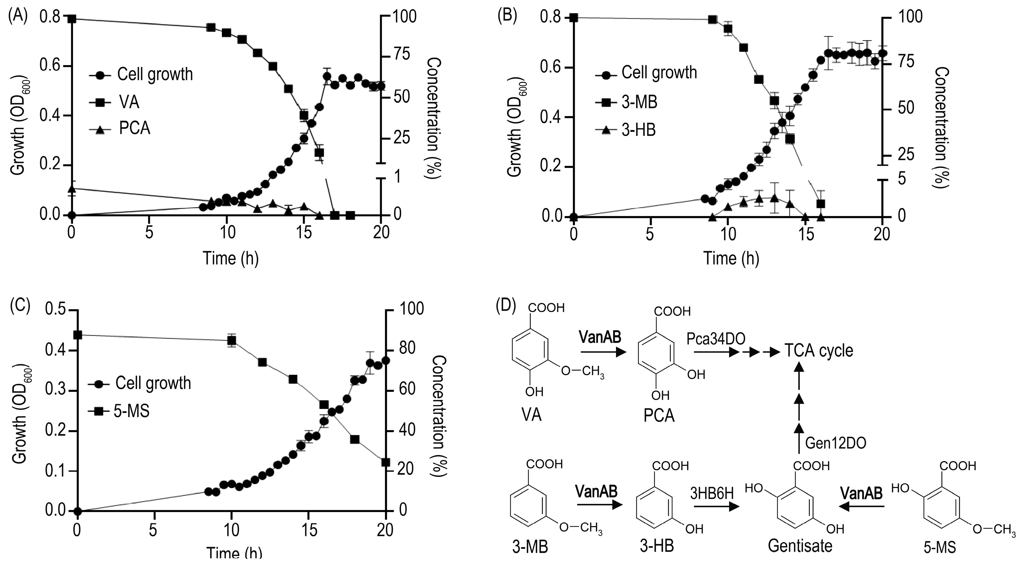
Surprisingly, the bacterium Rhodococcus ruber R1 only harbors VanAB proteins that are very distant from the well-known rhodococcal VanOD homologue described in Rhodococcus jostii RHA1 [11], showing only 36% amino acid identity for both subunits. Moreover, alternative tetrahydrofolate (H4folate)-dependent O-demethylases for VA, as those described in Sphingobium sp. SYK-6 [25], were not identified in its genome. On the other hand, PCA was transiently detected during growth of R1 strain in VA (Figure 1A), revealing that O-demethylation is occurring during its catabolism, and suggesting that a phylogenetically divergent VanOD encoded by putative vanAB genes (locus tag: E2561_01225 and E2561_01230) could be effectively responsible for VA catabolism in this bacterium. This interesting observation raises questions about possible differences among this divergent VanOD and the canonical one described in R. jostii RHA1 [11], its potential supporting role in the consumption of alternative substrates, and the distribution of close homologues in different Rhodococcus species.

2. Materials and Methods

2.1. Bacterial Strains, Plasmids, and Growth Conditions

Bacteria and plasmids used in this study are listed in Table 1. Rhodococcus species and Cupriavidus pinatubonensis JMP134 derivatives were grown at 30 °C in mineral salts medium previously reported [29], supplemented with 5 mM VA, PCA, 3-methoxybenzoate (3-MB), 5-methoxysalicylate (5-MS), gentisate, 3-hydroxybenzoate (3-HB), 3-methoxyphenylacetate, 3-hydroxyphenylacetate, 3-methoxysalicylate, 2,3-dihydroxybenzoate, syringate and homovanillate, as sole carbon and energy source. Escherichia coli Mach1 (Invitrogen, Carlsbad, CA, USA) was grown at 37 °C in Luria-Bertani (LB) medium. Growth was measured by optical density at 600 nm (OD600) using a spectrophotometer Spectroquant® Prove (Merck, Darmstadt, Germany), or a Synergy HTX Multi-Mode plate reader (BioTek, Winooski, VT, USA). At least three biological replicates were performed for each growth measurement.

2.2. Construction of a Plasmid Expressing vanAB Genes and Growth Tests of Strain Derivatives

To obtain pBS1-vanAB plasmid (Table 1), which contain the vanAB genes under the control of an L-arabinose-inducible promoter, a restriction enzymes approach was used. In brief, PCR product comprising vanAB genes (locus tags: E2561_01225–E2561_01230), was obtained using oligos FW_vanAB_R1_EcoRI (5′-TGACGAATTCGAAGGAACGACATGACCGATC-3′) and RV_vanAB_R1_XbaI (5′-GTACTCTAGATGTATCCGATGACCAGGCC-3′) including underlined restriction sites for EcoRI and XbaI enzymes. The amplified DNA fragment was purified and double digested to be ligated into EcoRI/XbaI restriction sites of pBS1 [30], forming pBS1-vanAB plasmid, that was electroporated into E. coli Mach1. Transformed cells were selected in LB medium supplemented with gentamycin 30 µg mL−1; and selected transformants were checked by PCR for proper insertion of the vanAB genes. The full-length gene construct was again checked by Sanger sequencing for errors, and the pBS1-derived plasmid was transferred into strain JMP134 for phenotypic analysis. To evaluate growth proficiency, derivatives of JMP134 strain carrying vanAB-expressing plasmid were grown overnight on LB medium, and then inoculated at 0.2% in cultures containing 5 mM VA, PCA, 3-MB, 3-HB, 5-MS or gentisate as the sole carbon and energy source. For expression of vanAB genes driven by the heterologous PBAD promoter, these derivatives were exposed to 1 mM L-arabinose in addition to growth substrates. The cultures were incubated in a 96-well microplate (Thermo Fisher Scientific, Rochester, NY, USA) at 30 °C and the OD600 was recorded in a Synergy HTX Multi-Mode plate reader (BioTek, Winooski, VT, USA).

2.3. Resting Cell Assays

Resting cells of strain JMP134 derivatives were grown on 5 mM VA or 3-HB plus 1 mM arabinose where appropriate. These cells were washed twice with 1 volume of phosphate buffer (14 g/L Na2HPO4·12H2O, 2 g/L KH2PO4), 5X concentrated, and subsequently incubated with 1 mM of each compound to be assayed where appropriate. Samples were obtained at different times, filtered (0.22 µm), and stored at −20 °C.

2.4. Analytical Methods

The presence of VA, PCA, 3-MB, 3-HB, 5-MS, gentisate, syringate, 3-O-methylgallate, 3-methoxysalicylate, homovanillate, isovanillate, 3-methoxyphenylacetate, 2-methoxybenzoate, 4-methoxybenzoate, and guaiacol was determined by high performance liquid chromatography (HPLC) using cell-free supernatants. Samples were injected into a JASCO liquid chromatograph LC-4000 (JASCO, Okhaloma City, OK, USA) equipped with a Kromasil 100-3.5-C18 4.6 mm diameter column. A gradient HPLC method was used, which consists of a mobile phase composed of solvent A (solution containing formic acid 0.1% v/v in water) and solvent B (methanol), at a flow rate of 0.8 mL min−1. The initial mobile phase composition was maintained at 25% solvent B for 11 min, changed linearly to 55% (11–13 min) and finally it was kept at 55% solvent B for 7 min (13–20 min). The column effluent was monitored at 295 nm for VA, PCA, 3-MB, 3-HB, and 2-methoxybenzoate; 275 nm for syringate, 3-O-methylgallate, 3-methoxyphenylacetate, homovanillate and guaiacol; 320 nm for 5-MS, 3-methoxysalicylate and gentisate; and 260 nm for isovanillate and 4-methoxybenzoate. Retention times for VA, 3-MB, 3-HB, 2-methoxybenzoate, syringate, 3-O-methylgallate, 3-methoxyphenylacetate, homovanillate, guaiacol, 5-MS, 3-methoxysalicylate, isovanillate, 4-methoxybenzoate, PCA, and gentisate were 9.3, 18.7, 10.9, 15.9, 10.5, 5.0, 17.7, 10.1, 15.8, 18.1, 16.6, 10.2, 18.1, 4.4, and 7.2 min, respectively.

2.5. Bioinformatic Tools

The vanAB gene sequences from different bacterial species were retrieved from non-redundant protein sequences database of GenBank (https://blast.ncbi.nlm.nih.gov/Blast.cgi, accessed on 8 September 2022) [31]. Only proteins displaying at least 60% amino acid identity with previously described VanA from Pseudomonas sp. HR199 [14], Rhodococcus jostii RHA1 [11], and Rhodococcus ruber R1 (this study) were recruited for analysis using BLAST software [32].
Evolutionary relationships were inferred by IQ-TREE web server tools (http://iqtree.cibiv.univie.ac.at/, accessed on 8 September 2022) proposed for estimate maximum-likelihood phylogenies [33] employing ModelFinder as model-selection method [34] and UFBoot2 for ultrafast bootstrap approximation [35] with the -m TEST, -bb 1000, and -alrt 1000 settings. Sequence alignments for phylogenetic reconstruction were calculated using MAFFT software online server (https://mafft.cbrc.jp/alignment/server/, accessed on 8 September 2022) employing Auto strategy (FFT-NS-1, FFT-NS-2, FFT-NS-i or L-INS-i; depending on data size) [36]. Edition and visualization of dendrograms was performed by the Interactive Tree of Life (iTOL) online tool (https://itol.embl.de/, accessed on 8 September 2022) [37].
Figure 1. Growth curves of Rhodococcus ruber R1 on VA and analogs as sole carbon and energy sources, and their channeling into catabolic routes after O-demethylation. Growth of R1 strain on 5 mM (A) VA, (B) 3-MB, and (C) 5-MS as sole carbon and energy sources. Detection of (A) PCA and (B) 3-HB indicates O-demethylation activity in degradation of VA and 3-MB, respectively. In (C) 5-MS-grown cells, its O-demethylation product (gentisate) was not detected, suggesting rapid and efficient gentisate turnover in R1 strain. (D) Catabolic pathways for VA, 3-MB, and 5-MS predicted from inspection of R. ruber R1 genome. The first step in the VA degradation pathway is O-demethylation into the dihydroxylated intermediate protocatechuate (PCA), employing the vanAB-encoded enzyme (VanAB) as reported in Rhodococcus jostii RHA1 [11]. On the other hand, the O-demethylation of 3-MB could generate 3-HB, that is converted by action of a flavoprotein hydroxylase (3HB6H) into gentisate; which in turn, is the putative product of 5-MS O-demethylation. The route for 3-HB turnover through gentisate has been previously described in R. jostii RHA1 [38,39]. VanAB, Vanillate O-demethylase; Pca34DO, protocatechuate 3,4-dioxygenase; 3HB6H, 3-hydroxybenzoate 6-hydroxylase; Gen12DO, gentisate 1,2-dioxygenase; TCA, tricarboxylic acid. Optical density at 600 nm (closed circles); and the concentrations (%) of VA, 3-MB or 5-MS (closed squares), and PCA or 3-HB (closed triangles) are depicted in the figure. Concentrations are represented as percentages of the initial substrate concentration. Three biological replicates were performed for growth measurements. Error bars indicate SEM.
Microorganisms 11 00078 g001

2.6. Chemicals

VA, PCA, 3-MB, 3-HB, 5-MS, gentisate, 2,3-dihydroxybenzoate, syringate, 3-O-methylgallate, 3-methoxysalicylate, homovanillate, isovanillate, 3-hydroxyphenylacetate, 3-methoxyphenylacetate, 2-methoxybenzoate, 4-methoxybenzoate, and guaiacol were purchased from Sigma-Aldrich (Steinheim, Germany). L(+)-arabinose was purchased from Merck (Darmstadt, Germany).

3. Results and Discussion

3.1. Heterologous Expression and Resting Cell Assays Suggest a Key Role of VanOD from Rhodococcus ruber R1 in meta-Methoxylated Aromatic Acids Degradation

Our original observation about the ability of R. ruber R1 to grow on VA as a sole carbon and energy source (Figure 1A), in the absence of a canonical VanOD as the one described in R. jostii RHA1 [11], prompted us to analyze its growth profile on other meta-methoxylated aromatic substrates, such as 3-methoxybenzoate (3-MB) and 5-methoxysalicylate (5-MS). Interestingly, 3-MB and 5-MS also supported cell proliferation (see Figure 1B,C for a detailed growth curve of R1 cells), suggesting O-demethylation of these substrates into 3-hydroxybenzoate (3-HB) and gentisate respectively, as depicted in Figure 1D. For 3-MB consumption, the O-demethylation activity was additionally suggested by the transient accumulation of 3-HB (Figure 1B). In the case of 5-MS growth, the absence of gentisate in the supernatant of R1 cell cultures would be correlated with a lower rate of substrate consumption, in comparison to VA and 3-MB consumption curves, as shown by Figure 1A–C; avoiding the accumulation of intermediates. Further catabolism of 3-HB and gentisate is correlated with the presence of genes encoding 3-hydroxybenzoate 6-hydroxylase (locus tag: E2561_07550) and gentisate 1,2-dioxygenase (locus tag: E2561_07565) enzymes, comprising the catabolic route for 3-HB via gentisate in strain R1, which are closely related to the enzymes described for R. jostii RHA1 [38,39]. The presence of O-demethylation activities for VA, 3MB, and 5-MS in R. ruber R1 raise the possibility that VanOD encoded in the genome of this strain would be responsible for all of them.
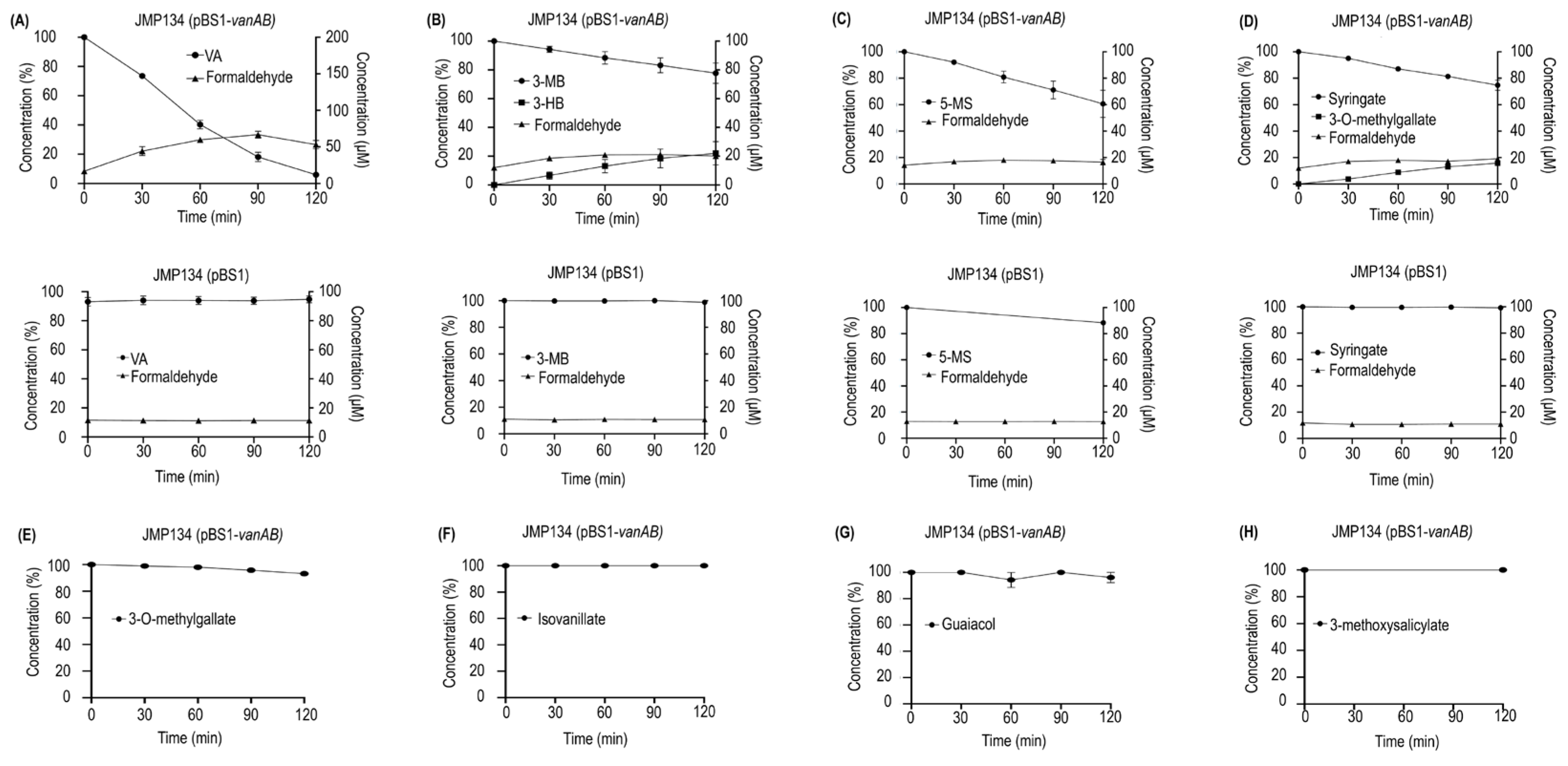
In order to gain comprehension about the whole function of the divergent VanOD from R. ruber R1, a plasmid construct containing the vanAB genes of this strain was introduced into C. pinatubonensis JMP134, a well-known aromatics-degrader bacterium unable to grow on VA, 3-MB, and 5-MS, but that harbors PCA, 3-HB, and gentisate degradation routes (Figure 2B,D,F) [40], allowing complementation of the catabolic abilities. The expression of the vanAB genes was controlled by the L-arabinose-inducible PBAD promoter, which was chosen since L-arabinose is non-toxic and is not a carbon source for C. pinatubonensis JMP134, permitting reliable growth tests in this strain [30,41]. Remarkably, the presence of vanAB genes was sufficient to allow L-arabinose-depending growth on VA, 3-MB, and 5-MS of JMP134 strain (Figure 2A,C,E), strongly suggesting that VanOD of R1 strain has O-demethylation activity toward the three meta-methoxylated aromatic substrates. It should be noted that, in the absence of L-arabinose as an inducer, no growth was observed (data not shown), and that the presence of the empty pBS1 vector has no effect on cell proliferation of JMP134 strain on these substrates (Figure 2A,C,E). Moreover, resting cell assays of JMP134 (pBS1-vanAB) cells previously grown on VA showed a sharp decrease in the concentration of VA, and a slower consumption rate for 3-MB and 5-MS, also detecting the occurrence of 3-HB in 3-MB-incubated cells (Figure 3A–C); which provides further support for VA/3-MB/5-MS O-demethylase activity encoded by R1 vanAB genes. This inference was additionally supported by detecting a small accumulation of formaldehyde in parallel to substrates consumption (Figure 3A–C), which is the by-product of O-demethylation by VanOD [26].
The presence of functional groups in the potential substrates of the divergent VanOD of R1 strain was the next interesting issue to be determined. Nishimura et al. [19] reported that a carboxylic acid on the benzene ring in conjunction with a hydroxyl group in para-orientation, as occurs in VA or syringate molecules, is required for efficient methoxy oxidation in meta-position of the VanAB substrates in Streptomyces sp. NL15-2K, which is homologous to VanAB from RHA1 (70% aa identity for oxygenase subunit). Recently, the properties of VanOD from Pseudomonas sp. HR199 were extensively examined, confirming that the presence of a carboxylic acid moiety is essential, and that catalysis occurs selectively at the meta-position relative to the –COOH group in the aromatic ring, although exposing specific differences in substrate recognition in comparison to VanAB from Streptomyces sp. NL15-2K [19,28]. To confirm that previous observations also apply to VanOD from R. ruber R1, resting cell assays considering additional potential substrates were performed in C. pinatubonensis JMP134 carrying the plasmid that contains the vanAB genes from R1 strain. The doubly meta-methoxylated syringate that carries a –OH group in the para-position relative to the carboxylic acid was a proper substrate for VanOD of the R1 strain; being 3-O-methylgallate, the partially demethoxylated analog, identified as the only conversion product of its catalysis (Figure 3D). Meanwhile, 3-O-methylgallate apparently was not recognized as a substrate by the VanOD of R1 strain (Figure 3E), similar to what was described for the VanAB from HR199 strain [28], but unlike VanAB of NL15-2K strain which is able to generate a mixture of 3-O-methylgallate and gallate, the fully demethoxylated analog, in the presence of syringate [19]. These results were supported by introduction of vanAB genes of R1 strain into Pseudomonas putida KT2440, which is unable to grow on syringate or 3-O-methylgallate but contains a functional gallate degradation pathway [42,43], being the product of two consecutive O-demethylations over syringate comprising 3-O-methylgallate as intermediate, as mentioned before. The P. putida KT2440 (pBS1-vanAB) strain was unable to grow on syringate as a sole carbon and energy source (data not shown), suggesting that inefficient O-demethylation of 3-O-methylgallate by VanOD from R1 could be the reason for this phenotype.
Additional compounds including differences in the key positions of functional groups for the recognition of substrates by this enzyme, such as 2-methoxybenzoate and 4-methoxybenzoate (methoxy group in ortho- or para-position in relation to –COOH group), isovanillate (methoxy group in para-position in relation to –COOH group with an adjacent –OH group in meta-position), homovanillate (VA analog with a –CH2COOH replacing –COOH group), 3-methoxyphenylacetate (3 MB analog with a –CH2COOH replacing –COOH group), 3-methoxysalicylate (methoxy group in meta-position in relation to –COOH group with an adjacent –OH group in ortho-position), and guaiacol (methoxy group with an adjacent –OH group but lacking –COOH group) were not degraded by resting cells of C. pinatubonensis JMP134 carrying the vanAB genes of R1 strain (see Figure 3F–H, for isovanillate, guaiacol, and 3-methoxysalicylate as representative examples).
In summary, the results of growth tests and resting cell assays suggest that divergent VanOD from strain R1 not only recognizes VA, but also 3-MB, 5-MS, and syringate as proper substrates to a lesser extent (Figure 3A–D). According to this, vanAB genes could be key not only on VA degradation, but also on the potential catabolism of 3-MB and 5-MS in additional Rhodococcus species that carry this divergent version of VanOD.

3.2. Rhodococcus Strains Carrying VanAB Homologues Closely Related to VanOD of R. ruber R1 Strain Are Able to Grow on VA, 3-MB, and 5-MS as a Sole Carbon and Energy Sources

To explore the phenotypic differences of selected Rhodococcus species carrying divergent VanOD related to VanAB from R. jostii RHA1 strain or VanAB from R. ruber R1 strain, we analyzed their growth profile on several meta-methoxylated substrates structurally related to VA, such as 3-MB, 5-MS, syringate, 3-methoxysalicylate, 3-methoxyphenylacetate, and homovanillate, in addition to some of the putative products of O-demethylation such as PCA, 3-HB, gentisate, 2,3-dihydroxybenzoate and 3-hydroxyphenylacetate (Figure 4).
Results showed that Rhodococcus strains harboring vanAB-like genes similar to R. ruber R1 as R. ruber DSM 43338T, R. ruber Chol-4 and R. pyridinivorans JCM 10940T [8,44,45], and vanAB genes comparable to R. jostii RHA1 as R. aetherivorans BCP1 [46,47] were able to grow on VA and its O-demethylation product, PCA (Figure 4), suggesting that all these strains contain proficient VanOD-encoded genes and a functional PCA pathway (Figure 1D). We also included in our growth profile assays marine-isolated Rhodococcus strains MS13 and H-CA8f as control [48,49], since they apparently do not harbor VanOD-encoded genes, even though they carry the classical PCA pathway [50], as revealed by BLAST searches and confirmed by growth on PCA as a sole carbon and energy source (Figure 4). Accordingly, both Rhodococcus strains of marine origin were unable to grow on VA (Figure 4), confirming the previous bioinformatic survey that revealed the absence of VanOD encoded genes, and suggesting that VA degradation activity could be linked to Rhodococcus species found mainly in soil or freshwater environments, probably correlated to lignin depolymerization [51,52,53].
Figure 4. Growth on meta-methoxylated aromatic acids and their O-demethylated products of different Rhodococcus species. Strains belonging to Rhodococcus genus were grown in mineral salt medium with 5 mM of several meta-methoxylated aromatic acids (methoxylated substrates; left side) and its O-demethylated products (right side) as sole carbon and energy sources. Shading indicates optical density (OD) at 600 nm after 40 h (average of three biological replicates). It is worth mentioning that O-demethylated products related to syringate (3-O-methylgallate and gallate) and homovanillate (homoprotocatechuate) were rapidly oxidized in solution yielding an intense dark brown color on the medium, precluding determination of optical density, and consequently were excluded of the study.
Microorganisms 11 00078 g004
Remarkably, R. ruber strains DSM 43338T and Chol-4 that harbor vanAB genes close related to R1 homologues were also able to use 3-MB and 5-MS, and its putative demethylation products, 3-HB and gentisate, respectively, as sole carbon and energy sources (Figure 4), suggesting that their VanOD enzymes are able to act on both meta-methoxylated substrates, and that they harbor the corresponding putative downstream pathways (Figure 1D). Notably, R. pyridinivorans JCM 10940T containing R1-like vanAB genes was unable to grow on 3-MB and 5-MS, and was also unable to grow on 3-HB and gentisate (Figure 4), which is in accordance with the absence of genes encoding 3-HB 6-hydroxylase and gentisate 1,2-dioxygenase enzymes. This suggests that lack of a functional 3-HB/gentisate pathway might impair its growth on such meta-methoxylated substrates, regardless of the presence of a proficient VanOD. Conversely, despite strains RHA1 and BCP1 harbor RHA1-like vanAB genes and contain a functional 3-HB/gentisate pathway, both were unable to grow on 3-MB and 5-MS (Figure 4), suggesting that the VanOD harbored by these Rhodococcus strains does not support the O-demethylation activities toward these meta-methoxylated substrates. These results could indicate that the in vivo range of substrate acceptance for R1-like and RHA1-like VanOD enzymes is not the same. Finally, all Rhodococcus strains tested were unable to use the remaining VA analogs assayed as sole carbon and energy sources, including those where –COOH group is replaced by –CH2COOH as 3-methoxyphenylacetate or homovanillate (Figure 4).

3.3. Two-Component Rieske-Type VanOD of Rhodococcus Species Are Allocated in Two Divergent Phylogenetic Clades

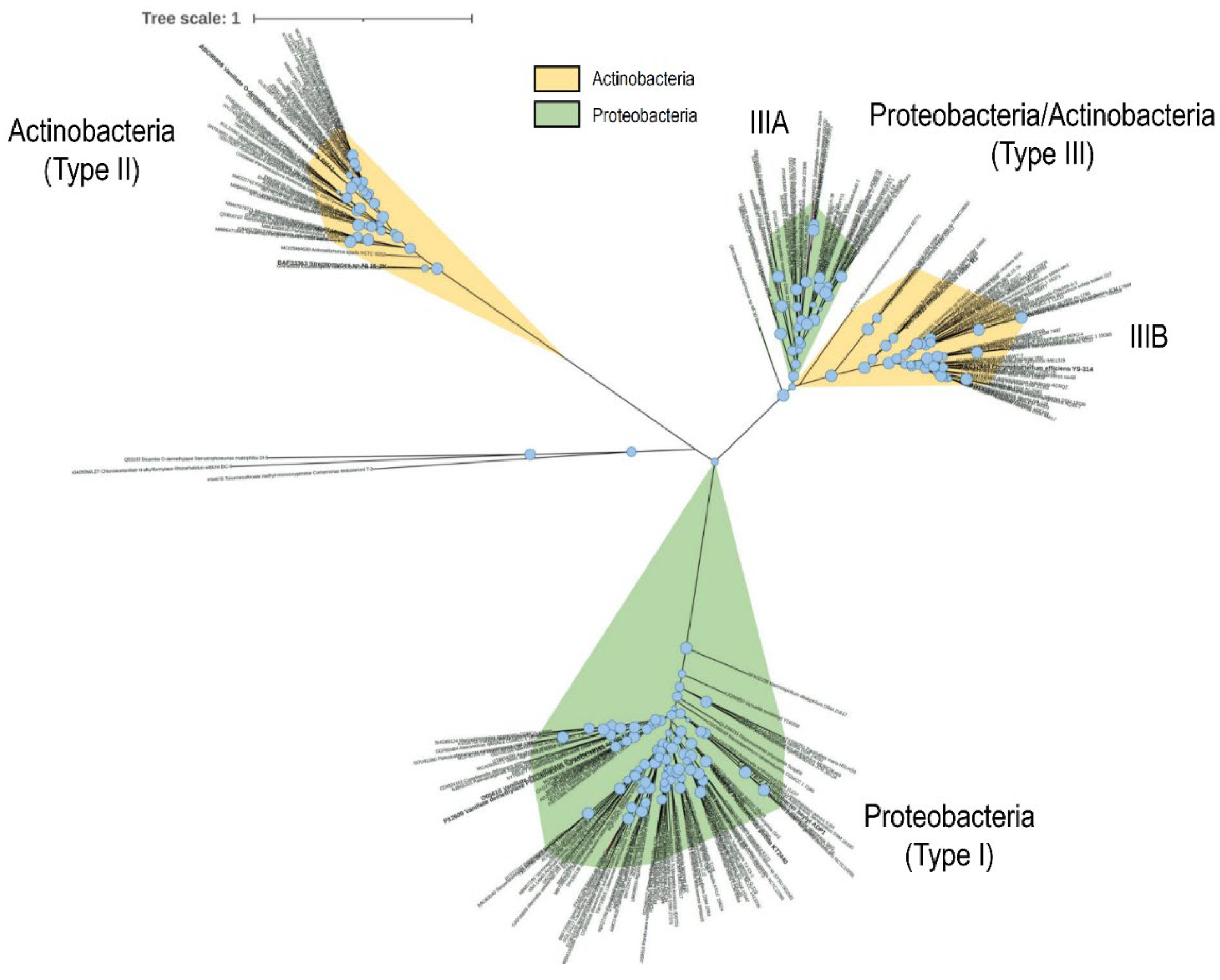
The existence of at least two distinct VanOD types in Rhodococcus species prompted us to evaluate the distribution of each kind in this genus and other actinobacterial and proteobacterial genomes. For that purpose, we chose as gene marker the VanA product, coding the oxygenase component of the enzyme, from R. jostii RHA1 [11], R. ruber R1, and also Pseudomonas sp. HR199 as bona fide representative of proteobacterial VanOD [14]. Then, we conducted a search in the non-redundant protein sequences database of GenBank as of September 2022, selecting VanA from bacterial species displaying at least 60% amino acid identity in order to establish phylogenetic relationships. As a result of a high number of redundant VanA sequences, we selected one representative VanA homologue belonging to each genus identified. The resulting VanA phylogenetic tree showed three clearly divergent groups, in which a precise partition was perceived between a proteobacterial clade (called type I), including the well-known VanA homologues from Acinetobacter baylyi ADP1, Pseudomonas sp. HR199, and Comamonas testosteroni BR6020 [14,16,18]; and an actinobacterial clade (type II), including the aforementioned VanA homologues from R. jostii RHA1 and Streptomyces sp. NL15-2K [11,19] (Figure 5). Interestingly, a distinct third clade was detected (type III), internally partitioned in two subclades including homologues from Proteobacteria (type IIIA) and Actinobacteria (type IIIB) (Figure 5). The last one included VanA from R. ruber R1, revealing that the VanOD reported in this work is the first member of this clade whose functionality and substrate range is analyzed in detail. It should be noted that a closer inspection of each clade reveals a predominance of β- and γ-proteobacterial VanA homologues among members of the type I clade, meanwhile only homologues from Actinobacteria representatives were found in type II (data not shown). Conversely, a prevalence of VanA homologues from α-proteobacteria subclass representatives (IIIA) in conjunction with Actinobacteria (IIIB) were observed in type III (data not shown).
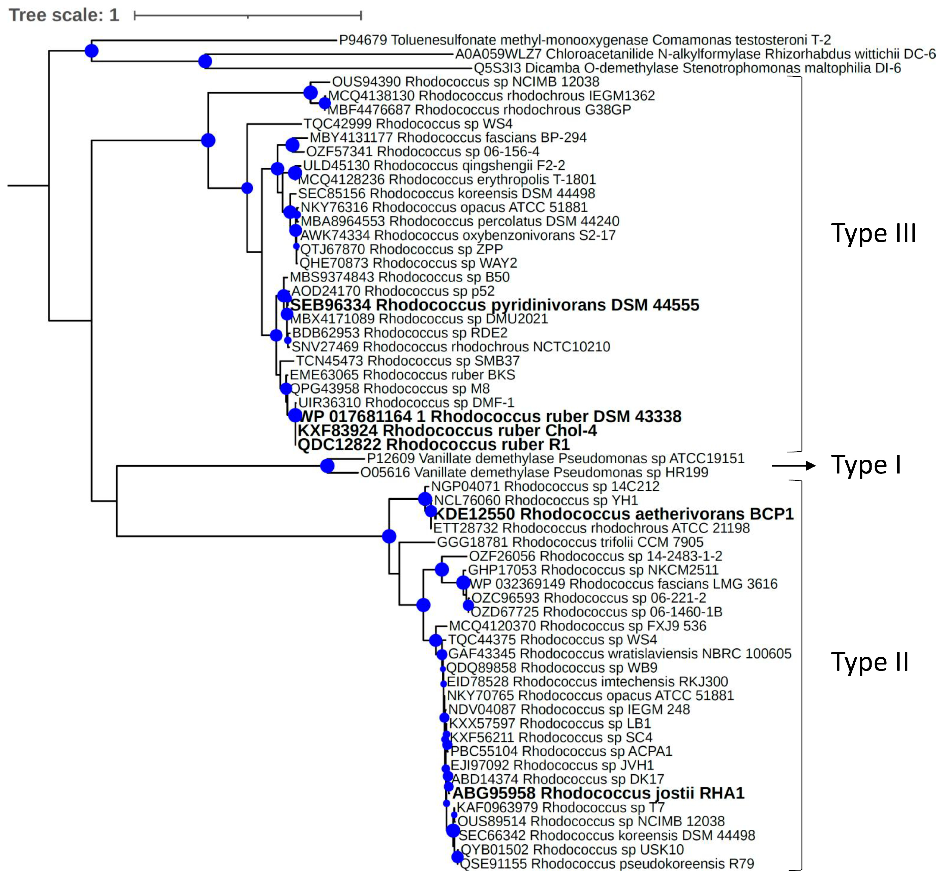
In order to gain a deeper understanding of the phylogenetic relationships between VanOD enzymes from Rhodococcus species, an additional phylogenetic tree was constructed including VanA homologues from a broader range of Rhodococcus species representatives (Figure 6). Similar to what was previously observed, it was shown that VanA from Rhodococcus species are grouped either in conjunction with VanA from RHA1 strain (type II clade) or VanA from R1 strain (type III clade). The number of VanA homologues from Rhodococcus species grouped in each clade was roughly similar, suggesting that both types of VanOD are numerically relevant in this actinobacterial genus. No VanA homologue of the Rhodococcus species considered in this study was located out of these clades.

4. Conclusions

Given the current interest in O-demethylation reactions for lignin conversion into renewable chemicals [54], this study aimed to shed light on Rhodococcus enzymes acting on meta-methoxylated aromatic compounds such as VA, one of the most prominent lignin-derived phenolics. This work revealed that Rhodococcus genus harbors at least two divergent types of VanOD-encoding genes represented by vanAB from Rhodococcus jostii RHA1 (Type II) and vanAB from Rhodococcus ruber R1 (Type III). Most interestingly, the VanOD from R1 strain is responsible for catabolism of additional meta-methoxylated phenolics such as 3-MB and 5-MS, as inferred from growth tests and resting cell assays of a heterologous strain expressing R1 vanAB genes, and from the substrate utilization pattern of Rhodococcus strains harboring close homologues of this enzyme. This expanded substrate specificity would be advantageous for metabolic engineering endeavors focused on bioconversion process toward renewable chemicals based on microbial demethylation of lignin monomers.
Table 1. Bacterial strains and plasmids used in this study.
Strain or PlasmidRelevant Phenotype and/or GenotypeReference or Source
Rhodococcus strains
R. aetherivorans BCP1VA+, 3-MB-, 5-MS-[47]
R. jostii RHA1VA+, 3-MB-, 5-MS-[46]
R. ruber R1VA+, 3-MB+, 5-MS+[8]
R. ruber Chol-4VA+, 3-MB+, 5-MS+[45]
R. ruber DSM 43338 TVA+, 3-MB+, 5-MS+DSMZ a
R. pyridinivorans JCM 10940 TVA+, 3-MB-, 5-MS-[44]
Rhodococcus sp. H-CA8fVA-, 3-MB, 5-MS-[48]
Rhodococcus sp. MS13VA-, 3-MB-, 5-MS-[49]
Other strains
E. coli Mach1∆recA1398 endA1 tonA Φ80∆lacM15 ∆lacX74 hsdR (rK- mK+)Invitrogen, Carlsbad, CA, USA
C. pinatubonensis JMP134
PCA+, Gentisate+, 3-HB+, VA-, 3-MB-, 5-MS-[40]
Plasmids
pBS1Broad host range vector, araC-PBAD, GmR[55]
pBS1-vanABpBS1 derivative expressing vanAB genes, GmRThis study
a DSMZ, Deutsche Sammlung von Mikroorganismen und Zellkulturen GmbH (Braunschweig, Germany). T: Type strain.

Author Contributions

Conceptualization, D.P.-P.; methodology, R.C., C.G.-C. and S.G.; software, R.A.D.; formal analysis, R.A.D. and D.P.-P.; writing—original draft preparation, R.D; writing—review and editing, D.P.-P.; supervision, D.P.-P.; funding acquisition, R.A.D. and D.P.-P.; project administration, R.A.D. and D.P.-P. All authors have read and agreed to the published version of the manuscript.

Funding

This work was funded by FONDECYT 1201741, FONDECYT 11220354 and ANID PIA/BASAL FB0002 grants of Chilean government, and the LE19-05 project supported by the Fund of Scientific and Technological Equipment, year 2019, Universidad Tecnológica Metropolitana.

Data Availability Statement

The data supporting the conclusions of this work are included within the manuscript and there were no large datasets generated or analyzed during the current study.

Acknowledgments

The authors acknowledge Beatriz Camara (Universidad Técnica Federico Santa María, Valparaíso, Chile) and Patricia Aguila-Torres (Universidad Austral de Chile, Puerto Montt, Chile) for providing the Rhodococcus sp. H-CA8f and Rhodococcus sp. MS13 strains respectively. Authors are indebted to Thomas Ledger (Universidad Adolfo Ibañez, Santiago, Chile) for careful reading of the manuscript.

Conflicts of Interest

The authors declare no potential conflict of interest.

References

  1. Warhurst, A.M.; Fewson, C.A. Biotransformations catalyzed by the genus Rhodococcus. Crit. Rev. Biotechnol. 1994, 14, 29–73. [Google Scholar] [CrossRef] [PubMed]
  2. Martínková, L.; Uhnáková, B.; Pátek, M.; Nesvera, J.; Kren, V. Biodegradation potential of the genus Rhodococcus. Environ. Int. 2009, 35, 162–177. [Google Scholar] [CrossRef] [PubMed]
  3. Yam, K.C.; Okamoto, S.; Roberts, J.N.; Eltis, L.D. Adventures in Rhodococcus—From steroids to explosives. Can. J. Microbiol. 2011, 57, 155–168. [Google Scholar] [CrossRef] [PubMed]
  4. Zampolli, J.; Zeaiter, Z.; Di Canito, A.; Di Gennaro, P. Genome analysis and -omics approaches provide new insights into the biodegradation potential of Rhodococcus. Appl. Microbiol. Biotechnol. 2019, 103, 1069–1080. [Google Scholar] [CrossRef] [PubMed]
  5. Busch, H.; Hagedoorn, P.L.; Hanefeld, U. Rhodococcus as a versatile biocatalyst in organic synthesis. Int. J. Mol. Sci. 2019, 20, 4787. [Google Scholar] [CrossRef] [Green Version]
  6. Ivshina, I.; Bazhutin, G.; Tyumina, E. Rhodococcus strains as a good biotool for neutralizing pharmaceutical pollutants and obtaining therapeutically valuable products: Through the past into the future. Front. Microbiol. 2022, 13, 967127. [Google Scholar] [CrossRef]
  7. Nazari, M.T.; Simon, V.; Machado, B.S.; Crestani, L.; Marchezi, G.; Concolato, G.; Ferrari, V.; Colla, L.M.; Piccin, J.S. Rhodococcus: A promising genus of actinomycetes for the bioremediation of organic and inorganic contaminants. J. Environ. Manag. 2022, 323, 116220. [Google Scholar] [CrossRef]
  8. Farkas, C.; Donoso, R.A.; Melis-Arcos, F.; Gárate-Castro, C.; Pérez-Pantoja, D. Complete Genome Sequence of Rhodococcus ruber R1, a Novel Strain Showing a Broad Catabolic Potential toward Lignin-Derived Aromatics. Microbiol. Resour. Announc. 2020, 9, e00905.19. [Google Scholar] [CrossRef] [Green Version]
  9. Jadan, A.P.; van Berkel, W.J.; Golovleva, L.A.; Golovlev, E.L. Purification and properties of p-hydroxybenzoate hydroxylases from Rhodococcus strains. Biochemistry 2001, 66, 898–903. [Google Scholar]
  10. Otani, H.; Lee, Y.E.; Casabon, I.; Eltis, L.D. Characterization of p-hydroxycinnamate catabolism in a soil Actinobacterium. J. Bacteriol. 2014, 196, 4293–4303. [Google Scholar] [CrossRef] [Green Version]
  11. Chen, H.P.; Chow, M.; Liu, C.C.; Lau, A.; Liu, J.; Eltis, L.D. Vanillin catabolism in Rhodococcus jostii RHA1. Appl. Environ. Microbiol. 2012, 78, 586–588. [Google Scholar] [CrossRef] [PubMed]
  12. Correll, C.C.; Batie, C.J.; Ballou, D.P.; Ludwig, M.L. Phthalate dioxygenase reductase: A modular structure for electron transfer from pyridine nucleotides to [2Fe-2S]. Science 1992, 258, 1604–1610. [Google Scholar] [CrossRef] [PubMed]
  13. Dehmel, U.; Engesser, K.H.; Timmis, K.N.; Dwyer, D.F. Cloning, nucleotide sequence, and expression of the gene encoding a novel dioxygenase involved in metabolism of carboxydiphenyl ethers in Pseudomonas pseudoalcaligenes POB310. Arch. Microbiol. 1995, 163, 35–41. [Google Scholar] [CrossRef] [PubMed]
  14. Priefert, H.; Rabenhorst, J.; Steinbüchel, A. Molecular characterization of genes of Pseudomonas sp. strain HR199 involved in bioconversion of vanillin to protocatechuate. J. Bacteriol. 1997, 179, 2595–2607. [Google Scholar] [CrossRef] [PubMed] [Green Version]
  15. Venturi, V.; Zennaro, F.; Degrassi, G.; Okeke, B.C.; Bruschi, C.V. Genetics of ferulic acid bioconversion to protocatechuic acid in plant-growth-promoting Pseudomonas putida WCS358. Microbiology 1998, 144, 965–973. [Google Scholar] [CrossRef] [Green Version]
  16. Morawski, B.; Segura, A.; Ornston, L.N. Substrate range and genetic analysis of Acinetobacter vanillate demethylase. J. Bacteriol. 2000, 182, 1383–1389. [Google Scholar] [CrossRef] [Green Version]
  17. Merkens, H.; Beckers, G.; Wirtz, A.; Burkovski, A. Vanillate metabolism in Corynebacterium glutamicum. Curr. Microbiol. 2005, 51, 59–65. [Google Scholar] [CrossRef]
  18. Providenti, M.A.; O’Brien, J.M.; Ruff, J.; Cook, A.M.; Lambert, I.B. Metabolism of isovanillate, vanillate, and veratrate by Comamonas testosteroni strain BR6020. J. Bacteriol. 2006, 188, 3862–3869. [Google Scholar] [CrossRef] [Green Version]
  19. Nishimura, M.; Nishimura, Y.; Abe, C.; Kohhata, M. Expression and substrate range of Streptomyces vanillate demethylase. Biol. Pharm. Bull. 2014, 37, 1564–1568. [Google Scholar] [CrossRef] [Green Version]
  20. Notonier, S.; Werner, A.Z.; Kuatsjah, E.; Dumalo, L.; Abraham, P.E.; Hatmaker, E.A.; Hoyt, C.B.; Amore, A.; Ramirez, K.J.; Woodworth, S.P.; et al. Metabolism of syringyl lignin-derived compounds in Pseudomonas putida enables convergent production of 2-pyrone-4,6-dicarboxylic acid. Metab. Eng. 2021, 65, 111–122. [Google Scholar] [CrossRef] [PubMed]
  21. Mueller, J.; Willett, H.; Feist, A.M.; Niu, W. Engineering Pseudomonas putida for improved utilization of syringyl aromatics. Biotechnol. Bioeng. 2022, 119, 2541–2550. [Google Scholar] [CrossRef] [PubMed]
  22. Berman, M.H.; Frazer, A.C. Importance of tetrahydrofolate and ATP in the anaerobic O-demethylation reaction for phenylmethylethers. Appl. Environ. Microbiol. 1992, 58, 925–931. [Google Scholar] [CrossRef] [PubMed] [Green Version]
  23. Kaufmann, F.; Wohlfarth, G.; Diekert, G. O-demethylase from Acetobacterium dehalogenans—Substrate specificity and function of the participating proteins. Eur. J. Biochem. 1998, 253, 706–711. [Google Scholar] [CrossRef] [PubMed]
  24. Naidu, D.; Ragsdale, S.W. Characterization of a three-component vanillate O-demethylase from Moorella thermoacetica. J. Bacteriol. 2001, 183, 3276–3281. [Google Scholar] [CrossRef] [PubMed] [Green Version]
  25. Abe, T.; Masai, E.; Miyauchi, K.; Katayama, Y.; Fukuda, M. A tetrahydrofolate-dependent O-demethylase, LigM, is crucial for catabolism of vanillate and syringate in Sphingomonas paucimobilis SYK-6. J. Bacteriol. 2005, 187, 2030–2037. [Google Scholar] [CrossRef] [Green Version]
  26. Hibi, M.; Sonoki, T.; Mori, H. Functional coupling between vanillate-O-demethylase and formaldehyde detoxification pathway. FEMS Microbiol. Lett. 2005, 253, 237–242. [Google Scholar] [CrossRef] [Green Version]
  27. Kohler, A.C.; Mills, M.J.L.; Adams, P.D.; Simmons, B.A.; Sale, K.L. Structure of aryl O-demethylase offers molecular insight into a catalytic tyrosine-dependent mechanism. Proc. Natl. Acad. Sci. USA 2017, 114, E3205–E3214. [Google Scholar] [CrossRef] [Green Version]
  28. Lanfranchi, E.; Trajković, M.; Barta, K.; de Vries, J.G.; Janssen, D.B. Exploring the Selective Demethylation of Aryl Methyl Ethers with a Pseudomonas Rieske Monooxygenase. ChemBioChem 2019, 20, 118–125. [Google Scholar] [CrossRef] [Green Version]
  29. Donoso, R.A.; González-Toro, F.; Pérez-Pantoja, D. Widespread distribution of hmf genes in Proteobacteria reveals key enzymes for 5-hydroxymethylfurfural conversion. Comput. Struct. Biotechnol. J. 2021, 19, 2160–2169. [Google Scholar] [CrossRef]
  30. Donoso, R.A.; Ruiz, D.; Gárate-Castro, C.; Villegas, P.; González-Pastor, J.E.; de Lorenzo, V.; González, B.; Pérez-Pantoja, D. Identification of a self-sufficient cytochrome P450 monooxygenase from Cupriavidus pinatubonensis JMP134 involved in 2-hydroxyphenylacetic acid catabolism, via homogentisate pathway. Microb. Biotechnol. 2021, 14, 1944–1960. [Google Scholar] [CrossRef]
  31. NCBI. Resource Coordinators. Database resources of the National Center for Biotechnology Information. Nucleic Acids Res. 2018, 46, D8–D13. [Google Scholar] [CrossRef] [PubMed]
  32. Johnson, M.; Zaretskaya, I.; Raytselis, Y.; Merezhuk, Y.; McGinnis, S.; Madden, T.L. NCBI BLAST: A better web interface. Nucleic Acids Res. 2008, 36, W5–W9. [Google Scholar] [CrossRef] [PubMed]
  33. Nguyen, L.T.; Schmidt, H.A.; von Haeseler, A.; Minh, B.Q. IQ-TREE: A fast and effective stochastic algorithm for estimating maximum-likelihood phylogenies. Mol. Biol. Evol. 2015, 32, 268–274. [Google Scholar] [CrossRef] [PubMed]
  34. Kalyaanamoorthy, S.; Minh, B.Q.; Wong, T.K.F.; von Haeseler, A.; Jermiin, L.S. ModelFinder: Fast model selection for accurate phylogenetic estimates. Nat. Methods 2017, 14, 587–589. [Google Scholar] [CrossRef] [PubMed] [Green Version]
  35. Hoang, D.T.; Chernomor, O.; von Haeseler, A.; Minh, B.Q.; Vinh, L.S. UFBoot2: Improving the Ultrafast Bootstrap Approximation. Mol. Biol. Evol. 2018, 35, 518–522. [Google Scholar] [CrossRef] [PubMed]
  36. Katoh, K.; Rozewicki, J.; Yamada, K.D. MAFFT online service: Multiple sequence alignment, interactive sequence choice and visualization. Brief. Bioinform. 2019, 20, 1160–1166. [Google Scholar] [CrossRef] [PubMed] [Green Version]
  37. Letunic, I.; Bork, P. Interactive Tree of Life (iTOL) v4: Recent updates and new developments. Nucleic Acids Res. 2019, 47, W256–W259. [Google Scholar] [CrossRef] [Green Version]
  38. Montersino, S.; van Berkel, W.J. Functional annotation and characterization of 3-hydroxybenzoate 6-hydroxylase from Rhodococcus jostii RHA1. Biochim. Biophys. Acta 2012, 1824, 433–442. [Google Scholar] [CrossRef]
  39. Sucharitakul, J.; Tongsook, C.; Pakotiprapha, D.; van Berkel, W.J.; Chaiyen, P. The reaction kinetics of 3-hydroxybenzoate 6-hydroxylase from Rhodococcus jostii RHA1 provide an understanding of the para-hydroxylation enzyme catalytic cycle. J. Biol. Chem. 2013, 288, 35210–35221. [Google Scholar] [CrossRef] [Green Version]
  40. Pérez-Pantoja, D.; De la Iglesia, R.; Pieper, D.H.; González, B. Metabolic reconstruction of aromatic compounds degradation from the genome of the amazing pollutant-degrading bacterium Cupriavidus necator JMP134. FEMS Microbiol. Rev. 2008, 32, 736–794. [Google Scholar] [CrossRef] [Green Version]
  41. Pérez-Pantoja, D.; Donoso, R.A.; Sánchez, M.A.; González, B. Genuine genetic redundancy in maleylacetate-reductase-encoding genes involved in degradation of haloaromatic compounds by Cupriavidus necator JMP134. Microbiology 2009, 155, 3641–3651. [Google Scholar] [CrossRef] [PubMed]
  42. Nogales, J.; Canales, A.; Jiménez-Barbero, J.; García, J.L.; Díaz, E. Molecular characterization of the gallate dioxygenase from Pseudomonas putida KT2440. The prototype of a new subgroup of extradiol dioxygenases. J. Biol. Chem. 2005, 280, 35382–35390. [Google Scholar] [CrossRef] [PubMed] [Green Version]
  43. Nogales, J.; Canales, A.; Jiménez-Barbero, J.; Serra, B.; Pingarrón, J.M.; García, J.L.; Díaz, E. Unravelling the gallic acid degradation pathway in bacteria: The gal cluster from Pseudomonas putida. Mol. Microbiol. 2011, 79, 359–374. [Google Scholar] [CrossRef] [PubMed]
  44. Yoon, J.H.; Kang, S.S.; Cho, Y.G.; Lee, S.T.; Kho, Y.H.; Kim, C.J.; Park, Y.H. Rhodococcus pyridinivorans sp. nov., a pyridine-degrading bacterium. Int. J. Syst. Evol. Microbiol. 2000, 50, 2173–2180. [Google Scholar] [CrossRef] [PubMed]
  45. Heras, L.F.D.L.; Fernández, E.G.; Llorens, J.M.N.; Perera, J.; Drzyzga, O. Morphological, physiological, and molecular characterization of a newly isolated steroid-degrading actinomycete, identified as Rhodococcus ruber strain Chol-4. Curr. Microbiol. 2009, 59, 548–553. [Google Scholar] [CrossRef]
  46. McLeod, M.P.; Warren, R.L.; Hsiao, W.W.L.; Araki, N.; Myhre, M.; Fernandes, C.; Miyazawa, D.; Wong, W.; Lillquist, A.L.; Wang, D.; et al. The complete genome of Rhodococcus sp. RHA1 provides insights into a catabolic powerhouse. Proc. Natl. Acad. Sci. USA 2006, 103, 15582–15587. [Google Scholar] [CrossRef] [PubMed] [Green Version]
  47. Cappelletti, M.; Di Gennaro, P.; D’Ursi, P.; Orro, A.; Mezzelani, A.; Landini, M.; Fedi, S.; Frascari, D.; Presentato, A.; Zannoni, D.; et al. Genome Sequence of Rhodococcus sp. Strain BCP1, a Biodegrader of Alkanes and Chlorinated Compounds. Genome Announc. 2013, 1, e00657.13. [Google Scholar] [CrossRef] [Green Version]
  48. Undabarrena, A.; Salvà-Serra, F.; Jaén-Luchoro, D.; Castro-Nallar, E.; Mendez, K.N.; Valencia, R.; Ugalde, J.A.; Moore, E.R.B.; Seeger, M.; Cámara, B. Complete genome sequence of the marine Rhodococcus sp. H-CA8f isolated from Comau fjord in Northern Patagonia, Chile. Mar. Genom. 2018, 40, 13–17. [Google Scholar] [CrossRef] [PubMed]
  49. Aguila-Torres, P.; Maldonado, J.; Gaete, A.; Figueroa, J.; González, A.; Miranda, R.; González-Stegmaier, R.; Martin, C.; González, M. Biochemical and Genomic Characterization of the Cypermethrin-Degrading and Biosurfactant-Producing Bacterial Strains Isolated from Marine Sediments of the Chilean Northern Patagonia. Mar. Drugs 2020, 18, 252. [Google Scholar] [CrossRef]
  50. Yamanashi, T.; Kim, S.Y.; Hara, H.; Funa, N. In vitro reconstitution of the catabolic reactions catalyzed by PcaHG, PcaB, and PcaL: The protocatechuate branch of the β-ketoadipate pathway in Rhodococcus jostii RHA1. Biosci. Biotechnol. Biochem. 2015, 79, 830–835. [Google Scholar] [CrossRef] [Green Version]
  51. Lee, S.; Monnappa, A.K.; Mitchell, R.J. Biological activities of lignin hydrolysate-related compounds. BMB Rep. 2012, 45, 265–274. [Google Scholar] [CrossRef] [PubMed]
  52. Li, J.; Wang, P.; Salam, N.; Li, X.; Ahmad, M.; Tian, Y.; Duan, L.; Huang, L.; Xiao, M.; Mou, X.; et al. Unraveling bacteria-mediated degradation of lignin-derived aromatic compounds in a freshwater environment. Sci. Total Environ. 2020, 749, 141236. [Google Scholar] [CrossRef] [PubMed]
  53. Suzuki, Y.; Otsuka, Y.; Araki, T.; Kamimura, N.; Masai, E.; Nakamura, M.; Katayama, Y. Lignin valorization through efficient microbial production of β-ketoadipate from industrial black liquor. Bioresour. Technol. 2021, 337, 125489. [Google Scholar] [CrossRef] [PubMed]
  54. Venkatesagowda, B.; Dekker, R.F.H. Microbial demethylation of lignin: Evidence of enzymes participating in the removal of methyl/methoxyl groups. Enzym. Microb. Technol. 2021, 147, 109780. [Google Scholar] [CrossRef]
  55. Bronstein, P.A.; Marrichi, M.; Cartinhour, S.; Schneider, D.J.; DeLisa, M.P. Identification of a twin-arginine translocation system in Pseudomonas syringae pv. tomato DC3000 and its contribution to pathogenicity and fitness. J. Bacteriol. 2005, 187, 8450–8461. [Google Scholar] [CrossRef]
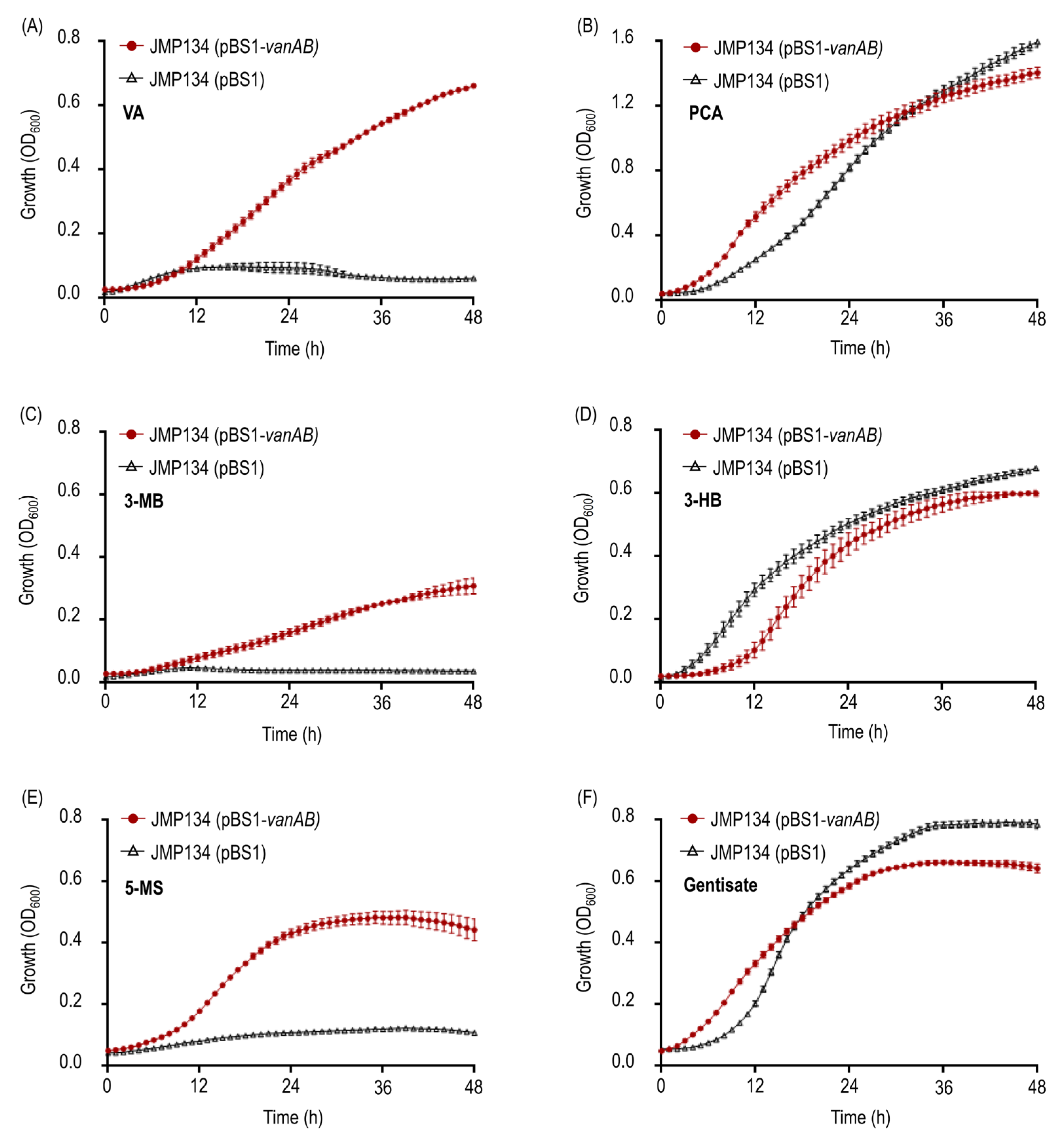
Figure 2. Growth on meta-methoxylated aromatic acids, VA, 3-MB, and 5-CS, and their O-demethylation products of Cupriavidus pinatubonensis JMP134 expressing vanAB genes of R. ruber R1. Growth of C. pinatubonensis JMP134 expressing vanAB genes driven by a heterologous PBAD promoter on 5 mM (A) VA, (B) PCA, (C) 3-MB, (D) 3-HB, (E) 5-MS, or (F) gentisate as sole carbon and energy sources was assayed in the presence of 1 mM L-arabinose as inducer. Three biological replicates were performed for growth measurements. Error bars indicate the SEM.
Microorganisms 11 00078 g002
Figure 3. Resting cell assays of Cupriavidus pinatubonensis JMP134 harboring vanAB genes in the presence of compounds structurally related to VA. Cells of C. pinatubonensis JMP134 expressing vanAB genes were grown on 5 mM VA plus 1 mM L-arabinose as inducer, washed, and subsequently exposed to 1 mM (A) VA, (B) 3-MB, (C) 5-MS, (D) syringate, (E) 3-O-methylgallate, (F) isovanillate, (G) guaiacol, and (H) 3-methoxysalicylate. Cells of C. pinatubonensis JMP134 lacking vanAB genes were grown on 5 mM 3HB plus 1 mM L-arabinose and treated as indicated previously for comparison. Three biological replicates were performed for substrate consumption measurements. Error bars indicate SEM.
Microorganisms 11 00078 g003
Figure 5. Evolutionary relationships among VanA homologues from bacteria. Maximum likelihood topology provided by IQ-TREE software [33] based on sequence alignments calculated using MAFFT software [36] is shown with SH-like approximate likelihood ratio support values (n = 1000) given at each node (values >70% are shown). Light orange, Actinobacteria phyla; green, Proteobacteria phyla.
Microorganisms 11 00078 g005
Figure 6. Evolutionary relationships among VanA homologues from Rhodococcus species. Maximum likelihood topology provided by IQ-TREE software [33] based on sequence alignments calculated using MAFFT software [36] with SH-like approximate likelihood ratio support values (n = 1000) given at each node (values >70% are shown). VanA homologues of Pseudomonas sp. ATCC 19151 and Pseudomonas sp. HR199 were included as representatives of the proteobacterial type I VanOD. Oxygenase components of Rieske-type 4-Toluene sulfonate methyl-monooxygenase from Comamonas testosteroni T-2, Chloroacetanilide N-alkylformylase from Rhizorhabdus wittichii DC-6, and Dicamba O-demethylase from Stenotrophomonas maltophilia DI-6 were used as outgroup. Sequences highlighted in bold belong to strains tested by their ability to grow in meta-methoxylated aromatic acids as a sole carbon and energy source.
Microorganisms 11 00078 g006
Disclaimer/Publisher’s Note: The statements, opinions and data contained in all publications are solely those of the individual author(s) and contributor(s) and not of MDPI and/or the editor(s). MDPI and/or the editor(s) disclaim responsibility for any injury to people or property resulting from any ideas, methods, instructions or products referred to in the content.