Article

Isolation and Identification of Microorganisms and Antibiotic Resistance Microorganisms from Beehives Located in Palm, Corn and Longan Plantations, Thailand

1
School of Health Sciences Research, Research Institute for Health Sciences, Chiang Mai University, Chiang Mai 50200, Thailand
2
Environmental Sciences Program, Faculty of Sciences, Chiang Mai University, Chiang Mai 50200, Thailand
3
Microbiology Unit, Diagnostic Laboratory, Maharaj Nakorn Chiang Mai Hospital, Faculty of Medicine, Chiang Mai University, Chiang Mai 50200, Thailand
4
School of Agriculture and Natural Resources, University of Phayao, Phayao 56000, Thailand
5
Faculty of Science, Energy and Environment, Rayong Campus, King Mongkut’s University of Technology North Bangkok, Bankhai 21120, Thailand
6
Meliponini and Apini Research Laboratory, Department of Entomology and Plant Pathology, Faculty of Agriculture, Chiang Mai University, Chiang Mai 50200, Thailand
7
Environmental, Occupational Health Sciences and NCD Center of Excellence, Research Institute for Health Sciences, Chiang Mai University, Chiang Mai 50200, Thailand
*
Authors to whom correspondence should be addressed.
Microorganisms 2023, 11(12), 2855; https://doi.org/10.3390/microorganisms11122855
Submission received: 26 October 2023 / Revised: 22 November 2023 / Accepted: 23 November 2023 / Published: 24 November 2023
(This article belongs to the Special Issue The Role of Microbes in Contaminated Environments)

Abstract

:
This study aims to determine the prevalence of microorganisms and antibiotic-resistant microorganisms in beehives located on different plantations in Thailand. Seventeen swabs immersed in transport media were utilized for samples from different zones within beehives. Traditional microbial culture-based methods, biochemical tests, MALDI-TOF MS (VITEK® MS, bioMerieux, Marcy-l’Étoile, France), and antibiotic drug susceptibility (disk-diffusion) tests were used to detect microorganism and antimicrobial resistance bacteria. The results from 16 beehive swabs found Gram-positive bacteria at 59.5%, Gram-negative bacteria at 35.1%, and fungi (yeast) at 5.4%. These organisms are classified as 11, 11, and 2 types of Gram-positive bacteria, Gram-negative bacteria, and fungi (yeast), respectively. Furthermore, no organism showed resistance to vancomycin or cefoxitin for antibiotic drug susceptibility testing. In contrast, all Acinetobacter spp. were susceptible to ciprofloxacin, levofloxacin, ceftazidime, cefotaxime, imipenem, and meropenem, except for Acinetobacter schindleri, which was resistant to ceftazidime and cefotaxime. For other organisms, due to the limitations of tests to identify some environmental microbial species, the antimicrobial susceptibility test results cannot be interpreted as resistant or susceptible to the drug for these organisms. The study’s findings will support prevention, healthcare services, and public health systems.

1. Introduction

Over the years, the problem of antimicrobial resistance in bacteria has become more severe and tends to increase continuously, which is a serious threat to health security. At present, the problem of drug resistance is not only affecting public health but also causing economic and social losses [1,2]. Microorganisms such as Pseudomonas aeruginosa, Acinetobacter baumannii, Klebsiella pneumoniae, and Escherichia coli are among the most threatening bacteria, significantly resistant to many antimicrobial agents, and only a small range of antibiotics, especially combinations working synergistically [3]. Efforts have been made to deal with this problem nationally and internationally for a long time. Collaboration between tripartite organizations on human, animal, and environmental health (Tri-partite), including the World Health Organization (WHO), the Office International des Epizooties (OIE), and the Agriculture Organization of the United Nations (FAO), have mandated that antimicrobial resistance be addressed. It is a major goal to develop and achieve a WHO global action plan on antimicrobial resistance [4]. The use of medicated feed in animal husbandry and the use of antibiotic drugs in plant-disease control have raised concerns about the emergence of antibiotic resistance in the agricultural sector, which may aid the spread of drug-resistant strains to humans [5]. Countries worldwide, including Thailand, have made efforts to develop and control the overuse of antibiotics in agriculture through various methods to prevent and mitigate the problem of antimicrobial resistance.
For foodborne pathogen identification, more investigations are necessary to prevent significant outbreaks. A study is necessary to locate and stop the spread of the infections before a significant outbreak. However, limited research has been conducted in Thailand on the microorganisms of beehives in agricultural regions.
Honey bees play a vital role as primary pollinators of plants [6]. Consequently, beekeeping has developed within Thailand’s agricultural community. Beekeeping demands significantly less land when compared to other livestock, and is environmentally responsible. Furthermore, beekeeping yields substantial returns by selling honey and bee-derived products to beekeepers. Honey bees make their food from plant materials [7]. These insect pollinators visit flowers time and time again to gather pollen and nectar, which they then share with other bees in their hive to make beebread and honey [8]. Beehives create a significant number of microorganisms throughout the process of making these products, including bacteria that originate from plants and other sections of the honey bee’s body [9]. In beebread, the majority of the microorganisms found in pollen come from the soil and phyllosphere, making this microorganism’s divergence even more evident [10,11]. Therefore, these uncommon organisms may affect the quality of honey and may infect honey bees and beekeepers who lack hygiene practices and mostly work with bare hands in this business. Nowadays, food safety and hygiene are more of a concern, but limited in-depth research has been conducted on microorganisms and antibiotic resistance microbial determinants from beehives.
Hence, this study aimed to determine the prevalence of microorganisms and antibiotic-resistant microorganisms within beehives in different agricultural settings in Thailand to prevent or reduce the spread of these organisms from natural sources to humans. This may affect the quality of human life and economic conditions in the future. In this study, microorganisms in beehives were examined at various locations using the methods of microorganisms culture in enrichment culture media, pathogens were identified by traditional biochemical tests and MALDI-TOF MS (VITEK® MS, bioMerieux, Marcy-l’Étoile, France) machines, and antimicrobial drug susceptibility tests were performed.

2. Materials and Methods

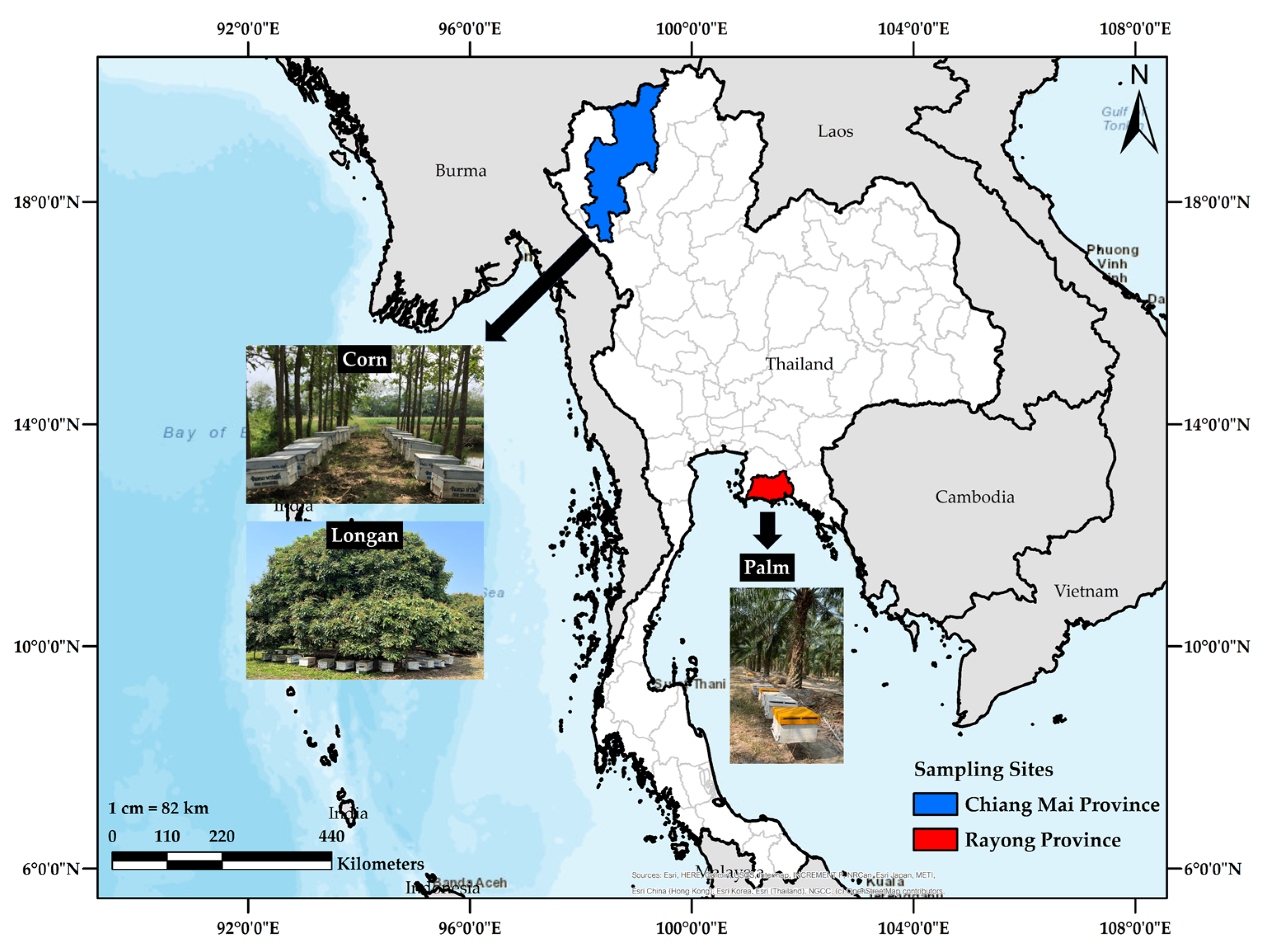
2.1. Sample Collection

For sample collection, Amies swabs were used to swab on the outer surface area of the beehive (1 swab for 1 beehive). Then, this swab was kept in Amies agar gel transport media and transported to the laboratory at room temperature.
In March 2023, 10 Amies agar gel transport swabs were obtained from palm orchards in Rayong Province, Thailand; meanwhile, in June 2023, 1 swab was taken from a longan orchard, and 6 were collected from corn fields in Chiang Mai Province. In addition, 17 swabs were collected from various plantations in Rayong and Chiang Mai Provinces (see Figure 1).

2.2. Microbial Culture and Identification

Seventeen Amies agar gel transport swabs that were swabbed on a beehive were used to perform bacterial culture and identification by the standard method. Briefly, samples were cultured in sheep blood and MacConkey agar and incubated at 37 °C for 18–24 h. All suspect colonies were sub-cultured and isolated on sheep blood agar plates. Those identified were further processed using conventional methods involving the Gram staining technique and other appropriate biochemical tests, including catalase, coagulase, oxidase, indole, motility tests, sugar fermentation tests, Triple sugar iron (TSI) agar, citrate utilization, and urease production. These were performed following the laboratory standard operating procedures adopted by the Clinical and Laboratory Standard Institute (CLSI) [12,13]. In addition, a MALDI-TOF MS (VITEK® MS, bioMerieux, Marcy-l’Étoile, France) machine was further used to identify the organism type.

2.3. Antimicrobial Susceptibility Test

The standardized antimicrobial sensitivity test was performed on Mueller–Hinton agar (MHA) plates using the disk diffusion Kirby–Bauer technique with 0.5 McFarland turbidity standard methods, and the results were interpreted according to the Standards for Antimicrobial Susceptibility of the Clinical Laboratory Standards Institute (CLSI) protocol [12,13]. Briefly, well-isolated bacterial colonies were selected from an agar plate culture and transferred into a broth culture until a slight visible turbidity appeared (similar to the 0.5 McFarland standard). A sterile swab was dipped into the standardized suspension of bacteria, and excess fluid was removed by pressing and rotating the swab firmly against the inside of the tube above the fluid level. The swab was used to streak on the MHA plate, then antibiotic discs appropriate for organisms were placed onto the media and incubated at 37 °C for 18–24 h. After that, each plate was examined, and inhibition zones were measured to the nearest millimeter at the back of the inverted culture plate. The measurements were then compared with a standard chart adopted by the Clinical and Laboratory Standard Institute (CLSI) to determine susceptibility or resistance.
Antibiotic discs, including glycopeptide, β-lactams, fluoroquinolone, third generation cephalosporins, and the carbapenems antibiotic group, were used. They comprised vancomycin (VA30), cefoxitin (oxacillin) (CX30), ciprofloxacin (CIP5), levofloxacin (LEV5), ceftazidime (CAZ30), cefotaxime (CTX30), imipenem (IMI10), and meropenem (MEM10).

2.4. Data Analysis

Data are described as frequencies (counts and percentages) using Microsoft Excel.

3. Results

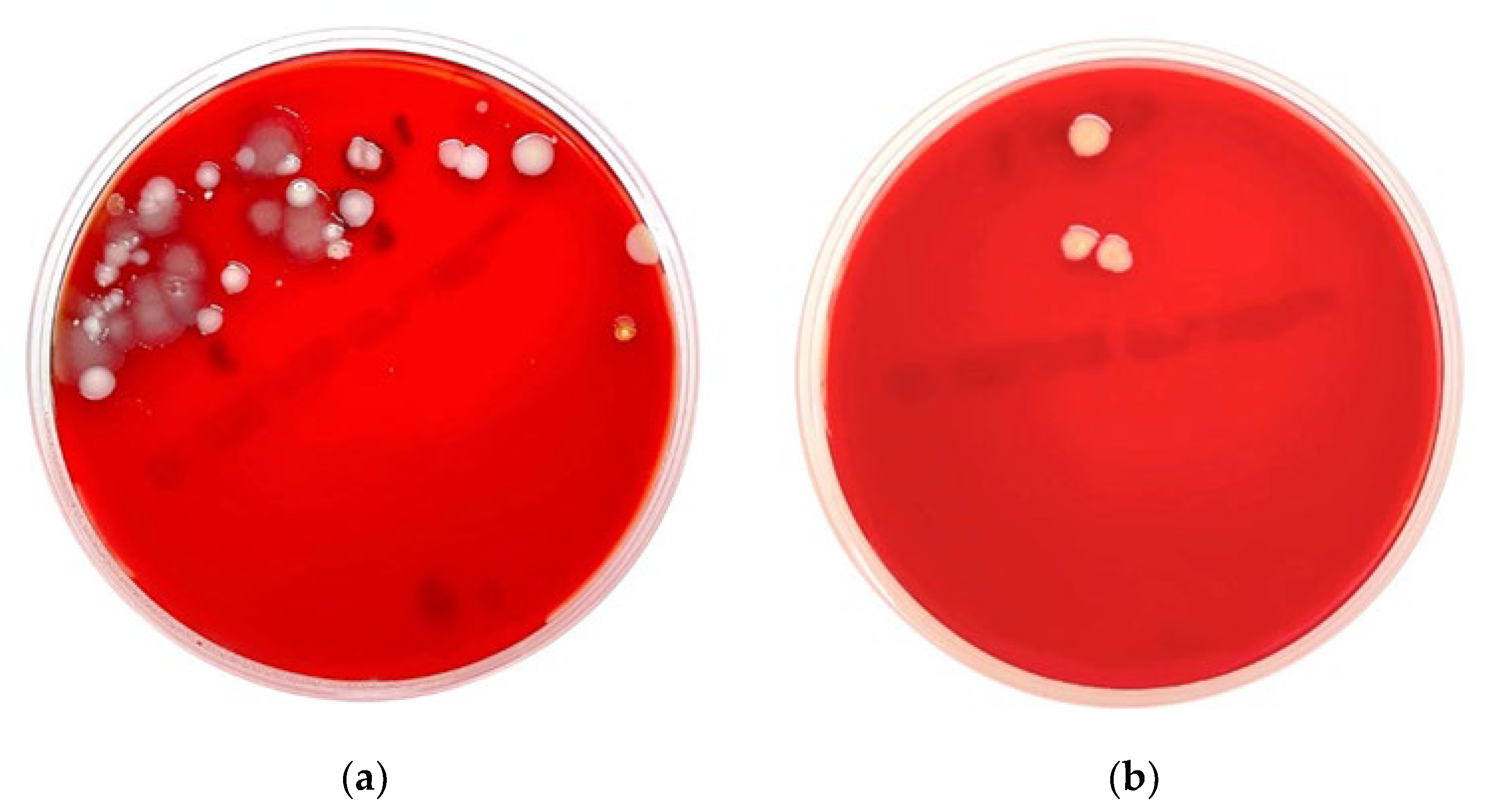
3.1. Microbial Culture and Identification

The 17 beehive swabs from different plantations in Rayong and Chiang Mai, Thailand, were used for bacterial culture and identification by biochemical tests using MALDI-TOF MS (VITEK® MS, bioMerieux, Marcy-l’Étoile, France). The results are shown in Table 1, and examples of the bacteria grown on blood agar are shown in Figure 2. One swab from the palm orchard had no growth (5.88%). The other 16 swabs showed growth (94.12%), with Gram-positive bacteria as 22 of the 37 colony sources, Gram-negative bacteria as 13 of the 37 colony sources, and fungi (yeast) as 2 of the 37 colony sources. These organisms were classified as different 11, 11, and 2 species of Gram-positive bacteria, Gram-negative bacteria, and fungi (yeast), respectively. The results of the prevalence of microbials detected on the beehives from different plantations are shown in Table 2.

3.2. Antimicrobial Drug Susceptibility Test

Overall, 37 organism isolates were detected on the swabs collected from the beehives. The antimicrobial susceptibility of isolates is shown in Table 3. These results were interpreted according to the Standards for Antimicrobial Susceptibility of the Clinical Laboratory Standards Institute (CLSI) protocol. Of the tested antimicrobials, no microbial organism resisted vancomycin or cefoxitin. And all Acinetobacter spp. were susceptible to ciprofloxacin (CIP5), levofloxacin (LEV5), ceftazidime (CAZ30), cefotaxime (CTX30), imipenem (IMI10), and meropenem (MEM10), except for Acinetobacter schindleri, which was resistant to ceftazidime (CAZ30) and cefotaxime (CTX30) (third generation cephalosporin antibiotic). The antimicrobial susceptibility test results for other organisms cannot be interpreted as resistant or susceptible to antibiotic drugs. The results provided only the inhibition zones of drug sensitivity, which were formed with each antibiotic agent (Table 3 and Figure 3).

4. Discussion

4.1. Bacterial Identification

Apiculture, or beekeeping, is widely pursued in Thailand’s agricultural sector, particularly in cultivating crops such as rambutan, longan, lychee, sunflower, palm, rubber, and other plant species. Honey bees are essential in maintaining ecological balance in natural ecosystems and providing economic advantages. Therefore, they are valuable insects for humanity. This study provided the results of the prevalence of antimicrobial resistance microorganisms found in the beehives placed in plantations of palm, longan, and corn in different provinces of Thailand.
For the results of the detection of contaminated microbials in the beehives from different plantations, we used culture-based methods and identified microbial organisms by Gram staining, traditional biochemical tests, and MALDI-TOF MS (VITEK® MS, bioMerieux, Marcy-l’Étoile, France) machines. From the 17 swab specimens, we found no microbial organism growth from swab no. 5 collected from the beehives in the palm orchard [5.9% (1/17)]. This specimen’s inability to detect bacterial contamination may be because there is no microbial contamination or very low amounts of microbial contamination, along with the limitation of the sensitivity of this culture-based method [14]. In 16 other swab specimens with 37 suspected colonies, microbial contaminations were detected in samples collected from the beehives in palm orchards, longan orchards, and cornfields. We found Gram-positive bacteria at 59.5% (22/37), Gram-negative bacteria at 35.1% (13/37), and fungi (yeast) at 5.4% (2/37).
The bacteria contaminated on beehive boxes from palm orchards were 60% Gram-positive bacteria, 30% Gram-negative bacteria, and 10% yeast. Staphylococcus spp. and Bacillus spp. were the most frequently found Gram-positive bacteria. Other bacteria were S. sciuri, S. gallinarum, B. altitudinis/pumilus, B. megaterium, and Exiguobacterium acetylicum; unidentified Coryneform bacteria; and unidentified coagulase-negative Staphylococcus spp. Meanwhile, for Gram-negative bacteria found in the beehives placed in palm orchards, Pantoea spp. were detected the most frequently, followed by A. junii and P. otitidis. C. orthopsilosis and C. guilliermondis/C. permentati were the three types of yeast detected on the beehives located in the palm orchard.
Nevertheless, for bacterial contamination in the beehives from cornfields, 60% were Gram-positive and 40% were Gram-negative bacteria. Bacillus spp. were the most-detected Gram-positive bacteria, followed by unidentified Coryneform bacteria and E. faecalis. In contrast, Acinetobacter spp. were the most-detected Gram-negative bacteria, followed by P. alcaligenes, C. gleum, and unidentified Gram-negative bacteria.
For longan orchards, we obtained only 1 swab specimen collected from the beehives. We found 2 suspected organism colonies, Gram-positive and Gram-negative, with unidentified Coryneform bacteria [100% (1/1)] and P. dispersa [100% (1/1)], respectively.
In this study, the overall results of bacterial contamination in the beehives from different plantations indicate that we mostly detected different organisms in different areas. However, most organisms detected were Gram-positive bacteria, followed by Gram-negative bacteria and fungi (yeast).
Some detected microorganisms are an essential cause of health effects for people who work in these businesses. For Gram-positive bacteria, Bacillus spp. (mostly found in the beehives in palm orchards and cornfields) are thermoduric bacteria, which can produce endospores (spore-forming bacteria). Bacilli are among the major causes of food spoilage (microbial spoilage) [15]. Some strains of Bacillus, including Bacillus cereus, which was detected in the beehives placed in cornfields, are pathogens that cause food poisoning intoxication, which is caused by consuming food that contains toxins produced by these bacteria. The most well-known symptom of such a condition is gastrointestinal illness, which can be brought on by this toxin when consumed. B. cereus-related gastrointestinal (GI) syndromes include diarrheal illness with little upper intestine symptoms and mostly upper GI syndrome with nausea and vomiting but no diarrhea. Additionally, B. cereus is linked to eye and respiratory system infections, and wounds [16,17].
However, for honey bees, Bacillus spp. are endemic bacteria or normal flora organisms that do not cause disease. They also play a biochemical role in preserving food stored in beehives and resisting disease in bees. Bacillus spp. were the predominant microorganisms in the feces of worker larvae [16,18]. From the feces of 20 larvae, 44 isolates of Bacillus spp. were obtained. Seventeen of these were B. megaterium, and 19 were B. subtilis. B. cereus and B. megaterium were the most common Bacillus spp. in the intestines of queen bees [18].
The other Gram-positive bacteria detected, Enterococcus faecalis, found within the beehives in cornfields, is a commensal bacterium inhabiting the gastrointestinal tracts of humans [19,20]. Like other species in the genus Enterococcus, E. faecalis is found in healthy humans and can be used as a probiotic. As an opportunistic pathogen, E. faecalis can cause life-threatening infections, especially in the nosocomial (hospital) environment, where the naturally high levels of antibiotic resistance found in E. faecalis contribute to its pathogenicity [20]. E. faecalis can also cause endocarditis, sepsis, urinary tract infections (UTIs), meningitis, and other human infections [21,22]. Several virulent factors are thought to contribute to E. faecalis infections.
Staphylococcus spp., which was only found in the beehives collected from palm orchards, was one of the leading pathogen infections in hospitals, and many strains of this bacterium are now resistant to antibiotics. Staphylococcus includes at least 43 species. Many species cannot cause disease and reside normally on humans’ and other animals’ skin and mucous membranes. Staphylococcus species are nectar-inhabiting microbes [23]. They are also a small component of the soil microbiome [24,25].
Coryneform bacteria, or Corynebacterium, which could be detected in the beehives positioned in palm orchards, longan orchards, and cornfields in this study, are thermoduric bacteria, which are an important bacteriua in food and the cause of spoilage (microbial spoilage) in many types of food such as meat, poultry, etc. Furthermore, Corynebacterium spp. can cause a few clinically important respiratory infections, especially in immunocompromised people or those with severe respiratory disorders [26].
Additionally, we detected Acinetobacter spp. In the beehives in palm orchards and cornfields as Gram-negative bacteria. In nature, there are many Acinetobacter spp., and they frequently appear in soil and water [27]. Some Acinetobacter spp. can survive in a hospital setting due to their capacity to survive on both wet and dry surfaces as well as exposure to both numerous common disinfectants [27]. In nosocomial infections, Acinetobacter is commonly isolated. It is particularly common in intensive care units, where widespread sporadic cases, epidemics, and endemic occurrences occur.
The other Gram-negative bacteria, Pantoea spp., was detected in the beehives in palm and longan orchards. They are bacteria usually isolated from soil, water, plants, seeds, fruits (e.g., pineapples, mandarin oranges), and the gastrointestinal tracts of humans and animals in dairy products, blood, and urine. Pantoea spp. causes infections in humans and plants. Some are plant pathogens and opportunistic in immunocompromised humans, causing wounds, bleeding, and urinary tract inflammation [28].
Pseudomonas spp. and P. alcaligenes were detected in the beehives located in cornfields. They are Gram-negative aerobic bacteriua usually used for the bioremediation of oil pollution, pesticide substances, and certain chemical substances, as they can degrade polycyclic aromatic hydrocarbons [29]. They can be human pathogens, but such occurrences are very rare [30,31]. Whereas P. otitidis, which was detected in the beehives placed in palm orchards, is a new Pseudomonas spp. that has recently been recognized in association with otitis infections in humans, including acute otitis externa, acute otitis media, and chronic suppurative otitis media [32].
Overall, we found several organisms that may affect beekeepers and related worker health. Therefore, the beekeepers who placed their beehives in palm orchards, longan orchards, and cornfields may be concerned about the risks and dangers of these pathogen infections when handling or interacting with them. Efforts should be made to minimize the introduction of additional microorganisms into the beehives.
Nevertheless, due to the limitations of the bacterial identification test in this study, we used matrix-assisted laser desorption-ionization time of flight mass spectrometry (MALDI-TOF MS) [33] to identify microorganisms in the swabs that were collected from the beehives in different locations and plantations. We found that some organisms could not be identified. Due to the limitations of these techniques, and as technology has evolved, the expansion of databases containing spectra of known organisms has allowed for the identification of species with similar phenotypic, genotypic, and biochemical properties that were not previously possible. However, inherent similarities between organisms and a limited number of spectra in the database can lead to poor discrimination between species and misidentifications. In these cases, obtaining an incorrect species-level identification or no identification is possible. These errors occur with relatively low frequency and can typically be overcome with supplemental testing [34]. For further study, adopting high-sensitivity and specific techniques, such as molecular identification methods like 16S rRNA Gene [28,35] or next-generation sequencing technologies, is a challenging way to solve these problems and obtain more information on the study’s bacterial identification results.

4.2. Antimicrobial Drug Susceptibility Test

Antimicrobial drug susceptibility tests determine a microbe’s vulnerability to antimicrobial drugs by exposing a standardized concentration of organisms to specific concentrations, according to a test of a bacteria’s resistance to an antibiotic. The benefits of these laboratory tests are used to direct doctors in choosing powerful antibiotics to treat patients [36]. Susceptibility testing can be performed for bacteria, fungi, and viruses. For some organisms, the results obtained with one drug predict results with similar drugs. Thus, not all potentially useful drugs are tested.
Based on the susceptibility test of bacteria to antimicrobial agents, this study interpreted the zone of inhibition results according to the Standards for Antimicrobial Susceptibility of the Clinical Laboratory Standards Institute (CLSI) protocol [12,13]. It is a laboratory test to identify which pathogenic isolated bacteria are susceptible or resistant to any antimicrobial agent and guide physicians in selecting effective antimicrobial agents to treat patients.
Nevertheless, this study used ten antibiotic discs for susceptibility tests with the disk diffusion Kirby–Bauer technique. These antibiotic groups, including glycopeptide (vancomycin), β-lactams [cefoxitin (oxacillin)], fluoroquinolone (ciprofloxacin and levofloxacin), third generation cephalosporins (ceftazidime and cefotaxime), and carbapenems (imipenem and meropenem), are usually and commonly used as antibiotic agents with high effectiveness [37] in our setting. In addition, according to the CLSI guidelines [12,13], each organism underwent antimicrobial drug susceptibility testing against each antibiotic agent according to their Gram stain results. In this study, for Gram-positive bacteria, vancomycin and cefoxitin were tested; meanwhile, ciprofloxacin, levofloxacin, ceftazidime, cefotaxime, imipenem, and meropenem were tested for Gram-negative bacteria. Therefore, bacteria which were not tested with a specific antibiotic based on their Gram-stain result, are given ND (Not done) results in Table 3.
The antimicrobial susceptibility of isolates is shown in Table 3. This study found that 37 organism isolates were detected on swabs collected from the beehives in palm orchards, longan orchards, and cornfields. Of the antimicrobials tested for Gram-positive bacteria, all Staphylococcus spp., which were only found in the beehives placed in palm orchards, were resistant to cefoxitin (oxacillin), with an inhibition zone less than 25 mm (the susceptible zone diameter is ≥25 mm). According to a previous study, Chalalai et al. (2017) revealed that coagulase-negative staphylococci were the main species of Staphylococcus consisting of S. gallinarum, S. lentus, S. sciuri, S. saprophyticus, S. arlettae, S. cohnii, S. simulans, S. carnosus, and S. kloosii. They also found that the majority of isolates showed resistance to oxacillin (87%), erythromycin (52%), and clindamycin (48%) [38].
All Acinetobacter spp., which are Gram-negative bacteria, found in the beehives located in palm orchards and cornfields, were susceptible to ciprofloxacin (CIP5) (susceptible zone diameter is ≥25 mm), levofloxacin (LEV5) (susceptible zone diameter is ≥25 mm), ceftazidime (CAZ30) (susceptible zone diameter is ≥25 mm), cefotaxime (CTX30) (Susceptible zone diameter is ≥25 mm), and imipenem (IMI10) (susceptible zone diameter is ≥25 mm) and meropenem (MEM10) (susceptible zone diameter is ≥25 mm), except for Acinetobacter schindleri, which was resistant to both of the third generation cephalosporin antibiotics. Ceftazidime (CAZ30) provided an inhibition zone at 16 mm and cefotaxime (CTX30) provided an inhibition zone at 20 mm.
Moreover, for Chryseobacterium gleum, this detected bacteria did not form inhibition zone sizes (at 0 mm) for cefotaxime (CTX30), which is a third generation cephalosporin antibiotic drug, or meropenem (MEM10), which is a carbapenems antibiotic group. Therefore, Chryseobacterium gleum was resistant to these antibiotic agents.
The antimicrobial drug susceptibility tests could not interpret the other detected organisms by using the CLSI guidelines because their organism names were not specified in this guidelines. Therefore, the antimicrobial susceptibility test results cannot be interpreted as resistant or susceptible to the drug. Nevertheless, they provided a different range of inhibition zone sizes of drug sensitivity for each antibiotic agent (see Table 3). These inhibition zones represent how each antibiotic agent can inhibit bacteria growth. The size of the inhibition zone can measure the effectiveness of an antibiotic. The larger the zone of inhibition, the more bacteria were killed, indicating a higher effectiveness of the substance [39].
Regarding the bacteria in the Bacillus spp. group and the coryneform bacteria, which are Gram-positive, the susceptibility of antimicrobials cannot be tested by the disk diffusion method. The Minimum Inhibitory Concentration method, or MIC method, is used to quantify the minimum concentration of antimicrobial drug required to inhibit or eradicate bacteria and to test the susceptibility of the bacteria to antimicrobials for the best treatment effect [12,13,30,31].

5. Conclusions

Overall, microorganisms were detected in the samples swabbed from beehives placed in plantations in Rayong and Chiang Mai provinces, Thailand. These detected organisms may affect the quality of honey and their product, and may also affect beekeepers’ health if they make contact the surface area of beehives. Hence, the findings of this study can be used to disseminate knowledge to beekeepers, healthcare units, and people involved in beekeeping businesses. Beekeepers must comprehensively understand microbial infections while touching or working with these materials. The utilization of personal protection equipment and hygiene in beehives throughout the process of bee management and honey harvesting is a crucial aspect of ensuring the safety of beekeepers and their colonies.

Author Contributions

Conceptualization, S.H. (Surat Hongsibsong), S.D. and B.C.; methodology, S.D., S.Y., S.H. (Sayamon Hongjaisee) and S.H. (Surat Hongsibsong); validation, S.D., B.C. and S.H. (Surat Hongsibsong); formal analysis, S.D., P.J., P.K., S.M. and S.Y.; investigation, K.D., B.C. and J.M.; writing—original draft preparation, S.D., B.C. and S.H. (Surat Hongsibsong); writing—review and editing, S.D., B.C. and S.H. (Surat Hongsibsong); visualization, S.D. and P.K.; supervision, S.H. (Surat Hongsibsong); project administration, S.Y.; funding acquisition, B.C. All authors have read and agreed to the published version of the manuscript.

Funding

This research is supported by NSRF via the Program Management Unit for Human Resources & Institutional Development, Research, and was funded by the Research Institute for Health Sciences, Chiang Mai University, Chiang Mai, Thailand.

Data Availability Statement

The data are contained within the article.

Acknowledgments

The authors are thankful to the NSRF via the Program Management Unit for Human Resources & Institutional Development, Research and Research Institute for Health Science, Chiang Mai University, for the support in the analysis.

Conflicts of Interest

The authors declare no conflict of interest.

References

  1. Serwecińska, L. Antimicrobials and Antibiotic-Resistant Bacteria: A Risk to the Environment and to Public Health. Water 2020, 12, 3313. [Google Scholar] [CrossRef]
  2. Dadgostar, P. Antimicrobial Resistance: Implications and Costs. Infect. Drug Resist. 2019, 12, 3903–3910. [Google Scholar] [CrossRef]
  3. Prestinaci, F.; Pezzotti, P.; Pantosti, A. Antimicrobial resistance: A global multifaceted phenomenon. Pathog. Glob. Health. 2015, 109, 309–318. [Google Scholar] [CrossRef]
  4. World Health Organization. Sixty-Eighth World Health Assembly; World Health Organization: Geneva, Switzerland, 2015. [Google Scholar]
  5. World Health Organization. Global Action Plan on Antimicrobial Resistance; WHO Document Production Services: Geneva, Switzerland, 2015. [Google Scholar]
  6. Khalifa, S.A.M.; Elshafiey, E.H.; Shetaia, A.A.; El-Wahed, A.A.A.; Algethami, A.F.; Musharraf, S.G.; AlAjmi, M.F.; Zhao, C.; Masry, S.H.D.; Abdel-Daim, M.M.; et al. Overview of Bee Pollination and Its Economic Value for Crop Production. Insects 2021, 12, 688. [Google Scholar] [CrossRef]
  7. Anderson, K.E.; Sheehan, T.H.; Mott, B.M.; Maes, P.; Snyder, L.; Schwan, M.R.; Walton, A.; Jones, B.M.; Corby-Harris, V. Microbial ecology of the hive and pollination landscape: Bacterial associates from floral nectar, the alimentary tract and stored food of honey bees (Apis mellifera). PLoS ONE 2013, 8, e83125. [Google Scholar] [CrossRef]
  8. Sinacori, M.; Francesca, N.; Alfonzo, A.; Cruciata, M.; Sannino, C.; Settanni, L.; Moschetti, G. Cultivable microorganisms associated with honeys of different geographical and botanical origin. Food Microbiol. 2014, 38, 284–294. [Google Scholar] [CrossRef]
  9. Santorelli, L.A.; Wilkinson, T.; Abdulmalik, R.; Rai, Y.; Creevey, C.J.; Huws, S.; Gutierrez-Merino, J. Beehives possess their own distinct microbiomes. Environ. Microbiome 2023, 18, 1. [Google Scholar] [CrossRef]
  10. Anderson, K.E.; Carroll, M.J.; Sheehan TI, M.; Mott, B.M.; Maes, P.; Corby-Harris, V. Hive-stored pollen of honey bees: Many lines of evidence are consistent with pollen preservation, not nutrient conversion. Mol. Ecol. 2014, 23, 5904–5917. [Google Scholar] [CrossRef]
  11. Corby-Harris, V.; Maes, P.; Anderson, K.E. The bacterial communities associated with honey bee (Apis mellifera) foragers. PLoS ONE 2014, 9, e95056. [Google Scholar] [CrossRef]
  12. Clinical and Laboratory Standards Institute. M100 Performance Standards for Antimicrobial Susceptibility Testing, 32nd ed.; Clinical and Laboratory Standards Institute: Wayne, NY, USA, 2022. [Google Scholar]
  13. Clinical and Laboratory Standards Institute. M45 Methods for Antimicrobial Dilution and Disk Susceptibility Testing of Infrequently Isolated or Fastidious Bacteria, 3rd ed.; Clinical and Laboratory Standards Institute: Wayne, NY, USA, 2015. [Google Scholar]
  14. Aarestrup, F.M.; Seyfarth, A.M.; Emborg, H.D.; Pedersen, K.; Hendriksen, R.S.; Bager, F. Effect of abolishment of the use of antimicrobial agents for growth promotion on occurrence of antimicrobial resistance in fecal enterococci from food animals in Denmark. Antimicrob. Agents Chemother. 2001, 45, 2054–2059. [Google Scholar] [CrossRef]
  15. Ahmadi, A.; Khezri, A.; Nørstebø, H.; Ahmad, R. A culture-, amplification-independent, and rapid method for identification of pathogens and antibiotic resistance profile in bovine mastitis milk. Front. Microbiol. 2023, 13, 1104701. [Google Scholar] [CrossRef]
  16. Lorenzo, J.M.; Munekata, P.E.; Dominguez, R.; Pateiro, M.; Saraiva, J.A.; Franco, D. Main Groups of Microorganisms of Relevance for Food Safety and Stability: General Aspects and Overall Description. Innov. Technol. Food Preserv. 2018, 13, 53–107. [Google Scholar]
  17. Nguyen, A.T.; Tallent, S.M. Screening food for Bacillus cereus toxins using whole genome sequencing. Food Microbiol. 2019, 78, 164–170. [Google Scholar] [CrossRef]
  18. Hölzel, C.S.; Tetens, J.L.; Schwaiger, K. Unraveling the Role of Vegetables in Spreading Antimicrobial-Resistant Bacteria: A Need for Quantitative Risk Assessment. Foodborne Pathog. Dis. 2018, 15, 671–688. [Google Scholar] [CrossRef]
  19. Martha, G. Identification and roles of non-pathogenic microflora associated with honey bees. FEMS Microbiol. Lett. 1997, 155, 1–10. [Google Scholar]
  20. de Almeida, C.V.; Taddei, A.; Amedei, A. The controversial role of Enterococcus faecalis in colorectal cancer. Ther. Adv. Gastroenterol. 2018, 11, 1756284818783606. [Google Scholar] [CrossRef]
  21. Ryan, K.J.; Ray, C.G. Sherris Medical Microbiology, 4th ed.; McGraw Hill: New York, NY, USA, 2004; pp. 294–295. [Google Scholar]
  22. Murray, B.E. The life and times of the Enterococcus. Clin. Microbiol. Rev. 1990, 3, 46–65. [Google Scholar] [CrossRef]
  23. Hidron, A.I.; Edwards, J.R.; Patel, J.; Horan, T.C.; Sievert, D.M.; Pollock, D.A.; Fridkin, S.K. NHSN annual update: Antimicrobial-resistant pathogens associated with healthcare-associated infections: Annual summary of data reported to the National Healthcare Safety Network at the Centers for Disease Control and Prevention, 2006–2007. Infect. Control Hosp. Epidemiol. 2008, 29, 996–1011. [Google Scholar] [CrossRef]
  24. Jacquemyn, H.; Lenaerts, M.; Brys, R.; Willems, K.; Honnay, O.; Lievens, B. Among-population variation in microbial community structure in the floral nectar of the bee-pollinated forest herb Pulmonaria officinalis L. PLoS ONE 2013, 8, e56917. [Google Scholar] [CrossRef]
  25. Madigan, M.; Martinko, J. Brock Biomlogy of Microorganisms, 11th ed.; Prentice Hall: New York, NY, USA, 2005; ISBN 978-0-13-144329-7. [Google Scholar]
  26. Sandot, A.; Grall, N.; Rodier, T.; Bunel, V.; Godet, C.; Weisenburger, G.; Tran-Dinh, A.; Montravers, P.; Mordant, P.; Castier, Y.; et al. Risk of Bronchial Complications After Lung Transplantation With Respiratory Corynebacteria. Results from a Monocenter Retrospective Cohort Study. Transpl. Int. 2023, 36, 10942. [Google Scholar] [CrossRef]
  27. Doughari, H.J.; Ndakidemi, P.A.; Human, I.S.; Benade, S. The ecology, biology and pathogenesis of Acinetobacter spp.: An overview. Microbes Environ. 2011, 26, 101–112. [Google Scholar] [CrossRef]
  28. Layla, S.A.; Mayyada, F.D. Prevalence and antibiotic susceptibility patterns of Pantoea spp. isolated form clinical and environmental sources in Iraq. Int. J. ChemTech Res. 2016, 9, 430–437. [Google Scholar]
  29. O’Mahony, M.M.; Dobson, A.D.; Barnes, J.D.; Singleton, I. The use of ozone in the remediation of polycyclic aromatic hydrocarbon contaminated soil. Chemosphere 2006, 63, 307–314. [Google Scholar] [CrossRef]
  30. Valenstein, P.; Bardy, G.H.; Cox, C.C.; Zwadyk, P. Pseudomonas alcaligenes endocarditis. Am. J. Clin. Pathol. 1983, 79, 245–247. [Google Scholar] [CrossRef]
  31. Suzuki, M.; Suzuki, S.; Matsui, M.; Hiraki, Y.; Kawano, F.; Shibayama, K. Genome Sequence of a Strain of the Human Pathogenic Bacterium Pseudomonas alcaligenes That Caused Bloodstream Infection. Genome Announc. 2013, 1, e00919-13. [Google Scholar] [CrossRef]
  32. Clark, L.L.; Dajc, J.J.; McLean, C.H.; Bartell, J.G.; Stroman, D.W. Pseudomonas otitidis sp. nov., isolated from patients with otic infections. Int. J. Syst. Evol. Microbiol. 2006, 56, 709–714. [Google Scholar] [CrossRef]
  33. Tsuchida, S.; Umemura, H.; Nakayama, T. Current Status of Matrix-Assisted Laser Desorption/Ionization-Time-of-Flight Mass Spectrometry (MALDI-TOF MS) in Clinical Diagnostic Microbiology. Molecules 2020, 25, 4775. [Google Scholar] [CrossRef]
  34. Grenga, L.; Pible, O.; Armengaud, J. Pathogen proteotyping: A rapidly developing application of mass spectrometry to address clinical concerns. Clin. Mass Spectrom. 2019, 14 Pt A, 9–17. [Google Scholar] [CrossRef]
  35. Church, D.L.; Cerutti, L.; Gürtler, A.; Griener, T.; Zelazny, A.; Emler, S. Performance and Application of 16S rRNA Gene Cycle Sequencing for Routine Identification of Bacteria in the Clinical Microbiology Laboratory. Clin. Microbiol. Rev. 2020, 33, e00053-19. [Google Scholar] [CrossRef]
  36. Ozma, M.A.; Abbasi, A.; Asgharzadeh, M.; Pagliano, P.; Guarino, A.; Köse, Ş.; Samadi Kafil, H. Antibiotic therapy for pan-drug-resistant infections. Infez. Med. 2022, 30, 525–531. [Google Scholar]
  37. Parmanik, A.; Das, S.; Kar, B.; Bose, A.; Dwivedi, G.R.; Pandey, M.M. Current Treatment Strategies Against Multidrug-Resistant Bacteria: A Review. Curr. Microbiol. 2022, 79, 388. [Google Scholar] [CrossRef]
  38. Chalalai, R.; Srisamai, V.; Suporn, T.; Phitsanu, T. Species Diversity and Antimicrobial Susceptibility Properties of Staphylococcus Isolated from Broiler Feces in Selected Farms, Thailand. J. Public Health 2017, 47, 44–55. [Google Scholar]
  39. Kirby-Bauer Disk Susceptibility Test. LibreTextTM. Available online: https://bio.libretexts.org/ (accessed on 3 October 2023).
Figure 1. Scale map of the location of the sample plantation collection.
Microorganisms 11 02855 g001
Figure 2. Examples of blood agar including (a) swab no. 16 with detection of Acinetobacter schindleri, Acinetobacter radioresistens, and Pseudomonas alcaligenesor; and (b) swab no. 7 with detection of Staphylococcus gallinarum and Staphylococcus sciuri.
Microorganisms 11 02855 g002
Figure 3. Examples of antimicrobial susceptibility tests on the MHA plate: (a) Pantoea dispersa from swab no. 11 collected from a beehive located in a longan orchard; (b) Pseudomonas alcaligenes from swab no. 16 collected from a beehive in a cornfield; (c) Chryseobacterium gleum from swab no. 17 collected from a beehive in a cornfield.
Microorganisms 11 02855 g003
Table 1. Identification of microorganisms from beehives located in palm, corn, and longan plantations.
Swab No.PlantationGram Stain MorphologyMicrobial Identification
Biochemical Tests MALDI-TOF MS
(VITEK® MS, bioMerieux, Marcy-l’Étoile, France)
1Palm Not performedYeastCandida orthopsilosis
2Palm Gram-positive cocciCoagulase-negative Staphylococcus spp.Staphylococcus sciuri
Gram-positive cocciCoagulase-negative Staphylococcus spp.Unidentified
3Palm Gram-positive bacilliCoryneform bacteria Unidentified
4Palm Gram-positive cocciCoagulase-negative Staphylococcus spp.Staphylococcus sciuri
5Palm No growthNo growthNot performed
6PalmGram-positive bacilliCoryneform bacteria Unidentified
Gram-negative bacilliPseudomonas spp.Pseudomonas otitidis
Gram-negative bacilliNon-fermentative
Gram-negative bacilli
Acinetobacter junii
7PalmGram-positive cocciCoagulase-negative Staphylococcus spp.Staphylococcus gallinarum
Gram-positive cocciCoagulase-negative Staphylococcus spp.Staphylococcus sciuri
Gram-negative bacilliPantoea spp.Pantoea dispersa
8Palm Gram-positive bacilli with sporeBacillus spp.Bacillus altitudinis/pumilus
Gram-positive bacilliBacillus spp.Bacillus megaterium
Not conductedYeastCandida guilliermondis +
Candida permentati
9Palm Gram-positive cocciCoagulase-negative Staphylococcus spp.Staphylococcus sciuri
Gram-positive cocciCoagulase-negative Staphylococcus spp.Staphylococcus gallinarum
Gram-negative bacilliPantoea spp.Pantoea ananatis
10Palm Gram-positive bacilliCoryneform bacteria Exiguobacterium acetylicum
Gram-negative bacilliPantoea spp.Unidentified
Gram-negative bacilliNon-fermentative
Gram negative bacilli
Acinetobacter junii
11Longan Gram-positive bacilliCoryneform bacteria Unidentified
Gram-negative bacilliPantoea spp.Pantoea dispersa
12Corn Gram-positive bacilliCoryneform bacteria Unidentified
13Corn Gram-positive bacilliBacillus spp.Bacillus megaterium
Gram-positive bacilliCoryneform bacteria Unidentified
14Corn Gram-positive bacilliBacillus spp.Unidentified
Gram-positive bacilliBacillus spp.Bacillus cereus gr.
15Corn Gram-positive bacilliBacillus spp.Bacillus cereus gr.
Gram-negative cocco-bacilliUnidentified Gram-negative bacteriaUnidentified
16Corn Gram-negative cocco-bacilliNon-fermentative
Gram-negative bacilli
Acinetobacter schindleri
Gram-negative cocco-bacilliNon-fermentative
Gram-negative bacilli
Acinetobacter radioresistens
Gram-negative bacilliPseudomonas spp.Pseudomonas alcaligenes
17Corn Gram-positive bacilliCoryneform bacteria Unidentified
Gram-negative cocco-bacilliNon-fermentative
Gram-negative bacilli
Acinetobacter lwoffii
Gram-positive bacilliBacillus spp.Bacillus flexus
Gram-positive cocco-bacilliEnterococcus faecalisEnterococcus faecalis
Gram-negative bacilliNon-fermentative
Gram-negative bacilli
Chryseobacterium gleum
Table 2. Prevalence of microorganism growth separated by type of plantations.
Plantation/No. of Colony Source
(n = 37)
Type of Microorganisms
Gram-Positive [59.5% (22/37)]Gram-Negative [35.1% (13/37)]Fungus (Yeast) [5.4% (2/37)]
Palm/
(n = 20)
Gram-positive [60% (12/20)]
Staphylococcus sciuri [33.3% [4/12)]
Staphylococcus gallinarum [16.7% (2/12)]
Bacillus altitudinis/pumilus [8.3% (1/12)]
Bacillus megaterium [8.3% (1/12)]
Exiguobacterium acetylicum [8.3% (1/12)]
Unidentified Coagulase-negative Staphylococcus spp. [8.3% (1/12)]
Unidentified Coryneform bacteria [16.7% (2/12)]
Gram-negative [30% (6/20)]
Acinetobacter junii [33.3% (2/6)]
Pantoea ananatis [16.7% (1/6)]
Pantoea dispersa [16.7% (1/6)]
Pseudomonas otitidis [16.7% (1/6)]
Unidentified Pantoea spp. [16.7% (1/6)]
Fungus (Yeast) (10% (2/20)]
Candida orthopsilosis [50% (1/2)]
Candida guilliermondis + Candida permentati [50% (1/2)]
Longan/
(n = 2)
Gram-positive [50% (1/2)]
Unidentified Coryneform bacteria [100% (1/1)]
Gram-negative [50% (1/2)]
Pantoea dispersa [100% (1/1)]
Not Found
Corn/
(n = 15)
Gram-positive [60% (9/15)]
Bacillus cereus gr. [22.2% (2/9)]
Bacillus megaterium [11% (1/9)]
Bacillus flexus [11% (1/9)]
Enterococcus faecalis [11% (1/9)]
Unidentified Bacillus spp. [11% (1/9)]
Unidentified Coryneform bacteria [33.3% (3/9)]
Gram-negative [40% (6/15)]
Acinetobacter schindleri [16.7% (1/6)]
Acinetobacter radioresistens [16.7% (1/6)]
Pseudomonas alcaligenes [16.7% (1/6)]
Acinetobacter lwoffii [16.7% (1/6)]
Chryseobacterium gleum [16.7% (1/6)]
Unidentified Gram-negative bacteria [16.7% (1/6)]
Not Found
Table 3. Antimicrobial drug-susceptibility testing (AST) of bacterial from beehive swabs.
Swab No.Plantation-Based Swab SourceMicroorganismAntibiotic Inhibition Zone Size (mm) 1
VA30 2CX30 3CIP5 4LEV5 4CAZ30 5CTX30 5IMI10 6MEM10 6
2Palm Staphylococcus sciuri21 23 (R)NDNDNDNDNDND
Unidentified Coagulase negative Staphylococcus spp.19 22 (R)NDNDNDNDNDND
4Palm Staphylococcus sciuri20 24 (R)NDNDNDNDNDND
6Palm Pseudomonas otitidisNDND383218181919
Acinetobacter juniiNDND34 (S)34 (S)32 (S)32 (S)40 (S)34 (S)
7PalmStaphylococcus gallinarum19 21 (R)NDNDNDNDNDND
Staphylococcus sciuri19 20 (R)NDNDNDNDNDND
Pantoea dispersaNDND403634303034
9Palm Staphylococcus sciuri18 22 (R)NDNDNDNDNDND
Staphylococcus gallinarum19 23 (R)NDNDNDNDNDND
Pantoea ananatisNDND363628343032
10Palm Acinetobacter juniiNDND34 (S)32 (S)28 (S)30 (S)26 (S)30 (S)
Unidentified Pantoea spp.NDND222021213422
11Longan Pantoea dispersaNDND383226282630
15Corn Unidentified Gram-negative bacteriaNDND444034323242
16CornAcinetobacter schindleriNDND26 (S)26 (S)16 (R)20 (R)30 (S)28 (S)
Acinetobacter radioresistensNDND26 (S)26 (S)23 (S)25 (S)28 (S)32 (S)
Pseudomonas alcaligenesNDND36281182314
17Corn Acinetobacter lwoffiiNDND28 (S)28 (S)19 (S)24 (S)36 (S)30 (S)
Enterococcus faecalis20 (S)NDNDNDNDNDNDND
Chryseobacterium gleumNDND2425190140
Note: S: Susceptible, R: Resistant, ND: Not Done, VA30: vancomycin, CX30: cefoxitin, CIP5: ciprofloxacin, LEV5: levofloxacin, CAZ30: ceftazidime, CTX30: cefotaxime, IMI10: imipenem, MEM10: meropenem. Superscript number: 1 CLINICAL AND LABORATORY STANDARDS INSTITUTE® M100, Performance Standards for Antimicrobial Susceptibility Testing, 32nd Edition guideline, 2 glycopeptide antibiotic, 3 β-lactams antibiotic, 4 fluoroquinolone antibiotic, 5 3rd generation cephalosporins antibiotic, 6 carbapenems antibiotic.
Disclaimer/Publisher’s Note: The statements, opinions and data contained in all publications are solely those of the individual author(s) and contributor(s) and not of MDPI and/or the editor(s). MDPI and/or the editor(s) disclaim responsibility for any injury to people or property resulting from any ideas, methods, instructions or products referred to in the content.

