Article

Antifungal Activity of Spent Coffee Ground Extracts

by
Daniela Calheiros
1,
Maria Inês Dias
2,3,
Ricardo C. Calhelha
2,3,
Lillian Barros
2,3,
Isabel C. F. R. Ferreira
2,
Chantal Fernandes
1,† and
Teresa Gonçalves
1,4,*,†
1
CNC—Center for Neuroscience and Cell Biology of Coimbra, 3004-504 Coimbra, Portugal
2
Centro de Investigação de Montanha (CIMO), Instituto Politécnico de Bragança, Campus de Santa Apolónia, 5300-253 Bragança, Portugal
3
Laboratório Associado Para a Sustentabilidade e Tecnologia em Regiões de Montanha (SusTEC), Instituto Politécnico de Bragança, Campus de Santa Apolónia, 5300-253 Bragança, Portugal
4
FMUC—Faculty of Medicine, University of Coimbra, 3004-504 Coimbra, Portugal
*
Author to whom correspondence should be addressed.
These authors contributed equally to this work.
Microorganisms 2023, 11(2), 242; https://doi.org/10.3390/microorganisms11020242
Submission received: 30 November 2022 / Revised: 14 January 2023 / Accepted: 14 January 2023 / Published: 18 January 2023
(This article belongs to the Special Issue Antimicrobial Activities of Natural Products)

Abstract

:
Coffee is one of the most popular and consumed products in the world, generating tons of solid waste known as spent coffee grounds (SCG), containing several bioactive compounds. Here, the antifungal activity of ethanolic SCG extract from caffeinated and decaffeinated coffee capsules was evaluated against yeasts and filamentous fungi. These extracts had antifungal activity against Candida krusei, Candida parapsilosis, Trichophyton mentagrophytes, and Trichophyton rubrum, all skin fungal agents. Moreover, SCG had fungicidal activity against T. mentagrophytes and T. rubrum. To understand the underlying mechanisms of the antifungal activity, fungal cell membrane and cell wall components were quantified. SCG caused a significant reduction of the ergosterol, chitin, and β-(1,3)-glucan content of C. parapsilosis, revealing the synthesis of this membrane component and cell wall components as possible targets of these extracts. These extracts were cytotoxic for the tumoral cell lines tested but not for the non-tumoral PLP2 cell line. The analysis of the phenolic compounds of these extracts revealed the presence of caffeoylquinic acid, feruloylquinic acid, and caffeoylshikimic acid derivatives. Overall, this confirmed the antifungal activity of spent coffee grounds, presenting a potential increase in the sustainability of the life cycle of coffee grounds, as a source for the development of novel antifungal formulations, especially for skin or mucosal fungal infections.

1. Introduction

Superficial infections of the skin, nails, hair, and mucosa are the most common fungal diseases in humans, affecting approximately a quarter of the global population [1]. These infections are mainly caused by dermatophytes, specifically, Trichophyton spp., Microsporum spp., and Epidermophyton spp., causing well-known conditions such as athlete’s foot, ringworm of the skin and scalp, and onychomycoses [2,3,4,5]. Mucosal infections of the genital and oral tracts are also extremely common, especially vulvovaginal candidiasis. About 75% of the women have had a vulvovaginal infection caused by Candida spp. during their lifetime [1,6]. Although superficial infections do not present a high mortality rate, they are unpleasant and affect the patient’s self-esteem and quality of life [1]. Moreover, heavy skin colonization/skin infections by yeasts are considered risk factors for the development of a life-threatening systemic infection in the case of a decreased immune condition of the host [7]. Candida species have been classified as human skin commensals since they are ubiquitous yeasts that can be found on the skin and mucosa normal microbiota. Species such as Candida albicans, Candida tropicalis, Candida parapsilosis, and Candida orthopsilosis, found on healthy skin, can become pathogenic [8].
Trichophyton rubrum and Trichophyton mentagrophytes are the main dermatophyte species responsible for causing onychomycosis, accounting about 90% of the toe infections, followed by non-dermatophyte yeasts, C. albicans and non-albicans Candida spp. [3].
The widespread use of antifungal drugs used both in human and animal health and in the protection of intensive agriculture are turning antifungal therapies to be less effective, due to the emergence of resistant strains. Moreover, some fungal infections require prolonged treatment leading to selective pressure, with fewer drug-susceptible variants and failure treatments as a consequence [9,10]. This turns essential the discovery of new and effective antifungals. Natural products are rich in bioactive compounds that provide unlimited opportunities for new drug development. It is scientifically proven that plants are enriched with many bioactive secondary metabolites such as saponins, alkaloids, and terpenoids, and other compounds containing physiologically active biochemicals that have great potential to have antifungal properties to be used for the treatment of both human and animal mycoses, but still only a few plants have been scientifically studied for quality, safety, and efficacy assessment [11].
Coffee is one of the most consumed beverages and traded commodities in the world and is also a major contributor to dietary antioxidant intake [12]. Coffea arabica and Coffea canephora var. robusta are the two main species cultivated for coffee commercial production [12,13,14,15]. Besides being a rich source of antioxidants, coffee also has several bioactive metabolites such as xanthines, phenolics, melanoidins, diterpenes, flavonoids, carotenoids, vitamin precursors, and chlorogenic acids, and its consumption is associated with less risk of developing neurodegenerative diseases and less oxidative stress [12,13,16]. Most of these beneficial properties have been associated with coffee but is not yet clear if these benefits are equally present in caffeinated and decaffeinated coffee. In fact, there is evidence that decaffeinated coffee may have identical properties, pointing to the relevant role of compounds other than caffeine [14,17]. The coffee fruit has been indicated as having antimicrobial activities, including against fungi. In fact, the fruit pulp [18,19,20], the skin [21], and the seed extracts [22,23], either roasted [24] or brewed [25,26], have been demonstrated to have bioactivity against fungi agents of animal/human infection and phytopathogens.
The production of coffee requires many processes. The treatment and processing of coffee cherries, the roasting of coffee beans, and coffee brewing generates huge volumes of biowaste contributing to environmental pollution, approximately six million tons of spent coffee grounds (SCG) per year [16,27]. SCG are residues obtained during the ground coffee brewing and they contain several organic compounds that can be exploited as a source of value-added products since only a few substances are extracted from the brewing process [15]. Despite the brewing, studies have investigated the SCG composition and exploited potential applications. SCG still has a high concentration of polysaccharides, oligosaccharides, lipids, aliphatic acids, amino acids, proteins, alkaloids, phenolics, minerals, lignin, melanoidins, and volatile compounds [16,27]. One of the most abundant phenolic compounds in SCG is chlorogenic acid, described to have many valuable properties such as antioxidant activity, anti-inflammatory, antibacterial, anticarcinogenic, antiviral, and hypoglycemic, which have potential to numerous applications in food, agriculture, and pharmaceutical areas [27,28,29,30]. However, the amount of chlorogenic acids depends on the coffee preparation or decaffeination, bean type, bean roasting, geographical origin, and coffee quality [16,17].
As a food by-product, SCG re-use and valorization reduce large amounts of industrial waste, contributing to a significant impact on sustainability [12,13]. In this context, here, we prepared ethanolic caffeinated and decaffeinated SCG extracts from commercial capsules, determined their phenolic profile, evaluated the antifungal activity against filamentous fungi and yeasts, and evaluated the anti-inflammatory activity on RAW 264.7 mouse macrophage cell lines and the cytotoxicity on tumor and non-tumor cell lines.

2. Materials and Methods

2.1. Fungal Strains and Growth Conditions

C. albicans YP0037, C. glabrata YP0937, C. parapsilosis YP0515, Candida krusei YP0338, T. rubrum IMF028, T. mentagrophytes IMF029, Aspergillus niger IMF005, Fusarium oxysporum IMF033, and Alternaria alternata IMF030 were obtained from the Clinical Yeast Collection—University of Coimbra (CYCUC) of the Institute of Microbiology of the Faculty of Medicine of the University of Coimbra. The fungal strains of Aspergillus fumigatus CBS 500.90 and Alternaria infectoria CBS 137.90 CBS were obtained from CBS-KNAW Fungal Biodiversity Centre (Utrecht, The Netherlands).
Yeast strains were grown on Yeast Extract Peptone Dextrose (YPD) agar plates with the following composition: 0.5% yeast extract (Panreac, Barcelona, Spain), 1% peptone (Panreac), 2% agar (VWR, Leuven, Belgium, and 2% glucose (Sigma-Aldrich, St. Louis, MO, USA) (w/v). Yeasts were inoculated and incubated at 30 °C for 24 h.
Filamentous fungi strains were grown on Potato Dextrose Agar (PDA; Difco). T. rubrum, T. mentagrophytes, A. fumigatus, A. niger, and F. oxysporum were inoculated and incubated at 30 °C for a week. A. alternata and A. infectoria were inoculated and incubated at 28 °C for a week, with alternating 16 h light and 8 h dark cycle under a BLB blacklight blue lamp (15 W), as previously described [31].

2.2. Coffee Spent Grounds Extracts Preparation

Capsules from caffeinated coffee (Delta Qalidus®) and decaffeinated coffee (Delta DeQafeinatus®) were purchased in local shops. These capsules were used in appropriate coffee machines to make the regular beverages and the capsules were collected and kept at 4 °C until further use (for no more than 48 h). Controls were made with unused capsules. Extracts were prepared following procedures described by Panusa et al. with modifications [15]. The capsules were opened, and the coffee grounds were dried at 35 °C for 5 days. Six different extracts were prepared by adding 2 g of dried SCG or GC (ground coffee) to 100 mL of the extraction solvent (sterile water or ethanol/water 70:30 and v/v), performing the following conditions: (1) caffeinated SCG extraction with water; (2) caffeinated SCG extraction with ethanol/water 70:30; (3) caffeinated GC extraction with ethanol/water 70:30; (4) decaffeinated SCG extraction with water; (5) decaffeinated SCG extraction and ethanol/water 70:30; (6) decaffeinated GC extraction with ethanol/water 70:30.
The extracts were placed in a water bath at 60 °C for 30 min under continuous stirring. The extracts were centrifuged at 5000× g for 5 min at 4 °C, the supernatant was collected, and the pellet was discarded. The ethanolic extracts were evaporated on a rotatory evaporator until about 20% of the initial volume had evaporated and were filtered by 0.2 µm sterile mixed cellulose ester filters (Whatman, Maidstone, UK).
To determine the extracts concentration, 1 mL of each extract was weighed after drying at 50 °C for 4 days.

2.3. Analysis of Phenolic Compounds

Phenolic compounds were analyzed in hydroethanolic, infused and decocted extracts, which were redissolved in methanol/water (80:20 and v/v) to a final concentration of 10 mg/mL and were filtered using 0.22 μm disposable filter disks. The analysis was performed in a HPLC system (Dionex Ultimate 3000 UPLC, Thermo Scientific, San Jose, CA, USA) coupled with a diode-array detector (DAD, using 280 and 370 nm as preferred wavelengths) and a Linear Ion Trap (LTQ XL) mass spectrometer (MS, Thermo Finnigan, San Jose, CA, USA) equipped with an electrospray ionization (ESI) source. Separation was made in a Waters Spherisorb S3 ODS-2 C18 column (3 µm, 4.6 mm × 150 mm; Waters, Milford, MA, USA). The operating conditions were previously described by Bessada et al., as well as the identification and quantification procedures [32]. The results were given as mg per g of extract.

2.4. Screening of the Antifungal Activity In Vitro

2.4.1. Antimicrobial Susceptibility Testing

Antimicrobial susceptibility testing using the microdilution broth technique was performed to determine the minimum inhibitory concentration (MIC) of the extracts, following the standard protocol M27-A2 for yeasts and the M38-A2 protocol for filamentous fungi Clinical and Laboratory Standards Institute (CLSI).

2.4.2. Minimum Fungicidal Concentration

Minimum fungicidal concentration (MFC) is defined as the lowest drug dilution that can yield a 99–99.5% killing of the fungal agent [33]. In that procedure, 20 µL from suspension of the well in which the inhibitory concentration was determined and from the wells with the two higher following concentrations and from the growth control (extract-free medium) were inoculated in the following culture medium: YPD for yeast and PDA for filamentous fungi, and incubated as described for the microdilution broth assay. The MFC considered in this study was the lowest drug concentration where no growth was observed.

2.5. Quantification of Membrane and Cell Wall Components

To study the mechanism of action of the extracts, the membrane and cell wall components of the species that showed growth inhibition were quantified.

2.5.1. Inoculum Preparation

The yeast inoculum was prepared by picking Candida spp. colonies of an overnight YPD agar plate. The colonies were suspended in sterile 0.85% saline solution and the OD600 was measured.
The suspension was used to inoculate 50 mL of Yeast Malt Extract (YME) with the following composition: 0.4% yeast extract (panreac), 1% glucose (Sigma-Aldrich), 1% malt extract agar (Difco) (w/v) to a final OD600 of 0.05 with and without the extracts B and D at the previously determined MIC. An additional control with itraconazole (0.0157 µg/mL) was performed. The cultures were incubated for 24 h with orbital shaking at 30 °C. The cells were collected by centrifugation at 5000× g for 5 min and resuspended in sterile water. The fungal dry weight was determined by drying 1 mL of the cell suspension at 50 °C (triplicates).
The filamentous fungi inocula were prepared by covering four–five days colonies with 1 mL of sterile 0.85% saline and gently probing the colonies with a sterile swab. The suspension resulted in a mixture of spores and hyphal fragments that were left to settle for five minutes. The conidia were counted in a haemocytometer and the concentration was adjusted to a final concentration of 105 spores/mL and 500 µL of the suspension were used to inoculate 100 mL of YME with and without the extracts at the previously determined MIC. A control with itraconazole (0.0157 µg/mL) was also performed. The cultures were incubated for 4 days with orbital shaking at 30 °C. The mycelia were isolated and washed with sterile water and then lyophilized.

2.5.2. Determination of the Ergosterol Contents

The total intracellular sterols were extracted following the alcoholic KOH method described by Breivik and Owades with modifications [34]. The spectral absorption pattern between 220 nm and 300 nm, representative of the ergosterol and 24(28)dehydroergosterol [24(28)-DHE] content of the samples, was obtained. Ergosterol and 24(28)-DHE absorbs at 281.5 nm while only 24(28)- DHE absorbs at 230 nm. Therefore, the ergosterol content was determined by subtracting the amount of 24(28)-DHE (OD230) from the total ergosterol plus 24(28)-DHE (OD281.5).
Ergosterol content was calculated with the following equations, where F is the dilution factor in ethanol 99.9% and 290 and 518 are the E values (in percent per centimetre) determined for crystalline ergosterol and 24(28)DHE, respectively:
%   ergosterol + %   24 28 DHE = A 281.5 290 × F dry   weight  
% 24 28 DHE = A 230 518 × F dry   weight
%   ergosterol = % ergosterol + %   24 28 DHE % 24 28 DHE

2.5.3. Determination the Cell Wall β-(1,3)-Glucan Content

The content of β-(1,3)-glucan in the cell walls was determined by aniline blue assay as described by Fernandes et al. [35]. The fluorescence was determined with a fluorometer (Spectra Max® Gemini EM, Molecular Devices, San Jose, CA, USA) at 405 nm excitation wavelength and 460 nm emission wavelength. Standard calibration curves were performed using curdlan (Sigma C7821).

2.5.4. Determination the Cell Wall Chitin Contents

The cell wall chitin content was determined by quantifying the glucosamine released by acid hydrolysis of purified cell walls as described before [35]. The OD520 was measured using SpectraMax® Plus 384 spectrophotometer. Standard calibration curves were performed using glucosamine solution (Sigma-Aldrich®, St. Louis, MO, USA).

2.6. Characterization of Morphological and Ultrastructural Changes

The ultrastructural changes induced by the caffeinated and decaffeinated SCG extracts in C. parapsilosis and T. rubrum were analyzed by Transmission Electron Microscopy (TEM).
To perform TEM analysis, the inoculum was prepared as described previously in section “Quantification of membrane and cell wall components”, except that the mycelia of filamentous fungi was not lyophilized. The samples were fixed with 2.5% glutaraldehyde in 0.1 M sodium cacodylate buffer (pH 7.2). Post-fixation was performed using 1% osmium tetroxide for 1 h. After rinsing with the buffer, the samples were dehydrated in a graded ethanol series (30 to 100%), impregnated, and embedded in epoxy resin (Fluka Analytical®). Ultrathin sections (80 nm) were mounted on copper grids (300 mesh) and stained with uranyl acetate 2% (15 min) and 0.2% lead citrate (10 min).
TEM images were obtained using FEI-Tecnai® G2 Spirit Bio Twin transmission electron microscope at 100 kV.

2.7. Cytotoxicity Activity

The following human tumor cell lines were used: AGS (gastric adenocarcinoma), CaCo2 (colorectal adenocarcinoma), MCF-7 (breast adenocarcinoma), and NCI-H460 (lung carcinoma). Non-tumor cell line was also tested: PLP2 (primary pig liver culture). All of them maintained in RPMI-1640 medium supplemented with 10% fetal bovine serum, glutamine (2 mM), penicillin (100 U/mL), and streptomycin (100 mg/mL). The culture flasks were incubated at 37 °C and with 5% CO2 under a humid atmosphere. The cells were used only when they had 70 to 80% confluence. A known mass of each of the extracts (8 mg) was dissolved in H2O (1 mL), in order to obtain the stock solutions with a concentration of 8 mg/mL. Successive dilutions were made to obtain the concentrations to be tested (0.125–8 mg/mL). Each of the extract concentrations (10 μL) were incubated with the cell suspension (190 μL) of the cell lines tested, in 96-well microplates for 72 h. The microplates were incubated at 37 °C and with 5% CO2 in a humid atmosphere, after checking the adherence of the cells. All cell lines were tested at a concentration of 10,000 cells/well. After the incubation period, the cells were treated with TCA (10% w/v; 100 μL) previously cooled, and plates were incubated for 1 h at 4 °C, washed with water, and after drying, an SRB solution (0.057%, w/v; 100 μL) was added, left to stand at room temperature for 30 min. To remove non-adhered SRB, plates were washed three times with a solution of acetic acid (1% v/v) and placed to dry. Finally, an adhered SRB was solubilized with Tris (10 mM, 200 μL) and the absorbance at a wavelength of 540 nm was read in the Biotek ELX800 microplate reader. A non-tumor cell culture (PLP2) was also used. This cell culture was prepared from a freshly harvested porcine liver obtained from a local slaughterhouse and submitted to the same procedure as the remaining cell cultures. The results are expressed in terms of the concentration of extracts with the ability to inhibit cell growth by 50%—GI50. As a positive control it was used an ellipticine.

2.8. Anti-Inflammatory Activity

The extracts were dissolved in H2O in order to obtain a final concentration of 8 mg/mL. Successive dilutions were carried out, obtaining the concentrations to be tested (0.125–8 mg/mL). The RAW 264.7 mouse macrophage cell line, obtained from DMSMZ-Leibniz-Institut DSMZ-Deutsche Sammlung von Mikroorganismen und Zellkulturen GmbH, was grown in DMEM medium, supplemented with heat-inactivated (SFB) fetal serum (10%), glutamine and antibiotics, and kept in an incubator at 37 °C, with 5% CO2 under a humid atmosphere. Cells were detached with a cell scraper. An aliquot of the cell suspension of macrophages (300 μL) with a cell density of 5 × 10⁵ cells/mL and with a proportion of dead cells below 5% according to the trypan blue exclusion test, was placed in each well. The microplate was incubated for 24 h under the conditions previously indicated, in order to allow an adequate adherence and multiplication of the cells. After that period, the cells were treated with different concentrations of extracts (15 μL and 0.125–8 mg/mL) and incubated for one hour, with the range of concentrations tested being 6.25–400 μg/mL. Stimulation was performed with the addition of 30 μL of the lipopolysaccharide solution—LPS (1 mL/mL), and incubated for an additional 24 h. Dexamethasone (50 mM) was used as a positive control and samples in the absence of LPS were used as a negative control. Quantification of nitric oxide was performed using a Griess reagent system kit (nitrophenamide, ethylenediamine, and nitrite solutions) through the nitrite calibration curve (100 mM sodium nitrite at 1.6 mM) prepared in a 96-well plate. The nitric oxide produced was determined by reading absorbances at 540 nm (ELX800 Biotek microplate reader, Bio-Tek Instruments, Inc., Winooski, VT, USA) and by comparison with the standard calibration line (y = 0.0066x + 0.1349; R2 = 0.9986). The results were calculated through the graphical representation of the percentage of inhibition of nitric oxide production versus the sample concentration and were expressed in relation to the concentration of each of the extracts that causes the 50% inhibition of nitric oxide production—IC50.

2.9. Statistical Analysis

Data analyses were performed using a GraphPad Prism software (Version 8.0.1, Dr. Harvey Motulsky, San Diego, CA, USA). Data are presented as means ± SEM. Statistical differences were determined using one-way or two-way ANOVA, with Dunnett’s post-hoc test for multiple comparison test and T-student test. Results with p < 0.05 were considered statistically significant.

3. Results

3.1. Extract Preparation and Total Phenol Analysis

Six extracts were prepared by adding 2 g of dried SCG or GC, caffeinated or decaffeinated, to 100 mL of the extraction solvent: water or ethanol/water 70:30 (v/v). Six conditions were tested. The yield of the extraction was 1.4% for caffeinated SCG extraction with water; 1.6% for caffeinated SCG extraction with ethanol/water 70:30; 35.4% for caffeinated GC extraction with ethanol/water 70:30; 1.9% for decaffeinated SCG extraction with water; 2.1% for decaffeinated SCG extraction and ethanol/water 70:30; and 28.3% decaffeinated GC extraction with ethanol/water 70:30.
The phenolic compounds profile of the ethanol caffeinated and decaffeinated SCG extracts were analyzed by HPLC-MS. The results were given as mg per g of extract and are presented in Table 1. The caffeinated SCG extract is rich in trans 5-O-caffeoylquinic acid, 4-O-feruloylquinic acid, caffeoylshikimic acid, and cis 5-O-caffeoylquinic acid. The decaffeinated SCG extract has more cis 5-O-caffeoylquinic acid and caffeoylshikimic acid than caffeinated SCG.

3.2. Antifungal Activity

Antimicrobial susceptibility testing was performed to determine the MIC of the SCG and GC extracts, caffeinated and decaffeinated, following standardized CLSI protocol M27-A2 for yeasts and M38-A2 for filamentous fungi. The aqueous extracts of SCG did not show growth inhibition in all the species tested, so it was not further used during the present work. Although the aim of this work was to study the antifungal activity of SCG, we used as a control the same coffee grounds, same origin, and same coffee capsules, but not previously brewed. The objective of using GC was to verify if the commercial coffee grounds inside the capsules had an antifungal effect, and if the brewing, with the recommended coffee machine, eliminated the bioactivity. The results showed that GC had a stronger fungal growth inhibition than SCG, with lower MIC values when compared with SCG extracts (Table 2). The species C. krusei, C. parapsilosis, T. mentagrophytes, and T. rubrum are the most susceptible to the tested extracts. The SCG did not show antifungal activity in the following species: A. alternata, A. infectoria, A. fumigatus, A. niger, C. albicans, C. glabrata, and F. oxysporum, so the following studies only proceeded on the most susceptible species. The most susceptible species to decaffeinated extracts were also C. krusei, C. parapsilosis, T. mentagrophytes, and T. rubrum. While the decaffeinated SCG MIC values were higher than the MIC values for caffeinated SCG, the opposite was obtained for the GC extracts (Table 2).
Although the CLSI M38-A2 guidelines do not define breakpoints for mold testing, working breakpoints were assigned for analytical purposes. Accordingly, the MIC determined for all the species tested are within the range of MIC breakpoints defined to be susceptible species, except for A. fumigatus, which is considered intermediate, and A. niger and F. oxysporum, which are considered resistant. To understand whether the effect of the extracts is fungicidal or fungistatic against the most susceptible species, the MFC was determined for the SCG extracts. Both SCG extracts were not fungicidal in C. krusei and C. parapsilosis but were fungicidal in T. mentagrophytes and T. rubrum (MFC = 137.50 μg/mL of caffeinated SCG and MFC = 300 μg/mL and 150 μg/mL of decaffeinated SCG to T. mentagrophytes and T. rubrum, respectively (Table 3).

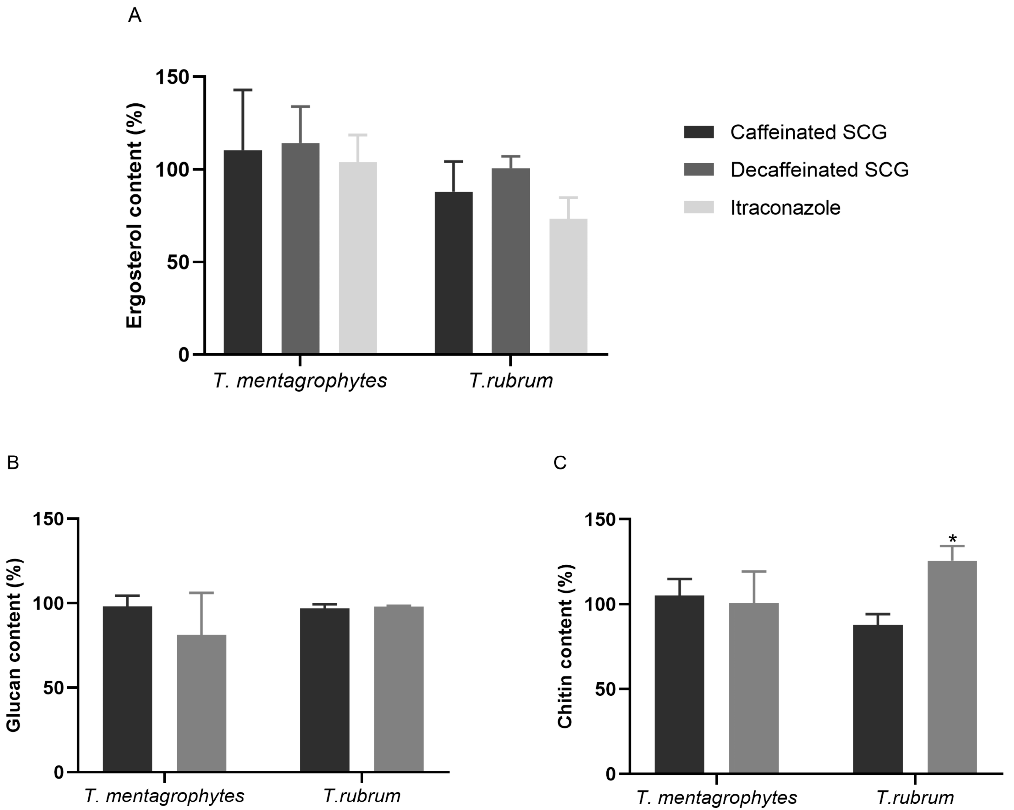
3.3. Cell Membrane and Cell Wall Components Modulation in Response to SCG

To elucidate a possible mechanism of action underlying the antifungal effect, ergosterol, β-(1,3)-glucan, and chitin contents of C. krusei, C. parapsilosis, T. mentagrophytes, and T. rubrum were determined after exposure to MIC of the SCG extracts (caffeinated and decaffeinated) and were expressed in a percentage in comparison to the non-treated fungal cells (control 100%). Ergosterol was also quantified after exposure to 0.016 μg/mL of itraconazole as a positive control. A lower concentration than the MIC value was used to allow fungal growth and to have enough biomass to extract and quantify ergosterol.
As seen in Figure 1, no significant differences in the ergosterol, chitin, or β-(1,3)-glucan after exposure of C. krusei to caffeinated or decaffeinated SCG extracts were detected. Yet, in C. parapsilosis, the caffeinated SCG extract reduced the ergosterol content by 49.77% with statistical significance (p < 0.01), similar to the reduction (52.32%) caused by itraconazole (p < 0.001). This extract also led to a statistically significant reduction of 14.83% of the amount of β-(1,3)-glucan (p < 0.01), and a reduction of 21.66% in the chitin content (p < 0.05). In C. parapsilosis, exposure to the decaffeinated SCG extract only caused a decrease in the chitin content by 38.62%, with statistical significance (p < 0.01).
In respect to filamentous fungi, the ergosterol content of T. mentagrophytes after exposure to caffeinated and decaffeinated SCG extracts showed no decrease, even when treated with itraconazole (0.016 μg/mL) (Figure 2A). Treatment with caffeinated SCG extracts reduced the total amount of ergosterol of T. rubrum by 12.05% but the decaffeinated SCG extracts had no impact. Itraconazole lowered the ergosterol content by 26.68%, with no statistical significance. The β-(1,3)-glucan content of T. mentagrophytes and T. rubrum (Figure 2B) had no decrease when exposed to caffeinated SCG extracts. The decaffeinated SCG extract only reduced T. mentagrophytes β-(1,3)-glucan by 18.75% but not with statistical significance. The quantification of chitin content of T. mentagrophytes revealed no significant alterations after treatment with caffeinated and decaffeinated SCG extracts (Figure 2C). The chitin levels of T. rubrum decreased by 12.16% after exposure to the caffeinated SCG extract, without statistical significance. Curiously, exposure to the decaffeinated SCG extract increased the total amount of chitin by 25.69% (p < 0.05).

3.4. Fungal Ultrastructural Modifications in Response to SCG Evaluated by TEM

Considering the modulation caused by the caffeinated SCG extract in the cell membrane and cell wall of C. parapsilosis and the potential fungicidal effect on T. rubrum, both these species were selected to elucidate the effect of caffeinated and decaffeinated SCG extracts at the ultrastructural level in comparison to untreated fungal cells. Fungal cells were exposed to a MIC concentration of both the extracts and a TEM study was performed.
C. parapsilosis untreated cells (Figure 3A,B) exhibited a well-defined surface and a regular and integral cell wall. However, there appear to be some structures that cross the cytoplasmic membrane and the cell wall to the outside (black arrows). C. parapsilosis treated with caffeinated SCG extracts (Figure 3C–E) and decaffeinated SCG extracts (Figure 3F–H) have a thinner (Figure 4) cell wall and more electrodense cell wall (a more electrodense line in the middle of the cell wall), and the same unknown structures that cross the cytoplasmic membrane are present but higher in number (black arrows).
Regarding T. rubrum, untreated cells (Figure 5A,B) have a smooth surface, a regular cell wall, a structured and regular organelles disposition, and visible mitochondria with well-defined cristae. In contrast, cells treated with the caffeinated SCG extract (Figure 5C–E) and decaffeinated SCG extract (Figure 5F–H) have organelle disorganization, especially in what concerns mitochondria, an increased number of and larger vacuoles, and there appear to be endomembrane systems suggesting autophagic structures.
To determine whether the SCG extract was associated with antiproliferative activities, its cytotoxicity was assessed using assays in human tumor cell lines AGS (gastric adenocarcinoma), CaCo2 (colorectal adenocarcinoma), MCF-7 (breast adenocarcinoma), and NCI-H460 (lung carcinoma), along with non-tumor cell lines PLP2 (primary pig liver culture). As depicted in Table 4, the SCG extracts exhibited cytotoxic activity in all cancer cell lines tested, at relatively low concentrations. Among the tumor cell lines, the gastric adenocarcinoma human (AGS) was more susceptible to the antiproliferative effects of SCG extracts with GI50 values of 55 and 52 µg/mL, for caffeinated and decaffeinated SCG extracts, respectively. Whereas in the normal cell line PLP2, SCG extracts exhibited little cytotoxicity with GI50 value > 400 µg/mL, compared to the control, ellipticine. These results are indicative of the potential of SCG extracts as an inhibitor of the growth of human cancer cell lines, especially AGS.
The anti-inflammatory activity of SCG extracts was evaluated by measuring the percent of inhibition of NO production, expressed in IC50 in LPS-activated RAW 264.7 cells. Cells were pre-treated with SCG extracts and incubated for 1 h, stimulated with 1 µg/mL of LPS, and incubated for 24 h. As shown in Table 4, the SCG extracts showed inhibitory effects on the NO production in LPS-activated RAW 264.7 with a IC50 > 400 µg/mL, which means that the NO production was not inhibited by the SCG extracts, so no anti-inflammatory activity of the SCG was obtained.

4. Discussion

The incidences of fungal infections have been increasing over the years and antifungal therapy is still limited. To overcome antifungal resistance, there is an urgent search for alternatives to conventional antifungal drugs; new and novel strategies with high antifungal potential are needed. SCG have been reported as a potential source of bioactive compounds with beneficial health effects. Caffeine is the most abundant compound, followed by chlorogenic acids and derivatives such as caffeoylquinic acids, feruloylquinic acids, p-coumaroylquinic acids, and mixed diesters of caffeic and ferulic acids with quinic acid, all described as powerful in vitro antioxidants [16,27,28,36].
In the present study, SCG from caffeinated and decaffeinated coffee were obtained from Delta Qalidus® and Delta deQafeinatus® capsules (Portuguese brand of coffee products), respectively. According to Delta Q® commercial online information, Delta Qalidus® is a fusion of arabica coffee beans from Honduras and robusta coffee from Angola and Cameroon while Delta deQafeinatus® is a mix of arabica coffee from Brazil and robusta coffee from Vietnam and Uganda. Aqueous and ethanolic extracts were prepared and microdilution broth was performed to study the susceptibility of common fungal agents of human infections. Only the ethanolic extracts showed antifungal activity. Mussato et al. quantified the total phenolic compounds and antioxidant activity of SCG extracts with different extraction solvents and obtained a higher yield using methanol in comparison with water, justifying that phenolic compounds are usually more soluble in organic solvents that are less polar than water [27]. Extraction with ethanol as the solvent recovered more than 90% of the phenolic compounds present in SCG [15,36].
The ethanolic caffeinated SCG extract obtained in the present study is abundant in phenolic compounds, namely in the chlorogenic acids derivatives, as trans 5-O-caffeoylquinic acid, 4-O-feruloylquinic acid, caffeoylshikimic acid, and cis 5-O- caffeoylquinic acid. The decaffeinated SCG extract is also abundant in the chlorogenic acid derivatives cis 5-O-caffeoylquinic acid and caffeoylshikimic acid. Chlorogenic acid and its derivatives have already been reported to have antifungal activity against C. albicans by impairing ergosterol biosynthesis and disrupting the cell membrane [37]. Another study showed the antifungal potential of chlorogenic acids against phytopathogenic fungi causing early membrane permeabilization of the spores [38]. Ma and Ma showed the antifungal activity of chlorogenic acid derivatives by inhibiting glucan synthase leading to a decrease in glucan content and cell wall disruption, suggesting their potential as antifungal agents [39]. The antifungal activity of 5-O-caffeoylquinic acid against Candida sp. and Aspergillus sp. has also been reported before [40,41].
SCG from caffeinated and decaffeinated coffee extracts showed antifungal activity against the same fungal species: C. krusei, C. parapsilosis, T. mentagrophytes, and T. rubrum, fungi associated with fungal skin infections and not to A. alternata, A. infectoria, A. fumigatus, A. niger, C. albicans, or C. glabrata fungi more associated with other human pathologies. This could suggest an interest for these extracts for topical application. Interestingly, the MIC determination of SCG from caffeinated and decaffeinated coffee extracts revealed to be fungicidal to T. mentagrophytes and T. rubrum, killing 99.9% of the fungal agents. The control using ground coffee (GC) extracts revealed that unused coffee grounds had antifungal activity, with lower MIC values. The brewing process of GC removes water-soluble compounds, making SCG less concentrated. Nonetheless, SCG extracts have antifungal activity against the same species as GC extracts. Despite being less concentrated, the bioactive compounds with antifungal activity remain present in SCG. Previous studies have highlighted the antimicrobial activity of SCG, mostly with a focus on bacteria [42,43,44,45,46,47]. Studies describing the antifungal effect of SCG are scarce. Several yeast species (C. krusei, C. parapsilosis, and C. albicans) have already been reported to be susceptible to SCG extracts [12,43]. The antifungal activity against filamentous fungi was also recently reported for a SCG extract obtained using isopropanol [45]. However, it is difficult to compare between studies due to different methodologies (type of coffee, extraction method, antifungal susceptibility tests, and fungal strains). Regardless, this is the first study describing the antifungal effect of SCG against fungal agents of dermatophytosis, T. mentagrophytes, and T. rubrum.
To identify the mechanism of action underlying the antifungal effect of the extracts, cell membrane and cell wall components were quantified. Ergosterol is the main sterol present in the fungal cell membrane, and it is responsible for the membrane’s structural integrity, fluidity, and permeability [48,49,50]. Chitin and glucan are fundamental in maintaining fungal cell integrity during growth and adaptation to stress [48,51,52]. Impairment in these components could lead the cell membrane or cell wall to disruption and consequently, cell death [45,50,51,52]. Most antifungal therapy available targets these components; however, conventional antifungals are associated with many side effects and drug to drug interactions, with hepatotoxicity, nephrotoxicity, and myelotoxicity being the most severe side effects [53,54,55].
Quantification of ergosterol, β-(1,3)-glucan, and chitin after exposure to the MIC of SCG from caffeinated and decaffeinated coffee extracts was performed. SCG caffeinated extracts induced significant impairment on ergosterol, β-(1,3)-glucan, and chitin contents of C. parapsilosis, while SCG from decaffeinated coffee, caused a non-significant reduction in ergosterol and β-(1,3)-glucan levels but a significant decrease in chitin content. Those might be the targets of these extracts in C. parapsilosis. However, it remains unknown where the ergosterol and chitin pathway might be affected by exposure to the extracts. Yet, in C. krusei, none of these contents were affected significantly by both extracts, although this species is more sensitive to both these extracts, indicating that another vital structure or physiological pathway might be impaired.
The decrease in the β-(1,3)-glucan and chitin content of C. parapsilosis and the thinner and more electrodense cell wall observed on TEM images may suggest a cell wall remodeling and changes to the chitin and β-(1,3)-glucan architecture after exposure to SCG extracts [56]. No scientific explanation was found to justify the structures that cross the cell wall present in C. parapsilosis un-treated cells, although treatment with SCG extracts seems to increase the number of these structures, which might be a response to stress induced by the extracts.
Although SCG from caffeinated and decaffeinated extracts were determined to be fungicidal against T. mentagrophytes and T. rubrum, the quantification of the cell membrane and cell wall components had no significant impact, except on the chitin content of T. rubrum when exposed to SCG from decaffeinated extracts. Curiously, chitin biosynthesis increased. Chitin build-up is generally associated with a compensatory mechanism triggered in response to cell wall perturbing agents as a defense mechanism [31,52,57]. According to these results, it was not possible to conclude a target underlying the antifungal activity of both extracts against these dermatophyte species. However, TEM images of T. rubrum treated with SCG extracts show disorganized, undefined, and perhaps dysfunctional mitochondria, in contrast to non-treated cells where it is possible to observe healthy, organized, and well-defined mitochondria. Mitochondria are related to antifungal tolerance; its dysfunction is associated with the susceptibility and resistance to cell membrane targeting drugs [58]. Besides that, after exposure to the extracts, it also possible to observe in T. rubrum TEM images an increase in the number and size of vacuoles and the presence of structures that resemble endomembrane systems, suggesting that autophagy is activated as a protective mechanism, possibly the mechanism behind the antifungal activity of SCG extracts.
In this study, we also reported that the SCG caffeinated and decaffeinated coffee extracts were cytotoxic for different cancer cell lines, without causing cytotoxicity on the non-tumoral cell line used. This is in accordance with previous works that reports a moderate cytotoxicity of SCG extracts on liver cancer cells (HepG2), which are also lower than the positive control used in the assay [45]. However, our results did not show anti-inflammatory activity of SCG from caffeinated and decaffeinated coffee extracts on the RAW 264.7 cell line. A recent publication reported that the SCG water extracts are more effective than extracts obtained with solvents [59]. Nevertheless, when considering the antifungal activity of the extracts obtained in this study in fungi agents of human skin infections together with the absence of cytotoxicity on a non-tumoral cell line, it can be anticipated as a possible utilization in the therapy of dermatomycosis.

5. Conclusions

Coffee consumption generates tons of solid waste SCG, containing several bioactive compounds with potential uses in the pharmaceutical, food, and cosmetic industries [16]. In this study, we show that the ethanolic SCG extracts from both caffeinated and decaffeinated coffee are cytotoxic for different cancer cell lines, indicating antiproliferative bioactivity, with no cytotoxicity on a non-tumoral cell line. Our results also clearly demonstrate that these extracts have a strong antifungal bioactivity, especially against fungi involved in skin infections. This indicates that a safe formulation of an SCG-based phytotherapeutic for topic administration could be an economic and eco-friendly alternative to treat skin fungal infections, promoting a coffee by-product and minimizing waste.

Author Contributions

Conceptualization, T.G., D.C. and C.F.; extracts preparation, antifungal assays, TEM, cell wall and membrane quantification methodologies, D.C.; validation, D.C. and C.F.; antioxidant, cytotoxic, and anti-inflammatory activity assays, and extract analysis, M.I.D., R.C.C., L.B. and I.C.F.R.F.; data curation, D.C.; writing—original draft preparation, D.C.; writing—review and editing, T.G., C.F. and D.C.; supervision, T.G. and C.F.; funding acquisition, T.G. All authors have read and agreed to the published version of the manuscript.

Funding

This work was financed by the European Regional Development Fund (ERDF), through the Centro 2020 Regional Operational Programme: project CENTRO-01-0145-FEDER- 000012- HealthyAging2020 and CENTRO-01-0145-FEDER- 022095: ViraVector; the COMPETE 2020–Operational Programme for Competitiveness and Internationalisation, and the Portuguese national funds via FCT–Fundação para a Ciência e a Tecnologia, I.P.:UIDB/04539/2020 and UIDP/04539/2020. The authors are also thankful to the Foundation for Science and Technology (FCT, Portugal) for financial support through national funds FCT/MCTES (PIDDAC) to CIMO (UIDB/00690/2020 and UIDP/00690/2020) and SusTEC (LA/P/0007/2020); national funding by FCT, P.I., through the institutional scientific employment program-contract for R.C.C., M.I.D., and L.B. contract.

Institutional Review Board Statement

Not applicable.

Informed Consent Statement

Not applicable.

Data Availability Statement

Not applicable.

Acknowledgments

The authors are thankful to NTfor the technical support at the Chemistry Department of the Faculty of Sciences and Technology of the University of Coimbra.

Conflicts of Interest

The authors declare no conflict of interest. The study was not funded by any commercial coffee brand.

References

  1. Brown, G.D.; Denning, D.W.; Gow, N.A.R.; Levitz, S.M.; Netea, M.G.; White, T.C. Hidden killers: Human fungal infections. Sci. Transl. Med. 2012, 4, 165rv13. [Google Scholar] [CrossRef] [Green Version]
  2. Faway, E.; de Rouvroit, C.L.; Poumay, Y. In vitro models of dermatophyte infection to investigate epidermal barrier alterations. Exp. Dermatol. 2018, 27, 915–922. [Google Scholar] [CrossRef] [Green Version]
  3. Garber, G. An overview of fungal infections. Drugs 2001, 61 (Suppl. S1), 1–12. [Google Scholar] [CrossRef]
  4. Ghannoum, M.A.; Isham, N.C. Dermatophytes and dermatophytoses. In Clinical Mycology, 2nd ed.; Elsevier: Amsterdam, The Netherlands, 2009; pp. 375–384. [Google Scholar]
  5. Tainwala, R.; Sharma, Y.K. Pathogenesis of dermatophytoses. Indian J. Dermatol. 2011, 56, 259–261. [Google Scholar] [CrossRef]
  6. Geddes-McAlister, J.; Shapiro, R.S. New pathogens, new tricks: Emerging, drug-resistant fungal pathogens and future prospects for antifungal therapeutics. Ann. N. Y. Acad. Sci. 2019, 1435, 57–78. [Google Scholar] [CrossRef]
  7. Alenazy, H.; Alghamdi, A.; Pinto, R.; Daneman, N. Candida colonization as a predictor of invasive candidiasis in non-neutropenic ICU patients with sepsis: A systematic review and meta-analysis. Int. J. Infect. Dis. 2021, 102, 357–362. [Google Scholar] [CrossRef]
  8. Kühbacher, A.; Burger-Kentischer, A.; Rupp, S. Interaction of Candida species with the skin. Microorganisms 2017, 5, 32. [Google Scholar] [CrossRef] [Green Version]
  9. Casadevall, A. Fungal diseases in the 21st century: The near and far horizons. Pathog. Immun. 2018, 3, 183–196. [Google Scholar] [CrossRef]
  10. Fisher, M.C.; Gurr, S.J.; Cuomo, C.A.; Blehert, D.S.; Jin, H.; Stukenbrock, E.H.; Stajich, J.E.; Kahmann, R.; Boone, C.; Denning, D.W.; et al. Threats Posed by the Fungal Kingdom to Humans, Wildlife, and Agriculture. mBio 2020, 11, e00449-20. [Google Scholar] [CrossRef]
  11. Mishra, K.K.; Kaur, C.D.; Sahu, A.K.; Panik, R.; Kashyap, P.; Mishra, S.P.; Dutta, S. Medicinal Plants Having Antifungal Properties. In Medicinal Plants-Use in Prevention and Treatment of Diseases; Hassan, B.A.R., Ed.; IntechOpen: London, UK, 2020. [Google Scholar]
  12. Sousa, C.; Gabriel, C.; Cerqueira, F.; Manso, M.C.; Vinha, A.F. Coffee industrial waste as a natural source of bioactive compounds with antibacterial and antifungal activities. In The Battle Against Microbial Pathogens: Basic Science, Technological Advances and Educational Programs; Méndez-Vilas, A., Ed.; Formatex Research Center: Badajoz, Spain, 2016; pp. 131–136. [Google Scholar]
  13. Campos-Vega, R.; Loarca-Piña, G.; Vergara-Castañeda, H.A.; Dave Oomah, B. Spent coffee grounds: A review on current research and future prospects. Trends Food Sci. Technol. 2015, 45, 24–36. [Google Scholar] [CrossRef]
  14. Nieber, K. The Impact of Coffee on Health Author Pharmacokinetics and Mode of Action Bioactive Components in Coffee. Planta Med. 2017, 83, 1256–1263. [Google Scholar]
  15. Panusa, A.; Zuorro, A.; Lavecchia, R.; Marrosu, G.; Petrucci, R. Recovery of natural antioxidants from spent coffee grounds. J. Agric. Food Chem. 2013, 61, 4162–4168. [Google Scholar] [CrossRef]
  16. Kovalcik, A.; Obruca, S.; Marova, I. Valorization of spent coffee grounds: A review. Food Bioprod. Process. 2018, 110, 104–119. [Google Scholar] [CrossRef]
  17. Colombo, R.; Papetti, A. Decaffeinated coffee and its benefits on health: Focus on systemic disorders. Crit. Rev. Food Sci. Nutr. 2021, 61, 2506–2522. [Google Scholar] [CrossRef]
  18. Duangjai, A.; Suphrom, N.; Wungrath, J.; Ontawong, A.; Nuengchamnong, N.; Yosboonruang, A. Comparison of antioxidant, antimicrobial activities and chemical profiles of three coffee (Coffea arabica L.) pulp aqueous extracts. Integr. Med. Res. 2016, 5, 324–331. [Google Scholar] [CrossRef] [Green Version]
  19. Alvarado-Ambriz, S.; Lobato-Calleros, C.; Hernández-Rodríguez, L.; Vernon-Carter, E. Wet processing coffee waste as an alternative to produce extracts with antifungal activity: In vitro and in vivo valorization. Rev. Mex. De Ing. Química 2020, 19 (Suppl. S1), 135–149. [Google Scholar] [CrossRef]
  20. Sangta, J.; Wongkaew, M.; Tangpao, T.; Withee, P.; Haituk, S.; Arjin, C.; Sringarm, K.; Hongsibsong, S.; Sutan, K.; Pusadee, T.; et al. Recovery of Polyphenolic Fraction from Arabica Coffee Pulp and Its Antifungal Applications. Plants 2021, 10, 1422. [Google Scholar] [CrossRef]
  21. Rodrigues, F.; Palmeira-de-Oliveira, A.; das Neves, J.; Sarmento, B.; Amaral, M.H.; Oliveira, M.B. Coffee silverskin: A possible valuable cosmetic ingredient. Pharm. Biol. 2015, 53, 386–394. [Google Scholar] [CrossRef]
  22. Mathur, I.; Shruthi, S.; Gandrakota, K.; Nisha, K.K. Comparative Evaluation of Antifungal Activity of Green Coffee and Green Tea Extract against Candida albicans: An In Vitro Study. World J. Dent. 2021, 12, 265–270. [Google Scholar] [CrossRef]
  23. Antoine, G.; Vaissayre, V.; Meile, J.-C.; Payet, J.; Conéjéro, G.; Costet, L.; Fock-Bastide, I.; Joët, T.; Dussert, S. Diterpenes of Coffea seeds show antifungal and anti-insect activities and are transferred from the endosperm to the seedling after germination. Plant Physiol. Biochem. 2023, 194, 627–637. [Google Scholar] [CrossRef]
  24. Mahajan, R.; Kapoor, N. Phytochemical analysis and antimicrobial activity of roasted beans of coffea robusta. Int. J. Pharm. Biol. Sci. 2018, 8, 89–95. [Google Scholar]
  25. Martínez-Tomé, M.; Jiménez-Monreal, A.M.; García-Jiménez Almela, L.L.; García-Diz, L.; Mariscal-Arcas, M.; Murcia, M.A. Assessment of antimicrobial activity of coffee brewed in three different ways from different origins. Eur. Food Res. Technol. 2011, 233, 497. [Google Scholar] [CrossRef]
  26. Mehta, V.; Rajesh, G.; Rao, A.; Shenoy, R.; Pai, M. Antimicrobial Efficacy of Punica granatum mesocarp, Nelumbo nucifera Leaf, Psidium guajava Leaf and Coffea Canephora Extract on Common Oral Pathogens: An In-vitro Study. J. Clin. Diagn. Res. 2014, 8, 65–68. [Google Scholar]
  27. Mussatto, S.I.; Ballesteros, L.F.; Martins, S.; Teixeira, J.A. Extraction of antioxidant phenolic compounds from spent coffee grounds. Sep. Purif. Technol. 2011, 83, 173–179. [Google Scholar] [CrossRef] [Green Version]
  28. Farah, A.; Donangelo, C.M. Phenolic compounds in coffee. Braz. J. Plant Physiol. 2006, 18, 23–36. [Google Scholar] [CrossRef]
  29. Murthy, P.S.; Naidu, M.M. Recovery of Phenolic Antioxidants and Functional Compounds from Coffee Industry By-Products. Food Bioprocess Technol. 2012, 5, 897–903. [Google Scholar] [CrossRef]
  30. Ramalakshmi, K.; Rao, L.J.M.; Takano-Ishikawa, Y.; Goto, M. Bioactivities of low-grade green coffee and spent coffee in different in vitro model systems. Food Chem. 2009, 115, 79–85. [Google Scholar] [CrossRef]
  31. Fernandes, C.; Anjos, J.; Walker, L.A.; Silva, B.M.A.; Cortes, L.; Mota, M.; Munro, C.A.; Gow, N.A.R.; Gonçalves, T. Modulation of Alternaria infectoria cell wall chitin and glucan synthesis by cell wall synthase inhibitors. Antimicrob. Agents Chemother. 2014, 58, 2894–2904. [Google Scholar] [CrossRef] [Green Version]
  32. Bessada, S.M.F.; Barreira, J.C.M.; Barros, L.; Ferreira, I.C.F.R.; Oliveira, M.B.P.P. Phenolic profile and antioxidant activity of Coleostephus myconis (L.) Rchb.f.: An underexploited and highly disseminated species. Ind. Crops Prod. 2016, 89, 45–51. [Google Scholar] [CrossRef] [Green Version]
  33. Espinel-Ingroff, A.; Chaturvedi, V.; Fothergill, A.; Rinaldi, M. Optimal testing conditions for determining MICs and minimum fungicidal concentrations of new and established antifungal agents for uncommon molds: NCCLS collaborative study. J. Clin. Microbiol. 2002, 40, 3776–3781. [Google Scholar] [CrossRef] [Green Version]
  34. Breivik, O.N.; Owades, J.L. Yeast analysis, spectrophotometric semimicrodetermination of ergosterol in yeast. J. Agric. Food Chem. 1957, 5, 360–363. [Google Scholar] [CrossRef]
  35. Fernandes, C.; Mota, M.; Barros, L.; Dias, M.I.; Ferreira, I.C.F.R.; Piedade, A.P.; Casadevall, A.; Gonçalves, T. Pyomelanin Synthesis in Alternaria alternata Inhibits DHN-Melanin Synthesis and Decreases Cell Wall Chitin Content and Thickness. Front Microbiol. 2021, 12, 691433. [Google Scholar] [CrossRef]
  36. Zuorro, A.; Lavecchia, R. Spent coffee grounds as a valuable source of phenolic compounds and bioenergy. J. Clean. Prod. 2012, 34, 49–56. [Google Scholar] [CrossRef]
  37. Sung, W.S.; Lee, D.G. Antifungal action of chlorogenic acid against pathogenic fungi, mediated by membrane disruption. Pure Appl. Chem. 2010, 82, 219–226. [Google Scholar] [CrossRef]
  38. Martínez, G.; Regente, M.; Jacobi, S.; Del Rio, M.; Pinedo, M.; de la Canal, L. Chlorogenic acid is a fungicide active against phytopathogenic fungi. Pestic. Biochem. Physiol. 2017, 140, 30–35. [Google Scholar] [CrossRef]
  39. Ma, J.-N.; Ma, C.-M. Antifungal inhibitory activities of caffeic and quinic acid derivatives. In Coffee in Health and Disease Prevention; Preedy, V.R., Ed.; Elsevier Inc.: Amsterdam, The Netherlands; Academic Press: London, UK, 2015; pp. 635–641. [Google Scholar]
  40. Kalinowska, M.; Bajko, E.; Matejczyk, M.; Kaczyński, P.; Łozowicka, B.; Lewandowski, W. The Study of Anti-/Pro-Oxidant, Lipophilic, Microbial and Spectroscopic Properties of New Alkali Metal Salts of 5-O-Caffeoylquinic Acid. Int. J. Mol. Sci. 2018, 19, 463. [Google Scholar] [CrossRef] [Green Version]
  41. Suárez-Quiroz, M.L.; Alonso Campos, A.; Valerio Alfaro, G.; González-Ríos, O.; Villeneuve, P.; Figueroa-Espinoza, M.C. Anti-Aspergillus activity of green coffee 5-O-caffeoyl quinic acid and its alkyl esters. Microb. Pathog. 2013, 61–62, 51–56. [Google Scholar] [CrossRef] [PubMed]
  42. Almeida, A.A.P.; Farah, A.; Silva, D.A.; Nunan, E.A.; Glória, M.B.A. Antibacterial activity of coffee extracts and selected coffee chemical compounds against enterobacteria. J. Agric. Food Chem. 2006, 54, 8738–8743. [Google Scholar] [CrossRef]
  43. Monente, C.; Bravo, J.; Vitas, A.I.; Arbillaga, L.; De Peña, M.P.; Cid, C. Coffee and spent coffee extracts protect against cell mutagens and inhibit growth of food-borne pathogen microorganisms. J. Funct. Foods 2015, 12, 365–374. [Google Scholar] [CrossRef]
  44. Díaz-Hernández, G.C.; Alvarez-Fitz, P.; Maldonado-Astudillo, Y.I.; Jiménez-Hernández, J.; Parra-Rojas, I.; Flores-Alfaro, E.; Salazar, R.; Ramírez, M. Antibacterial, Antiradical and Antiproliferative Potential of Green, Roasted, and Spent Coffee Extracts. Appl. Sci. 2022, 12, 1938. [Google Scholar] [CrossRef]
  45. Badr, A.N.; El-Attar, M.M.; Ali, H.S.; Elkhadragy, M.F.; Yehia, H.M.; Farouk, A. Spent Coffee Grounds Valorization as Bioactive Phenolic Source Acquired Antifungal, Anti-Mycotoxigenic, and Anti-Cytotoxic Activities. Toxins 2022, 14, 109. [Google Scholar] [CrossRef] [PubMed]
  46. Rawangkan, A.; Siriphap, A.; Yosboonruang, A.; Kiddee, A.; Pook-In, G.; Saokaew, S.; Sutheinkul, O.; Duangjai, A. Potential Antimicrobial Properties of Coffee Beans and Coffee By-Products Against Drug-Resistant Vibrio cholerae. Front. Nutr. 2022, 9, 865684. [Google Scholar] [CrossRef] [PubMed]
  47. Bouhlal, F.; Aqil, Y.; Chamkhi, I.; Belmaghraoui, W.; Labjar, N.; El Hajjaji, S.; Benabdellah, G.A.; Aurag, J.; Lotfi, E.M.; Mahi, M.E. GC-MS Analysis, Phenolic Compounds Quantification, Antioxidant, and Antibacterial Activities of the Hydro-alcoholic Extract of Spent Coffee Grounds. J. Biol. Act. Prod. Nat. 2020, 10, 325–337. [Google Scholar] [CrossRef]
  48. Latgé, J.P. Tasting the fungal cell wall. Cell. Microbiol. 2010, 12, 863–872. [Google Scholar] [CrossRef] [PubMed]
  49. Money, N.P. Fungal Cell Biology and Development. In The Fungi, 3rd ed.; Elsevier Ltd.: Amsterdam, The Netherlands, 2016. [Google Scholar]
  50. Jordá, T.; Puig, S. Regulation of Ergosterol Biosynthesis in Saccharomyces cerevisiae. Genes 2020, 11, 795. [Google Scholar] [CrossRef]
  51. Bowman, S.M.; Free, S.J. The structure and synthesis of the fungal cell wall. BioEssays 2006, 28, 799–808. [Google Scholar] [CrossRef]
  52. Gow, N.A.R.; Latgé, J.P.; Munro, C.A. The fungal cell wall: Structure, biosynthesis, and function. Microbiol. Spectr. 2017, 5, 267–292. [Google Scholar] [CrossRef] [Green Version]
  53. Kuranda, K.; Leberre, V.; Sokol, S.; Palamarczyk, G.; François, J. Investigating the caffeine effects in the yeast Saccharomyces cerevisiae brings new insights into the connection between TOR, PKC and Ras/cAMP signalling pathways. Mol. Microbiol. 2006, 61, 1147–1166. [Google Scholar] [CrossRef]
  54. Perfect, J.R. The antifungal pipeline: A reality check. Nat. Rev. Drug Discov. 2017, 16, 603–616. [Google Scholar] [CrossRef] [Green Version]
  55. Scorzoni, L.; de Paula e Silva, A.C.A.; Marcos, C.M.; Assato, P.A.; de Melo, W.C.M.A.; de Oliveira, H.C.; Costa-Orlandi, C.B.; Mendes-Giannini, M.J.S.; Fusco-Almeida, A.M. Antifungal therapy: New advances in the understanding and treatment of mycosis. Front. Microbiol. 2017, 8, 1–23. [Google Scholar] [CrossRef] [Green Version]
  56. Fernandes, C.; Gow, N.A.R.; Gonçalves, T. The importance of subclasses of chitin synthase enzymes with myosin-like domains for the fitness of fungi. Fungal Biol. Rev. 2016, 30, 1–14. [Google Scholar] [CrossRef] [Green Version]
  57. Arias, P.; Díez-Muñiz, S.; García, R.; Nombela, C.; Rodríguez-Peña, J.M.; Arroyo, J. Genome-wide survey of yeast mutations leading to activation of the yeast cell integrity MAPK pathway: Novel insights into diverse MAPK outcomes. BMC Genom. 2011, 12, 390. [Google Scholar] [CrossRef] [PubMed] [Green Version]
  58. Shingu-Vazquez, M.; Traven, A. Mitochondria and fungal pathogenesis: Drug tolerance, virulence, and potential for antifungal therapy. Eukaryot. Cell 2011, 10, 1376–1383. [Google Scholar] [CrossRef] [PubMed]
  59. Angeloni, S.; Freschi, M.; Marrazzo, P.; Hrelia, S.; Beghelli, D.; Juan-García, A.; Juan, C.; Caprioli, G.; Sagratini, G.; Angeloni, C. Antioxidant and Anti-Inflammatory Profiles of Spent Coffee Ground Extracts for the Treatment of Neurodegeneration. Oxidative Med. Cell. Longev. 2021, 2021, 6620913. [Google Scholar] [CrossRef]
Figure 1. Effects of caffeinated and decaffeinated SCG extracts on cell membrane and cell wall components of yeasts. C. krusei treated with 137.50 μg/mL of caffeinated SCG extract, 150 μg/mL of decaffeinated SCG extract, and 0.016 μg/mL of itraconazole. C. parapsilosis treated with 275 μg/mL of caffeinated SCG extract, 150 μg/mL of decaffeinated SCG extract, and 0.016 μg/mL of itraconazole. (A) Ergosterol, (B) β-(1,3)-glucan, and (C) chitin contents. Results are expressed as mean ± standard error and are a representation of three independent experiments performed in triplicate, * p < 0.05, ** p < 0.01, and *** p < 0.001 (Dunnett’s post-hoc test). Data were normalized to control values.
Microorganisms 11 00242 g001
Figure 2. Effects of caffeinated and decaffeinated SCG extracts on cell membrane and cell wall components of filamentous fungi. T. mentagrophytes treated with 137.5 μg/mL of caffeinated SCG extract, 300 μg/mL of decaffeinated SCG extract, and 0.016 μg/mL of itraconazole. T. rubrum treated with 137.5 μg/mL of caffeinated extract, 150 μg/mL of decaffeinated extract, and 0.016 μg/mL of itraconazole. (A) Ergosterol, (B) β-(1,3)-glucan, and (C) chitin contents. Results are expressed as mean ± standard error and a representation of three independent experiments performed in triplicate, * p < 0.05 (Dunnett’s post-hoc test). Data were normalized to control values.
Microorganisms 11 00242 g002
Figure 3. Transmission electron microscopic observations of C. parapsilosis. (A,B) non-treated fungal cells, (A) scale bar 1000 nm, (B) scale bar 2000 nm; (CE) treated with caffeinated SCG extract (275 μg/mL), scale bar: 1000 nm; (FH) treated with decaffeinated SCG extract (150 μg/mL), (F) scale bar: 1000 nm, and (G,H) scale bar: 2000 nm. Black arrows indicate structures crossing the membrane and cell wall.
Microorganisms 11 00242 g003
Figure 4. Measurement of C. parapsilosis cell wall thickness of non-treated fungal cells (control), treated with caffeinated SCG extract (275 μg/mL), and treated with decaffeinated SCG extract (150 μg/mL). Results are the mean ± the standard error of the mean of one experiment, **** p < 0.0001 (Dunnett’s post-hoc test).
Microorganisms 11 00242 g004
Figure 5. Transmission electron microscopic observations of T. rubrum. (A,B) Non-treated fungal cells, scale bar: 2000 nm; (CE) treated with caffeinated SCG extract (137.5 μg/mL), scale bar: 2000 nm; (FH) treated with decaffeinated SCG extract (150 μg/mL), (F,E) scale bar: 1000 nm, (H) scale bar 2000 nm*—autophagic-like structures; black arrow—vacuoles; open arrows—mitochondria.
Microorganisms 11 00242 g005
Table 1. Phenolic profile of caffeinated and decaffeinated SCG ethanol extracts.
PeakRt (min)λmax (nm)[M-H] (m/z)MS2 (m/z)Tentative IdentificationQuantification
(mg/g Extract)
t-Students
Test
p-Value
SCG CaffeinatedSCG Decaffeinated
14.64316353191(100), 179(78), 173(11), 135(14).3-O-Caffeoylquinic acid12.1 ± 0.18 ± 1<0.001
25.23319353191(49), 179(72), 173(100), 135(11).4-O-Caffeoylquinic acid5.26 ± 0.0312.1 ± 0.3<0.001
35.77323353191(100), 179(49), 173(52), 135(12).cis 5-O-Caffeoylquinic acid16.6 ± 0.524 ± 1<0.001
46.44323353191(100), 179(28), 173(35), 135(5).trans 5-O-Caffeoylquinic acid24 ± 114 ± 1<0.001
59.66325367193(23), 191(5), 173(100), 161(10), 149(4).4-O-Feruloylquinic acid21.17 ± 0.022.49 ± 0.04<0.001
612.01326335179/13), 161(100), 134(41).Caffeoylshikimic acid17.2 ± 0.120.3 ± 0.5<0.001
717.87324515MS2: 353(100), 335(12), 317(5), 299(12), 203(10), 179(14), 173(13). MS3:191(52), 179(70), 173(100).1,4-O-diCaffeoylquinic acid11.3 ± 0.510.2 ± 0.3<0.001
819.1324515MS2: 353(100). MS3: 335(12), 191(100), 179(81), 173(142).1,3-O-diCaffeoylquinic acid7.02 ± 0.114.7 ± 0.2<0.001
921.64324515MS2: 353(100), 335(9), 317(5), 299(21), 203(9), 173(5). MS3:191(32), 179(51), 173(100).3,4-O-diCaffeoylquinic acid8.3 ± 0.26.8 ± 0.5<0.001
1023.27325529MS2: 367(100), 335(20), 193(14). MS3: 173(100).4-Feruloyl-5-caffeoylquinic acid1.87 ± 0.031.5 ± 0.1<0.001
1127.29325529MS2: 367(100), 193(14). MS3: 191(100).3-Feruloyl-5-caffeoylquinic acid2.1 ± 0.22.7 ± 0.1<0.001
Total Phenolic Compounds127.39 ± 1.03107 ± 1<0.001
Standard calibration curves: chlorogenic acid (y = 168823x − 161172, R2 = 0.9999, LOD = 0.20 µg/mL; LOQ = 0.68 µg/mL, peaks 1, 2, 3, 4, 5, 6, 7, 8, and 9); ferulic acid (y = 633126x − 185462, R2 = 0.999; LOD = 0.20 μg/mL; LOQ = 1.01 μg/mL, peaks 5, 10, and 11).
Table 2. Minimum inhibitory concentration (MIC) of caffeinated and decaffeinated extracts and itraconazole for different species (ug/mL). Three independent experiments were performed.
SpeciesCaffeinated Coffee
Extract
Decaffeinated Coffee
Extract
Itraconazole
GCSCGGCSCG
A. alternata>1770.00>1100.00>1415.00>2400.000.50
A. fumigatus>1770.00>1100.00>1415.00>2400.002.00
A. infectoria>1770.00>1100.00>1415.00>2400.000.50
A. niger>1770.00>1100.00>1415.00>2400.00>16.00
C. albicans>1770.00>1100.00>1415.00>2400.000.06
C. glabrata>1770.00>1100.00>1415.00>2400.000.01
C. krusei55.31137.5044.22150.000.25
C. parapsilosis110.63275.0044.22150.000.03
F. oxysporum>1770.00>1100.00>1415.00>2400.00>16.00
T. mentagrophytes27.66137.5022.11300.000.03
T. rubrum27.66137.5022.11150.000.03
Table 3. Minimum fungicidal concentration of caffeinated and decaffeinated SCG extracts against T. mentagrophytes and T. rubrum. Three independent experiments were performed.
SpeciesMFC (μg/mL)
Caffeinated SCG ExtractDecaffeinated SCG Extract
T. mentagrophytes137.50300
T. rubrum137.50150
Table 4. Cytotoxic and anti-inflammatory activity of the caffeinated and decaffeinated SCG extracts.
Caffeinated SCG
Extract
Decaffeinated SCG ExtractPositive Control
Cytotoxic activity (GI50 and µg/mL) Ellipticine
AGS55 ± 452 ± 21.20 ± 0.03
CaCo2214 ± 9179 ± 101.20 ± 0.02
MCF-7207 ± 1675.7 ± 0.31.00 ± 0.02
NCI-H460217 ± 18228 ± 101.21 ± 0.02
Cytotoxic activity (GI50 and µg/mL)
PLP2>400240 ± 251.4 ± 0.1
Anti-inflammatory activity (IC50 and µg/mL) Dexametasone
RAW 264.7>400>4006.3 ± 0.4
Disclaimer/Publisher’s Note: The statements, opinions and data contained in all publications are solely those of the individual author(s) and contributor(s) and not of MDPI and/or the editor(s). MDPI and/or the editor(s) disclaim responsibility for any injury to people or property resulting from any ideas, methods, instructions or products referred to in the content.

