Review

Pleiotropic Functions of Nitric Oxide Produced by Ascorbate for the Prevention and Mitigation of COVID-19: A Revaluation of Pauling’s Vitamin C Therapy

Faculty of Science, University of the Ryukyus, Nishihara 903-0213, Okinawa, Japan
*
Author to whom correspondence should be addressed.
Microorganisms 2023, 11(2), 397; https://doi.org/10.3390/microorganisms11020397
Submission received: 16 December 2022 / Revised: 30 January 2023 / Accepted: 31 January 2023 / Published: 3 February 2023

Abstract

:
Linus Pauling, who was awarded the Nobel Prize in Chemistry, suggested that a high dose of vitamin C (l-ascorbic acid) might work as a prevention or treatment for the common cold. Vitamin C therapy was tested in clinical trials, but clear evidence was not found at that time. Although Pauling’s proposal has been strongly criticized for a long time, vitamin C therapy has continued to be tested as a treatment for a variety of diseases, including coronavirus infectious disease 2019 (COVID-19). The pathogen of COVID-19, SARS-CoV-2, belongs to the β-coronavirus lineage, which includes human coronavirus, severe acute respiratory syndrome (SARS), and Middle East respiratory syndrome (MERS). This review intends to shed new light on vitamin C antiviral activity that may prevent SARS-CoV-2 infection through the chemical production of nitric oxide (NO). NO is a gaseous free radical that is largely produced by the enzyme NO synthase (NOS) in cells. NO produced by upper epidermal cells contributes to the inactivation of viruses and bacteria contained in air or aerosols. In addition to enzymatic production, NO can be generated by the chemical reduction of inorganic nitrite (NO2), an alternative mechanism for NO production in living organisms. Dietary vitamin C, largely contained in fruits and vegetables, can reduce the nitrite in saliva to produce NO in the oral cavity when chewing foods. In the stomach, salivary nitrite can also be reduced to NO by vitamin C secreted from the epidermal cells of the stomach. The strong acidic pH of gastric juice facilitates the chemical reduction of salivary nitrite to produce NO. Vitamin C contributes in multiple ways to the host innate immune system as a first-line defense mechanism against pathogens. Highlighting chemical NO production by vitamin C, we suggest that controversies on the therapeutic effects of vitamin C in previous clinical trials may partly be due to less appreciation of the pleiotropic functions of vitamin C as a universal bioreductant.

1. Introduction

COVID-19 is the coronavirus infectious disease that has brought about a global pandemic since December 2019. As of December 2022, more than six million people have died worldwide due to COVID-19. This pandemic is caused by severe acute respiratory syndrome coronavirus 2 (SARS-CoV-2), which binds to a cell surface receptor of the host, called angiotensin converting enzyme 2 (ACE2) [1]. In a critical presentation of COVID-19, lung injury often progresses rapidly with acute respiratory distress syndrome (ARDS), followed by multiple organ failure due to a “cytokine storm”, a condition of uncontrolled systemic hyperinflammation induced by excessive cytokines [2]. Acute hypoxemic respiratory failure (AHRF) is the most common form of organ failure [3], contributing to over 90% of COVID-19-related deaths in intensive care unit (ICU) patients [4]. The incidence of COVID-19-related AHRF reached 15% of hospitalized patients [5]. Such severe patients with COVID-19 require invasive mechanical ventilation or ECMO (extracorporeal membrane oxygenation) [6].
The pandemic has allowed for continuous mutations of SARS-CoV-2, especially in the spike (S) gene [7], generating further diversity with an evolutionary rate between 0.0004 and 0.002 mutations per nucleotide per year [8]. The World Health Organization (WHO) designated the variants of concern (VOCs) using the Greek alphabet from Alpha, and now it is Omicron [9]. Until recently, a variety of drugs have been applied to cure moderate to severe COVID-19 patients [10,11,12]. Many types of vaccines have been administered to millions of people to suppress the outbreak [13,14]. To date, however, there are few drugs or vaccines available that are specific to new variants.
The antiviral drug remdesivir [10], along with dexamethasone (a systemic steroid) [15], is commonly used in patients with severe forms of COVID-19. Unfortunately, antiviral drugs that are currently available to clinicians have little effect on mortality, the length of in-hospital stay, and the need for mechanical ventilation [5]. COVID-19 and the common cold both present a syndrome of disease states. It may be difficult to rely on a single drug or chemical to cure the disease. High-dose intravenous vitamin C (HDIVC) treatment has been reported to be effective in decreasing days of hospitalization, ICU stay and mortality [16,17,18]. Vitamin C therapy has been known for several decades to be a safe adjunctive treatment that has been examined for a wide variety of diseases, including severe acute respiratory syndrome (SARS) caused by SARS-CoV [19]. Despite its long research background, there are controversies regarding the therapeutic effects of HDIVC treatment [20]. Here, we review ascorbate-dependent nitric oxide (NO) production, which may contribute to mitigating COVID-19 symptoms, as well as preventing SARS-CoV-2 infection. Revisiting Pauling’s vitamin C therapy proposed in the 1970s, the potential therapeutic roles of NO, nitrate, nitrite, vitamin C, and vitamin P are discussed in relation to their pleiotropic functions that have sometimes led to states of confusion.

2. NO Therapy for COVID-19 Patients

Inhaled nitric oxide gas (iNO) is considered a safe treatment for hospitalized patients [5,21]. NO is a free radical that plays an important pathophysiological role in the human cardiovascular and immune systems [22,23,24]. The use of iNO was first approved in 1999 by the U.S. Food and Drug Administration (FDA) as a treatment for neonatal pulmonary hypertension [25,26]. In 2003, iNO was tested in severe acute respiratory syndrome (SARS) patients. Clinical trials showed beneficial effects such as decreased pulmonary hypertension, improved arterial oxygenation, and a reduced spread and density of lung infiltrates [27]. Therefore, it was a logical rationale to investigate the therapeutic effects of iNO for COVID-19 as a preventive measure, a treatment against COVID-19, and a rescue treatment to improve arterial oxygenation against ARDS [22,28].
Pregnant patients are at risk of developing AHRF due to pregnancy. A recent cohort study reported that iNO was associated with a reduced need for oxygen supplementation and a shorter hospital stay for pregnant patients with COVID-19 pneumonia in the absence of adverse events in mothers or infants [29], supporting the safety of iNO therapy. Although there have yet to be active debates on the effect of iNO on the clinical outcomes of COVID-19 patients [30,31], a number of studies have confirmed that iNO therapy is associated with improved oxygenation parameters in COVID-19 patients [5,21,32]. The combination use of iNO with other drugs has also been tested as a rescue therapy in COVID-19 patients with ARDS [32].
In iNO therapy, NO can be delivered directly to the pulmonary vasculature by inhalation with a commercially available NO delivery system [23]. In healthy conditions, a sufficient amount of NO should be synthesized by the endogenous enzyme NO synthase (NOS; EC 1.14.13.39) [33]. The principle of iNO therapy can be attributed to a supplementation of NO gas to the pulmonary vasculature to improve the local NO bioavailability. In addition to the inhalation of NO gas, other clinical options to increase NO levels in COVID-19 patients have been investigated to date: the administration of the NOS substrates l-arginine [34,35] and l-citrulline [36] and the administration of NO chemical donors such as nitroglycerin [37], S-nitroso-N-acetylpenicillamine (SNAP) [38], sodium nitroprusside (SNP) [39], and 1,2,5-oxadiazole-2-oxide (furoxan) [40]. Viagra (sildenafil citrate) is the first commercially available drug for male erectile dysfunction (ED) [41]. Sildenafil, in principle, inhibits phosphodiesterase type 5 (PDE5) which breaks the second messenger cyclic GMP (cGMP), thereby allowing for the accumulation of cGMP in cells even under conditions of low NO input [41]. In many countries, sildenafil has been approved as a treatment for pulmonary hypertension in neonates [42], and its therapeutic effects have also been tested in COVID-19 patients [43,44,45]. These clinical trials have focused on the potent vasodilation activity of NO, which may reduce the risk of respiratory failure.

3. Antiviral Activity of NO

3.1. Smokers’ Paradox

Asthma and cigarette smoking, as well as obesity, diabetes, and chronic heart disease, are considered high-risk factors for acquiring COVID-19 or poorer outcomes. Early in the pandemic, however, there were few asthma patients with severe cases of COVID-19 [46,47]. A recent meta-analysis study also supported that people with asthma have a lower risk of SARS-CoV-2 infection than those without asthma [48]. The use of inhaled corticosteroids might partly account for a protective effect against SARS-CoV-2 infection, due to decreased ACE2 in asthma patients [49]. It is important to note that NO emission is generally high due to eosinophilic airway inflammation in asthmatic patients. In fact, the fraction exhaled NO (FENO) has been adopted as a non-invasive indicator of the type 2 airway inflammation of asthma [50]. Although non-allergic asthma (non-type 2) seems to have a greater risk, there were fewer asthma patients with COVID-19 in many countries [47], implying that high NO emission from the airway may protect against SARS-CoV-2 infection.
Cigarette smoking has also been listed as a risk factor for contracting COVID-19. In general, cigarette smoking is associated with relatively poor outcomes in respiratory infectious diseases [51]. In 2020, the WHO and the FDA released statements warning that smoking may increase the risk and severity of COVID-19. However, only a low proportion of smokers suffered from SARS-CoV-2 infection [52]. This is referred to as the “smokers’ paradox” [53]. Although smoking cannot be recommended as a protective measure for COVID-19, the underlying mechanism for the smokers’ paradox may give a clue for our consideration of preventing SARS-CoV-2 infection. Farsalinos et al. proposed that nicotine intake could be the reason for the low prevalence of smoking among hospitalized patients [51], whereas Hedenstierna et al. hypothesized that the short burst of concentrated NO (approximately 250 to 1350 ppm per puff) contained in cigarette smoke may prevent SARS-CoV-2 infection [54], an explanation similar to that given for why asthmatic patients are less likely to contract COVID-19.

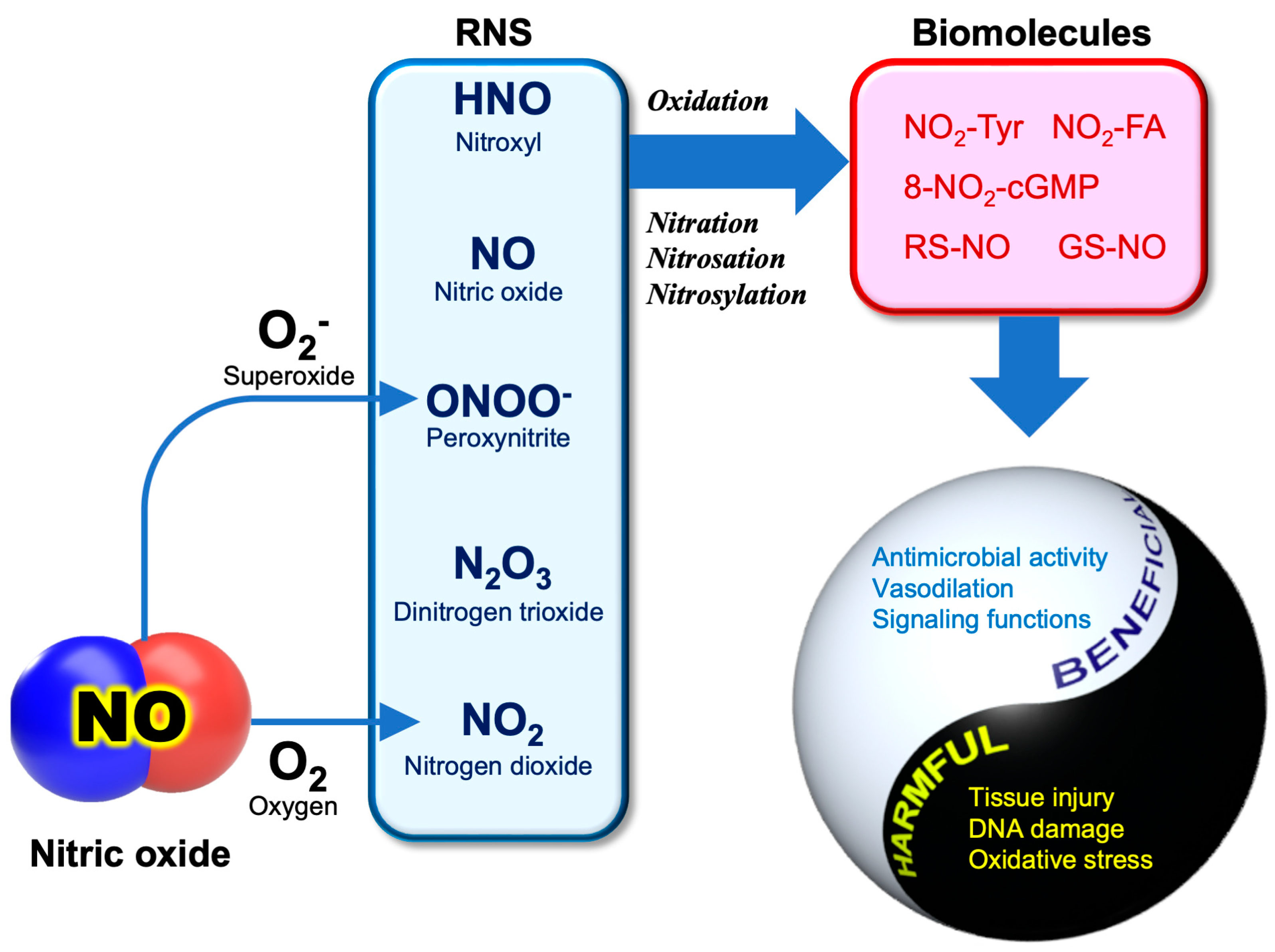
3.2. RNS Biochemistry

Apart from its potent actions on cardiovascular systems, NO is involved in innate immunological host defense. The innate immune response is also mediated by reactive oxygen species (ROS), including O2, H2O2, and hypochlorite anion (OCl), which are produced by phagocytic cells such as neutrophils and activated macrophages [55]. Its action is non-specific, and potentially inactivates a broad range of pathogens, including parasites, fungi, bacteria, and viruses [56]. As ROS is the term for a group of reactive molecules derived from O2, reactive molecules originating from NO are referred to as “reactive nitrogen species (RNS)” [57]. Figure 1 illustrates the pathophysiological conditions associated with the major RNS.
Like ROS, RNS are highly reactive oxidants that are associated with oxidative stress in living organisms. Compared with ROS, the chemistry of RNS-related reactions is much more complicated, especially under in vivo conditions, and most of them are not fully understood. Basically, the reactions of NO are involved in the oxidation, nitration (the addition of NO2), nitrosation (the addition of NO+), and nitrosylation (the addition of NO) of biomolecules [58]. An uncontrolled situation of these reactions may cause “nitrative” or “nitrosative stress”, leading to cellular damage or cell death.
Most classes of biomolecules, including proteins, nucleic acids, and lipids, can be nitrated, generating products such as nitro-tyrosine (NO2-Tyr) [59], 8-nitroguanosine 3′,5′-cyclclic monophosphate (8-nitro-cGMP) [60], nitro-fatty acids (NO2-FA) [61], and nitro-phenolics [62]. NO+ could directly react with the thiols (RSH) of cysteine residues or the reduced form of glutathione (GSH) to produce S-nitrosothiols (RS-NO, GS-NO). Regarding RS-NO chemical generation in biological systems, several possible mechanisms have been proposed, but currently none of them have reached a consensus [63]. The antipathogenic activity of NO relies on these unique RNS reactions (nitration, nitrosation, and nitrosylation) that are capable of inactivating or killing pathogens through the modification of biomolecules, including enzyme proteins. It is important to remember that these reactions are non-specific, thereby also causing cellular damage to the host cells during inflammation [64]. It appears that the use of RNS as a countermeasure against pathogens is a risky business for hosts.
Under oxidative stress conditions where ROS are overproduced, such as during inflammation, peroxynitrite (ONOO) can be produced as the reaction product between NO and O2, an important interplay between ROS and RNS [65].
The rate constant for the reaction between NO and O2 is near diffusion controlled (Equation (1)), which is faster than the superoxide dismutase (SOD) reaction that removes O2. The product ONOO is stable at pH 12 in the absence of target molecules. At physiological pH, ONOO is in rapid equilibrium with its conjugated acid peroxynitrous acid (ONOOH, pKa 6.8) (Equation (2)), which is a short-lived molecule that spontaneously decays to nitrate (Equation (3)) [66]. Due to its high reactivity, ONOO- is considered the major cytotoxic agent in RNS.
NO + O2 → ONOO
ONOO + H+ → ONOOH
ONOOH → NO3 + H+
ONOO and ONOOH are strong oxidants capable of oxidizing various molecules, such as thiols, sulfides, ascorbate, and phenols [67]. In addition to the oxidation of molecules, ONOO can chemically nitrate aromatics, with a reaction being facilitated in the presence of bicarbonate anion (HCO3) [68]. The dysfunction of proteins or enzymes may occur due to the formation of nitro-tyrosine residues (NO2-Tyr). ONOO- is also involved in DNA fragmentation [67] and RNA viral mutation [69] through deamination of the bases.

3.3. Anti-SARS-CoV-2 Activity of NO

The antiviral activity of NO has been reported for many types of viruses, most typically, DNA viruses such as murine poxvirus, herpesviruses, and some RNA viruses [55]. The direct action of NO as an antiviral agent involves the inhibition of viral replication and viral entry into the host [70,71]. In 1999, Saura et al. demonstrated that the in vitro replication of the RNA virus coxsackievirus is suppressed by NO-dependent S-nitrosylation that causes the inactivation of viral cysteine protease, an enzyme necessary for replication [72]. The S-nitrosylation of the cysteine-containing enzymes of viruses is thought to be a general mechanism for the antiviral activity of NO [73].
SARS-CoV-2 is a positive-sense RNA virus belonging to the family Coronaviridae, which includes severe acute respiratory syndrome coronavirus (SARS-CoV), the pathogen that caused the SARS outbreak. In 2005, Akerstrom et al. reported that the NO chemical donor SNAP inhibits the in vitro replication cycle and the protein and RNA synthesis of SARS-CoV [74]. This inhibitory effect was not observed with SNP (sodium nitroprusside), another chemical NO donor [38]. Likewise, NO released from SNAP was reported to inhibit the replication of SARS-CoV-2 in Vero E6 cells through the inhibition of the SARS-CoV-2 3CL cysteine protease [75].
Macrophages are multifunctional innate immune cells that play an essential role in the clearance of pathogens and control inflammatory responses. Recent studies have suggested that S-palmitoylation is a key reaction for control macrophages in the processes of endocytosis [76]. Interestingly, NO was reported to suppress the palmitoylation of the spike (S) proteins that is needed for their binding to ACE2 [77]. The spike (S) proteins of coronaviruses are receptor-binding proteins that are synthesized in the endoplasmic reticulum (ER), followed by complex post-translational modification in the host Golgi apparatus [78,79,80]. S-Palmitoylation is one of the post-translational modifications in the Golgi apparatus where palmitoyl acyltransferase (PAT) adds the saturated fatty acid palmitate (C16:0) to the cysteine thiol group (-SH) of proteins [78,80]. Protein modification causes an increase in the hydrophobicity of the proteins, which is essential for cell-cell fusion activity [79,81]. Endothelial NO synthase (eNOS), an isoform of the host’s NO-producing enzyme, can be modified by palmitoylation, and its activity is decreased by the modification [82]. S-nitrosylation of the SARS-CoV spike (S) protein with NO may reduce cell-cell fusion activity through decreased amounts of spike (S) protein palmitoylation [77]. It is presumable that the disturbance of the cysteine palmitoylation of the spike (S) proteins is also involved in the mechanism for antiviral activity of NO against the coronavirus [83].

4. Diversity of NO Generating Mechanisms

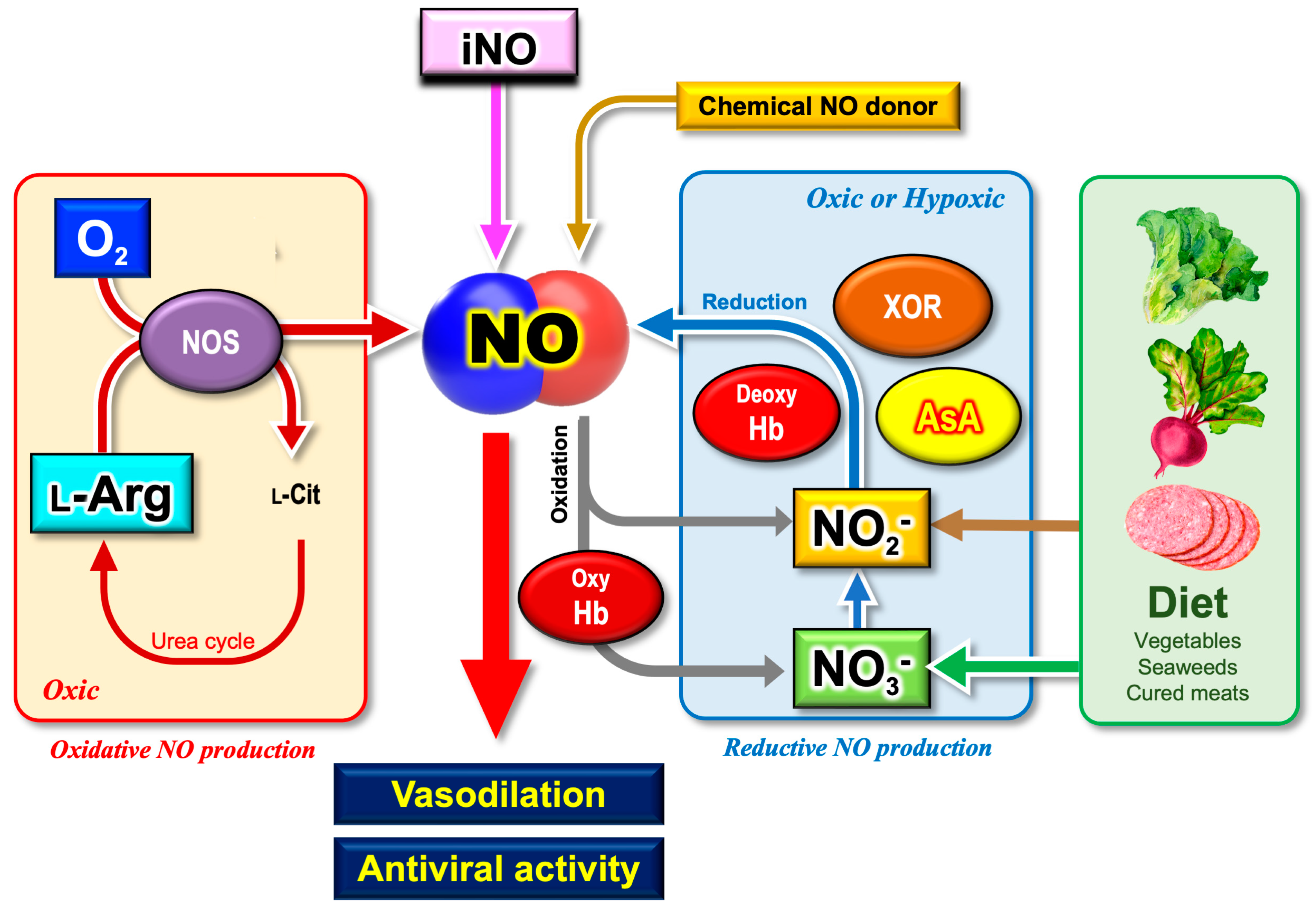
4.1. Oxidative and Reductive Mechanisms for NO Generation

It is evident that NO exhibits substantial antiviral activity against many types of viruses, including SARS-CoV-2. In mammals, NOS plays a central role in endogenous NO supply. Notably, the NOS reaction is not the only endogenous source of NO [84,85]. There are multiple mechanisms for biological NO production other than NOS. Such distinct mechanisms have been called the “nitrate-nitrite-NO pathway” [86,87,88], “NOS-independent mechanism” [89], “nitrite-dependent NO production pathway” [65,90], “nitrite pathway” [91], or the “reductive pathway” [92].
It has been demonstrated by many researchers that inorganic nitrite (NO2) is reduced to NO by redox enzymes or reductants. This alternative mechanism for NO production (hereafter the reductive mechanism) appears to be less appreciated than the NOS-dependent NO production mechanism (hereafter the oxidative mechanism) [92]. Figure 2 summarizes our current knowledge on the multiple sources of NO, relevant to the pathophysiology of COVID-19.

4.2. Nitrite as a Degradation Product of NO

NOS utilizes the amino acid l-arginine (l-Arg) and molecular oxygen (O2) as substrates and requires nicotinamide-adenine-dinucleotide phosphate (NADPH), flavin adenine dinucleotide (FAD), flavin mononucleotide (FMN), and (6R-)5,6,7,8-tetrahydro-l-biopterin (BH4) as cofactors [33] (Equation (4)):
l-arginine + NADPH + H+ + O2 → l-citrulline +1/2 NADP+ + NO + H2O
There are three isoforms of NOS in mammals: neural NOS (nNOS, NOS1), inducible NOS (iNOS, NOS2), and endothelial NOS (eNOS, NOS3) [33]. nNOS is expressed in specific neurons of the central nervous system (CNS), where NO acts as an atypical neurotransmitter associated with learning and memory formation [33]. NO formed in the CNS by nNOS is involved in the central regulation of blood pressure. The inhibition of nNOS activity in the medulla and hypothalamus results in systemic hypertension [66]. eNOS plays a key role in vascular functions, which mediates basal vasodilation [93]. NO generated by iNOS exhibits beneficial antiviral activity, whereas aberrant iNOS induction is also associated with many types of diseases, including asthma, arthritis, multiple sclerosis, colitis, neurodegenerative diseases, tumor development, and septic shock [94].
Although the autoxidation of NO is a kinetically slow reaction [92], nitrite (NO2) can be produced as an oxidation product of NO in the presence of O2 (Equation (5)):
4NO + O2 +2H2O → 4NO2 + 4H+
The Griess method has been widely used to quantify NOS activities [95]. The method was originally designed to detect nitrite (NO2), as well as nitrate (NO3), in a solution [96], and it was later applied to estimating NO production in biological samples [97]. In addition to the autoxidation of NO, nitrate (NO3) can be produced by NO-dioxygenation, which is mediated by blood oxyhemoglobin (Oxy-Hb) [92] (Equation (6)):
NO + Hb (FeII-O2) → NO3 + Hb (FeIII)
Here, it should be emphasized that inorganic nitrite (NO2) and nitrate (NO3) are produced as the degradation products of NO, which is why the Griess method has been widely used as an index parameter of NOS activity.

4.3. Nitrite as a Precursor of NO

Antimicrobial technology is important for food industries in the prevention of food poisoning. In many countries, nitrite is ordinarily contained as a food additive in cured meats such as in sausage, ham, or bacon. When meats are processed with nitrite salt, the meat can be stored at room temperature for a long time without degrading. The mechanism for the antibacterial activity of nitrite was uncovered by the discovery of NO production from nitrite. It is not nitrite but NO that functions as an antibacterial agent that prevents the food poisoning of cured meats.
The inorganic nitrite ion (NO2) exists in equilibrium with its conjugate acid, nitrous acid (HNO2, pKa 3.1–3.5) (Equations (7)–(8)). HNO2 spontaneously produces dinitrogen trioxide (N2O3) as a result of the disproportionation reaction (Equation (9)). NO can be generated by the chemical decomposition of N2O3 [98] (Equation (10):
NO2 + H+ ←→ HNO2
3HNO2 → 2NO + HNO3 + H2O
2HNO2 → N2O3 + H2O
N2O3 → NO + NO2
Since spontaneous NO production from nitrite under acidic conditions is relatively slow [65], it may have minimal physiological relevance, such as in gastric juice [99].

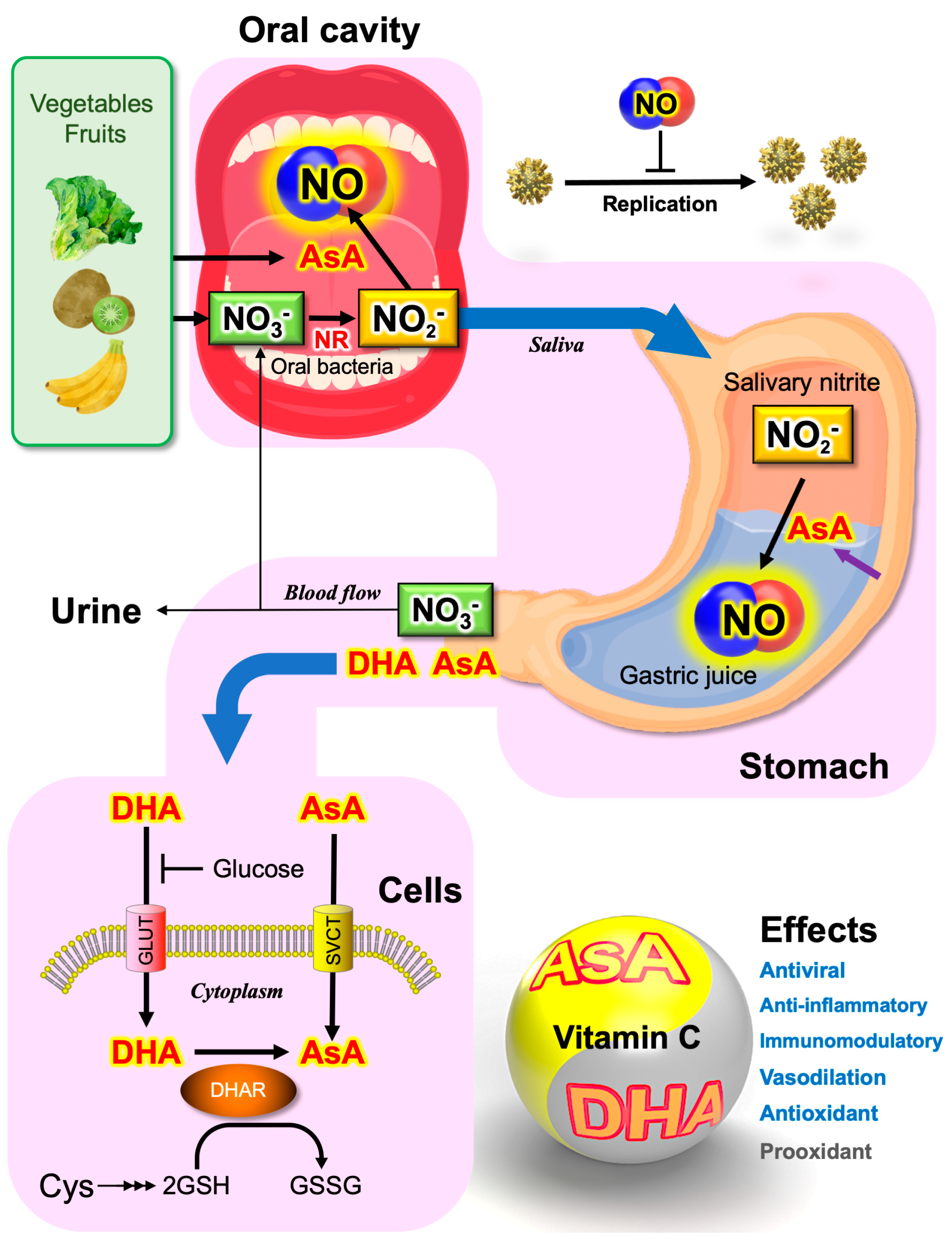
4.4. Saliva as the Major Source of Nitrite

The presence of endogenous nitrite formation in the human body was first noted by Mitchell et al. (1916) more than a century ago [100], but the endogenous source of this nitrite had remained unknown until the 1980s [90]. The discovery of NOS reactions has provided an answer to Mitchell’s observation (Equation 5). In healthy conditions, the consumption of a high-nitrate meal results in a huge increase in the nitrate concentration in plasma, as well as in urine [101]. The exogenously supplied nitrate can be converted to nitrite by the nitrate reductase activity of symbiotic bacteria (Equation (11)):
NO3 + 2e + 2H+ → NO2 + H2O
In humans, nitrate (NO3) is highly concentrated by the salivary glands and secreted into saliva [102]. The nitrate concentration in saliva was reported to be a few hundred µmoles/L in basal conditions, but their concentrations increase to a few mmoles/L after the consumption of nitrate [101]. In 2012, Qin et al. demonstrated that sialin (SLC17A5) expressed in the plasma membrane of salivary gland acinar cells functions as a nitrate transporter [103]. Sialin (SLC17A5) was originally identified as the cause of sialic acid (SA) storage disease or Salla disease [104,105], and the protein has been known to mediate SA/H+ cotransport [103]. They found that sialin (SLC17A5) is also capable of transporting NO3 and H+ via an electrogenic cotransport mechanism [103].
The oral ingestion of potassium nitrate (KNO3) solution quickly increases the salivary nitrite (NO2) concentration [99], which cannot be observed after using a mouthwash to sterilize oral bacteria [106]. The oral cavity has the second largest and diverse microbiota, harboring over 700 species of bacteria [107]. Many commensal bacteria are capable of reducing nitrate (NO3) to nitrite (NO2) by their nitrate reductase (NR) activity [106]. Approximately 5% to 7% of dietary nitrate is converted to nitrite in the oral cavity by facultative anaerobic bacteria on the posterior part of the tongue [108]. Through spontaneous chemical reactions (Equations 7–10), swallowed salivary nitrite is then converted to NO under the strong acidic conditions of gastric juice in the stomach [109]. Responding to salivary nitrite concentrations, a huge amount of NO can be generated in the stomach by the ascorbate-dependent reductive mechanism described below. This series of reactions has been considered to inactivate swallowed pathogens [98]. Nitrite and acidic conditions are the key factors in antimicrobial activity.

5. Ascorbate-Dependent NO Production

5.1. Antibacterial Activity of Vitamin C

Urinary acidification has long been used as an aid for the treatment of urinary tract infections (UTI). In 1959, McDonald and Murphy suggested the use of ascorbate as an acidifying agent in the treatment of UTIs. Since UTIs are one of the most frequent complications during pregnancy (between 8 and 23%), even today, daily intake of ascorbate (vitamin C) is recommended, particularly for pregnant women [110]. However, the mechanism for the antibacterial action of vitamin C has long been unclear.
It has long been known in chemistry that NO can be generated by the reduction of nitrite in the presence of iodide or ferrocyanide in an acidified solution. Historically, this reaction was used to quantify nitrate/nitrite in urine [100]. This chemical production of NO via the reductive mechanism is almost stoichiometric, thereby being widely used as a convenient method to calibrate NO concentrations in a solution, even today [90]. Ascorbate (vitamin C, l-ascorbic acid, AsA), a potent natural antioxidant contained in fruits and vegetables [111], has also been reported to induce the chemical NO production from nitrite [112]. The one-electron reduction of nitrite produces NO and the short-lived monodehydroascorbate (MDA) radical (Equation (12)):
2HNO2 + 2AsA → 2NO + 2MDA + 2H2O
Two molecules of MDA spontaneously disproportionate to ascorbate and dehydroascorbate (DHA) (Equation (13)):
2MDA → AsA + DHA
Plants have evolved to reduce MDA radicals with the MDA reductase (MADR) enzyme to regenerate ascorbate [113], but no specific enzyme for MDA reduction has been identified in animal cells. As a result of these reactions, two molecules of NO can be produced by ascorbate (Equation (14)):
2HNO2 + AsA → 2NO + DHA + 2H2O
The antibacterial function of ascorbate in urine may include the chemical NO generation from the nitrite contained in urine [114].

5.2. Ascorbate Secreted from the Stomach

Ascorbate is known to be secreted into the stomach, leading to gastric juice concentrations greater than those in plasma [115]. It is also known that the secretion of ascorbate is impaired in the presence of chronic gastritis [116]. It has long been puzzled what the physiological functions of the secretion of ascorbate into gastric juice might be. Epidemiological investigations have suggested that ascorbate in gastric juice protects us from gastric cancer [116]. It was hypothesized that ascorbate in gastric juice may be required for the chemical reduction of carcinogenic N-nitroso compounds, such as the nitroso amines implicated in gastric carcinogenesis, a possible mechanism for the prevention of stomach cancer by ascorbate [117,118].
NO has been detected in the gastric headspace after nitrate ingestion [109]. Its concentration was reported to be between 14.8 and 89.4 ppm after potassium nitrate (KNO3) ingestion [98]. Taking the presence of nitrite in saliva fluid into account, one can expect that ascorbate secreted into gastric juice facilitates the efficient chemical reduction of salivary nitrite to generate a ppm level of NO in the stomach [119]. A strong acidity (approx. pH 1 in fasting conditions) of gastric juice is considered the first defense mechanism against the oral transmission of pathogens. However, this strong acidity cannot be retained after the ingestion of foods. Chemical NO production with nitrite and ascorbate may further ensure the inactivation of pathogens through the antimicrobial activity of NO, even after the ingestion of foods.

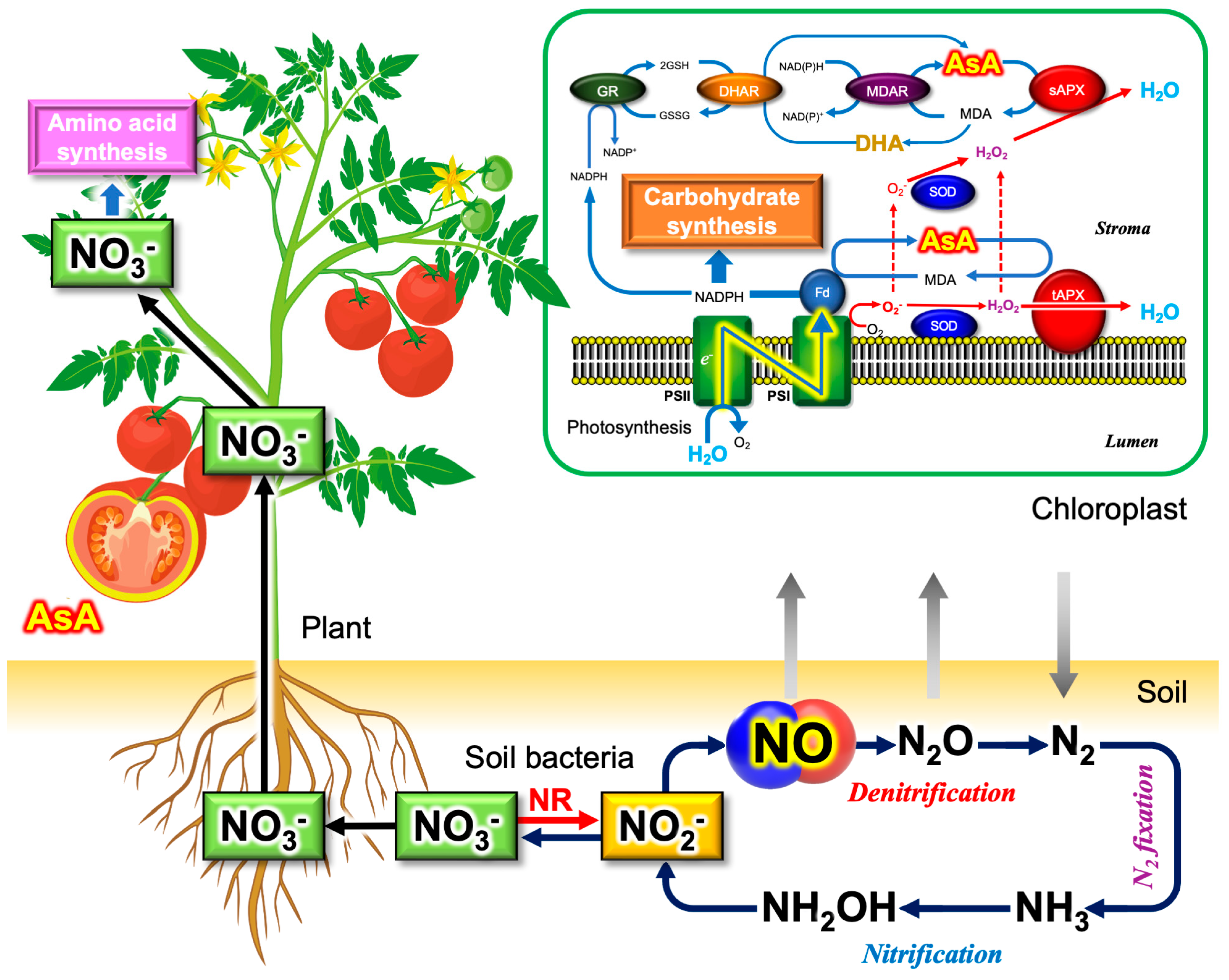
5.3. Plants as the Major Dietary Source of Nitrate and Vitamin C

Vegetables are the major dietary sources of nitrate for humans (Figure 2). Approximately 80% of ingested nitrate can be attributed to vegetables [108]. Absorbed by roots in soils and translocated through the plant vascular system, nitrate can become highly concentrated in plant leaves, i.e., green leafy vegetables [120]. Plants synthesize the amino acids in leaves, where nitrite transported into the chloroplasts is further reduced by light energy to synthesize glutamate (nitrogen assimilation). Nitrate contents vary widely between plant species [121], and even within the same tissue types of the same species due to the differences in nitrate fertilization during cultivation [120]. A hundredfold variation in average nitrate levels was found in cooked greens, ranging from 4850 mg kg−1 in English spinach to 48 mg kg−1 in iceberg lettuce [122]. Hence, fresh green leafy vegetables are a dietary source of nitrate.
Vitamin C can be acquired from green leafy vegetables and fruits. In the 1920s to 1930s, Albert Szent-Györgyi identified and isolated ascorbate from the fruits of Hungarian red pepper [111]. Since humans do not synthesize ascorbate due to the loss of l-gulono-1,4-lactone oxidase (GULO), diets are extremely important to acquire vitamin C. As with nitrate content, ascorbate content also varies to a large extent between species and tissues [123]. Figure 3 illustrates nitrate and vitamin C in plants (vegetables and fruits).
Plants synthesize ascorbate mainly through the Smirnoff–Wheeler pathway, which is dominant in photosynthetic tissues, i.e., green leafy vegetables [111]. However, the reason why plants accumulate high concentrations of ascorbate in leaves had been unknown for a long time. In 1976, Foyer and Halliwell proposed that ascorbate may be involved in a mechanism for the removal of ROS in plants [134]. After the discovery of ascorbate peroxidase (APX) and ascorbate recycling systems, including monodehydroascorbate reductase (MDAR) and dehydroascorbate reductase (DHAR) [135], it became evident that high concentrations of ascorbate are essential to operate the O2-evolving photosynthetic electron transport system. Ascorbate is used to detoxify the ROS produced in chloroplasts under illumination [136]. This unique plant ascorbate-dependent ROS scavenging mechanism is referred to as the ascorbate–glutathione cycle [137] or the water‒water cycle [132] (Figure 3). The reason why fruits, which are not photosynthetic organs, accumulate ascorbate remains a subject to be investigated [111].

5.4. Vitamin P and NO

In 1936, Szent-Györgyi and coworkers demonstrated that the flavonoid glycosides rich in citrus fruits behave similarly to ascorbate in maintaining capillary permeability. Based on his observation, Szent-Györgyi proposed that plant flavonoids are essential nutrients as vitamin P (permeability) [138], a short-lived vitamin concept [139]. His idea that flavonoids can complement the function of ascorbate has been renewed with the development of the antioxidant hypothesis of phytochemicals.
Flavonoids are representative secondary metabolites of plants, accumulating in the epidermal cells of organs such as flowers, leaves, stems, roots and fruits [139]. Flavonoids can be classified into subgroups: chalcone, flavanone, flavone, isoflavone, flavonol, and anthocyanin. Most of these subgroups exhibit a yellowish coloration, whereas anthocyanins show multiple colorations depending on their structures, the presence of metals, their pH and their conjugation with other molecules [139]. In the 1980s, the anthocyanins contained in red wine were highlighted to account for the paradoxical epidemiological observation called the “French paradox”; French people have a relatively low incidence of coronary heart disease even though they consume a diet relatively rich in saturated fats [140]. Researchers were interested in the anthocyanins or “polyphenols” contained in red wine that may suppress heart disease through their antioxidant activities [140]. It is now widely appreciated that flavonoids or “polyphenols” are beneficial for human health largely due to their non-specific antioxidant functions [141]. In eNOS-dependent NO production (the oxidative mechanism), the flavonoids quercetin and catechin contained in red wine were demonstrated to increase the bioavailability of NO by scavenging O2 [142]. In plants, phenolic secondary metabolites, including flavonoid pigments, not only contribute to the plant colorations of flowers and fruits but also suppress oxidative damage of the cells through enzymatic [143] and non-enzymatic [144] mechanisms, complementing the ascorbate antioxidant systems [128].
If plant flavonoids and polyphenolic compounds are functionally equivalent to ascorbate, NO can be chemically generated from nitrite in the presence of plant pigments (Equation (15)):
HNO2 + Phe-OH → NO + Phe-O˙ + H2O
where Phe-OH and Phe-O˙ are the parent reduced form of phenolic compounds and their phenoxyl radicals, respectively. Gago et al. (2007) explored the involvement of anthocyanins in the reductive mechanism for NO generation in the stomach [145]. Following the consumption of red wine, a higher NO in the air expelled from the stomach (30,215 ppb) was detected than that under fasting conditions (1196 ppb), with a peak at 15 min. This wine effect was not observed in the absence of exogenous nitrate consumption [145]. Rocha et al. (2009) showed in vitro evidence that several classes of flavonoids can reduce nitrite to produce NO at pH 2 [146]. It should be noted that phenoxyl radicals can be efficiently reduced back to their parent compounds by ascorbate [147], implying that complicated redox interplays between vitamin P and vitamin C. Although there have been a limited number of studies available to date [146,148], it appears to be obvious that vitamin P also contributes to the reductive generation of NO in humans.
Plant (vegetable and fruit) coloration is attributed to the spectral properties of flavonoids, mostly anthocyanins. The plant pigment betalains that are included in spinach, chard, and beetroot are exceptional. The term “betalain” comes from the Latin name of the common beet (Beta vulgaris) from which betalains were first extracted [139]. Betalains are a class of tyrosine-derived pigments that are distributed in only 13 families of the Caryophyllales order, such as red beet (Amaranthaceae) and cactus (Cactaceae), and in some fungi [149]. To date, anthocyanins and betalains have never been detected jointly in plant tissues [149]. The biological meaning of the mutually exclusive relationship between betalains and anthocyanidins is still unknown [150]. Beetroot juice has recently attracted much attention from researchers as a natural nitrate (NO3) supplement [151]. In addition to nitrate supplementation and the antioxidant activity of betalains, the chemical NO-generating activity of betalains with nitrite is of great interest to understand the beneficial effects of beetroot juice [152,153].

6. Possible Roles of Nitrate, Nitrite, and Vitamin C in the Prevention of COVID-19

6.1. Toxicity of Nitrite and Nitrate

Nitrite has long been recognized as an inorganic toxic compound by food professionals as well as the public, mostly for two reasons: methemoglobinemia (cyanosis or blue baby syndrome) and the formation of nitrosamines. Water supplies, especially near agricultural areas, are often contaminated with nitrate derived from nitrogen fertilizer. Nitrite may be produced in such contaminated water by ambient bacteria via their nitrate reductase activities. In 1945, the pediatric resident Hunter Comly revealed that the use of nitrate-contaminated well water is the direct cause of blue baby syndrome, a condition characterized by cyanosis or blueness of the skin in babies [154]. Meanwhile, another concern regarding the toxicity of nitrite arose in food industries. Nitrite is an important meat preservative, but carcinogenic nitrosamines might be produced during the cooking of cured meats that contain nitrite. Because of these two reasons, the contents of nitrite and nitrate in drinking water, vegetables and cured meats have been strictly regulated by governments and authorities in many countries.
Contrary to common sense, many recent animal and epidemiological studies have not indicated such cytotoxicity, except for in infants. Rather, nitrite is now considered a potential therapeutic agent for the prevention and reversal of cerebral vasospasm after subarachnoid hemorrhage (SAH), the prevention of reperfusion injury associated with myocardial infarction, the elicitation of therapeutic angiogenesis, the treatment of pulmonary hypertension, and the enhanced preservation of tissues for transplantation [155]. Scientists’ appreciation of nitrite (nitrate) in life science has drastically changed from a toxin to a bioactive molecule. The prompt excretion of nitrite and nitrate into urine further supports the safety of nitrite and nitrate. Hence, it is now conclusive that the oral ingestion of nitrate is quite safe, at least as long as it is taken from vegetables and fruits.

6.2. Prevention of SARS-CoV-2 Transmission

Since 2019, most studies have focused on SARS-CoV-2 infection in the respiratory tract due to the close association of respiratory failure with COVID-19-related death, as mentioned in the introduction. However, ACE2 is not only expressed in the lungs but also in multiple organs implicated in COVID-19 complications. A number of studies have reported gastrointestinal symptoms in patients with COVID-19 [156]. The small intestine is one of the organs where high ACE2 expression occurs [157]. A meta-analysis of 4243 patients with COVID-19 from six countries reported that 17.6% of COVID-19 patients had gastrointestinal symptoms, and 48.1% of fecal samples from COVID-19 patients tested positive for viral RNA [158]. It is important to note that the prolonged shedding of viral RNA in stool rather than in respiratory samples was observed in 70.3% of the patients, which could be up to 33 days from illness onset [158]. During the SARS outbreak in 2003, a vertical spread of the outbreak in buildings was reported in the Amoy Gardens, Hong Kong. Researchers have speculated that the sewage system of the Amoy Gardens may have served as the major source of infection from patients excreting SARS-CoV [159,160]. Although there are ongoing active debates on fecal-oral transmission in COVID-19 [156,161], potential gastrointestinal infection should be carefully considered in preventing the outbreak [156,162]. To limit the oral transmission of viruses, as well as aerosol or airborne transmission, we suggest a reductive NO generation strategy for preventing and mitigating COVID-19, as shown in Figure 4.
Gastric juice acts as the first line of defense to avoid gastrointestinal infection by pathogens [165]. Its pH is near 1 (equivalent to 0.16 M HCl [166]). However, this strongly acidic pH can be measured only under fasting conditions. The pH may go up to a near neutral pH after the ingestion of foods. The strong acidity of gastric juice is sustained by the delivery of H+ into the lumen of the gastric gland, along with secreted Cl [166]. H+ extrusion is mediated by H+, K+-ATPase, an important drug target for gastric hyperacidity [167,168]. In 2017, Zhou et al. compared the gastrointestinal fluid tolerance of viruses between MERS-CoV, human coronavirus hCoV-229E, and EV71, a prototype of human enterovirus [169]. They suggested that MERS-CoV cannot be inactivated at a higher pH and that the intestinal tract may serve as an alternative infection route for MERS-CoV, which can be facilitated by the ingestion of foods or inhibition of the H+-pump [169].
In 2010, Kopic et al. reported that 2% ethanol significantly suppressed acid secretion through the inhibition of the AMP-activated protein kinase (AMPK), implying that a low dose of ethanol may elevate the pH of gastric juice [168]. There has been a great deal of controversy as to the effects of ethanol on gastric acid secretion, from being a mild stimulant to being a potent inhibitor [166]. Although the impact of ethanol intake on the oral transmission of pathogens has not yet been clarified [170,171], dinner with alcoholic beverages can be considered a risk factor in the oral transmission of pathogens. It is a logical speculation that the combination of ascorbate and nitrite can inactivate pathogens through reductive NO chemical generation, and is presumably effective even after the ingestion of foods (increased pH) and alcoholic beverages (inhibition of the H+-pump).

7. Vitamin C Therapy in COVID-19

7.1. Free Radical Storm

“It’s not stress that kills us; it is our reaction to it” (Hans Selye, 1956) [172].
The Nobel laureate Hans Selye was a physiologist who established his theory on stress [173]. The stress theory had long been a philosophical concept that was rather difficult to explain on a molecular basis. Infectious diseases, including COVID-19, can be categorized as “biotic stress” due to the biological pathogen-host interactions. According to stress theory, the immune response can be interpreted as a stress response against biological stressors. Indeed, SARS-CoV-2 itself does not kill us, but our immune response “cytokine storm” or hyperinflammation accompanying the overproduction of NO and ROS kills us through organ failure.
NO and ROS are so called “double-edged swords”. In small amounts, they function as signaling molecules in cells, but continuous exposure at high concentrations sometimes damages the host cells themselves. In viral infection, proinflammatory cytokines, such as interferon-γ (INF- γ), upregulate iNOS, which potentially overproduces NO in the pathogenesis of viral infection [55]. NO overproduction may be directly linked to the pathogenesis of viral pneumonia [55]. In fact, the administration of the NOS inhibitor Nw-monomethyl-l-arginine (l-NMMA) or the O2 scavenger superoxide dismutase (SOD) protected and recovered influenza virus-infected mice [174,175]. Lab mice deficient in iNOS showed reduced morbidity, mortality, and diminished cytokine production in the lung tissue following an influenza virus challenge [176]. These early experiments clearly indicated that NO, as well as ROS, contribute not only to the host defense but also to the severity of viral pathogenic diseases. As Wu (2020) pointed out, it is not a “cytokine storm” but a “free radical storm” that may be the direct cause of organ failure or cell death in COVID-19 [177].

7.2. High-Dose Intravenous Vitamin C Treatment

Along with anti-inflammatory drugs, the use of potent antioxidants may become a clinical option to calm the “free radical storm” in critical conditions. Vitamin C therapy has been known for several decades as a safe adjunctive treatment that has been examined in a wide variety of diseases, including cancer [178], obesity [179], and SARS [19]. Sepsis is a medical emergency that occurs due to the hyper-immunological response of patients. ARDS is a devastating complication. There has recently been emerging evidence for the use of vitamin C as a treatment for sepsis [20]. Similarly, high-dose intravenous vitamin C (HDIVC) treatment has been reported to be effective in decreasing days of hospitalization, ICU stay and mortality in patients with COVID-19 [16,17,18,36]. Bypassing the limitations of vitamin C uptake through a sodium-dependent vitamin C transporter (SVCT1), HDVIC can achieve 70-fold higher plasma concentrations of vitamin C compared to oral administration [180]. Despite its long research background, however, there are controversies regarding the therapeutic effects of HDIVC treatment [20].
Early in the pandemic, the number of COVID-19 patients was apparently low in Japan in contrast with Europe and the U.S., despite its high population density, large percentage of high-risk individuals over 65 years old, and the absence of restricted social activities [181]. The paradoxical phenomenon was often called the “Japanese paradox” [182]. The Japanese paradox received much attention from researchers, as well as from the public at that time. Many hypotheses have been proposed to account for the Japanese paradox in terms of cultural differences, the preacquisition of immunity, genetic background (ACE2 and HLA), and BCG vaccination [183]. As discussed above, it is likely that nitrate-rich dietary foods are involved in the prevention of SARS-CoV-2 infection through a reductive mechanism (Figure 2). In good agreement with this prospect, the Japanese diet containing abundant nitrate has been reported to improve hypertension and other vascular diseases [183]. Despite the promising therapeutic effects of nitrate on NO generation, a recent randomized clinical trial did not support our expectations [184]. Moreover, Lorente et al. (2022) reported higher blood nitrate and nitrite levels in non-survivor COVID-19 patients than in healthy subjects [185], presumably due to the overproduction of NO that produces nitrate and nitrite as the degradation product (Section 4.2). Similar to vitamin C therapy, there have been contradictory results on the therapeutic effects of nitrate and nitrite. It is likely that ascorbate therapy alone, or nitrate (or nitrite) therapy alone, may be insufficient to drive the reductive mechanism. A combination of these factors, as well as the appropriate mode of delivery (intravenous administration or oral administration), could be required to maximize the effects, which is a possible reason why HDIVC treatment in the ICU is sometimes ineffective or has effects opposite to our expectations [18].

8. Updating Pauling’s Vitamin C Therapy

8.1. Long-Lasting Debates on the Pharmacological Effects of Vitamin C

Ascorbate was proven by Szent-Györgyi to function as vitamin C against scurvy [186], but its function against infectious diseases had been suggested as early as this discovery [187,188,189]. Nonetheless, vitamin C may be more strongly linked to the name of Linus Pauling. In 1971, Pauling proposed that the oral intake of vitamin C may reduce the incidence and morbidity of the common cold based on his own meta-analysis (which is one of the earliest ones), using clinical trial data available at that time [190,191]. Almost simultaneously, Pauling advocated in 1970 the efficacy of vitamin C on the common cold to the public in his book, Vitamin C and the Common Cold [192]. Since the clinical trial data that Pauling used were limited, unavoidably biased results were included in these publications, one of the many reasons for the active scientific and political debates on the efficacy of vitamin C on the common cold and other diseases since the 1970s.
Even half a century later, active debates on this issue are still ongoing [193], a seeming “never ending story”. Vitamin C therapy is rejected by many medical professionals today. In discussing why medical professionals are not enthusiastic about the observation that vitamin C decreases the incidence of the common cold by 31%, Pauling wrote, “In the search for a drug to combat a disease the effort is usually made to find one that is 100 percent effective” [194]. He continued, “Also, there seems to have existed a feeling that the intake of vitamin C should be kept as small as possible, even though this vitamin is known to have extremely low toxicity” [194]. Pharmaceutical drugs, including antiviral drugs, are expected to have a high efficacy at low concentrations, because drugs are usually highly toxic at higher concentrations. High-dose administration of drugs is often dangerous and even fatal due to potential side effects. Even at relatively low concentrations, serious side effects do not seem to be uncommon. For example, drug-induced liver damage is problematic in the treatment of COVID-19 [195,196,197]. There is also the case for vaccines [198]. In contrast, vitamin C has virtually no toxicity, even at saturating concentrations in the body [194].

8.2. High Dose Necessary for Pleotropic Function of Vitamin C

Pauling emphasized the importance of maintaining high vitamin C concentrations in the body to maintain general health, for example, to prevent the common cold [194]. However, the recommended daily allowance (RDA) issued by the FDA was much less than the amounts that Pauling and his forerunners proposed. In his book, Pauling explained that the RDA value (1980 edition from the FDA) was 60 mg for adult males, but the recommendations of his forerunners and himself were more than 1000 mg [194]. The large difference between these recommended amounts stems from the fact that the RDA value issued by the FDA was set just to prevent scurvy. Szent-Györgyi and Pauling shared the opinion that the optimum intake of vitamin C is much larger than the RDA. Responding to an inquiry from Pauling, Szent-Gyorgyi wrote: “Scurvy is not the first sign of the deficiency but a premortal syndrome” [194]. Unfortunately, the misconception that vitamin C is only for scurvy has prevailed, which appears to have hampered awareness of the pleotropic physiological functions of vitamin C. Now, numerous lines of evidence have accumulated for the pleotropic functions of vitamin C other than prevention of scurvy [199,200,201,202]. Reflecting the diverse functions of vitamin C, together with individual variation, the high levels of vitamin C doses recommended by Pauling (1986) (1000–18,000 mg) [194] cannot be considered unreasonable. The precise values of the recommended daily intake of vitamin C for a given individual should vary based on genetic background, quality of diet, and severity of illness.

8.3. Updating Pauling’s Concept

Pauling discovered the molecular mechanisms of sickle cell anemia and coined the term “molecular disease” [203,204]. The seminal paper, “Sickle cell anemia, a molecular disease” [203], was published in Science in 1949, which opened an entirely new field of molecular biology and medicine. However, Pauling did not pay much attention to “molecular medicine”, probably because he had noticed how difficult it is to achieve effective molecular treatments based on molecular mechanisms.
According to the current paradigm in pharmacology, pathogenic microorganisms should be targeted by synthetic drugs that interact with the specific molecules of pathogens to kill them. This paradigm is based on Koch’s postulates [205], which were established more than a century ago. This Koch’s postulates-based paradigm may further make vitamin C less acceptable for medical professionals because Pauling’s vitamin C therapy is based on a paradigm entirely different from the current theories.
In “Western or modern” medicine, a disease can be defined as dysfunction of a physiological mechanism. Based on this concept, a drug in general is presumed to act on a specific component of a physiological mechanism. This way of treating a disease is molecular medicine. In many cases, these are inhibitors of enzymes or transporters, showing the “one-to-one” relationship between the drug and target molecule, in accordance with a structure-function relationship. The “one-to-one” philosophy in medicine and pharmacology works well if the cause of a disease is ascribed to a single component, such as a protein or an enzyme. However, most diseases that are difficult to prevent and cure are syndromes that are governed by multiple components with complicated interactions. COVID-19 is a good example.
Looking back on a long research history, one can notice that our society’s recognition of the simple molecules (nitrite, nitrate, NO and O2) has changed over time: harmful or beneficial? There have always been contradictory findings and interpretations regarding the physiological functions of those ubiquitous molecules. Vitamin C also exhibits harmful effects through the chemical Fenton reaction that produces highly toxic ROS (•OH, hydroxyl radicals) [178]. The excessive intake of DHA (the oxidized form of ascorbate) into the cells may cause oxidative stress conditions where the reductants GSH and NADPH are used up for ascorbate regeneration [206], which might be avoided by the supplementation of the glutathione precursor cysteine [207]. Two opposing effects of those simple molecules may lead non-specialists to a state of confusion: is it good for us or bad for us? In addition, the circularity of the relationship, such as between nitrite and NO (Figure 2 and Figure 3), gives rise to the chicken and egg issue: substate or product?
In biochemistry and molecular biology, major target molecules are considered relatively stable, large, and unique in structure, such as proteins, sugars, lipids, DNA, or RNA. These molecules may serve as excellent targets of molecular medicine. The nature of NO and nitrite are opposed to these conventional biomolecules: unstable, small, simple, and ubiquitous. Owing to these properties, conventional approaches are generally not applicable to understanding the physiological functions of these ubiquitous molecules. According to Pauling [131,194], “optimal molecular concentrations of biological substances that are normally present in the body, such as vitamin C, are critical for the function of organs”. Pauling further argued that the optimal concentrations may vary from individual to individual [131,194].
For understanding the physiological or pharmacological functions of those simple and universal biomolecules, knowing their fine balance is more valuable than the strict categorization of their effects or targets. Fritjof Capra, a physicist, introduced the parallelism between modern physics and Eastern philosophes in his book The Tao of Physics [208]. Chinese medicine is based on traditional Chinese philosophies that are mutually exclusive to “Western” science [209]. Pauling appears to share a similar philosophy with such Eastern philosophies: knowing the fine balance would be the ultimate answer.
Recent studies have revealed that H2S/HS- is also involved in the regulation of physiological processes through cysteine thiol persulfidation (Cys-SSH) [210] and polysulfidation (Cys-RSS(n)H) [211]. Such reactive sulfur molecules are termed reactive sulfur species (RSS) [57,91]. Complicated chemical interactions between ROS, RNS, and RSS are pronounced in cysteine thiols [57,210]. It is interesting to note that, in addition to nitrite and ascorbate, the sulfur-containing compound thiocyanate (SCN-) is present in saliva and gastric juice [212,213,214]. The H2S chemical donor isothiocyanates are rich in cruciferous vegetables (Cruciferae or Brassicaceae), such as cabbage, and they have been reported to reduce many types of diseases [215]. It is obvious that RSS also plays an important role in the optimization of the redox balance [91,127]. Updating Pauling’s concept, we propose that the optimal concentrations of ROS, RNS, and RSS that are normally present in the body, such as H2O2, NO and H2S, are critical for the function of organs, especially in vascular systems and immune systems.

9. Conclusions

Continuous exposure to a high concentration of NO may be harmful to host cells, as seen during inflammation. In contrast, a short burst of NO at high concentrations (hundreds ppm) should have maximal antiviral effects on COVID-19 without damaging the host cells, as recently discussed by Del Sorbo et al. (2022) [216]. We conclude that an intermittent NO burst, chemically generated by the combination of nitrite (or nitrate) and vitamin C (or vitamin P, phytophenolics and betalains), is a potential therapeutic treatment to prevent and mitigate COVID-19. Thus, the reductive NO-generating mechanism is worthy of consideration for developing clinical treatments and for designing and interpreting data from clinical trials [217]. It is important to note that chronic diseases associated with the dysfunction of vascular systems, such as hypertension [218], are also expected to be improved by the application of the reductive NO generation strategy. This can be achieved by the daily consumption of fresh green leafy vegetables (nitrate) along with fruits (rich in vitamins C and P and organic acids).
In the application of the reductive NO-generating strategy for clinal treatments, knowing an optimal dose balance of the key molecules (ascorbate, nitrate, nitrite, cysteine) for individuals will be critical to gain the maximum therapeutic effects. The gut microbiota has been interpreted in terms of a metabolic organ that influences the host through reciprocal interactions, encompassing the NO-nitrite-nitrate metabolic and immune pathways in a diet-dependent manner that shapes all aspects of host physiology [219]. Hence, redox metabolism can be strongly influenced by the bacterial communities colonized in the oral cavity and the gut, a situation analogous to the dynamics of bacterial flora in soils [220]. Unfortunately, we have not yet found measurable parameters to monitor the redox balance in cells, tissues, organs, and the whole body. A recent pilot study reported that dogs can distinguish the respiratory secretions of patients with COVID-19 from those of healthy controls, with high rates of sensitivity and specificity [221], implying that there are differences in the volatile compounds between them. To monitor an internal redox status of individuals in a noninvasive manner, real-time analysis of exhaled gases and/or volatile organic compounds (VOCs) will be a hopeful candidate in personalized medicine [222].

Author Contributions

Conceptualization and supervision, writing original draft preparation, H.Y.; writing and editing, J.M.O., A.T. and H.I. All authors have read and agreed to the published version of the manuscript.

Funding

This research received no external funding.

Data Availability Statement

Not applicable.

Acknowledgments

We thank Michael Cohen and George Iwama for their critical reading of the manuscript.

Conflicts of Interest

The authors declare no conflict of interest.

References

  1. Devaux, C.A.; Rolain, J.M.; Raoult, D. ACE2 receptor polymorphism: Susceptibility to SARS-CoV-2, hypertension, multi-organ failure, and COVID-19 disease outcome. J. Microbiol. Immunol. Infect. 2020, 53, 425–435. [Google Scholar] [CrossRef] [PubMed]
  2. Kim, J.S.; Lee, J.Y.; Yang, J.W.; Lee, K.H.; Effenberger, M.; Szpirt, W.; Kronbichler, A.; Shin, J.I. Immunopathogenesis and treatment of cytokine storm in COVID-19. Theranostics 2021, 11, 316–329. [Google Scholar] [CrossRef] [PubMed]
  3. Perkins, G.D.; Ji, C.; Connolly, B.A.; Couper, K.; Lall, R.; Baillie, J.K.; Bradley, J.M.; Dark, P.; Dave, C.; De Soyza, A.; et al. Effect of noninvasive respiratory dtrategies on intubation or mortality among patients with acute hypoxemic respiratory failure and COVID-19: The RECOVERY-RS randomized clinical trial. JAMA 2022, 327, 546–558. [Google Scholar] [CrossRef] [PubMed]
  4. Ginestra, J.C.; Mitchell, O.J.L.; Anesi, G.L.; Christie, J.D. COVID-19 critical illness: A data-driven review. Annu. Rev. Med. 2022, 73, 95–111. [Google Scholar] [CrossRef] [PubMed]
  5. Kamenshchikov, N.O.; Berra, L.; Carroll, R.W. Therapeutic effects of inhaled nitric oxide therapy in COVID-19 patients. Biomedicines 2022, 10, 369. [Google Scholar] [CrossRef]
  6. Lorusso, R.; Combes, A.; Lo Coco, V.; De Piero, M.E.; Belohlavek, J.; Euro, E.C.-W.; Euro, E.S.C. ECMO for COVID-19 patients in Europe and Israel. Intensive Care Med. 2021, 47, 344–348. [Google Scholar] [CrossRef]
  7. Liu, X.; Liu, X.; Zhou, J.; Dong, Y.; Jiang, W.; Jiang, W. Rampant C-to-U deamination accounts for the intrinsically high mutation rate in SARS-CoV-2 spike gene. RNA 2022, 28, 917–926. [Google Scholar]
  8. Tao, K.; Tzou, P.L.; Nouhin, J.; Gupta, R.K.; de Oliveira, T.; Kosakovsky Pond, S.L.; Fera, D.; Shafer, R.W. The biological and clinical significance of emerging SARS-CoV-2 variants. Nat. Rev. Genet. 2021, 22, 757–773. [Google Scholar] [CrossRef]
  9. Otaki, J.M.; Nakasone, W.; Nakamura, M. Nonself mutations in the spike protein suggest an increase in the antigenicity and a decrease in the virulence of the Omicron variant of SARS-CoV-2. COVID 2022, 2, 407–418. [Google Scholar] [CrossRef]
  10. Beigel, J.H.; Tomashek, K.M.; Dodd, L.E.; Mehta, A.K.; Zingman, B.S.; Kalil, A.C.; Hohmann, E.; Chu, H.Y.; Luetkemeyer, A.; Kline, S.; et al. Remdesivir for the treatment of COVID-19—Final report. N. Engl. J. Med. 2020, 383, 1813–1826. [Google Scholar] [CrossRef]
  11. Cully, M. A tale of two antiviral targets—And the COVID-19 drugs that bind them. Nat. Rev. Drug Discov. 2022, 21, 3–5. [Google Scholar] [CrossRef] [PubMed]
  12. Bernal, A.J.; Gomes da Silva, M.M.; Musungaie, D.B.; Kovalchuk, E.; Gonzalez, A.; Delos Reyes, V.; Martin-Quiros, A.; Caraco, Y.; Williams-Diaz, A.; Brown, M.L.; et al. Molnupiravir for oral treatment of COVID-19 in nonhospitalized patients. N. Engl. J. Med. 2022, 386, 509–520. [Google Scholar] [CrossRef]
  13. Krammer, F. SARS-CoV-2 vaccines in development. Nature 2020, 586, 516–527. [Google Scholar] [CrossRef]
  14. Otaki, J.M.; Nakasone, W.; Nakamura, M. Self and nonself short constituent sequences of amino acids in the SARS-CoV-2 proteome for vaccine development. COVID 2021, 1, 555–574. [Google Scholar] [CrossRef]
  15. Group, R.C.; Horby, P.; Lim, W.S.; Emberson, J.R.; Mafham, M.; Bell, J.L.; Linsell, L.; Staplin, N.; Brightling, C.; Ustianowski, A.; et al. Dexamethasone in hospitalized patients with COVID-19—Preliminary report. N. Engl. J. Med. 2021, 384, 693–704. [Google Scholar] [CrossRef]
  16. Cheng, R.Z. Can early and high intravenous dose of vitamin C prevent and treat coronavirus disease 2019 (COVID-19)? Med. Drug Disc. 2020, 5, 100028. [Google Scholar]
  17. Bhowmik, K.K.; Barek, M.A.; Aziz, M.A.; Islam, M.S. Impact of high-dose vitamin C on the mortality, severity, and duration of hospital stay in COVID-19 patients: A meta-analysis. Health Sci. Rep. 2022, 5, e762. [Google Scholar] [CrossRef]
  18. Olczak-Pruc, M.; Swieczkowski, D.; Ladny, J.R.; Pruc, M.; Juarez-Vela, R.; Rafique, Z.; Peacock, F.W.; Szarpak, L. Vitamin C supplementation for the treatment of COVID-19: A systematic review and meta-analysis. Nutrients 2022, 14, 4217. [Google Scholar] [CrossRef] [PubMed]
  19. Boretti, A.; Banik, B.K. Intravenous vitamin C for reduction of cytokines storm in acute respiratory distress syndrome. Pharmanutrition 2020, 12, 100190. [Google Scholar] [CrossRef]
  20. Kashiouris, M.G.; L’Heureux, M.; Cable, C.A.; Fisher, B.J.; Leichtle, S.W.; Fowler, A.A. The emerging role of vitamin C as a treatment for sepsis. Nutrients 2020, 12, 292. [Google Scholar] [CrossRef]
  21. Alqahtani, J.S.; Aldhahir, A.M.; Al Ghamdi, S.S.; AlBahrani, S.; AlDraiwiesh, I.A.; Alqarni, A.A.; Latief, K.; Raya, R.P.; Oyelade, T. Inhaled nitric oxide for clinical management of COVID-19: A systematic review and meta-analysis. Int. J. Environ. Res. Public Health 2022, 19, 12803. [Google Scholar] [CrossRef]
  22. Ichinose, F.; Roberts, J.D., Jr.; Zapol, W.M. Inhaled nitric oxide: A selective pulmonary vasodilator: Current uses and therapeutic potential. Circulation 2004, 109, 3106–3111. [Google Scholar] [CrossRef] [PubMed]
  23. Steudel, W.; Hurford, W.E.; Zapol, W.M. Inhaled nitric oxide: Basic biology and clinical applications. Anesthesiology 1999, 91, 1090. [Google Scholar] [CrossRef] [PubMed]
  24. Ignarro, L.J. Inhaled nitric oxide and COVID-19. Br. J. Pharmacol. 2020, 177, 3848–3849. [Google Scholar] [CrossRef] [PubMed]
  25. Fortenberry, J.D. Inhaled nitric oxide for pediatric acute respiratory distress syndrome: Another brick in the wall? Crit. Care Med. 2018, 46, 1879–1880. [Google Scholar] [CrossRef]
  26. Roberts, J.D.; Fineman, J.R.; Morin, F.C.; Shaul, P.W.; Rimar, S.; Schreiber, M.D.; Polin, R.A.; Zwass, M.S.; Zayek, M.M.; Gross, I.; et al. Inhaled nitric oxide and persistent pulmonary hypertension of the newborn. N. Engl. J. Med. 1997, 336, 605–610. [Google Scholar] [CrossRef]
  27. Chen, L.N.; Liu, P.; Gao, H.; Sun, B.; Chao, D.S.; Wang, F.; Zhu, Y.J.; Hedenstierna, R.; Wang, C.G. Inhalation of nitric oxide in the treatment of severe acute respiratory syndrome: A rescue trial in Beijing. Clin. Infect. Dis. 2004, 39, 1531–1535. [Google Scholar] [CrossRef]
  28. Martel, J.; Ko, Y.F.; Young, J.D.; Ojcius, D.M. Could nasal nitric oxide help to mitigate the severity of COVID-19? Microbes Infect. 2020, 22, 168–171. [Google Scholar] [CrossRef]
  29. Valsecchi, C.; Winterton, D.; Safaee Fakhr, B.; Collier, A.Y.; Nozari, A.; Ortoleva, J.; Mukerji, S.; Gibson, L.E.; Carroll, R.W.; Shaefi, S.; et al. High-dose inhaled nitric oxide for the treatment of spontaneously breathing pregnant patients with severe coronavirus disease 2019 (COVID-19) pneumonia. Obstet. Gynecol. 2022, 140, 195–203. [Google Scholar] [CrossRef]
  30. Al Sulaiman, K.; Korayem, G.B.; Altebainawi, A.F.; Al Harbi, S.; Alissa, A.; Alharthi, A.; Kensara, R.; Alfahed, A.; Vishwakarma, R.; Al Haji, H.; et al. Evaluation of inhaled nitric oxide (iNO) treatment for moderate-to-severe ARDS in critically ill patients with COVID-19: A multicenter cohort study. Crit. Care 2022, 26, 304. [Google Scholar] [CrossRef]
  31. Shei, R.J.; Baranauskas, M.N. More questions than answers for the use of inhaled nitric oxide in COVID-19. Nitric Oxide 2022, 124, 39–48. [Google Scholar] [CrossRef] [PubMed]
  32. Bagate, F.; Tuffet, S.; Masi, P.; Perier, F.; Razazi, K.; de Prost, N.; Carteaux, G.; Payen, D.; Mekontso Dessap, A. Rescue therapy with inhaled nitric oxide and almitrine in COVID-19 patients with severe acute respiratory distress syndrome. Ann. Intensive Care 2020, 10, 151. [Google Scholar] [CrossRef] [PubMed]
  33. Forstermann, U.; Sessa, W.C. Nitric oxide synthases: Regulation and function. Eur. Heart J. 2012, 33, 829–837. [Google Scholar] [CrossRef] [PubMed]
  34. Fiorentino, G.; Coppola, A.; Izzo, R.; Annunziata, A.; Bernardo, M.; Lombardi, A.; Trimarco, V.; Santulli, G.; Trimarco, B. Effects of adding l-arginine orally to standard therapy in patients with COVID-19: A randomized, double-blind, placebo-controlled, parallel-group trial. Results of the first interim analysis. Eclinicalmedicine 2021, 40, 101125. [Google Scholar] [CrossRef] [PubMed]
  35. Adebayo, A.; Varzideh, F.; Wilson, S.; Gambardella, J.; Eacobacci, M.; Jankauskas, S.S.; Donkor, K.; Kansakar, U.; Trimarco, V.; Mone, P.; et al. L-Arginine and COVID-19: An update. Nutrients 2021, 13, 3951. [Google Scholar] [CrossRef]
  36. Horie, S.; McNicholas, B.; Rezoagli, E.; Pham, T.; Curley, G.; McAuley, D.; O’Kane, C.; Nichol, A.; Dos Santos, C.; Rocco, P.R.M.; et al. Emerging pharmacological therapies for ARDS: COVID-19 and beyond. Intensive Care Med. 2020, 46, 2265–2283. [Google Scholar] [CrossRef]
  37. Daxon, B.T.; Lark, E.; Matzek, L.J.; Fields, A.R.; Haselton, K.J. Nebulized nitroglycerin for coronavirus disease 2019-associated acute respiratory distress syndrome: A case report. A A Pract. 2021, 15, e01376. [Google Scholar] [CrossRef] [PubMed]
  38. Keyaerts, E.; Vijgen, L.; Chen, L.; Maes, P.; Hedenstierna, G.; Van Ranst, M. Inhibition of SARS-coronavirus infection in vitro by S-nitroso-N-acetylpenicillamine, a nitric oxide donor compound. Int. J. Infect. Dis. 2004, 8, 223–226. [Google Scholar] [CrossRef]
  39. Ellis, G.R.; Anderson, R.A.; Chirkov, Y.Y.; Morris-Thurgood, J.; Jackson, S.K.; Lewis, M.J.; Horowitz, J.D.; Frenneaux, M.P. Acute effects of vitamin c on platelet responsiveness to nitric oxide donors and endothelial function in patients with chronic heart failure. J. Cardiovasc. Pharm. 2001, 37, 564–570. [Google Scholar] [CrossRef]
  40. Al-Sehemi, A.G.; Pannipara, M.; Parulekar, R.S.; Patil, O.; Choudhari, P.B.; Bhatia, M.S.; Zubaidha, P.K.; Tamboli, Y. Potential of NO donor furoxan as SARS-CoV-2 main protease (M(pro)) inhibitors: In silico analysis. J. Biomol. Struct. Dyn. 2021, 39, 5804–5818. [Google Scholar] [CrossRef]
  41. Terrett, N.K.; Bell, A.S.; Brown, D.; Ellis, P. Sildenafil (VIAGRA(TM)), a potent and selective inhibitor of type 5 cGMP phosphodiesterase with utility for the treatment of male erectile dysfunction. Bioorg. Med. Chem. Lett. 1996, 6, 1819–1824. [Google Scholar] [CrossRef]
  42. Kelly, L.E.; Ohlsson, A.; Shah, P.S. Sildenafil for pulmonary hypertension in neonates. Cochrane Database Syst. Rev. 2017, 8, CD005494. [Google Scholar] [CrossRef]
  43. Mario, L.; Roberto, M.; Marta, L.; Teresa, C.M.; Laura, M. Hypothesis of COVID-19 therapy with sildenafil. Int. J. Prev. Med. 2020, 11, 76. [Google Scholar] [CrossRef] [PubMed]
  44. Santamarina, M.G.; Beddings, I.; Lomakin, F.M.; Boisier Riscal, D.; Gutierrez Claveria, M.; Vidal Marambio, J.; Retamal Baez, N.; Pavez Novoa, C.; Reyes Allende, C.; Ferreira Perey, P.; et al. Sildenafil for treating patients with COVID-19 and perfusion mismatch: A pilot randomized trial. Crit. Care 2022, 26, 1. [Google Scholar] [CrossRef]
  45. McFadyen, C.; Garfield, B.; Mancio, J.; Ridge, C.A.; Semple, T.; Keeling, A.; Ledot, S.; Patel, B.; Samaranayake, C.B.; McCabe, C.; et al. Use of sildenafil in patients with severe COVID-19 pneumonitis. Br. J. Anaesth. 2022, 129, e18–e21. [Google Scholar] [CrossRef]
  46. Zhang, J.J.; Dong, X.; Cao, Y.Y.; Yuan, Y.D.; Yang, Y.B.; Yan, Y.Q.; Akdis, C.A.; Gao, Y.D. Clinical characteristics of 140 patients infected with SARS-CoV-2 in Wuhan, China. Allergy 2020, 75, 1730–1741. [Google Scholar] [CrossRef] [PubMed]
  47. Lombardi, C.; Gani, F.; Berti, A.; Comberiati, P.; Peroni, D.; Cottini, M. Asthma and COVID-19: A dangerous liaison? Asthma Res. Pract. 2021, 7, 9. [Google Scholar] [CrossRef]
  48. Sunjaya, A.P.; Allida, S.M.; Di Tanna, G.L.; Jenkins, C. Asthma and risk of infection, hospitalization, ICU admission and mortality from COVID-19: Systematic review and meta-analysis. J. Asthma 2022, 59, 866–879. [Google Scholar] [CrossRef]
  49. Morais-Almeida, M.; Aguiar, R.; Martin, B.; Ansotegui, I.J.; Ebisawa, M.; Arruda, L.K.; Caminati, M.; Canonica, G.W.; Carr, T.; Chupp, G.; et al. COVID-19, asthma, and biologic therapies: What we need to know. World Allergy Organ. J. 2020, 13, 100126. [Google Scholar] [CrossRef]
  50. Sandrini, A.; Taylor, D.R.; Thomas, P.S.; Yates, D.H. Fractional exhaled nitric oxide in asthma: An update. Respirology 2010, 15, 57–70. [Google Scholar] [CrossRef]
  51. Farsalinos, K.; Barbouni, A.; Poulas, K.; Polosa, R.; Caponnetto, P.; Niaura, R. Current smoking, former smoking, and adverse outcome among hospitalized COVID-19 patients: A systematic review and meta-analysis. Ther. Adv. Chronic Dis. 2020, 11, 2040622320935765. [Google Scholar] [CrossRef] [PubMed]
  52. Miyara, M.; Tubach, F.; Pourcher, V.; Morelot-Panzini, C.; Pernet, J.; Haroche, J.; Lebbah, S.; Morawiec, E.; Gorochov, G.; Caumes, E.; et al. Low rate of daily active tobacco smoking in patients with symptomatic COVID-19. Qeios 2020, WPP19W.4. [Google Scholar] [CrossRef]
  53. Usman, M.S.; Siddiqi, T.J.; Khan, M.S.; Patel, U.K.; Shahid, I.; Ahmed, J.; Kalra, A.; Michos, E.D. Is there a smoker’s paradox in COVID-19? BMJ Evidence-Based Med. 2021, 26, 279–284. [Google Scholar] [CrossRef] [PubMed]
  54. Hedenstierna, G.; Chen, L.; Hedenstierna, M.; Lieberman, R.; Fine, D.H. Nitric oxide dosed in short bursts at high concentrations may protect against COVID 19. Nitric Oxide 2020, 103, 1–3. [Google Scholar] [CrossRef] [PubMed]
  55. Akaike, T.; Maeda, H. Nitric oxide and virus infection. Immunology 2000, 101, 300–308. [Google Scholar] [CrossRef]
  56. DeGroote, M.A.; Fang, F.C. Antimicrobial properties of nitric oxide. In Nitric Oxide and Infection; Fang, F.C., Ed.; Kluwer Academic/Plenum Publishers: New York, NY, USA, 2002; pp. 231–261. [Google Scholar]
  57. Cortese-Krott, M.M.; Koning, A.; Kuhnle, G.G.C.; Nagy, P.; Bianco, C.L.; Pasch, A.; Wink, D.A.; Fukuto, J.M.; Jackson, A.A.; van Goor, H.; et al. The reactive rpecies Iinteractome: Evolutionary emergence, biological significance, and opportunities for redox metabolomics and personalized medicine. Antioxid. Redox Signal. 2017, 27, 684–712. [Google Scholar] [CrossRef]
  58. Patel, R.P.; McAndrew, J.; Sellak, H.; White, C.R.; Jo, H.; Freeman, B.A.; Darley-Usmar, V.M. Biological aspects of reactive nitrogen species. Biochim. Biophys. Acta 1999, 1411, 385–400. [Google Scholar] [CrossRef]
  59. Nag, T.C.; Kathpalia, P.; Gorla, S.; Wadhwa, S. Localization of nitro-tyrosine immunoreactivity in human retina. Ann. Anat. 2019, 223, 8–18. [Google Scholar] [CrossRef]
  60. Akaike, T.; Okamoto, S.; Sawa, T.; Yoshitake, J.; Tamura, F.; Ichimori, K.; Miyazaki, K.; Sasamoto, K.; Maeda, H. 8-nitroguanosine formation in viral pneumonia and its implication for pathogenesis. Proc. Natl. Acad. Sci. USA 2003, 100, 685–690. [Google Scholar] [CrossRef]
  61. Villacorta, L.; Gao, Z.; Schopfer, F.J.; Freeman, B.A.; Chen, Y.E. Nitro-fatty acids in cardiovascular regulation and diseases: Characteristics and molecular mechanisms. Front. Biosci. 2016, 21, 873. [Google Scholar] [CrossRef]
  62. Sakihama, Y.; Tamaki, R.; Shimoji, H.; Ichiba, T.; Fukushi, Y.; Tahara, S.; Yamasaki, H. Enzymatic nitration of phytophenolics: Evidence for peroxynitrite-independent nitration of plant secondary metabolites. FEBS Lett. 2003, 553, 377–380. [Google Scholar] [CrossRef] [PubMed]
  63. Fukuto, J.M.; Perez-Ternero, C.; Zarenkiewicz, J.; Lin, J.; Hobbs, A.J.; Toscano, J.P. Hydropersulfides (RSSH) and nitric oxide (NO) signaling: Possible effects on S-nitrosothiols (RS-NO). Antioxidants 2022, 11, 169. [Google Scholar] [CrossRef] [PubMed]
  64. Dedon, P.C.; Tannenbaum, S.R. Reactive nitrogen species in the chemical biology of inflammation. Arch. Biochem. Biophys. 2004, 423, 12–22. [Google Scholar] [CrossRef]
  65. Yamasaki, H. Nitrite-dependent nitric oxide production pathway: Implications for involvement of active nitrogen species in photoinhibition in vivo. Philos. Trans. R. Soc. B Biol. Sci. 2000, 355, 1477–1488. [Google Scholar] [CrossRef]
  66. Toda, N.; Ayajiki, K.; Okamura, T. Control of systemic and pulmonary blood pressure by nitric oxide formed through neuronal nitric oxide synthase. J. Hypertens. 2009, 27, 1929–1940. [Google Scholar] [CrossRef]
  67. Szabo, C. Multiple pathways of peroxynitrite cytotoxicity. Toxicol. Lett. 2003, 140-141, 105–112. [Google Scholar] [CrossRef] [PubMed]
  68. Sanchez, A.G.; Ibargoyen, M.N.; Mastrogiovanni, M.; Radi, R.; Keszenman, D.J.; Peluffo, R.D. Fast and biphasic 8-nitroguanine production from guanine and peroxynitrite. Free Radic. Biol. Med. 2022, 193, 474–484. [Google Scholar] [CrossRef]
  69. Akaike, T.; Fujii, S.; Kato, A.; Yoshitake, J.; Miyamoto, Y.; Sawa, T.; Okamoto, S.; Suga, M.; Asakawa, M.; Nagai, Y.; et al. Viral mutation accelerated by nitric oxide production during infectionin vivo. FASEB J. 2000, 14, 1447–1454. [Google Scholar] [CrossRef]
  70. Oza, P.P.; Kashfi, K. Utility of NO and H2S donating platforms in managing COVID-19: Rationale and promise. Nitric Oxide 2022, 128, 72–102. [Google Scholar] [CrossRef]
  71. Klingstrom, J.; Akerstrom, S.; Hardestam, J.; Stoltz, M.; Simon, M.; Falk, K.I.; Mirazimi, A.; Rottenberg, M.; Lundkvist, A. Nitric oxide and peroxynitrite have different antiviral effects against hantavirus replication and free mature virions. Eur. J. Immunol. 2006, 36, 2649–2657. [Google Scholar] [CrossRef]
  72. Saura, M.; Zaragoza, C.; McMillan, A.; Quick, R.A.; Hohenadl, C.; Lowenstein, J.M.; Lowenstein, C.J. An antiviral mechanism of nitric oxide: Inhibition of a viral protease. Immunity 1999, 10, 21–28. [Google Scholar] [CrossRef] [PubMed]
  73. Colasanti, M.; Persichini, T.; Venturini, G.; Ascenzi, P. S-nitrosylation of viral proteins: Molecular bases for antiviral effect of nitric oxide. IUBMB Life 1999, 48, 25–31. [Google Scholar] [CrossRef]
  74. Akerstrom, S.; Mousavi-Jazi, M.; Klingstrom, J.; Leijon, M.; Lundkvist, A.; Mirazimi, A. Nitric oxide inhibits the replication cycle of severe acute respiratory syndrome coronavirus. J. Virol. 2005, 79, 1966–1969. [Google Scholar] [CrossRef]
  75. Akaberi, D.; Krambrich, J.; Ling, J.; Luni, C.; Hedenstierna, G.; Jarhult, J.D.; Lennerstrand, J.; Lundkvist, A. Mitigation of the replication of SARS-CoV-2 by nitric oxide in vitro. Redox Biol. 2020, 37, 101734. [Google Scholar] [CrossRef]
  76. Guns, J.; Vanherle, S.; Hendriks, J.J.A.; Bogie, J.F.J. Protein lipidation by palmitate controls macrophage function. Cells 2022, 11, 565. [Google Scholar] [CrossRef] [PubMed]
  77. Akerstrom, S.; Gunalan, V.; Keng, C.T.; Tan, Y.J.; Mirazimi, A. Dual effect of nitric oxide on SARS-CoV replication: Viral RNA production and palmitoylation of the S protein are affected. Virology 2009, 395, 1–9. [Google Scholar] [CrossRef]
  78. Wong, N.A.; Saier, M.H., Jr. The SARS-coronavirus infection cycle: A survey of viral membraneproteins, their functional interactions and pathogenesis. Int. J. Mol. Sci. 2021, 22, 1308. [Google Scholar] [CrossRef] [PubMed]
  79. Li, D.; Liu, Y.; Lu, Y.; Gao, S.; Zhang, L. Palmitoylation of SARS-CoV-2 S protein is critical for S-mediated syncytia formation and virus entry. J. Med. Virol. 2022, 94, 342–348. [Google Scholar] [CrossRef]
  80. Main, A.; Fuller, W. Protein S-palmitoylation: Advances and challenges in studying a therapeutically important lipid modification. FEBS J. 2022, 289, 861–882. [Google Scholar] [CrossRef]
  81. Gordon, D.E.; Jang, G.M.; Bouhaddou, M.; Xu, J.; Obernier, K.; White, K.M.; O’Meara, M.J.; Rezelj, V.V.; Guo, J.Z.; Swaney, D.L. A SARS-CoV-2 protein interaction map reveals targets for drug repurposing. Nature 2020, 583, 459–468. [Google Scholar] [CrossRef]
  82. Fernandez-Hernando, C.; Fukata, M.; Bernatchez, P.N.; Fukata, Y.; Lin, M.I.; Bredt, D.S.; Sessa, W.C. Identification of Golgi-localized acyl transferases that palmitoylate and regulate endothelial nitric oxide synthase. J. Cell Biol. 2006, 174, 369–377. [Google Scholar] [CrossRef]
  83. Li, X.; Yuan, H.; Li, X.; Wang, H. Spike protein mediated membrane fusion during SARS-CoV-2 infection. J. Med. Virol. 2022, 95, e28212. [Google Scholar] [CrossRef]
  84. Bender, D.; Schwarz, G. Nitrite-dependent nitric oxide synthesis by molybdenum enzymes. FEBS Lett. 2018, 592, 2126–2139. [Google Scholar] [CrossRef]
  85. Gladwin, M.T.; Raat, N.J.; Shiva, S.; Dezfulian, C.; Hogg, N.; Kim-Shapiro, D.B.; Patel, R.P. Nitrite as a vascular endocrine nitric oxide reservoir that contributes to hypoxic signaling, cytoprotection, and vasodilation. Am. J. Physiol. Circ. Physiol. 2006, 291, H2026–H2035. [Google Scholar] [CrossRef]
  86. Weitzberg, E.; Lundberg, J.O. Novel aspects of dietary nitrate and human health. Annu. Rev. Nutrit. 2013, 33, 129–159. [Google Scholar] [CrossRef]
  87. Lundberg, J.O.; Carlström, M.; Weitzberg, E. Metabolic effects of dietary nitrate in health and disease. Cell Metab. 2018, 28, 9–22. [Google Scholar] [CrossRef] [PubMed]
  88. Lundberg, J.O.; Weitzberg, E.; Gladwin, M.T. The nitrate-nitrite-nitric oxide pathway in physiology and therapeutics. Nat. Rev. Drug Discov. 2008, 7, 156–167. [Google Scholar] [CrossRef]
  89. Bryan, N.S.; Calvert, J.W.; Gundewar, S.; Lefer, D.J. Dietary nitrite restores NO homeostasis and is cardioprotective in endothelial nitric oxide synthase-deficient mice. Free Radic. Biol. Med. 2008, 45, 468–474. [Google Scholar] [CrossRef] [PubMed]
  90. Yamasaki, H.; Watanabe, N.S.; Fukuto, J.; Cohen, M.F. Nitrite-dependent nitric oxide production pathway: Diversity of NO production systems. In Studies on Pediatric Disorders; Tsukahara, H., Kaneko, K., Eds.; Oxidative Stress in Applied Basic Research and Clinical Practice; Springer: New York, NY, USA, 2014; pp. 35–54. [Google Scholar]
  91. Yamasaki, H. The NO world for plants: Achieving balance in an open system. Plant Cell Environ. 2005, 28, 78–84. [Google Scholar] [CrossRef]
  92. DeMartino, A.W.; Kim-Shapiro, D.B.; Patel, R.P.; Gladwin, M.T. Nitrite and nitrate chemical biology and signalling. Br. J. Pharmacol. 2019, 176, 228–245. [Google Scholar] [CrossRef]
  93. Huang, P.L.; Huang, Z.H.; Mashimo, H.; Bloch, K.D.; Moskowitz, M.A.; Bevan, J.A.; Fishman, M.C. Hypertension in mice lacking the gene for endothelial nitric oxide synthase. Nature 1995, 377, 239–242. [Google Scholar] [CrossRef]
  94. Kleinert, H.; Art, J.; Pautz, A. Regulation of the expression of inducible nitric oxide synthase. In Nitric Oxide; Elsevier: Amsterdam, The Netherlands, 2010; pp. 211–267. [Google Scholar]
  95. Sun, J.; Zhang, X.; Broderick, M.; Fein, H. Measurement of nitric oxide production in biological systems by using Griess reaction assay. Sensors 2003, 3, 276–284. [Google Scholar] [CrossRef] [Green Version]
  96. Stuehr, D.J.; Marletta, M.A. Mammalian nitrate biosynthesis: Mouse macrophages produce nitrite and nitrate in response to Escherichia coli lipopolysaccharide. Proc. Natl. Acad. Sci. USA 1985, 82, 7738–7742. [Google Scholar] [CrossRef]
  97. Yamasaki, H.; Watanabe, N.S.; Sakihama, Y.; Cohen, M.F. An overview of methods in plant nitric oxide (NO) research: Why do we always need to use multiple methods? In Plant Nitric Oxide: Methods and Protocols; Gupta, K.J., Ed.; Methods in Molecular Biology; Springer Science+Business Media: New York, NY, USA, 2016; Volume 1424, pp. 1–14. [Google Scholar]
  98. McKnight, G.; Smith, L.; Drummond, R.; Duncan, C.; Golden, M.; Benjamin, N. Chemical synthesis of nitric oxide in the stomach from dietary nitrate in humans. Gut 1997, 40, 211–214. [Google Scholar] [CrossRef]
  99. Benjamin, N.; O’Driscoll, F.; Dougall, H.; Duncan, C.; Smith, L.; Golden, M.; McKenzie, H. Stomach NO synthesis. Nature 1994, 368, 502. [Google Scholar] [CrossRef]
  100. Mitchell, H.H.; Shonle, H.A.; Grindley, H.S. The origin of the ntirate in the urine. J. Biol. Chem. 1916, 24, 461–490. [Google Scholar] [CrossRef]
  101. Pannala, A.S.; Mani, A.R.; Spencer, J.P.; Skinner, V.; Bruckdorfer, K.R.; Moore, K.P.; Rice-Evans, C.A. The effect of dietary nitrate on salivary, plasma, and urinary nitrate metabolism in humans. Free Radic. Biol. Med. 2003, 34, 576–584. [Google Scholar] [CrossRef]
  102. Duncan, C.; Dougall, H.; Johnston, P.; Green, S.; Brogan, R.; Leifert, C.; Smith, L.; Golden, M.; Benjamin, N. Chemical generation of nitric oxide in the mouth from the enterosalivary circulation of dietary nitrate. Nat. Med. 1995, 1, 546–551. [Google Scholar] [CrossRef]
  103. Qin, L.; Liu, X.; Sun, Q.; Fan, Z.; Xia, D.; Ding, G.; Ong, H.L.; Adams, D.; Gahl, W.A.; Zheng, C.; et al. Sialin (SLC17A5) functions as a nitrate transporter in the plasma membrane. Proc. Natl. Acad. Sci. USA 2012, 109, 13434–13439. [Google Scholar] [CrossRef]
  104. Elsaba, Y.; Khan, A.; Galal, M.; Lakshmanadoss, L.; Bolkini, M. Infantile free sialic acid storage disease presenting as non-immune hydrops fetalis. J. Pediat. Neon. Individ. Med. 2019, 8, e080114. [Google Scholar]
  105. Huizing, M.; Hackbarth, M.E.; Adams, D.R.; Wasserstein, M.; Patterson, M.C.; Walkley, S.U.; Gahl, W.A.; Consortium, F. Free sialic acid storage disorder: Progress and promise. Neurosci. Lett. 2021, 755, 135896. [Google Scholar] [CrossRef]
  106. Pignatelli, P.; Fabietti, G.; Ricci, A.; Piattelli, A.; Curia, M.C. How periodontal disease and presence of nitric oxide reducing oral bacteria can affect blood pressure. Int. J Mol. Sci. 2020, 21, 7538. [Google Scholar] [CrossRef]
  107. Deo, P.N.; Deshmukh, R. Oral microbiome: Unveiling the fundamentals. J. Oral Maxillofac. Pathol. 2019, 23, 122–128. [Google Scholar] [CrossRef]
  108. Ma, L.; Hu, L.; Feng, X.; Wang, S. Nitrate and nitrite in health and disease. Aging Dis. 2018, 9, 938–945. [Google Scholar] [CrossRef]
  109. Lundberg, J.; Weitzberg, E.; Lundberg, J.; Alving, K. Intragastric nitric oxide production in humans: Measurements in expelled air. Gut 1994, 35, 1543–1546. [Google Scholar] [CrossRef]
  110. Ochoa-Brust, G.J.; Fernandez, A.R.; Villanueva-Ruiz, G.J.; Velasco, R.; Trujillo-Hernandez, B.; Vasquez, C. Daily intake of 100 mg ascorbic acid as urinary tract infection prophylactic agent during pregnancy. Acta Obstet. Gynecol. Scand. 2007, 86, 783–787. [Google Scholar] [CrossRef]
  111. Fenech, M.; Amaya, I.; Valpuesta, V.; Botella, M.A. Vitamin C content in fruits: Biosynthesis and regulation. Front. Plant Sci. 2018, 9, 2006. [Google Scholar] [CrossRef]
  112. Weitzberg, E.; Lundberg, J. Nonenzymatic nitric oxide production in humans. Nitric Oxide 1998, 2, 1–7. [Google Scholar] [CrossRef]
  113. Sakihama, Y.; Mano, J.; Sano, S.; Asada, K.; Yamasaki, H. Reduction of phenoxyl radicals mediated by monodehydroascorbate reductase. Biochem. Biophys. Res. Commun. 2000, 279, 949–954. [Google Scholar] [CrossRef]
  114. Carlsson, S.; Wiklund, N.P.; Engstrand, L.; Weitzberg, E.; Lundberg, J.O. Effects of pH, nitrite, and ascorbic acid on nonenzymatic nitric oxide generation and bacterial growth in urine. Nitric Oxide 2001, 5, 580–586. [Google Scholar] [CrossRef]
  115. Rathbone, B.; Johnson, A.; Wyatt, J.I.; Kelleher, J.; Heatley, R.; Losowsky, M. Ascorbic acid: A factor concentrated in human gastric juice. Clin. Sci. 1989, 76, 237–241. [Google Scholar] [CrossRef]
  116. Waring, A.; Drake, I.; Schorah, C.; White, K.; Lynch, D.; Axon, A.; Dixon, M. Ascorbic acid and total vitamin C concentrations in plasma, gastric juice, and gastrointestinal mucosa: Effects of gastritis and oral supplementation. Gut 1996, 38, 171–176. [Google Scholar] [CrossRef]
  117. Sobala, G.M.; Schorah, C.J.; Sanderson, M.; Dixon, M.F.; Tompkins, D.S.; Godwin, P.; Axon, A.T. Ascorbic acid in the human stomach. Gastroenterology 1989, 97, 357–363. [Google Scholar] [CrossRef]
  118. Tuo, B.G.; Yan, Y.H.; Ge, Z.L.; Ou, G.W.; Zhao, K. Ascorbic acid secretion in the human stomach and the effect of gastrin. World J. Gastroenterol. 2000, 6, 704. [Google Scholar] [CrossRef]
  119. Bahadoran, Z.; Mirmiran, P.; Kashfi, K.; Ghasemi, A. Lost-in-translation of metabolic effects of inorganic nitrate in type 2 diabetes: Is ascorbic acid the answer? Int. J. Mol. Sci. 2021, 22, 4735. [Google Scholar] [CrossRef]
  120. Zhen, R.; Leigh, R. Nitrate accumulation by wheat (Triticum aestivum) in relation to growth and tissue N concentrations. Plant Soil 1990, 124, 157–160. [Google Scholar] [CrossRef]
  121. Colla, G.; Kim, H.-J.; Kyriacou, M.C.; Rouphael, Y. Nitrate in fruits and vegetables. Sci. Hortic-Amst. 2018, 237, 221–238. [Google Scholar] [CrossRef]
  122. Hsu, J.; Arcot, J.; Alice Lee, N. Nitrate and nitrite quantification from cured meat and vegetables and their estimated dietary intake in Australians. Food Chem. 2009, 115, 334–339. [Google Scholar] [CrossRef]
  123. Hassimotto, N.M.A.; Genovese, M.I.; Lajolo, F.M. Antioxidant activity of dietary fruits, vegetables, and commercial frozen fruit pulps. J. Agric. Food Chem. 2005, 53, 2928–2935. [Google Scholar] [CrossRef]
  124. Watanabe, N.; Yamasaki, H. Dynamics of nitrite content in fresh spinach leaves: Evidence for nitrite formation caused by microbial nitrate reductase activity. J. Nutrit. Food Sci. 2017, 7, 572. [Google Scholar] [CrossRef]
  125. Cohen, M.F.; Mazzola, M.; Yamasaki, H. Nitric oxide research in agriculture. In Abiotic Stress Tolerance in Plants; Rai, K., Takabe, T., Eds.; Springer: Dordrecht, The Netherlands, 2006; pp. 71–90. [Google Scholar]
  126. Cohen, M.F.; Lamattina, L.; Yamasaki, H. Nitric oxide signaling by plant-associated bacteria. In Nitric Oxide in Plant Physiology; Hyat, S., Miri, M., Pichel, J., Ahmad, A., Eds.; Wiley-VCH: Weinheim, Germany, 2010; pp. 161–172. [Google Scholar]
  127. Yamasaki, H.; Cohen, M.F. Biological consilience of hydrogen sulfide and nitric oxide in plants: Gases of primordial earth linking plant, microbial and animal physiologies. Nitric Oxide 2016, 55, 91–100. [Google Scholar] [CrossRef]
  128. Sakihama, Y.; Cohen, M.F.; Grace, S.C.; Yamasaki, H. Plant phenolic antioxidant and prooxidant activities: Phenolics-induced oxidative damage mediated by metals in plants. Toxicology 2002, 177, 67–80. [Google Scholar] [CrossRef]
  129. Yamasaki, H.; Sakihama, Y.; Takahashi, S. An alternative pathway for nitric oxide production in plants: New features of an old enzyme. Trend. Plant Sci. 1999, 4, 128–129. [Google Scholar] [CrossRef]
  130. Yamasaki, H.; Cohen, M.F. NO signal at the crossroads: Polyamine-induced nitric oxide synthesis in plants? Trend. Plant Sci. 2006, 11, 522–524. [Google Scholar] [CrossRef]
  131. Pauling, L. Orthomolecular psychiatry. Science 1968, 160, 265–271. [Google Scholar] [CrossRef]
  132. Asada, K. The water-water cycle in chloroplasts: Scavenging of active oxygens and dissipation of excess photons. Annu. Rev. Plant Physiol. Plant Mol. Biol. 1999, 50, 601–639. [Google Scholar] [CrossRef]
  133. Badejo, A.A.; Wada, K.; Gao, Y.; Maruta, T.; Sawa, Y.; Shigeoka, S.; Ishikawa, T. Translocation and the alternative D-galacturonate pathway contribute to increasing the ascorbate level in ripening tomato fruits together with the D-mannose/L-galactose pathway. J. Exp. Bot. 2012, 63, 229–239. [Google Scholar] [CrossRef]
  134. Foyer, C.H.; Halliwell, B. The presence of glutathione and glutathione reductase in chloroplasts: A proposed role in ascorbic acid metabolism. Planta 1976, 133, 21–25. [Google Scholar] [CrossRef]
  135. Asada, K. Production and scavenging of reactive oxygen species in chloroplasts and their functions. Plant Physiol. 2006, 141, 391–396. [Google Scholar] [CrossRef]
  136. Takahashi, S.; Tamashiro, A.; Sakihama, Y.; Yamamoto, Y.; Kawamitsu, Y.; Yamasaki, H. High-susceptibility of photosynthesis to photoinhibition in the tropical plant Ficus microcarpa L. f. cv. Golden Leaves. BMC Plant Biol. 2002, 2, 2. [Google Scholar] [CrossRef]
  137. Roychoudhury, A.; Basu, S. Ascorbate-glutathione and plant tolerance to various abiotic stresses. In Oxidative Stress in Plants: Causes, Consequences and Tolerance; Umar, S., Naser, A.A., Eds.; IK International Publishers: New Delhi, India, 2012; pp. 177–258. [Google Scholar]
  138. Rusznyak, S.; Szent-Györgyi, A. Vitamin P: Flavonols as vitamins. Nature 1936, 138, 27. [Google Scholar] [CrossRef]
  139. Sakihama, Y.; Yamasaki, H. Phytochemical antioxidants: Past, present and future. In Antioxidants; Waisundara, V., Ed.; IntechOpen Limited: London, UK, 2021. [Google Scholar]
  140. Nijveldt, R.J.; Van Nood, E.; Van Hoorn, D.E.; Boelens, P.G.; Van Norren, K.; Van Leeuwen, P.A. Flavonoids: A review of probable mechanisms of action and potential applications. Am. J. Clin. Nutr. 2001, 74, 418–425. [Google Scholar] [CrossRef]
  141. Bondonno, C.P.; Croft, K.D.; Ward, N.; Considine, M.J.; Hodgson, J.M. Dietary flavonoids and nitrate: Effects on nitric oxide and vascular function. Nutr. Rev. 2015, 73, 216–235. [Google Scholar] [CrossRef]
  142. Benito, S.; Lopez, D.; Saiz, M.; Buxaderas, S.; Sanchez, J.; Puig-Parellada, P.; Mitjavila, M. A flavonoid-rich diet increases nitric oxide production in rat aorta. Br. J. Pharmacol. 2002, 135, 910–916. [Google Scholar] [CrossRef]
  143. Yamasaki, H. A function of colour. Trend. Plant Sci. 1997, 2, 7–8. [Google Scholar] [CrossRef]
  144. Yamasaki, H.; Uefuji, H.; Sakihama, Y. Bleaching of the red anthocyanin induced by superoxide radical. Arch. Biochem. Biophys. 1996, 332, 183–186. [Google Scholar] [CrossRef]
  145. Gago, B.; Lundberg, J.O.; Barbosa, R.M.; Laranjinha, J. Red wine-dependent reduction of nitrite to nitric oxide in the stomach. Free Radic. Bio. Med. 2007, 43, 1233–1242. [Google Scholar] [CrossRef]
  146. Rocha, B.S.; Gago, B.; Barbosa, R.M.; Laranjinha, J. Dietary polyphenols generate nitric oxide from nitrite in the stomach and induce smooth muscle relaxation. Toxicology 2009, 265, 41–48. [Google Scholar] [CrossRef]
  147. Yamasaki, H.; Grace, S.C. EPR detection of phytophenoxyl radicals stabilized by zinc ions: Evidence for the redox coupling of plant phenolics with ascorbate in the H2O2 peroxidase system. FEBS Lett. 1998, 422, 377–380. [Google Scholar] [CrossRef]
  148. Morina, F.; Takahama, U.; Yamauchi, R.; Hirota, S.; Veljovic-Jovanovic, S. Quercetin 7-O-glucoside suppresses nitrite-induced formation of dinitrosocatechins and their quinones in catechin/nitrite systems under stomach simulating conditions. Food Funct. 2015, 6, 219–229. [Google Scholar] [CrossRef]
  149. Miguel, M.G. Betalains in some species of the Amaranthaceae family: A review. Antioxidants 2018, 7, 53. [Google Scholar] [CrossRef]
  150. Li, G.; Meng, X.; Zhu, M.; Li, Z. Research progress of betalain in response to adverse stresses and evolutionary relationship compared with anthocyanin. Molecules 2019, 24, 3078. [Google Scholar] [CrossRef]
  151. Dominguez, R.; Cuenca, E.; Mate-Munoz, J.L.; Garcia-Fernandez, P.; Serra-Paya, N.; Estevan, M.C.; Herreros, P.V.; Garnacho-Castano, M.V. Effects of beetroot juice supplementation on cardiorespiratory endurance in athletes. a systematic review. Nutrients 2017, 9, 43. [Google Scholar] [CrossRef] [Green Version]
  152. Nemzer, B.; Pietrzkowski, Z.; Hunter, J.; Robinson, J.; Fink, B. Betalain-rich dietary supplement, but not PETN, increases vasodilation and nitric oxide: A comparative, single-dose, randomized, placebo-controlled, blinded, crossover pilot study. J. Food Res. 2020, 10, 26. [Google Scholar] [CrossRef]
  153. Torrens, C.; Feelisch, M. How to beet hypertension in pregnancy: Is there more to beetroot juice than nitrate? J. Physiol. 2020, 598, 3823–3824. [Google Scholar] [CrossRef]
  154. Comly, H.H. Cyanosis in infants caused by nitrates in well water. J. Am. Med. Assoc. 1945, 129, 112–116. [Google Scholar] [CrossRef]
  155. Oldfield, E.H.; Loomba, J.J.; Monteith, S.J.; Crowley, R.W.; Medel, R.; Gress, D.R.; Kassell, N.F.; Dumont, A.S.; Sherman, C. Safety and pharmacokinetics of sodium nitrite in patients with subarachnoid hemorrhage: A phase IIa study. J. Neurosurg. 2013, 119, 634–641. [Google Scholar] [CrossRef]
  156. Guo, M.; Tao, W.; Flavell, R.A.; Zhu, S. Potential intestinal infection and faecal-oral transmission of SARS-CoV-2. Nat. Rev. Gastroenterol. Hepatol. 2021, 18, 269–283. [Google Scholar] [CrossRef]
  157. Vabret, N.; Britton, G.J.; Gruber, C.; Hegde, S.; Kim, J.; Kuksin, M.; Levantovsky, R.; Malle, L.; Moreira, A.; Park, M.D.; et al. Immunology of COVID-19: Current state of the science. Immunity 2020, 52, 910–941. [Google Scholar] [CrossRef]
  158. Cheung, K.S.; Hung, I.F.; Chan, P.P.; Lung, K.; Tso, E.; Liu, R.; Ng, Y.; Chu, M.Y.; Chung, T.W.; Tam, A.R. Gastrointestinal manifestations of SARS-CoV-2 infection and virus load in fecal samples from a Hong Kong cohort: Systematic review and meta-analysis. Gastroenterology 2020, 159, 81–95. [Google Scholar] [CrossRef]
  159. Chan, K.H.; Poon, L.L.; Cheng, V.; Guan, Y.; Hung, I.; Kong, J.; Yam, L.Y.; Seto, W.H.; Yuen, K.Y.; Peiris, J.S.M. Detection of SARS coronavirus in patients with suspected SARS. Emerg. Infect. Dis. 2004, 10, 294. [Google Scholar] [CrossRef]
  160. Lee, S.-H. The SARS epidemic in Hong Kong. J. Epidemiol. Commun. Health 2003, 57, 652–654. [Google Scholar] [CrossRef]
  161. Albert, S.; Ruíz, A.; Pemán, J.; Salavert, M.; Domingo-Calap, P. Lack of evidence for infectious SARS-CoV-2 in feces and sewage. Eur. J. Clin. Microbiol. 2021, 40, 2665–2667. [Google Scholar] [CrossRef]
  162. Dancer, S.J.; Li, Y.; Hart, A.; Tang, J.W.; Jones, D.L. What is the risk of acquiring SARS-CoV-2 from the use of public toilets? Sci. Total Environ. 2021, 792, 148341. [Google Scholar] [CrossRef]
  163. Corpe, C.P.; Eck, P.; Wang, J.; Al-Hasani, H.; Levine, M. Intestinal dehydroascorbic acid (DHA) transport mediated by the facilitative sugar transporters, GLUT2 and GLUT8. J. Biol. Chem. 2013, 288, 9092–9101. [Google Scholar] [CrossRef]
  164. Gupta, R.; Ghosh, A.; Singh, A.K.; Misra, A. Clinical considerations for patients with diabetes in times of COVID-19 epidemic. Diabetes Metab. Syndr. 2020, 14, 211. [Google Scholar] [CrossRef]
  165. Peterson, W.L.; Mackowiak, P.A.; Barnett, C.C.; Marling-Cason, M.; Haley, M.L. The human gastric bactericidal barrier: Mechanisms of action, relative antibacterial activity, and dietary influences. J. Infect. Dis. 1989, 159, 979–983. [Google Scholar] [CrossRef]
  166. Kopic, S.; Corradini, S.; Sidani, S.; Murek, M.; Vardanyan, A.; Föller, M.; Ritter, M.; Geibel, J.P. Ethanol inhibits gastric acid secretion in rats through increased AMP-kinase activity. Cell. Physiol. Biochem. 2010, 25, 195–202. [Google Scholar] [CrossRef]
  167. Sachs, G.; Carlsson, E.; Lindberg, P.; Wallmark, B. Gastric H, K-ATPase as therapeutic target. Annu. Rev. Pharm. Toxicol. 1988, 28, 269–284. [Google Scholar] [CrossRef]
  168. Kopic, S.; Geibel, J.P. Update on the mechanisms of gastric acid secretion. Curr. Gastro. Rep. 2010, 12, 458–464. [Google Scholar] [CrossRef]
  169. Zhou, J.; Li, C.; Zhao, G.; Chu, H.; Wang, D.; Yan, H.H.-N.; Poon, V.K.-M.; Wen, L.; Wong, B.H.-Y.; Zhao, X. Human intestinal tract serves as an alternative infection route for Middle East respiratory syndrome coronavirus. Sci. Adv. 2017, 3, eaao4966. [Google Scholar] [CrossRef]
  170. Trevejo-Nunez, G.; Kolls, J.K.; De Wit, M. Alcohol use as a risk factor in infections and healing: A clinician’s perspective. Alcohol Res. Curr. Rev. 2015, 37, 177. [Google Scholar]
  171. Dai, X.-J.; Tan, L.; Ren, L.; Shao, Y.; Tao, W.; Wang, Y. COVID-19 risk appears to vary across different alcoholic beverages. Front. Nutr. 2022, 8, 772700. [Google Scholar] [CrossRef]
  172. Wagner, G.P.; Erkenbrack, E.M.; Love, A.C. Stress-induced evolutionary innovation: A mechanism for the origin of cell types. Bioessays 2019, 41, e1800188. [Google Scholar] [CrossRef]
  173. Tan, S.Y.; Yip, A. Hans Selye (1907–1982): Founder of the stress theory. Singap. Med. J. 2018, 59, 170–171. [Google Scholar] [CrossRef]
  174. Akaike, T.; Noguchi, Y.; Ijiri, S.; Setoguchi, K.; Suga, M.; Zheng, Y.M.; Dietzschold, B.; Maeda, H. Pathogenesis of influenza virus-induced pneumonia: Involvement of both nitric oxide and oxygen radicals. Proc. Natl. Acad. Sci. USA 1996, 93, 2448–2453. [Google Scholar] [CrossRef]
  175. Oda, T.; Akaike, T.; Hamamoto, T.; Suzuki, F.; Hirano, T.; Maeda, H. Oxygen radicals in influenza-induced pathogenesis and treatment with pyran polymer-conjugated SOD. Science 1989, 244, 974–976. [Google Scholar] [CrossRef]
  176. Perrone, L.A.; Belser, J.A.; Wadford, D.A.; Katz, J.M.; Tumpey, T.M. Inducible nitric oxide contributes to viral pathogenesis following highly pathogenic influenza virus infection in mice. J. Infect. Dis. 2013, 207, 1576–1584. [Google Scholar] [CrossRef]
  177. Wu, J. Tackle the free radicals damage in COVID-19. Nitric Oxide 2020, 102, 39–41. [Google Scholar] [CrossRef]
  178. Mussa, A.; Mohd Idris, R.A.; Ahmed, N.; Ahmad, S.; Murtadha, A.H.; Tengku Din, T.; Yean, C.Y.; Wan Abdul Rahman, W.F.; Mat Lazim, N.; Uskokovic, V.; et al. High-Dose Vitamin C for Cancer Therapy. Pharmaceuticals 2022, 15, 711. [Google Scholar] [CrossRef]
  179. Garcia-Diaz, D.F.; Lopez-Legarrea, P.; Quintero, P.; Martinez, J.A. Vitamin C in the treatment and/or prevention of obesity. J. Nutr. Sci. Vitaminol. 2014, 60, 367–379. [Google Scholar] [CrossRef]
  180. Padayatty, S.J.; Sun, H.; Wang, Y.; Riordan, H.D.; Hewitt, S.M.; Katz, A.; Wesley, R.A.; Levine, M. Vitamin C pharmacokinetics: Implications for oral and intravenous use. Ann. Intern. Med. 2004, 140, 533–537. [Google Scholar] [CrossRef]
  181. Iwasaki, A.; Grubaugh, N.D. Why does Japan have so few cases of COVID-19? EMBO Mol. Med. 2020, 12, e12481. [Google Scholar] [CrossRef]
  182. Inoue, H. Japanese strategy to COVID-19: How does it work? Glob. Health Med. 2020, 2, 131–132. [Google Scholar] [CrossRef]
  183. Sobko, T.; Marcus, C.; Govoni, M.; Kamiya, S. Dietary nitrate in Japanese traditional foods lowers diastolic blood pressure in healthy volunteers. Nitric Oxide 2010, 22, 136–140. [Google Scholar] [CrossRef]
  184. Sundqvist, M.L.; Larsen, F.J.; Carlstrom, M.; Bottai, M.; Pernow, J.; Hellenius, M.L.; Weitzberg, E.; Lundberg, J.O. A randomized clinical trial of the effects of leafy green vegetables and inorganic nitrate on blood pressure. Am. J. Clin. Nutr. 2020, 111, 749–756. [Google Scholar] [CrossRef]
  185. Lorente, L.; Gomez-Bernal, F.; Martin, M.M.; Navarro-Gonzalvez, J.A.; Argueso, M.; Perez, A.; Ramos-Gomez, L.; Sole-Violan, J.; Marcos, Y.R.J.A.; Ojeda, N.; et al. High serum nitrates levels in non-survivor COVID-19 patients. Med. Intensiv. 2020, 46, 132–139. [Google Scholar] [CrossRef]
  186. Svirbely, J.L.; Szent-Györgyi, A. The chemical nature of vitamin C. Biochem. J. 1932, 26, 865–870. [Google Scholar] [CrossRef] [PubMed]
  187. Hess, A.F. Diet, nutrition and infection. N. Engl. J. Med. 1932, 207, 637–648. [Google Scholar] [CrossRef]
  188. Clausen, S.W. The influence of nutrition upon resistance to infection. Physiol. Rev. 1934, 14, 309–350. [Google Scholar] [CrossRef]
  189. Robertson, E.C. The vitamins and resistance to infection. Medicine 1934, 13, 190–206. [Google Scholar] [CrossRef]
  190. Pauling, L. The significance of the evidence about ascorbic acid and the common cold. Proc. Natl. Acad. Sci. USA 1971, 68, 2678–2681. [Google Scholar] [CrossRef]
  191. Pauling, L. Ascorbic acid and the common cold. Am. J. Clin. Nutr. 1971, 24, 1294–1299. [Google Scholar] [CrossRef]
  192. Pauling, L. Vitamin C and the Common Cold; Freeman: San Francisco, CA, USA, 1970. [Google Scholar]
  193. Hemilä, H. Bias against vitamin C in mainstream medicine: Examples from trials of vitamin C for infections. Life 2022, 12, 62. [Google Scholar] [CrossRef]
  194. Pauling, L. How to Live Longer and Feel Better; Freeman: San Francisco, CA, USA, 1986. [Google Scholar]
  195. Zhai, G.; Li, M.; Wang, Y.; Wu, J. Drug-induced liver disturbance during the treatment of COVID-19. Front. Pharmacol. 2021, 12, 719308. [Google Scholar] [CrossRef] [PubMed]
  196. Ortis, G.X.; Lenhart, G.; Becker, M.W.; Schwamback, K.H.; Tovo, C.V.; Blatt, C.R. Drug-induced liver injury and COVID-19: A review for clinical practice. World J. Hepatol. 2021, 13, 1143–1153. [Google Scholar] [CrossRef] [PubMed]
  197. Vitrone, M.; Mele, F.; Durante-Mangoni, E.; Zampino, R. Drugs and liver injury: A not to be overlooked binomial in COVID-19. J. Chemother. 2022, 34, 207–220. [Google Scholar] [CrossRef]
  198. Tun, G.S.Z.; Gleeson, D.; Al-Joudeh, A.; Dube, A. Immune-mediated hepatitis with the Moderna vaccine, no longer a coincidence but confirmed. J. Hepatol. 2022, 76, 747–749. [Google Scholar] [CrossRef]
  199. Traber, M.G.; Stevens, J.F. Vitamins C and E: Beneficial effects from a mechanistic perspective. Free Radic. Biol. Med. 2011, 51, 1000–1013. [Google Scholar] [CrossRef] [PubMed]
  200. Das, S.; Reddy, R.C.; Chadchan, K.S.; Patil, A.J.; Biradar, M.S.; Das, K.K. Nickel and oxidative stress: Cell signaling mechanisms and protective role of vitamin C. Endocr. Metab. Immune 2020, 20, 1024–1031. [Google Scholar] [CrossRef] [PubMed]
  201. May, J.M. How does ascorbic acid prevent endothelial dysfunction? Free Radic. Biol. Med. 2000, 28, 1421–1429. [Google Scholar] [CrossRef] [PubMed]
  202. Dresen, E.; Lee, Z.-Y.; Hill, A.; Notz, Q.; Patel, J.J.; Stoppe, C. History of scurvy and use of vitamin C in critical illness: A narrative review. Nutr. Clin. Pract. 2022, 2022, 46–54. [Google Scholar] [CrossRef]
  203. Pauling, L.; Itano, H.A.; Singer, S.J.; Wells, I.C. Sickle cell anemia, a molecular disease. Science 1949, 110, 543–548. [Google Scholar] [CrossRef] [PubMed]
  204. Eaton, W.A. Hemoglobin S polymerization and sickle cell disease: A retrospective on the occasion of the 70th anniversary of Pauling’s Science paper. Am. J. Hematol. 2010, 95, 205–211. [Google Scholar] [CrossRef] [PubMed] [Green Version]
  205. Evans, A.S. Causation and disease: The Henle-Koch postulates revisited. Yale J. Biol. Med. 1976, 49, 175–195. [Google Scholar]
  206. Yun, J.; Mullarky, E.; Lu, C.; Bosch, K.N.; Kavalier, A.; Rivera, K.; Roper, J.; Chio, I.I.C.; Giannopoulou, E.G.; Rago, C. Vitamin C selectively kills KRAS and BRAF mutant colorectal cancer cells by targeting GAPDH. Science 2015, 350, 1391–1396. [Google Scholar] [CrossRef]
  207. Lu, S.C. Glutathione synthesis. Biochim. Biophys. Acta. 2013, 1830, 3143–3153. [Google Scholar] [CrossRef]
  208. Capra, F. The Tao of Physics: An Exploration of the Parallels between Modern Physics and Eastern Mysticism; Shambhala: Boston, MA, USA, 1975. [Google Scholar]
  209. Yamasaki, H. Nitric oxide research in plant biology: Its past and future. In Nitric Oxide Signaling in Higher Plants; Magalhaes, J.R., Singh, R.P., Passos, L.P., Eds.; Studium Press: Houston, TX, USA, 2004; pp. 1–23. [Google Scholar]
  210. Filipovic, M.R.; Zivanovic, J.; Alvarez, B.; Banerjee, R. Chemical biology of H2S signaling through persulfidation. Chem. Rev. 2018, 118, 1253–1337. [Google Scholar] [CrossRef]
  211. Akaike, T.; Ida, T.; Wei, F.Y.; Nishida, M.; Kumagai, Y.; Alam, M.M.; Ihara, H.; Sawa, T.; Matsunaga, T.; Kasamatsu, S.; et al. Cysteinyl-tRNA synthetase governs cysteine polysulfidation and mitochondrial bioenergetics. Nat. Commun. 2017, 8, 1177. [Google Scholar] [CrossRef]
  212. Jalil, R.A. Concentrations of thiocyanate and hypothiocyanite in the saliva of young adults. J. Nihon Univ. Sch. Dent. 1994, 36, 254–260. [Google Scholar] [CrossRef]
  213. Cegolon, L.; Mirandola, M.; Salaris, C.; Salvati, M.V.; Mastrangelo, G.; Salata, C. Hypothiocyanite and hypothiocyanite/lactoferrin mixture exhibit virucidal activity in vitro against SARS-CoV-2. Pathogens 2021, 10, 233. [Google Scholar] [CrossRef] [PubMed]
  214. Ruddell, W.; Blendis, L.; Walters, C. Nitrite and thiocyanate in the fasting and secreting stomach and in saliva. Gut 1977, 18, 73–77. [Google Scholar] [CrossRef] [PubMed]
  215. Tang, L.; Paonessa, J.D.; Zhang, Y.; Ambrosone, C.B.; McCann, S.E. Total isothiocyanate yield from raw cruciferous vegetables commonly consumed in the United States. J. Funct. Foods 2013, 5, 1996–2001. [Google Scholar] [CrossRef] [PubMed]
  216. Sorbo, L.D.; Michaelsen, V.S.; Ali, A.; Wang, A.; Ribeiro, R.V.P.; Cypel, M. High doses of inhaled nitric oxide as an innovative antimicrobial strategy for lung infections. Biomedicines 2022, 10, 1525. [Google Scholar] [CrossRef]
  217. Green, S.J. COVID-19 accelerates endothelial dysfunction and nitric oxide deficiency. Microbes Infect. 2020, 22, 149–150. [Google Scholar] [CrossRef] [PubMed]
  218. Kinoshita, M.; Sato, K.; Vellingiri, B.; Green, S.J.; Tanaka, M. Inverse association between hypertension treatment and COVID-19 prevalence in Japan. Int. J. Infect. Dis. 2021, 108, 517–521. [Google Scholar] [CrossRef] [PubMed]
  219. Rocha, B.S.; Laranjinha, J. Nitrate from diet might fuel gut microbiota metabolism: Minding the gap between redox signaling and inter-kingdom communication. Free Radic. Biol. Med. 2020, 149, 37–43. [Google Scholar] [CrossRef]
  220. Cohen, M.F.; Yamasaki, H.; Mazzola, M. Brassica napus seed meal soil amendment modifies microbial community structure, nitric oxide production and incidence of Rhizoctonia root rot. Soil Biol. Biochem. 2005, 37, 1215–1227. [Google Scholar] [CrossRef]
  221. Jendrny, P.; Schulz, C.; Twele, F.; Meller, S.; von Kockritz-Blickwede, M.; Osterhaus, A.; Ebbers, J.; Pilchova, V.; Pink, I.; Welte, T.; et al. Scent dog identification of samples from COVID-19 patients—A pilot study. BMC Infect. Dis. 2020, 20, 536. [Google Scholar] [CrossRef] [PubMed]
  222. Popov, T.A. Human exhaled breath analysis. Ann. Allergy Asthma Immunol. 2011, 106, 451–456. [Google Scholar] [CrossRef]
Figure 1. NO and RNS in COVID-19. NO and its derived reactive molecules are frequently referred to as “reactive nitrogen species” (RNS). Peroxynitrite (ONOO) is a reaction product between NO and superoxide (O2). RNS potentially mediate the oxidation, nitration, nitrosation and nitrosylation of biomolecules. Those reactions exhibit both beneficial and harmful effects. NO2-Tyr, nitro-tyrosine; 8-NO2-cGMP, 8-nitroguanosine 3′,5′-cyclic monophosphate; NO2-FA, nitro-fatty acids; RS-NO, S-nitrosothiol; GS-NO, S-nitrosoglutathine.
Microorganisms 11 00397 g001
Figure 2. Diversity of NO generation mechanisms. NO displays multiple physiological functions in humans, such as vasodilation effects. It has also been suggested that NO inhibits the replication of viruses, including SARS-CoV, thereby preventing viral infection. There are two distinct mechanisms for NO synthesis, namely, NOS-dependent and NOS-independent NO generating mechanisms. Regardless of the pathways, inorganic nitrate (NO3) and/or nitrite (NO2) are produced as the oxidation product of NO. In humans, NO3 is supplied by daily diets, including green vegetables or seaweeds, which may help to support NO bioavailability. NO, nitric oxide; iNO, inhaled nitric oxide; NOS, nitric oxide synthase; oxy-Hb, oxy-hemoglobin; deoxy-Hb, deoxy-hemoglobin; XOR, xanthine oxidoreductase; NO2, inorganic nitrite; NO3, inorganic nitrate; AsA, l-ascorbic acid (vitamin C).
Microorganisms 11 00397 g002
Figure 3. Nitrate and vitamin C in vegetables and fruits. On Earth, plants and soil bacteria sustain the global nitrogen (N) cycle, which is essential for recycling the N contained in biomolecules, including proteins and nucleic acids. NO is involved in this global cycle through the denitrification activities of soil bacteria. Both denitrifying and nitrifying bacteria produce inorganic nitrite (NO2) as an intermediate metabolite [124,125,126]. NO3 is absorbed by the roots, followed by being delivered to the green leaves where photosynthetic CO2 assimilation takes place. NO3 is reduced to NO2 in the cytosol and further reduced to glutamate using light energy. In addition to nitrogen assimilation, chloroplasts are involved in the synthesis of cysteine using light energy. During the three major assimilation metabolisms, ROS, RNS, and RSS are produced as the byproducts [127], which is a strong reason why green leaves contain potent antioxidant systems [128]. In plant cells, NO is produced by reductive mechanisms [91,129,130]. Ascorbate accumulates in fruits and leaves. In leaves, ascorbate is present at high concentrations in the chloroplast stroma, such as 25 mM [131]. Ascorbate in the chloroplast is essential to detoxify ROS produced during the photosynthetic electron transport. Ascorbate is used to remove H2O2 by ascorbate peroxidase (APX), and the oxidized ascorbate monodehydroascorbate (MDA) and dehydroascorbate (DHA) are quickly reduced to regenerate ascorbate using light energy. The inset shows a schematic diagram for the water‒water cycle [132]. Ascorbate also accumulates in fruits at high concentrations. In tomato fruits, the ascorbate content in red fruits is higher than that in green fruits [133]. The localization of ascorbate in a tomato fruit is shown in yellow. AsA, l-ascorbic acid (vitamin C); MDA, monodehydroascorbate; DHA, dehydroascorbate; tAPX, thylakoidal ascorbate peroxidase; sAPX, stromal ascorbate peroxidase; MDAR, monohydroascorbate reductase; DHAR, dehydroascorbate reductase; GR, glutathione reductase; SOD, superoxide dismutase; Fd, ferredoxin, PSI, photosystem I complex; PSII, photosystem II complex; NR, nitrate reductase.
Microorganisms 11 00397 g003
Figure 4. Possible functions of orally-taken ascorbate and nitrate in the prevention and mitigation of COVID-19. Plants (vegetables and fruits) contain abundant nitrate (NO3) and vitamin C (l-ascorbate) in their tissues. When chewing those diets with saliva, NO can be generated by the reductive mechanism. Organic acids contained in fruits and vegetables facilitate the chemical production of NO in the oral cavity. The oral cavity harbors a huge diversity of symbiotic bacteria that can reduce the nitrate secreted from saliva glands to nitrite (NO2). If powdery vitamin C is taken orally, a high concentration of NO could be generated in the oral cavity by the reductive mechanism. When saliva, including nitrite, is mixed with gastric juice, similarly, chemical NO generation occurs. The strong acidity of the gastric juice facilitates chemical NO generation. NO3 is absorbed in the small intestine and is concentrated in the saliva glands. The vitamin C in plasma is incorporated into cells by sodium-dependent Vit C transporters (SVCTs) [20]. In parallel, dehydroascorbic acid (DHA), an oxidized form of vitamin C, is taken up through glucose transporters (GLUTs) [20]. Since glucose competes with DHA on transporters [163], vitamin C availability in cells may be limited in high sugar diets and high blood sugar conditions, a potential reason for the pathological severity of COVID-19 in diabetes patients [164]. Ascorbate and DHA show direct and indirect pharmaceutical effects that are sometimes opposed. AsA, l-ascorbic acid or vitamin C; DHAR, dehydroascorbic acid reductase; GSH, reduced form of glutathione; GSSG, oxidized form of glutathione; Cys, l-cysteine.
Microorganisms 11 00397 g004
Disclaimer/Publisher’s Note: The statements, opinions and data contained in all publications are solely those of the individual author(s) and contributor(s) and not of MDPI and/or the editor(s). MDPI and/or the editor(s) disclaim responsibility for any injury to people or property resulting from any ideas, methods, instructions or products referred to in the content.

