Article

Developmentally Programmed Switches in DNA Replication: Gene Amplification and Genome-Wide Endoreplication in Tetrahymena

by
Xiangzhou Meng
1,2,
Hung Quang Dang
1,3 and
Geoffrey M. Kapler
1,*
1
Department of Cell Biology and Genetics, Texas A&M University Health Science Center, Bryan, TX 77807, USA
2
State Key Laboratory of Microbial Metabolism, School of Life Sciences and Biotechnology, Shanghai Jiao Tong University, Shanghai 200240, China
3
Alstem Bioscience, Richmond, CA 94806, USA
*
Author to whom correspondence should be addressed.
Microorganisms 2023, 11(2), 491; https://doi.org/10.3390/microorganisms11020491
Submission received: 9 December 2022 / Revised: 29 December 2022 / Accepted: 9 February 2023 / Published: 16 February 2023

Abstract

:
Locus-specific gene amplification and genome-wide endoreplication generate the elevated copy number of ribosomal DNA (rDNA, 9000 C) and non-rDNA (90 C) chromosomes in the developing macronucleus of Tetrahymena thermophila. Subsequently, all macronuclear chromosomes replicate once per cell cycle during vegetative growth. Here, we describe an unanticipated, programmed switch in the regulation of replication initiation in the rDNA minichromosome. Early in development, the 21 kb rDNA minichromosome is preferentially amplified from 2 C to ~800 C from well-defined origins, concurrent with genome-wide endoreplication (2 C to 8–16 C) in starved mating Tetrahymena (endoreplication (ER) Phase 1). Upon refeeding, rDNA and non-rDNA chromosomes achieve their final copy number through resumption of just the endoreplication program (ER Phase 2). Unconventional rDNA replication intermediates are generated primarily during ER phase 2, consistent with delocalized replication initiation and possible formation of persistent RNA-DNA hybrids. Origin usage and replication fork elongation are affected in non-rDNA chromosomes as well. Despite the developmentally programmed 10-fold reduction in the ubiquitous eukaryotic initiator, the Origin Recognition Complex (ORC), active initiation sites are more closely spaced in ER phases 1 and 2 compared to vegetative growing cells. We propose that initiation site selection is relaxed in endoreplicating macronuclear chromosomes and may be less dependent on ORC.

1. Introduction

The conventional cell cycle of eukaryotes consists of four phases: G1, S, G2, and M. Gap phases (G1, G2) constitute the major periods for protein biosynthesis and cell growth, while S and M are devoted to DNA synthesis and chromosome segregation (mitosis or meiosis), respectively. Cell cycle regulation is imparted in large part by the biogenesis, post-translational modification, and/or degradation of cyclins, cyclin-dependent kinases, and proteins that are required to initiate DNA replication and segregate chromosomes (reviewed in [1,2]). Cell cycle checkpoints maintain genic balance by assuring that chromosomes are duplicated once and only once during the S phase and are properly segregated during mitosis.
Deviations from the conventional cell cycle are critical for metazoan development and increase gene dosage in a global or locus-specific manner. Genome-wide endoreplication consists of repeated Gap-S-Gap-S phases without cell division, and typically occurs in specialized terminally differentiated cells (reviewed in [3,4]). The net result is the biogenesis of polyploid nuclei that, among other things, enhance metabolic output. Heterochromatic regions can be under-replicated due to replication fork stalling, a trigger for genome stability in mitotic cycling cells [5].
By comparison, gene amplification is restricted to relatively small segments of the genome that selectively retain the ability to recruit the Origin Recognition Complex (ORC) and MCM2-7 replicative helicase to assemble pre-replicative complexes (pre-RCs) [6,7]. Whereas DNA replication normally initiates from thousands of sites in chromosomes (origins of replication) during conventional S phases, virtually all replication origins are silenced during locus-specific gene amplification. The few origins that remain active re-initiate DNA replication multiple times to elevate the copy number of neighboring genes. Developmentally programmed gene amplification is a highly regulated process, best illustrated in Drosophila melanogaster follicle cells, where several discrete loci are amplified to different levels at different stages of embryonic development [7,8]. In normal metazoan tissues, endoreplication precedes gene amplification ([9] and references therein). This is not the case in cancer cells that amplify protooncogenes or genes that confer drug-resistance (reviewed in [10]).
Tetrahymena thermophila provides unique opportunities to studying DNA replication and cell cycle checkpoint control. Like all ciliated protozoa, T. thermophila harbors two functionally distinct nuclei within its cytoplasm: the transcriptionally silent, diploid ‘germline’ micronucleus and the transcriptionally active, polyploid ‘somatic’ macronucleus (reviewed in [11]). ORC-dependent DNA replication is coordinately regulated in vegetative growing Tetrahymena, such that the heterochromatic micronucleus and euchromatic macronucleus replicate their chromosomes at non-overlapping stages of the cell cycle (reviewed in [12,13]). Development is much more complex: multiple rounds of micro- and macronuclear DNA replication, meiosis, and mitosis occur in parental gametes and post-zygotic progeny. They all occur within the same cytoplasm in the absence of cell division where micro- and macronuclear DNA replication are uncoupled. In addition, programmed nuclear death (PND) destroys 3 of the 4 haploid pronuclei and the parental macronucleus [14,15].
The early stages of conjugation (0–8 h) are exclusively devoted to the establishment of germline pronuclei and the transmission of micronuclear chromosomes between mating cells. The genesis of haploid pronuclei for reciprocal exchange between mating partners and production of four genetically identical progeny micronuclei requires five rounds of micronuclear DNA replication. Micronuclei do not resume DNA replication until two post-zygotic nuclei differentiate into macronuclei. As progeny inhabit the cytoplasm of their parent, the parental macronucleus must be destroyed to confer the phenotype of their progeny.
Macronuclear anlagen formation involves massive genome reorganization. One third of the genome is eliminated by RNA-guided removal of internally eliminated sequences (IESs) [16]. Site-specific DNA fragmentation reproducibly converts the five micronuclear chromosomes into ~180 discrete macronuclear chromosomes [17]. Through endoreplication, macronuclear chromosomes achieve a final copy number of ~90 C (Figure 1A) [18]. Furthermore, the single-copy 10.3 kb ribosomal DNA locus is excised from its parental chromosome, rearranged into a 21 kb palindromic minichromosome, and selectively over-replicated to ~9000 C. Once development is complete, Tetrahymena enters the vegetative phase of its life cycle, where micro- and macronuclear chromosomes replicate once per cell division. Consequently, the integrity of macronuclear chromosomes must be sufficient to minimize genome instability during amitotic vegetative transmission.
Recent studies have revealed an unexpected relationship between Tetrahymena ORC and the demands for DNA replication. Whereas very modest reductions in Orc1p trigger genome instability in the diploid mitotic micronucleus and polyploid amitotic macronucleus during vegetative growth [19], transient reductions of a greater magnitude are readily tolerated in response to DNA replication stress. For example, ORC levels decline 50-fold in hydroxyurea-arrested vegetative cells; however, upon removal of the drug, cells immediately enter the macronuclear S phase and initiate DNA replication prior to ORC replenishment [20]. ORC-dependent rDNA origins are inactivated in HU-treated cells and replication initiates proximal to the ribosomal RNA promoter during the recovery phase. Furthermore, ORC protein levels naturally fluctuate in mating cells, peaking early in development when four haploid pronuclei are generated and declining during macronuclear anlagen formation, when the demand for DNA replication is greatest [19].
In the work presented here, we determine the relationship between genome-wide endoreplication and locus-specific gene amplification in the developing Tetrahymena macronucleus. We provide evidence that elevated rDNA copy number is generated by two mechanisms rather than one. When ORC levels are high, the rDNA is amplified concurrent with endoreplication of non-rDNA macronuclear chromosomes. When ORC levels decline, the amplification program is shut down, and the rDNA is endoreplicated along with its non-rDNA counterparts. We present quantitative data for the differential regulation of replication initiation and fork elongation in endoreplicating non-rDNA chromosomes as well.

2. Materials and Methods

2.1. Tetrahymena Culture and Strains

Tetrahymena strains were cultured in 2% PPYS media (2% proteose peptone, 0.2% yeast extract, 0.003% sequestrine) at 30 °C, 250 μg/mL of penicillin, 100 μg/mL of streptomycin, and 250 ng/mL of amphotericin B (Antibiotic-Antimycotic, Life Technologies). For starvation, Tetrahymena cells were collected from log phase culture, washed twice with 10 mM Tris-HCl (pH 7.4), and starved in the same buffer supplemented with 250 μg/mL of penicillin, 100 μg/mL of streptomycin, and 250 ng/mL of amphotericin B for 18 h. For mating, Tetrahymena cells were starved for 18 h as described, mixed with an equal number at the final cell density of 2.5 × 10⁵ cells/mL, and then incubated under stationary conditions at 30 °C. All Tetrahymena strains used in this study are listed in Table 1.

2.2. 5-Ethynyl-2′-deoxyuridine (EdU) Labeling

Click-iT EdU Alexa Fluor 594 Imaging Kit (Life Technologies) was used to identify cells undergoing DNA replication using the EdU labeling assay. Basically, EdU was added to the mating culture at the final concentration of 100 μM and mating cells were labeled at 30 °C for 20 min. The cells were then washed, fixed, and reacted with Click-It reagent according to the manufacturer’s instructions (Life Technologies, Carlsbad, CA, USA). DAPI was used to stain the nuclei. Both DAPI staining and EdU labeling were detected by fluorescence microscopy.

2.3. Quantitative PCR (qPCR)

Genomic DNA was isolated from Tetrahymena cells according to a previously described protocol [21]. qPCR was performed in 96-well plates on the StepOnePlus Real-Time PCR Systems (Applied Biosystems). qPCR was performed in a 10 μL reaction including 5 μL of Power SYBR® Green PCR Master Mix (Applied Biosystems), with 1–5 ng of genomic DNA as a template, and 200 nM forward and reverse primers. Each DNA sample was assayed in triplicate. Water was used to replace genomic DNA for the negative control. Heterokaryon parental strains contained B rDNA in their macronucleus and C3 rDNA in their micronucleus. The abundance of the C3 rDNA allele in developing progeny macronuclei was determined by qPCR using primers that hybridize to a 42 bp sequence that is present in C3 rDNA and absent in B rDNA [22]. Similarly, the gfp tag was used to selectively amplify the histone g-H3.2+GFP-3 allele in the developing macronucleus. The rDNA qPCR and histone H3-gfp primers were: macronuclear rDNA: C3-2-For: TTTGCAACTTTTGAGACTTCGTG; C3-2-Rev: TCACACAAAATCTAAGCGCAATC; micronuclear rDNA locus: CBS 5-4-For: CTGACTAAACCAACCTCTTAAAAACG, CBS 5-4-Rev: TGAGTTCTCAGTGTGAAAGTTAAGAG; H3-GFP (non-rDNA): H3-GFP-1-For: GATGGCCCTGTCCTTTTACCA, H3-GFP-1-Rev: TTTTCGTTGGGATCTTTCGAA.

2.4. Flow Cytometry

DNA content of micronuclei and anlagen macronuclei of mating cells was quantified by flow cytometry [20]. A number of 5 × 10⁵ cells from mating cultures were collected by centrifugation at 3000× g rpm for 3 min. Cells were washed once using 10 mM Tris-HCl (pH 7.4) and re-pelleted by centrifugation. Am amount of 0.5 mL of TMS buffer (10 mM Tris-HCl [pH 7.4], 10 mM MgCl2, 3 mM CaCl2, 0.25 M sucrose, 0.2% NP-40) was added to lyse cells under condition that keeps nuclei intact. Propidium iodide (PI) and RNase A were then added to the samples at a final concentration of 20 μg/mL and 200 μg/mL, respectively, and samples were incubated in the dark at RT for 30 min. Samples were analyzed on a Becton Dickinson FACSAria II flow cytometer (BD Biosciences). A total of twenty thousand events were collected for each sample for data analysis. Data analyses were performed using Flowjo version 10 software. The number of events versus the relative DNA content was plotted in histograms.

2.5. DNA Fiber Analysis

DNA fiber analysis was performed as previously described [19]. Tetrahymena cells were pulse-labeled with 400 μM IdU (Sigma) at 30 °C for 10 min. Then, cells were washed once with 1×PBS. Cells were resuspended in pre-warmed fresh media (for vegetative cells) or 10 mM Tris-HCl (pH 7.4) (for mating cells) with 100 μM CldU (MP Biomedicals) and labeled for 10 min. After two washes with PBS, the cell density was adjusted to 1 × 10⁶ cells/mL. Preparation and immunostaining of DNA fibers were performed as previously described [23,24,25] with the following modifications. Briefly, after fixation and HCl treatment, slides were washed three times with 1×PBS, and 5% BSA in PBS was used to block slides for 30 min. Mouse α-BrdU (1:50, Becton Dickson), which recognizes IdU, and rat α-BrdU (1:100, Accurate Chemical), which recognizes CldU, in 5% BSA were then added onto slides. After 1 h of incubation, the slides were washed three times with 1×PBS and incubated for 30 min with secondary antibodies: Alexa Fluor 568 goat anti-mouse IgG (1:100, Invitrogen/Molecular probes) and Alexa Fluor 488 goat anti-rat IgG (1:100, Invitrogen/Molecular probes). Next, slides were washed three times with 1×PBS, dehydrated with an ethanol series, and mounted with SlowFade Gold antifade (Invitrogen). During immunostaining, all antibodies were diluted in 5% BSA in 1×PBS, all incubations were performed at 37 °C, and all wash steps were performed at RT.
DNA fiber images were taken by a Nikon A1R+ confocal microscope at 600× magnification. Measurements of track length were performed with Nikon NIS-Elements software. Inter-origin distance was defined as the distance between the centers of two red segments in either green-red-green-red-green or green-red-gap-red-green tracks. Fork velocity was determined by measuring the length of the green segment in a red-green track or red segments in a green-red-gap-red-green track. GraphPad Prism software was used to analyze the statistical significance, and p-values were determined by the Mann–Whitney test for single comparisons or the Kruskal–Wallis test for multiple comparisons.

2.6. Two-Dimensional (2D) Gel Electrophoresis of DNA Replication Intermediates (RIs)

Total genomic DNA was prepared as previously described [21]. An amount of 200 μg of genomic DNA was digested with restriction enzymes at 37 °C for 4 h, precipitated by ethanol, and resuspended in 400 μL of TNE buffer (100 mM NaCl, 10 mM Tris, 1 mM EDTA, pH 8.0). The digested DNA was then applied to 200 μL of benzoylated naphthoylated DEAE (BND)-cellulose (Sigma-Aldrich). RIs were bound to BND-cellulose, and non-specific binding DNA fragments were washed out with 400 μL of TNE buffer five times. An amount of 200 μL of 1.8% caffeine in TNE was used to elute RIs from BND-cellulose. DNA was then precipitated with isopropanol, washed with 70% ethanol, and resuspended in TE buffer. For B rDNA strains, the enriched RIs were directly applied onto gel electrophoresis. For strains containing C3 rDNA in the developing MAC and B rDNA in the parental MAC, RIs were further digested by SphI to separate parental B rDNA from progeny C3 rDNA 5′ NTS fragments.
Neutral–neutral 2D gel electrophoresis was performed according to a previous description [21]. Typically, 5–10 μg of BND cellulose-enriched RIs was loaded on a 0.4% agarose gel, and the one-dimensional gel was run in 1×TAE buffer at 1.5 V/cm for 18 h at RT. The gel was visualized by ethidium bromide staining, and a gel slice from each lane was cut with the correct size range of RIs and rotated 90 degrees for two-dimensional gel electrophoresis. Gel slices were inserted into 1% agarose gel, and the two-dimensional gel electrophoresis was performed in 1×TBE buffer (89 mM Tris, 89 mM boric acid, 2 mM EDTA, pH 8.0) containing 0.5 μg/mL of ethidium bromide at 3 V/cm for 18 h at 4 °C. Southern blot analysis was carried out to detect the patterns of RIs as previously described [21].

3. Results

3.1. Quantitative Analysis of rDNA Gene Amplification in the Developing Macronucleus

All eukaryotes contain multiple copies of ribosomal RNA genes to meet the high demands for protein synthesis. Virtually all species encode large tandem rDNA repeat arrays in one or more chromosomes (reviewed in [26]). In Tetrahymena, this is achieved by the developmentally programmed excision of the single-copy rRNA gene, formation of a palindromic 21 kb minichromosome, and re-replication to a final copy number ~9000 C. Meanwhile, the remainder of the genome is endoreplicated to a final copy number of 45 C. To obtain new insights into the production of rDNA minichromosomes, we used quantitative PCR (q-PCR) to examine the temporal increase in rDNA and non-rDNA copy number during development. Persistence of the parental macronucleus (PM) in progeny until late stages of macronuclear development obscured our ability to assess de novo synthesis of rDNA minichromosomes. To circumvent this problem for the rDNA, we mated heterokaryon strains SB1934 and SB2402, both of which are homozygous for the C3 rDNA allele in the diploid germline micronucleus but contain B rDNA in the polyploid somatic macronucleus (Table 1). Mating efficiency was monitored by light microscopy. Pair formation was detected at 2.5 h post-mixing and plateaued at 6 h (85% of cells formed mating pairs). C3-specific primers were used to quantify the increase in abundance of rDNA minichromosomes in developing macronuclei, while a second primer set amplified the single-integrated rDNA gene in the micronucleus. The 2−∆∆Ct method was used for quantification of macronuclear rDNA copy number [27]. Genome-wide endoreplication was assessed by flow cytometry.
A wave of rDNA synthesis was detected in the developing macronucleus between 13 and 16 h (from 4 C to ~800 C), followed by a plateau in starved mating cells (Figure 1B, linear graph plot). One round of endoreplication was achieved at this time (Figure 1C, 8 C PI peak). Whereas the rDNA copy number did not increase further in starved mating cells (18 h to 24 h), a significant subpopulation completed another round of endoreplication by 24 h (Figure 1C). rDNA replication resumed upon re-feeding of mated cells at 24 h, achieving a copy number of >4000 C by 8 h after re-feeding (RF) (Figure 1B). The majority of re-fed mating cells achieved a ploidy of 32–64 C (Figure 1C, RF 1–8 h). The periodicity of PI flow cytometry peaks is consistent with previous Feulgen staining analyses of individual cells [28]. These oscillations are expected for genome-wide endoreplication cycles (Gap-S-Gap-S) [19]. They are not consistent with continuous re-replication in a single, extended macronuclear S phase. The peak profiles suggest that developmental synchrony within the population is high.
To assess genome-wide endoreplication at the cellular level, the nucleoside analog of thymidine, 5-ethynyl-2′-deoxyuridine (EdU), was used to pulse-label cells to measure active DNA synthesis. Cells were collected at hourly intervals, fixed, and subjected to click chemistry for microscopic examination or flow cytometry (to measure bulk DNA content in the population). EdU labeling was detectable in macronuclear anlagen at 14 h post-mixing (Figure 1D), near the onset of rDNA gene amplification (Figure 1C). EdU incorporation increased dramatically during the 15 h and 16 h pulses (Figure 1E). Flow cytometry analysis data showed a similar pattern: the change in anlagen DNA content was evident at 14 h and increased significantly at 15 h to 16 h (Supplemental Figure S1). An amount of ~30% of mating cells were EdU-positive following a 20 min pulse label during the 4 C to 8 C endoreplication window, consistent with endocycling within with an S phase of ~40 min (Figure 1E, 18 h). The 2 h Gap-S-Gap-S endocycle in starved mating populations was asynchronous, as anticipated based on differences in the timing for mating pair formation. By comparison, the macronuclear S phase constitutes about 1/3 of the ~3 h vegetative cell cycle [21].

3.2. Kinetics of rDNA and Non-rDNA Copy Number Increases during Development

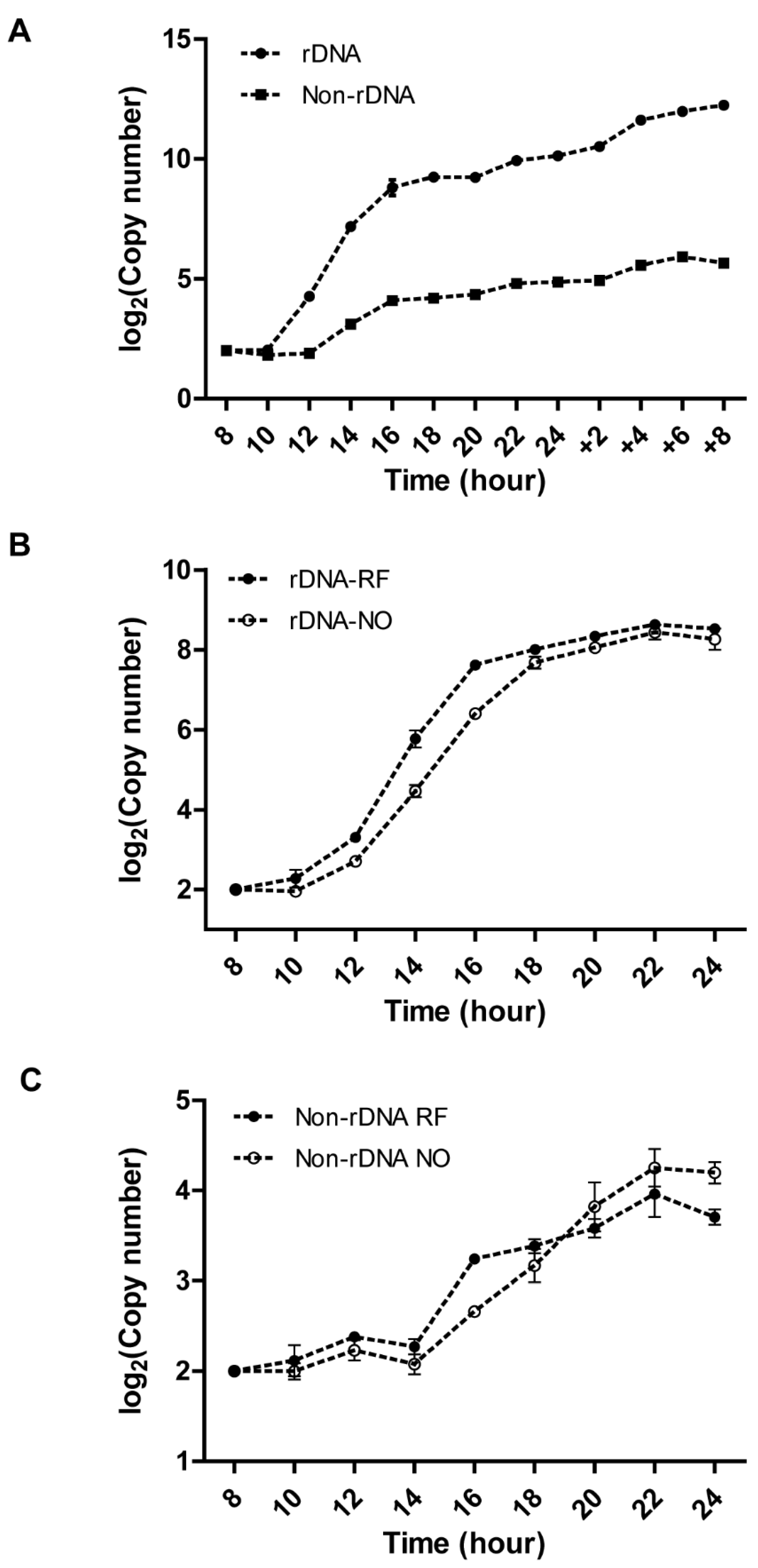
A limitation of our initial approach is that rDNA and non-rDNA abundance were assessed by different methods. To overcome this, we initiated a cross to assess rDNA and non-rDNA copy number changes by the same method. A C3 rDNA heterokaryon strain (SB1934: C3 rDNA micronucleus, B rDNA macronucleus) was mated to a histone H3 heterokaryon strain (g-H3.2+GFP-3: gfp-histone H3 micronucleus; wildtype histone H3 macronucleus), in which the coding sequence of the micronuclear HHT2 gene was fused to green fluorescent protein (GFP) (Table 1; Supplemental Figure S2). Allele-specific q-PCR was used to exclusively monitor C3 rDNA and HHT2-GFP copy number in the developing macronucleus of progeny. Four important pieces of information were obtained. First, rDNA amplification initiated prior to endoreplication of the non-rDNA HHT2 locus (Figure 2A, T = 12 h). Second, in contrast to all other developmental systems, gene amplification and endoreplication occurred concurrently. Unexpectedly, rDNA amplification was restricted to a relatively short developmental window in starved mating cells (Figure 2A; T = 10–16 h). Third, within the limits of resolution, there appeared to be a limiting factor or environmental cue that concurrently downregulates both DNA replication programs (Figure 2A; transition point: T = 16 h). Finally, upon re-feeding, mated cells resumed replication of rDNA and non-rDNA at chromosomes at comparable rates. We conclude that rDNA replication initiation is subjected to multiple levels of regulation: cell cycle control during the vegetative S phase, gene amplification during early macronuclear development, and endoreplication during the later stages of macronuclear development. As previously reported, the increased demand for DNA replication during macronuclear development is inversely correlated with the abundance of ORC [19]. Gene amplification accounts for ~20% of the final rDNA copy number and endoreplication generates the rest.

3.3. Effect of Re-Feeding on Endoreplication Phases 1 and 2

One possible explanation for the plateau in rDNA and non-rDNA replication in starved mating cells (Figure 1B,C and Figure 2A) is that re-feeding is required to generate new proteins and/or DNA precursors to sustain further rounds of DNA replication. We asked whether early re-feeding might eliminate the endoreplication plateau and possibly support more extensive rDNA gene amplification as well. To explore these possibilities, we used qPCR to assess rDNA and non-rDNA copy number in mating cells that were re-fed early (10 h post-mixing) and cells subjected to our normal mating regiment (refeeding at 24 h). Subtle differences were observed. For example, rDNA amplification was modestly accelerated in early re-fed mating cells, but there was no net increase in copy number (Figure 2B). The amplification ‘window’ was neither shortened nor lengthened: rDNA copy number plateaued in the 18–24 h interval, regardless of whether mating cells were re-fed. Early re-feeding had no obvious impact on the endoreplication program as well, as assessed through copy number analysis of the HHT2-gfp allele (Figure 2C). The collective data indicate that there are other underlying factors that control the temporal/developmentally program for overreplication of rDNA and non-rDNA chromosomes.

3.4. Replication Initiation and Elongation in Endoreplicating Non-rDNA Chromosomes

In previously published work, we discovered that ORC protein levels are dynamically regulated during development [19]. The observed changes are counterintuitive for ORC-driven replication initiation. During the period of meiotic and post-zygotic micronuclear DNA replication (0–9 h), Orc1p levels are elevated threefold relative to vegetative G1/S cultures, despite an ~25-fold reduction in the amount of DNA synthesis. Orc1p levels subsequently decline ~30-fold (0.1× vegetative level), reaching a minimum during endocycle phase II, when the developmental DNA replication load is greatest [19]. Mcm6 protein levels correlate well with Orc1p during development and are similarly diminished during the vegetative S phase in ORC1 knockdown mutants, suggesting that the abundance of these pre-RC components is coordinately regulated.
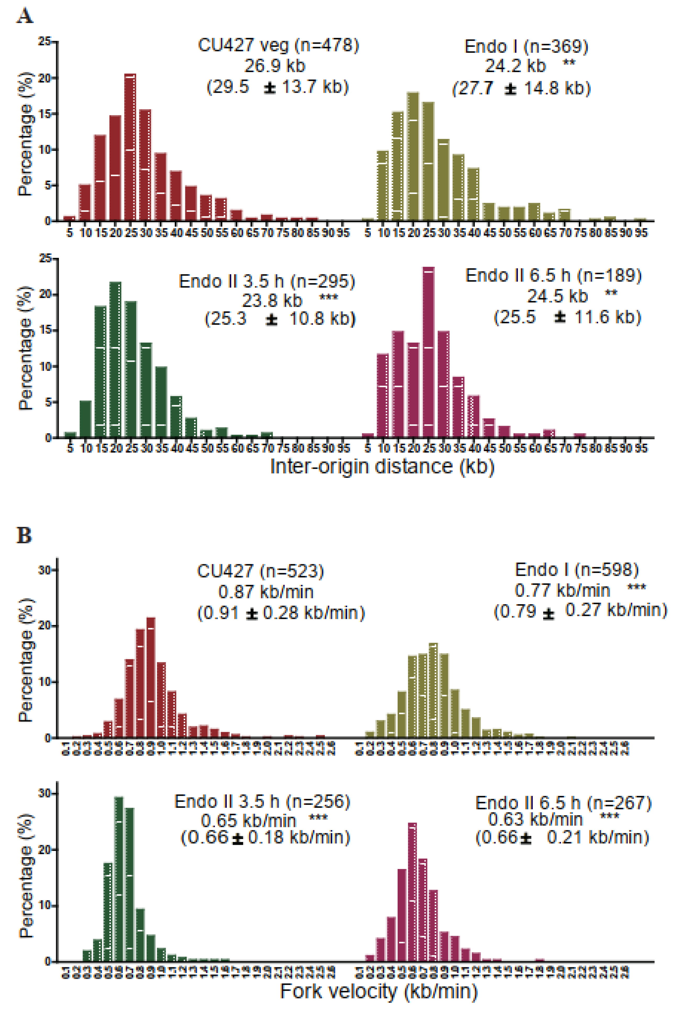
To examine DNA replication in the developing macronucleus, we employed DNA fiber imaging to measure origin activity and fork elongation rates in endoreplicating non-rDNA chromosomes relative to vegetative DNA synthesis. In a previous study, this method detected changes in replication fork elongation rates in a vegetative growing ORC knockdown mutant population [19]. Our data here revealed a 12% reduction in the average inter-origin distance in endocycling cells (vegetative S phase: 26.9 kb; endoreplication phase I: 24.2 kb; endoreplication phase 2: (early) 23.8 kb and (late) 24.5 kb (Figure 3A). Hence, more initiation events occur in endoreplicating macronuclei, despite the decreased abundance of ORC and MCM proteins. Consistent with a reduction in the abundance of the MCM2-7 replicative helicase, we observed a >25% decrease in the rate of replication fork elongation (Figure 3B; vegetative S phase, 0.87 kb/min; endoreplication phase I, 0.77 kb/min; endocycle phase 2 (early and late), 0.65 kb/min. These data are indicative of compensatory changes in replication initiation and elongation in endocycling cell populations.

3.5. Localization of Aberrant Replication Intermediates in Endoreplicating rDNA Minichromosomes

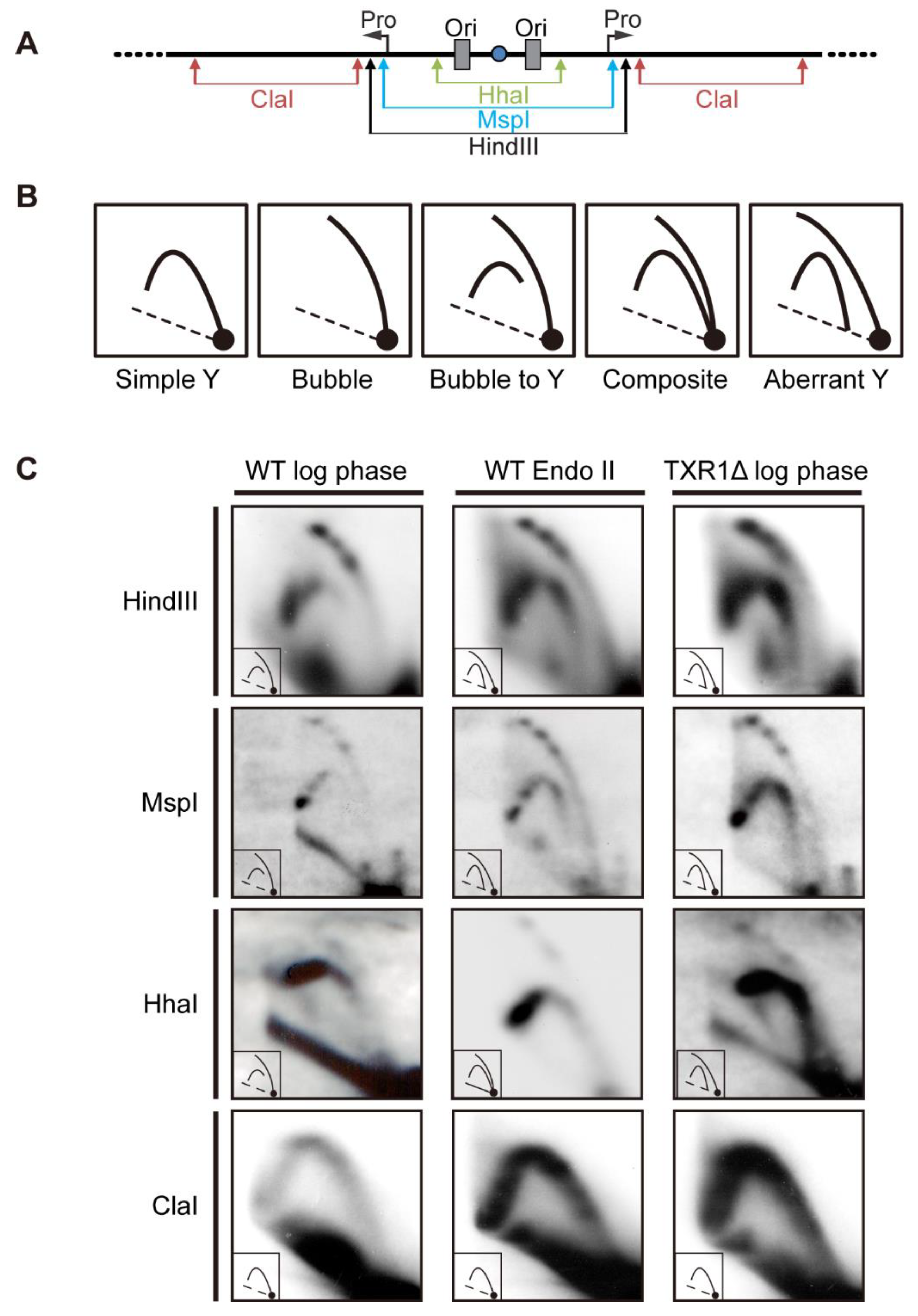
Two-dimensional (2D) gel electrophoresis of rDNA replication intermediates (RIs) previously demonstrated that the origins that amplify the rDNA in the developing macronucleus also control DNA replication during vegetative cell cycles (Figure 4A schematic) [21,29]. In the vegetative S phase, replication initiates exclusively from origins in the 5′ non-transcribed spacer (NTS), as evidenced by the presence of bubble-to-Y arc RIs, and the absence of complete simple Y arcs in HindIII-digested DNA (Figure 4A, restriction map; Figure 4B, upper left panel). Mutations in 26T RNA, a unique integral ORC subunit, inhibit ORC binding to origin-proximal type I elements and block initiation from these 5′ NTS origins [30,31]. We previously identified unusual replication intermediates (RIs) that form in wild-type Tetrahymena during endoreplication phase 2 in re-fed mating cells (Figure 4B,C (HindIII), middle panel) [19]. Aberrantly migrating simple Y arcs suggest that the known 5′ NTS origins are passively replicated in a subpopulation of molecules. The aberrant Y arcs pattern is consistent with extensive DNA unwinding and/or accumulation of persistent RNA:DNA hybrids, similar to what has been reported in S. cerevisiae senetaxin mutants [32]. The aberrant RIs (Figure 4C, (HindIII, right panel) and their sensitivity to mung-bean nuclease were reported in the vegetative Tetrahymena TXR1 knockout strain, defective in histone H3K27 monomethylation [19,33].
In an effort to localize the region responsible for the aberrant RIs, we performed 2D gel analysis on DNA digested with different restriction enzymes (Figure 4A). The vegetative TXR1 mutant was used as a reference point for comparative analysis to wild-type vegetative and endocycling cells. We specifically set out to determine whether the rDNA promoter region (responsible for rRNA biogenesis) or upstream non-coding sequences were involved. MspI cleaves 18 bp downstream of the rRNA start site, and HhaI digests just downstream of the tandem ORC binding sites (ORI). Aberrant simple Y arcs were detected in the MspI 5′ NTS fragment, but not in the HhaI-digested sample (Figure 4B, simple Y vs. aberrant Y; Figure 4C). Furthermore, normal migrating simple Y arcs were detected in the ClaI coding region fragment. The close proximity of the MspI site to the rRNA start site (18 bp) in conjunction with normal migrating ClaI coding region RIs support the idea that stalled 35S rRNA transcripts are not responsible aberrant RI production. The collective data suggest that rDNA replication initiates downstream of the nucleosome-free 5′ NTS origins that are bound by Watson:Crick RNA-DNA base pairing to the ORC [30], and upstream of the rRNA promoter [34] in a large fraction of replicating molecules. This interval is comprised of repetitive DNA sequences, the type II a-m elements. The implications are discussed below.

4. Discussion

In conventional mitotic cell cycles (G1-S-G2-M), DNA content oscillates by a factor of two. Chromosomes duplicate once in the S phase and are transmitted equally to daughter cells. Developmentally programmed endoreplication (Gap-S-Gap-S) increases DNA content on a genome-wide scale, enhancing a cell’s capacity to increase in size and/or perform specialized functions ([35]; reviewed in [36]) chromosomes, it achieves a similar objective, illustrated by the amplified Drosophila chorion genes that direct the production of essential eggshell proteins that protect the developing embryo [8]. In the case of the ciliated protozoan, Stentor coeruleus, single-cell sequencing demonstrated that chromosome copy number scales with the size of individual organisms [37].
The partitioning of germline and somatic functions into diploid, heterochromatic micronuclei and polyploid, euchromatic macronuclei, respectively, is a defining feature of Ciliophora. Synchronized mass mating has been used to study molecular events and biochemical pathways for genome reorganization in the developing macronucleus. In the work presented here, we mated Tetrahymena thermophila heterokaryons to study endoreplication and gene amplification. The copy number of marked rDNA and non-rDNA chromosomes was determined in progeny macronuclei. These studies revealed a more complex DNA replication program than previously realized.
Endoreplication in Tetrahymyena occurs in two distinct phases, separated by a prolonged gap phase that is experimentally terminated by re-feeding (Figure 1 and Figure 2). However, early refeeding is not sufficient to override cell cycle arrest of endoreplication phase I at the 8 C stage (Figure 2). We propose that the activation of nutrient signaling pathways is required for endoreplication phase 2, similar to TOR and SNAIL signaling in Drosophila [38], but that this is not sufficient to complete macronuclear development. Potential internal drivers for the transition to endoreplication phase 2 include programmed DNA rearrangement (reviewed in [39]), genome-wide conversion of heterochromatin into euchromatin ([33]; reviewed in [40]), new zygotic gene expression, modulation of the ASI2 signal transduction pathway, which is required for initiation of the endoreplication program [28,41], and degradation of the parental macronucleus [42].
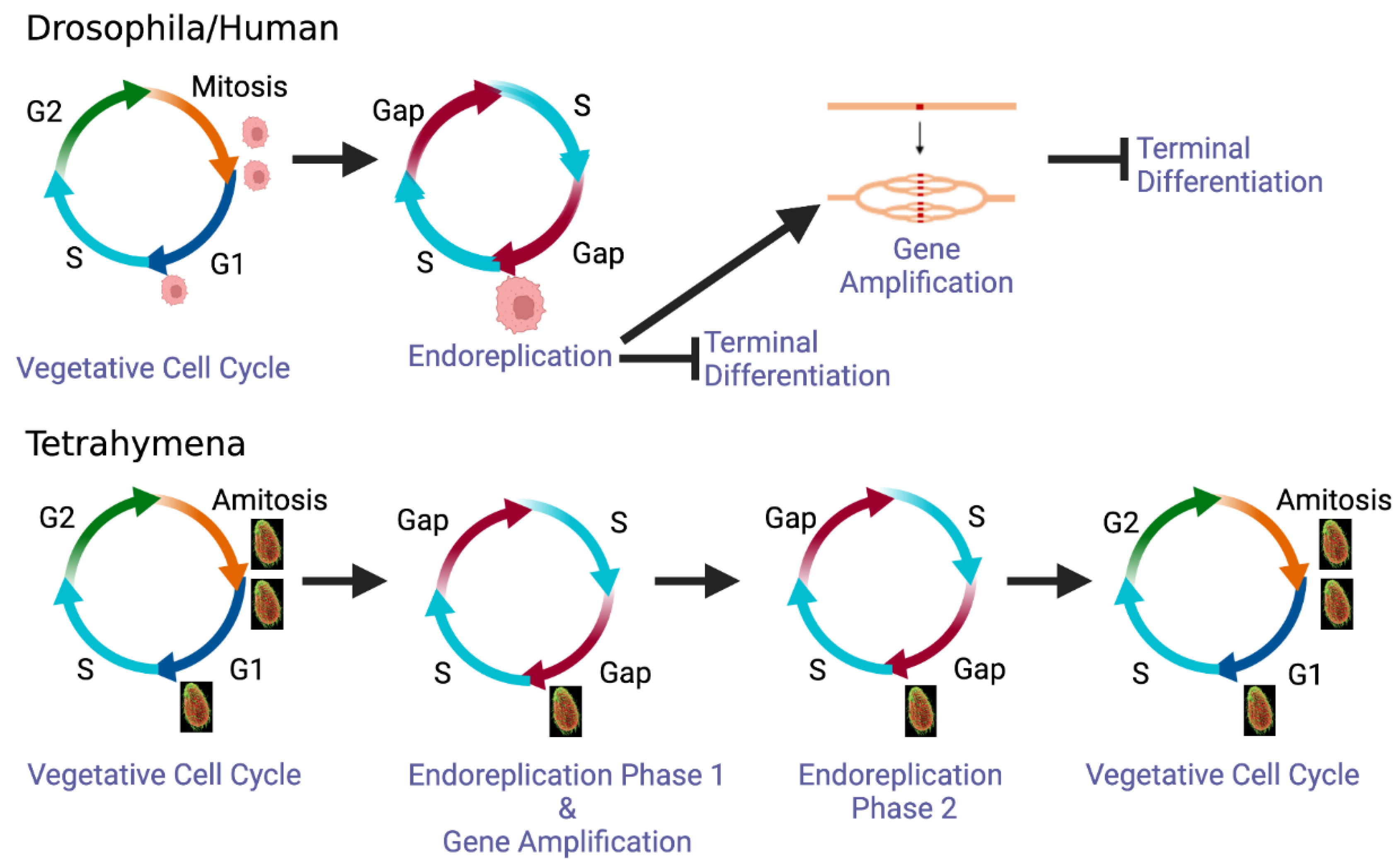
A distinguishing feature endoreplication and gene amplification in ciliates is that these DNA replication programs are not associated with terminal differentiation. Chromosomes are subsequently propagated during ‘vegetative’ cell cycles, when macronuclear chromosomes replicate once and only once in the S phase (Figure 5). With the exception of Drosophila adult rectal papillae cells that have a limited, error-prone proliferative lifespan [43], developmentally programmed endoreplication and gene amplification in metazoa occur in terminal differentiated cells. Chromosome integrity can be relaxed as chromosomes are not partitioned to daughter cells [44,45]. This situation is illustrated in the Drosophila salivary gland endocycle where cytologically visible DNA segments are under-replicated [5,46]. Furthermore, ‘onionskin’ DNA structures/stalled forks generated during amplification of Drosophila chorion gene loci are tolerated as these cells do not divide [7,47]. When both phenomena occur in metazoa, they are sequentially ordered—endoreplication precedes locus-specific gene amplification (Figure 5).
The only known Tetrahymena gene devoted to endoreplication is ASI2, a putative transmembrane signal transduction protein that is required to sustain early and late rounds of endoreplication in the developing macronucleus [41]. For comparison, Notch signaling plays a critical role in the mitosis-to-endocycle transition in Drosophila in follicle cells [48]. Perturbations of intrinsic signaling by 14-3-3 gamma, an inhibitor of the G2-to-M transition, or the local tumor environment promote endoreplication in human cancer cells [49,50]. DNA double-strand breaks (DSBs) trigger endoreplication in normal Arabidopsis thaliana endosperm and are thought to inhibit the G2-to-M transition [51]. Like metazoa, DNA replication in macronuclear anlagen involves a switch from a mitotic cell cycle, albeit an altered one, in which micronuclear DNA replication and mitosis (nuclear division without cytokinesis) precedes endoreplication phase 1.
Early in macronuclear development, sequence-specific double-strand breakage occurs to fragment the 5 germline chromosomes into 180 macronuclear chromosomes. An RNA-guide mechanism eliminates over 5000 internal DNA segments by breakage and joining reactions. It is plausible that DSB formation events could trigger the mitosis-to-endocycle transition in Tetrahymena, similar to Arabidopsis endosperm. Maternal expression of PDD1, the nucleating factor for programmed DNA elimination, is required for endoreplication [52] and PDD1-associated proteins are required for ligation of DSBs that, among other things, removes repetitive DNA sequences, including retrotransposons [53]. The precision of these processes appears to be adequate to create intact chromosomes for subsequent propagation during the vegetative phase of the life cycle.
Existing data suggest that rDNA amplification and genome-wide endoreplication do not induce genome instability in Tetrahymena. Consequently, aberrant DNA structures or collapsed replication forks are either not generated, are subsequently resolved, or are simply tolerated. Our 2D gel data clearly demonstrate the production of aberrant replication intermediates (Figure 4). Activation of the ATR intra-S phase checkpoint is consistent with DNA damage/fork arrest resolution. Toleration is plausible based on prior studies on vegetative knockdown mutations of ORC1 and TIF1, both of which activate the ATR intra-S phase checkpoint response but do not arrest cell cycle progression [19,54]. Lagging macronuclear chromosomes are observed in these mutants, slowing vegetative cell cycle progression. While these strains can be propagated indefinitely, their diploid micronucleus undergoes DNA deletions and rearrangements, and even the loss of entire chromosomes.
Four features of macronuclear chromosomes may factor into Tetrahymena’s ability to continuously propagate endoreplicated and amplified chromosomes. First, macronuclear chromosomes do not contain late replicating DNA and associated heterochromatic barriers to fork progression (L Zhang and GM Kapler, unpublished results). With the exception of a few rare minichromosomes that are only transiently propagated during early vegetative growth, there are no known regions that block fork elongation and render macronuclear chromosome sensitive to chromosome loss [55]. By analogy, endoreplicating Drosophila salivary gland chromosomes undergo fork collapse at many sites [5,46]. Genome instability severely limits the proliferative lifespan of the rare cell type that can propagate after endoreplication—Drosophila rectal papillae [43]. Second, in contrast to amplified regions in Drosophila and Sciara chromosomes [8,56], the Tetrahymena rDNA locus resides on a small autonomous minichromosome. The demands for bidirectional fork progression are minimal, as diverging forks only need to traverse only 10–12 kb to propagate the entire rDNA minichromosome [21]. In contrast, stalled/collapsed forks generated during chorion gene amplification produces onionskin chromosomes that cannot be partitioned to daughter cells [47]. Third, all macronuclear chromosomes lack centromeres and segregate by amitosis. Consequently, anaphase bridges do not form—breakage-bridge-fusion (BFB) cycles are averted. Whereas lagging macronuclear chromosomes are generated at a low frequency in wild-type Tetrahymena macronuclei, they are inconsequential. In contrast, BFB cycles in mitotic germline micronuclei render cells sterile [19,54]. Finally, a ‘self-correcting’ DNA copy number control mechanism somehow maintains the macronuclear genic balance [17,57]. Aberrant chromosomes, if generated, may be eliminated through a surveillance mechanism, analogous to programmed DNA elimination in the early developing macronucleus (reviewed in [39]).
As anticipated from previous work, endoreplication and gene amplification occur concurrently; rDNA amplification precedes endoreplication by 1–2 h. Unexpectedly, our data indicate that the rDNA minichromosome is only transiently amplified in starved mated cells. It is subsequently constrained to replicate at a comparable rate as non-rDNA chromosomes upon re-feeding. The switch from rDNA amplification to rDNA endoreplication might be sensitive to the abundance of ORC or a labile amplification-specific trans-acting factor, possibly a protein that is bound to a region that creates a virtually impermeable replication fork barrier whose strength diminishes over time [21]. Dynamic changes in rDNA replication coincide with the previously reported down-regulation of ORC and MCM proteins in the developing macronucleus [19].
Several studies in mammals and flies raise the possibility of an ORC-independent DNA replication program. Homozygous D. melanogaster Orc1−/− salivary gland cells endoreplicate to comparable levels as their wild-type counterparts (10 endocycles) [58]. Similar results were obtained for the Orc2 k43γ4 allele. Disruption of the ORC 1 gene in diploid mouse tissues blocks DNA replication; however, ORC1 is not required in polyploid extraembryonic trophoblasts and hepatocytes [59]. Finally, p53−/− HCC116 human colon cancer cells initiate DNA replication in the absence of ORC1, ORC2, or ORC5 subunits [60,61]. Whether the site for MCM2-7 recruitment is marked by an aberrant DNA structure (hairpin or quadraplex) or an alternative nucleation factor (non-ORC protein, non-coding RNA, stable RNA-DNA hybrid) remains to be determined. Compelling arguments for ORC-independent DNA replication in HU-arrested and -released vegetative Tetrahymena [20] are consistent with the relaxed requirements for ORC during macronuclear development.
Finally, in this study, we determined that aberrant RIs generated during rDNA endoreplication emanate from a relatively small region, upstream of the rRNA transcription start site (Figure 4). Inactivation of the known 5′ NTS origins has been observed in ORC mutants that are defective in rDNA origin binding [30] and in HU-treated Tetrahymena, where ORC protein levels are reduced 50-fold [20]. In both cases, an amorphous, alternate site was used, analogous to what we report here in endocycling wild-type Tetrahymena and the vegetative TXR1 mutant. As the rRNA coding region is unaffected in vegetative TXR1 knockout and endoreplicating wild-type strains (Figure 4), rRNA transcripts are not implicated. The most likely source for aberrant RI production is the type 2 element array, comprised of thirteen near-perfect tandem 21 bp direct repeats (type 2 a-m) of unknown function [62]. Type 2 elements are located between Domains 1 and 2 (the conventional 5′NTS origins) and the rRNA promoter (Figure 4A). Transient DNA melting of this AT-rich region could produce out-of-register Watson:Crick strand reassociations, generating unpaired single-strand gaps or hairpin structures on opposite DNA strands—analogous to structures that are proposed for regions of the human genome that undergo trinucleotide repeat expansions (reviewed in [63]). The prediction is that two structures would form on each DNA molecule, one on each strand—somewhat like the bidirectional replication fork. Could these hypothetical structures serve as molecular beacons for the DNA replication machinery when ORC is rate-limiting?

Supplementary Materials

The following supporting information can be downloaded at: https://www.mdpi.com/article/10.3390/microorganisms11020491/s1, Figure S1: Flow cytometry profile at 1h intervals; Figure S2: Schematic of mating between heterokaryons strains.

Author Contributions

X.M.: experimental design, execution and interpretation, manuscript writing, and graphics. H.Q.D.: experimental design, execution and interpretation, manuscript editing, and graphics. G.M.K.: experimental design and interpretation, manuscript writing, graphics, and funding. All authors have read and agreed to the published version of the manuscript.

Funding

This work was funded by NSF grant MCB-0132675 and the Tom and Jean McMullin Endowed Professorship to GMK.

Data Availability Statement

All relevant data can be found within this paper and accompanying Supplementary Figures S1 and S2.

Acknowledgments

We thank Doug Chalker for providing the histone H3-gfp tagged strain. This work was supported by NSF grant MCB-0132675 and the Tom and Jean McMullin Endowed Professorship to GMK.

Conflicts of Interest

The authors have declared that no conflict of interest.

References

  1. Morgan, D.O. Cyclin-Dependent kinases: Engines, Clocks, and Microprocessors. Annu. Rev. Cell Dev. Biol. 1997, 13, 261–291. [Google Scholar] [CrossRef] [PubMed]
  2. Dolson, A.; Sauty, S.M.; Shaban, K.; Yankulov, K. Dbf4-Dependent Kinase: DDK-ated to post-initiation events in DNA replication. Cell Cycle 2021, 20, 2348–2360. [Google Scholar] [CrossRef] [PubMed]
  3. Orr-Weaver, T.L. When bigger is better: The role of polyploidy in organogenesis. Trends Genet. 2015, 31, 307–315. [Google Scholar] [CrossRef] [PubMed] [Green Version]
  4. Shu, Z.; Row, S.; Deng, W.-M. Endoreplication: The Good, the Bad, and the Ugly. Trends Cell Biol. 2018, 28, 465–474. [Google Scholar] [CrossRef] [PubMed]
  5. Nordman, J.T.; Kozhevnikova, E.N.; Verrijzer, C.P.; Pindyurin, A.V.; Andreyeva, E.N.; Shloma, V.V.; Zhimulev, I.F.; Orr-Weaver, T.L. DNA Copy-Number Control through Inhibition of Replication Fork Progression. Cell Rep. 2014, 9, 841–849. [Google Scholar] [CrossRef] [Green Version]
  6. Austin, R.J.; Orr-Weaver, T.L.; Bell, S. Drosophila ORC specifically binds to ACE3, an origin of DNA replication control element. Genes Dev. 1999, 13, 2639–2649. [Google Scholar] [CrossRef]
  7. Claycomb, J.; MacAlpine, D.M.; Evans, J.G.; Bell, S.; Orr-Weaver, T.L. Visualization of replication initiation and elongation in Drosophila. J. Cell Biol. 2002, 159, 225–236. [Google Scholar] [CrossRef]
  8. Spradling, A.C.; Mahowald, A.P. Amplification of genes for chorion proteins during oogenesis in Drosophila melanogaster. Proc. Natl. Acad. Sci. USA 1980, 77, 1096–1100. [Google Scholar] [CrossRef] [Green Version]
  9. Calvi, B.R.; Lilly, M.A. Spradling AC Cell cycle control of chorion gene amplification. Genes Dev. 1998, 12, 734–744. [Google Scholar] [CrossRef] [Green Version]
  10. Albertson, D.G. Gene amplification in cancer. Trends Genet. 2006, 22, 447–455. [Google Scholar] [CrossRef]
  11. Karrer, K.M. Nuclear dualism. Methods Cell Biol. 2012, 109, 29–52. [Google Scholar] [PubMed]
  12. Cole, E.; Sugai, T. Developmental progression of Tetrahymena through the cell cycle and conjugation. Methods Cell Biol. 2012, 109, 177–236. [Google Scholar] [PubMed]
  13. Zhang, L.; Cervantes, M.D.; Pan, S.; Lindsley, J.; Dabney, A.; Kapler, G.M. Transcriptome analysis of the binucleate ciliate Tetrahymena thermophila with asynchronous nuclear cell cycles. Mol Biol Cell. 2023. [Google Scholar] [CrossRef] [PubMed]
  14. Cole, E.S.; Soelter, T.A. A Mutational Analysis of Conjugation in Tetrahymena thermophila 2. Phenotypes affecting middle and late development: Third prezygotic nuclear division, pronuclear exchange, pronuclear fusion, and postzygotic development. Dev. Biol. 1997, 189, 233–245. [Google Scholar] [CrossRef] [Green Version]
  15. Yakisich, J.S.; Kapler, G.M. Role of the PI3-kinase pathway in programmed nuclear death and the fate of pharmacologically-induced survival nuclei in Tetrahymena thermophila. Cell Death Differ. 2004, 10, 1146–1149. [Google Scholar] [CrossRef]
  16. Yao, M.C.; Fuller, P.; Xi, X. Programmed DNA deletion as an RNA-guided system of genome defense. Science 2003, 300, 1581–1584. [Google Scholar] [CrossRef]
  17. Eisen, J.A.; Coyne, R.S.; Wu, M.; Wu, D.; Thiagarajan, M.; Wortman, J.R.; Badger, J.H.; Ren, Q.; Amedeo, P.; Jones, K.M.; et al. Macronuclear Genome Sequence of the Ciliate Tetrahymena thermophila, a Model Eukaryote. PLoS Biol. 2006, 4, e286. [Google Scholar] [CrossRef]
  18. Zhou, Y.; Fu, L.; Mochizuki, K.; Xiong, J.; Miao, W.; Wang, G. Absolute quantification of chromosome copy numbers in the polyploid macronucleus of Tetrahymena thermophila at the single-cell level. J. Eukaryot. Microbiol. 2022, 69, 12907. [Google Scholar] [CrossRef]
  19. Lee, P.-H.; Meng, X.; Kapler, G.M. Developmental Regulation of the Tetrahymena thermophila Origin Recognition Complex. PLoS Genet. 2015, 11, e1004875. [Google Scholar] [CrossRef] [Green Version]
  20. Sandoval, P.Y.; Lee, P.-H.; Meng, X.; Kapler, G.M. Checkpoint Activation of an Unconventional DNA Replication Program in Tetrahymena. PLoS Genet. 2015, 11, e1005405. [Google Scholar] [CrossRef] [Green Version]
  21. Zhang, Z.; Macalpine, D.M.; Kapler, G.M. Developmental regulation of DNA replication: Replication fork barriers and programmed gene amplification in Tetrahymena thermophila. Mol. Cell Biol. 1997, 17, 6147–6156. [Google Scholar] [CrossRef] [PubMed] [Green Version]
  22. Larson, D.D.; Blackburn, E.H.; Yaeger, P.C.; Orias, E. Control of rDNA replication in Tetrahymena involves a cis-acting upstream repeat of a promoter element. Cell 1986, 47, 229–240. [Google Scholar] [CrossRef] [PubMed]
  23. Chastain, P.D.; Nakamura, J.; Swenberg, J.; Kaufman, D. Nonrandom AP site distribution in highly proliferative cells. FASEB J. 2006, 20, 2612–2614. [Google Scholar] [CrossRef] [PubMed]
  24. Schwab, R.A.; Niedzwiedz, W. Visualization of DNA Replication in the Vertebrate Model System DT40 using the DNA Fiber Technique. J. Vis. Exp. 2011, 1, e3255. [Google Scholar] [CrossRef]
  25. Stewart, J.A.; Wang, F.; Chaiken, M.F.; Kasbek, C.; Chastain, P.D., 2nd; Wright, W.E.; Price, C.M. Human CST promotes telomere duplex replication and general replication restart after fork stalling. EMBO J. 2012, 31, 3537–3549. [Google Scholar] [CrossRef] [Green Version]
  26. Iida, T.; Kobayashi, T. How do cells count multi-copy genes?: “Musical Chair” model for preserving the number of rDNA copies. Curr. Genet. 2019, 65, 883–885. [Google Scholar] [CrossRef]
  27. Livak, K.J.; Schmittgen, T.D. Analysis of relative gene expression data using real-time quantitative PCR and the 2−ΔΔCT Method. Methods 2001, 25, 402–408. [Google Scholar] [CrossRef]
  28. Li, S.; Yin, L.; Cole, E.S.; Udani, R.A.; Karrer, K.M. Progeny of germ line knockouts of ASI2, a gene encoding a putative signal transduction receptor in Tetrahymena thermophila, fail to make the transition from sexual reproduction to vegetative growth. Dev. Biol. 2006, 295, 633–646. [Google Scholar] [CrossRef] [Green Version]
  29. MacAlpine, D.M.; Zhang, Z.; Kapler, G.M. Type I elements mediate replication fork pausing at conserved upstream sequences in the Tetrahymena thermophila rDNA minichromosome. Mol. Cell. Biol. 1997, 17, 4517–4525. [Google Scholar] [CrossRef] [Green Version]
  30. Mohammad, M.M.; Smith, A.G.; Donti, T.R.; Kapler, G.M. Tetrahymena ORC contains a ribosomal RNA fragment that participates in rDNA origin recognition. EMBO J. 2007, 26, 5048–5060. [Google Scholar] [CrossRef]
  31. Donti, T.R.; Datta, S.; Sandoval, P.Y.; Kapler, G.M. Differential targeting of Tetrahymena ORC to ribosomal DNA and non-rDNA replication origins. EMBO J. 2009, 28, 223–233. [Google Scholar] [CrossRef] [PubMed]
  32. Alzu, A.; Bermejo, R.; Begnis, M.; Lucca, C.; Piccini, D.; Carotenuto, W.; Saponaro, M.; Brambati, A.; Cocito, A.; Foiani, M.; et al. Senataxin Associates with Replication Forks to Protect Fork Integrity across RNA-Polymerase-II-Transcribed Genes. Cell 2012, 151, 835–846. [Google Scholar] [CrossRef] [PubMed] [Green Version]
  33. Gao, S.; Xiong, J.; Zhang, C.; Berquist, B.R.; Yang, R.; Zhao, M.; Molascon, A.J.; Kwiatkowski, S.Y.; Yuan, D.; Qin, Z.; et al. Impaired replication elongation in Tetrahymena mutants deficient in histone H3 Lys 27 monomethylation. Genes Dev. 2013, 27, 1662–1679. [Google Scholar] [CrossRef] [PubMed] [Green Version]
  34. Pan, W.J.; Gallagher, R.C.; Blackburn, E.H. Replication of an rRNA gene origin plasmid in the Tetrahymena thermophila macronucleus is prevented by transcription through the origin from an RNA polymerase I promoter. Mol. Cell. Biol. 1995, 15, 3372–3381. [Google Scholar] [CrossRef] [Green Version]
  35. Berendes, H.D. Salivary gland function and chromosomal puffing patterns in Drosophila hydei. Chromosoma 1965, 17, 35–77. [Google Scholar] [CrossRef]
  36. Cross, J. How to make a placenta: Mechanisms of trophoblast cell differentiation in mice—A Review. Placenta 2005, 26, S3–S9. [Google Scholar] [CrossRef]
  37. Slabodnick, M.M.; Ruby, J.G.; Reiff, S.B.; Swart, E.C.; Gosai, S.; Prabakaran, S.; Witkowska, E.; Larue, G.E.; Fisher, S.; Freeman, R.M.; et al. The Macronuclear Genome of Stentor coeruleus Reveals Tiny Introns in a Giant Cell. Curr. Biol. 2017, 27, 569–575. [Google Scholar] [CrossRef] [Green Version]
  38. Zeng, J.; Huynh, N.; Phelps, B.; King-Jones, K. Snail synchronizes endocycling in a TOR-dependent manner to coordinate entry and escape from endoreplication pausing during the Drosophila critical weight checkpoint. PLoS Biol. 2000, 18, e3000609. [Google Scholar] [CrossRef] [Green Version]
  39. Noto, T.; Mochizuki, K. Whats, hows and whys of programmed DNA elimination in Tetrahymena. Open Biol. 2017, 7, 170–172. [Google Scholar] [CrossRef] [Green Version]
  40. Chalker, D.L. Dynamic nuclear reorganization during genome remodeling of Tetrahymena. Biochim. Biophys. Acta (BBA)-Mol. Cell Res. 2008, 1783, 2130–2136. [Google Scholar] [CrossRef] [Green Version]
  41. Yin, L.; Gater, S.T.; Karrer, K.M. A Developmentally Regulated Gene, ASI2, Is Required for Endocycling in the Macronuclear Anlagen of Tetrahymena. Eukaryot. Cell 2010, 9, 1343–1353. [Google Scholar] [CrossRef] [PubMed] [Green Version]
  42. Akematsu, T.; Pearlman, R.E.; Endoh, H. Gigantic macroautophagy in programmed nuclear death ofTetrahymena thermophila. Autophagy 2010, 6, 901–911. [Google Scholar] [CrossRef] [Green Version]
  43. Fox, D.T.; Gall, J.G.; Spradling, A.C. Error-prone polyploid mitosis during normal Drosophila development. Genes Dev. 2010, 24, 2294–2302. [Google Scholar] [CrossRef] [PubMed] [Green Version]
  44. Hassel, C.; Zhang, B.; Dixon, M.; Calvi, B.R. Induction of endocycles represses apoptosis independently of differentiation and predisposes cells to genome instability. Development 2014, 141, 112–123. [Google Scholar] [CrossRef] [PubMed] [Green Version]
  45. Mehrotra, S.; Maqbool, S.B.; Kolpakas, A.; Murnen, K.; Calvi, B.R. Endocycling cells do not apoptose in response to DNA rereplication genotoxic stress. Genes Dev. 2008, 22, 3158–3171. [Google Scholar] [CrossRef] [PubMed] [Green Version]
  46. Yarosh, W.; Spradling, A.C. Incomplete replication generates somatic DNA alterations within Drosophila polytene salivary gland cells. Genes Dev. 2014, 28, 1840–1855. [Google Scholar] [CrossRef] [PubMed] [Green Version]
  47. Osheim, Y.N.; Miller, O.L., Jr.; Beyer, A.L. Visualization of Drosophila melanogaster chorion genes undergoing amplification. Mol. Cell Biol. 1998, 8, 2811–2821. [Google Scholar]
  48. Sun, J.; Smith, L.; Armento, A.; Deng, W.-M. Regulation of the endocycle/gene amplification switch by Notch and ecdysone signaling. J. Cell Biol. 2008, 182, 885–896. [Google Scholar] [CrossRef] [Green Version]
  49. Gomes, C.J.; Centuori, S.M.; Harman, M.W.; Putnam, C.W.; Wolgemuth, C.W.; Martinez, J.D. The induction of endoreduplication and polyploidy by elevated expression of 14-3-3γ. Genes Cancer 2017, 8, 771–783. [Google Scholar] [CrossRef] [Green Version]
  50. Tan, Z.; Chu, D.Z.V.; Chan, Y.J.A.; Lu, Y.E.; Rancati, G. Mammalian Cells Undergo Endoreduplication in Response to Lactic Acidosis. Sci. Rep. 2018, 8, 2890. [Google Scholar] [CrossRef]
  51. Adachi, S.; Minamisawa, K.; Okushima, Y.; Inagaki, S.; Yoshiyama, K.; Kondou, Y.; Kaminuma, E.; Kawashima, M.; Toyoda, T.; Matsui, M.; et al. Programmed induction of endoreduplication by DNA double-strand breaks in Arabidopsis. Proc. Natl. Acad. Sci. USA 2011, 108, 10004–10009. [Google Scholar] [CrossRef] [PubMed] [Green Version]
  52. Coyne, R.S.; A Nikiforov, M.; Smothers, J.F.; Allis, C.; Yao, M.-C. Parental Expression of the Chromodomain Protein Pdd1p Is Required for Completion of Programmed DNA Elimination and Nuclear Differentiation. Mol. Cell 1999, 4, 865–872. [Google Scholar] [CrossRef] [PubMed]
  53. Xu, J.; Yuan, Y.; Liang, A.; Wang, W. Chromodomain protein Tcd1 is required for macronuclear genome rearrangement and repair in Tetrahymena. Sci. Rep. 2015, 5, 10243. [Google Scholar] [CrossRef] [PubMed] [Green Version]
  54. Yakisich, J.S.; Sandoval, P.Y.; Morrison, T.L.; Kapler, G.M. TIF1 activates the intra-S phase checkpoint response in the diploid micronucleus and amitotic polyploid macronucleus of Tetrahymena. Mol. Biol. Cell 2006, 17, 5185–5197. [Google Scholar] [CrossRef] [Green Version]
  55. Feng, L.; Wang, G.; Hamilton, E.P.; Xiong, J.; Yan, G.; Chen, K.; Chen, X.; Dui, W.; Plemens, A.; Khadr, L.; et al. A germline-limited piggyBac transposase gene is required for precise excision in Tetrahymena genome rearrangement. Nucleic Acids Res. 2017, 45, 9481–9502. [Google Scholar] [CrossRef] [Green Version]
  56. Liang, C.; Spitzer, J.D.; Smith, H.S.; Gerbi, S.A. Replication initiates at a confined region during DNA amplification in Sciara DNA puff II/9A. Genes Dev. 1993, 7, 1072–1084. [Google Scholar] [CrossRef] [Green Version]
  57. Doerder, F.P.; Ditaranto, J.; DeBault, L.E. Evolutionary constraints on quantitative variation and regulation of macronuclear DNA content in the genus Tetrahymena. J. Cell Sci. 1981, 49, 177–193. [Google Scholar] [CrossRef]
  58. Park, S.Y.; Asano, M. The origin recognition complex is dispensable for endoreplication in Drosophila. Proc. Natl. Acad. Sci. USA 2008, 105, 12343–12348. [Google Scholar] [CrossRef] [Green Version]
  59. Okano-Uchida, T.; Kent, L.N.; Ouseph, M.M.; McCarty, B.; Frank, J.J.; Kladney, R.; Cuitino, M.C.; Thompson, J.C.; Coppola, V.; Asano, M.; et al. Endoreduplication of the mouse genome in the absence of ORC1. Genes Dev. 2018, 32, 978–990. [Google Scholar] [CrossRef] [Green Version]
  60. Shibata, E.; Kiran, M.; Shibata, Y.; Singh, S.; Kiran, S.; Dutta, A. Two subunits of human ORC are dispensable for DNA replication and proliferation. Elife 2016, 5, 19084. [Google Scholar] [CrossRef]
  61. Shibata, E.; Dutta, A. A human cancer cell line initiates DNA replication normally in the absence of ORC5 and ORC2 proteins. J. Biol. Chem. 2020, 295, 16949–16959. [Google Scholar] [CrossRef] [PubMed]
  62. Challoner, P.B.; Amin, A.A.; Pearlman, R.E.; Blackburn, E.H. Conserved arrangements of repeated DNA sequences in nontranscribed spacers of ciliate ribosomal RNA genes: Evidence for molecular coevolution. Nucleic Acids Res. 1985, 11, 2661–2680. [Google Scholar] [CrossRef] [PubMed] [Green Version]
  63. Polleys, E.J.; House, N.C.M.; Freudenreich, C.H. Role of recombination and replication fork restart in repeat instability. DNA Repair 2017, 56, 156–165. [Google Scholar] [CrossRef] [PubMed]
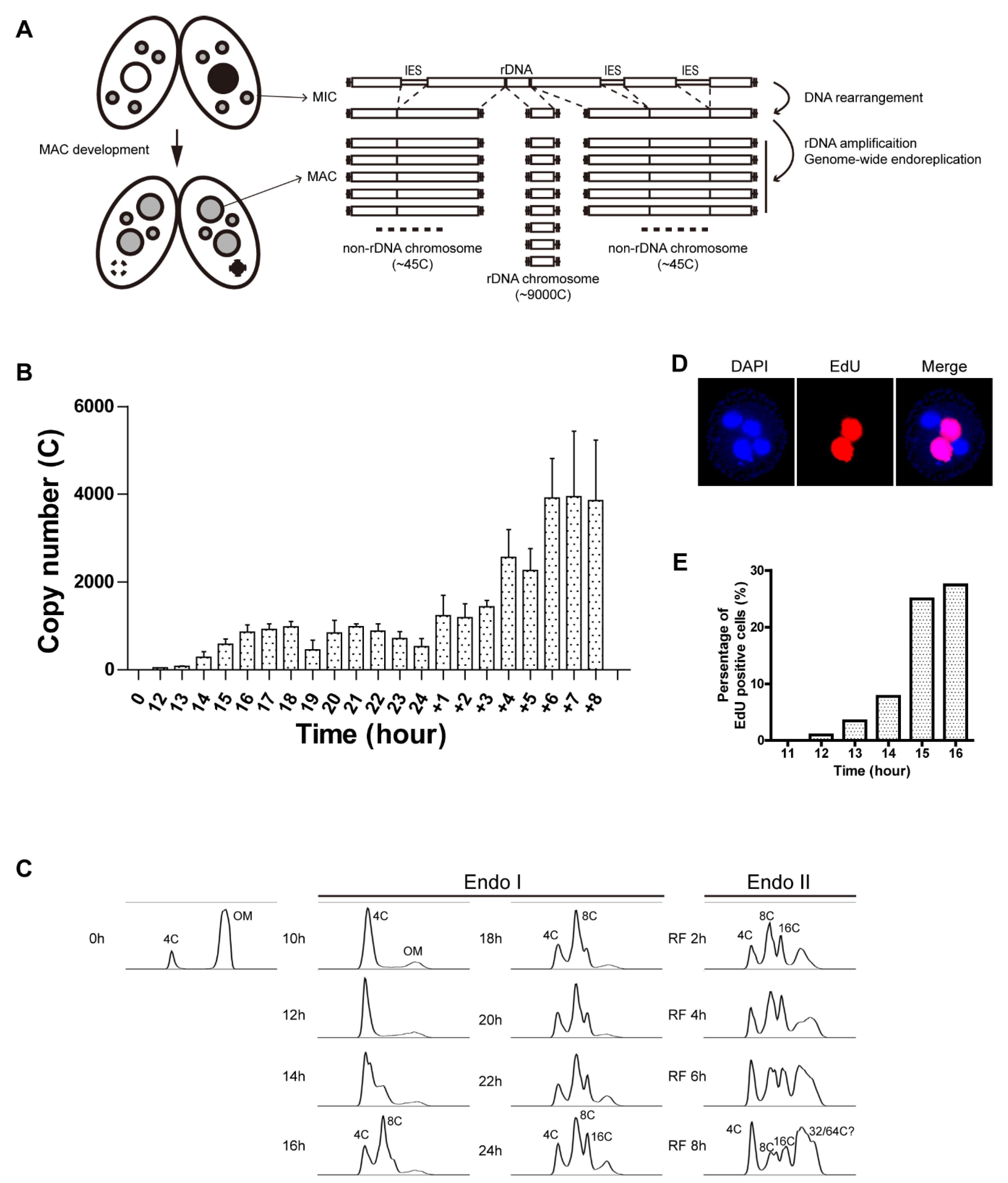
Figure 1. rDNA gene amplification and endoreplication. (A) Schematic of mating and subsequent macronuclear development. Upper image: Paired meiotic parental cells of different mating type at an early stage with 4 pronuclei (3 of which will degrade one that will undergo 1 round of DNA replication and nuclear division) and the parental macronucleus. Lower image: exconjugant progeny at a stage with two micronuclei, two developing macronuclei, and the degraded parental macronucleus. See text for details on chromosomal changes associated with macronuclear development. (B) qPCR analysis on C3 rDNA copy number in the developing macronucleus in a mating between strains SB1934 and SB4202. Samples were collected at 0 h and 12–24 h post-mixing. At 24 h, cells were re-fed, and samples were collected for an additional 8 h. rDNA copy number of each sample was normalized to the sample at 0 h post-mixing (integrated micronuclear rDNA locus). Micronuclear rDNA copy number was used as an internal control for the 2−∆∆Ct method. (C) Flow cytometry analysis on genomic DNA content during Endoreplication Phase I (mated for 0–24 h) and Endoreplication Phase 2 (mated for 24 h and re-fed for 1–8 h). OM: old (parental macronucleus). (D) A representative image of EdU labeling of anlagen nuclei. Nuclei: DAPI/blue color, active DNA replication: EdU/red. Cells during Endoreplication Phase I have 4 nuclei in each cell. Developing macronuclear anlagen are EdU-positive. (E) Quantification of EdU-labeled cells as a function of time in mating labeling image showing DNA replication during Endo I.
Microorganisms 11 00491 g001
Figure 2. Q-PCR analysis of rDNA and non-rDNA (H3-GFP) chromosome copy number changes during macronuclear development. Heterokaryon strains SB1934 and g-H3.2+GFP-3 were mated. Q-PCR analysis was performed with C3 rDNA and H3-gfp locus-specific primer sets to quantify DNA copy number in the newly developing macronucleus. The copy number of both rDNA and non-rDNA at 0 h was set to 2 C. (A) Log2 scale of copy number changes of rDNA and non-rDNA chromosomes in mated Tetrahymena that were re-fed at 24 h post-mixing. (B) Log2 scale of copy number change of rDNA copy number in mated Tetrahymena that were re-fed at 10 h versus 24 h post-mixing. RF: early re-fed at 10 h; NO: normal re-fed at 24 h. (C) Log2 scale of copy number change of non-DNA (H3-gfp locus) copy number in mated Tetrahymena that were re-fed at 10 h or 24 h post-mixing. RF: early re-fed at 10 h; NO: normal re-fed at 24 h.
Microorganisms 11 00491 g002
Figure 3. DNA fiber analysis on endoreplicating macronuclear anlagen. Mated cell progeny from a CU428 × CU427 cross were collected at different times and processes for DNA fiber analysis. Endo I: mate cell progeny were pulsed with CldU and chased with IdU at 15.5 h post-mixing. Endo II: mated cell progeny were re-fed at 24 h and sequentially labeled with CldU and IdU at 3.5 h or 6.5 h after refeeding. A log phase CU427 vegetative culture was used as a control. (A) Inter-origin distance (IOD) comparison between log phase and endoreplicating cells. (B) Fork velocity comparison between log phase and endoreplicating cells. The frequency distribution of DNA fibers with different IODs or fork velocities was graphed. For each group, the median IOD or fork velocity was calculated. The mean with S.D. is indicated in parentheses. N: the total number of DNA fibers scored. Statistical significance was determined using the Kruskal–Wallis test followed by Dunn’s test. Each sample from endoreplicating cells was compared to log phase CU427. **, p < 0.01; ***, p < 0.001.
Microorganisms 11 00491 g003
Figure 4. Two-dimensional gel analysis of rDNA replication intermediates (RIs) in endoreplicating macronuclei. (A) Schematic of the rDNA 5′NTS fragments generated by restriction digestion for 2D gel analysis with rDNA 5′ NTS or coding region probes. H3, Hind III sites are at nucleotide (nt) position 2132 in the palindromic 21 kb rDNA minichromosome; MspI: nt 1906; HhaI: nt 1347; ClaI: nt 2169–7621. The two origins (ORI) reside within the tandem 430 bp duplication designated as Domain I and Domain II (ORC binding sites: type IA and type IB elements (nt 762–794 and nt 1193–1224, respectively), Pro: 35S rRNA precursor start site (nt 1887) of the rDNA minichromosome. (B) Schematic of normal and aberrant DNA replication intermediate patterns. Different types of RIs may be detected by 2D gel analysis. Simple Y arc: replication from outside the examined DNA interval (passive replication). Bubble arc or composite bubble-to-Y arc pattern: replication initiation within the examined region. Composite with complete bubble and Y arcs: both active and passive replication within the examined region. Aberrant Y: replication intermediate that fails to intersect the 1N spot corresponding to unreplicated duplex DNA. Diagonal dashed line, migration of bulk linear duplex DNA fragments. (C). Two-dimensional gel analysis of wild-type log-phase vegetative cells (CU428), endoreplicating mated cells (SB1934 × SB4204 cross, mated 24 h and re-fed for 6 h), and a vegetative log-phase TXR1 knockout mutant (generated by macronuclear gene disruption and phenotypic assortment) [33].
Microorganisms 11 00491 g004
Figure 5. Comparative profiling of endoreplication and gene amplification developmental programs in metazoa and Tetrahymena. Upper panel—sequential DNA replication programs in terminally differentiated cells in flies and humans. Lower panel—sequential DNA replication programs in the developing macronucleus and subsequent entry into the amitotic vegetative cell cycle.
Microorganisms 11 00491 g005
Table 1. T. thermophila strains used in this study.
StrainMicronuclear GenotypeMacronuclear Phenotype
CU427chx1-1/chx1-1paromomycin-sensitive
cycloheximide-sensitive
CU428mpr1-1/mpr1-1paromomycin-sensitive
6-methylpurine-sensitive
SB1934rDNA [C3-1]/rDNA [C3-1];
mpr1-1/mpr1-1
paromomycin-sensitive
6-methylpurine-sensitive
rDNA [B]
SB4202pmr1[C3-1/pmr1[C3-1];
CHX1[C3]/CHX1[C3]
paromomycin-sensitive
cycloheximide-resistant
rDNA [B]
g-H3.2+GFP-3hht2[3′neo2, GFPc]
hht2[3′neo2, GFPc]
paromomycin-sensitive
Disclaimer/Publisher’s Note: The statements, opinions and data contained in all publications are solely those of the individual author(s) and contributor(s) and not of MDPI and/or the editor(s). MDPI and/or the editor(s) disclaim responsibility for any injury to people or property resulting from any ideas, methods, instructions or products referred to in the content.