Article

Oral Microbiota in Children and Adolescents with Type 1 Diabetes Mellitus: Novel Insights into the Pathogenesis of Dental and Periodontal Disease

1
Department of Diagnostic and Public Health, Microbiology Section, University of Verona, 37134 Verona, Italy
2
School of Health Statistics and Biometrics, University of Verona, Strada Le Grazie 8, 37134 Verona, Italy
3
Section of Pediatric Diabetes and Metabolism, Department of Surgery, Dentistry, Pediatrics and Gynecology, University of Verona, 37126 Verona, Italy
4
Department of Surgery, Dentistry, Paediatrics and Gynecology, University of Verona, 37134 Verona, Italy
*
Author to whom correspondence should be addressed.
These authors contributed equally to this work.
Microorganisms 2023, 11(3), 668; https://doi.org/10.3390/microorganisms11030668
Submission received: 1 February 2023 / Revised: 28 February 2023 / Accepted: 1 March 2023 / Published: 6 March 2023

Abstract

:
The oral microbiota can be influenced by multiple factors, but only a few studies have focused on the role of glycemic control in determining early alterations of oral microbiota and their association with pathogenesis of both periodontitis and caries. The aim of this study is to evaluate the interplay between bacteria composition, oral hygiene, and glycemic control in a cohort of children with T1D. A total of 89 T1D children were enrolled (62% males, mean age: 12.6 ± 2.2 years). Physical and clinical characteristics, glucometabolic parameters, insulin treatment, and oral hygiene habits data were collected. Microbiological analysis was performed from saliva samples. A high prevalence of cariogenic and periodontopathogens bacteria in our cohort was detected. In particular, in all subjects Actinomyces spp., Aggregatibacter actinomycetemcomitans, Prevotella intermedia, and Lactobacillus spp. were isolated. S. mutans was found in about half of the analyzed sample (49.4%), in particular in patients with imbalance values of glycemic control. Moreover, a higher presence of both S. mutans and Veillonella spp. was detected in subjects with poorer glycemic control, in terms of HbA1c, %TIR and %TAR, even adjusting for age, sex, and hygiene habits as covariates. Virtuous oral hygiene habits, such as frequency of toothbrush changes and professional oral hygiene, negatively correlated with the simultaneous presence of Tannerella forsythia, Treponema denticola, and Porphyromonas gingivalis, red complex bacteria. Our study shows it is crucial to pay attention to glycemic control and regular oral hygiene to prevent the establishment of an oral microbiota predisposing to dental and periodontal pathology in subjects with T1D since childhood.

1. Introduction

Type 1 diabetes (T1D) is a complex autoimmune disease caused by the destruction of pancreatic beta cells that leads to both acute and chronic complications [1]. An inadequate glycemic control is a major risk factor for the development of chronic complications, including cardiovascular disease, peripheral vascular disease, retinopathy, nephropathy, and neuropathy [2,3,4,5]. Periodontal disease (PD) is classified as the “sixth complication” of diabetes [6]. This chronic inflammation of periodontal tissues is characterized by the progressive destruction of the supporting structures of the teeth induced by a state of dysbiosis promoting host inflammatory response [7,8]. According to the new classification of PD, although there is sufficient evidence to believe that PD observed in the context of systemic disease that severely impair the immune response should be considered a periodontal manifestation of systemic disease, there is currently insufficient evidence to sustain that PD observed in poorly controlled diabetes is characterized by a unique pathophysiology [9]. The incidence of PD in patients with T1D is higher than in the healthy population and is significantly associated with a longer duration of diabetes and poor glycemic control [10,11]. Indeed, the latter together with changes in host response and differences in the composition of the oral microbiota are suggested as determinants of increased susceptibility of T1D patients to the development of PD and caries [12]. Two meta-analyses have recently confirmed the association between diabetes and periodontal disease, indicating a positive, bidirectional association between these two disorders [13,14]. On one hand, diabetes increases the risk and severity of inflammatory PD; on the other, periodontitis can trigger inflammatory host immune responses locally and systemically, affecting glucometabolic control in patients with T1D [15,16,17]. This relationship impacts on early onset of gingival disease and increased periodontal disease even in children and adolescents with T1D [10,11].
Different combinations of bacterial species are involved in periodontitis, such as the concomitant presence of Aggregatibacter actinomycetemcomitans and Prevotella intermedia in saliva [18]. In addition, the “red complex”, consisting of three strictly anaerobic bacteria, i.e., Porphyromonas gingivalis, Tannerella forsythia, and Treponema denticola, is associated with severe forms of periodontal disease [19,20].
Moreover, a relationship between T1D and an increased risk of dental caries has been suggested. This link is influenced by diabetes-induced changes in saliva composition and levels of glycemic control [21,22]. As regards microbial composition, Streptococcus mutans and Lactobacillus are the most cariogenic bacteria because of their ability to survive in an acidic environment and form biofilm [23]. A molecular study found that the simultaneous presence of Veillonella spp. and Streptococcus spp. can promote the development and progression of dental caries [24,25].
In the general population, oral microbiome is suspected to be affected by several variables including host genetics, geography, age, cohabitation, and familial relationship. In particular, the salivary microbiota of youths, aged 3 to 18 years, is still maturing [26,27], while, as they age, the composition of the oral microbiome changes and periodontal pathogens increase in abundance, leading to increased susceptibility to oral disease [28]. Generally, children and adolescents have an oral microbiota characterized by bacteria that are protective against periodontal disease and caries, whereas people > 50 years of age show changes in the microenvironment that lead to an increase in certain bacterial species that predispose to periodontitis. For example, the study by Rodenburg et al. showed that the prevalence of periodontopathogens such as Porphyromonas gingivalis in individuals with periodontitis increases with age [29]. Proper education of the patient with T1D in hygiene maintenance with professional oral hygiene sessions with special tools could positively affect the maintenance of oral and dental health by preventing oral dysbiosis [30].
However, the oral microbiota composition may be influenced by additional multiple modifiable factors: dietary habits, oral hygiene, and use of drugs or antibiotics. Currently, few studies have focused on the role of glycemic control, analyzing different CGM metrics, in determining alterations in the oral microbiota of subjects with T1D, and, to date, its association with pathogenesis of both periodontitis and caries remains controversial [31,32,33]. A few data are available in children and adolescents with T1D. Clarifying the relationship between the oral microbiota and the dental and metabolic health of T1D individuals from an early age is crucial in order to develop novel, effective, preventative, and therapeutic strategies.
Therefore, the aim of this study was to assess the presence of cariogenic and periodontopathogenic bacteria through saliva sample analysis and evaluate their potential roles in the interplay with oral hygiene and glycemic control in a cohort of children and adolescents with T1D.

2. Materials and Methods

2.1. Study Population

Eighty-nine children and adolescents with T1D (age: 12.6 ± 2.2 years, 55 boys) were consecutively recruited at the Regional Center for Pediatric Diabetes of the University Hospital, Verona (Italy) during a follow-up visit between December 2020 and February 2022. Inclusion criteria were diagnosis of T1D for at least one year, confirmed by positivity of at least two diabetes-associated autoantibodies (GADA, ZnT8A, IAA or IA–2A), and an age between 9 and 15 years. Exclusion criteria were chronic diseases other than T1D requiring pharmacotherapy, presence of other related genetic diseases, intake of drugs that alter salivary secretion, fixed orthodontic appliances, use of antibiotics or probiotics three months prior to inclusion, use of probiotic-containing food and medical conditions believed to affect oral and gut microbiota. Written informed consent to participate in the study was obtained from the parents/guardians of the children and adolescents. The Ethical Committee of the University Hospital of Verona approved the study, in accordance with the World Medical Association Declaration of Helsinki (approval number: Prog. 2722CESC, Prot n.29192, 25/05/2020).

2.2. Clinical Data Collection

Clinical and demographic parameters were recorded at enrollment: age, gender, age of onset, and duration of T1D and anthropometric measurements (i.e., body height, body weight, pubertal status determined using Tanner stages I–V [34], according to standard procedures, as previously reported [34,35]. Body mass index (BMI) was calculated using the formula: body weight (kg)/body height (m2), values were then standardized (BMI z-score) calculating age and sex-specific BMI percentiles according to World Health Organization (WHO) child growth standards [36]. The systolic blood pressure (SBP) and diastolic blood pressure (DBP) were measured by a physician three times on the left arm with the subject sitting, using a manual sphygmomanometer and a cuff of appropriate size [37]. Other clinical data such as daily insulin dosages (total, basal) and type of treatment (multiple daily insulin injections or continuous subcutaneous insulin infusion) were also recorded. Moreover, a questionnaire on oral hygiene habits was administered the same day as the visit and saliva sample collection. The two-page self-completion questionnaire was developed following a scoping review of the literature. The following topics were covered in the questionnaire: frequency of professional and daily oral hygiene, oral health advice received, current dental care, and oral hygiene behavior.

2.3. Glucometabolic Parameters

Glucometabolic control parameters (glycated hemoglobin (HbA1c) and continuous glucose monitoring (CGM) metrics of glycemic control and glucose variability) were collected. HbA1c was measured using the high-performance liquid chromatography technique and standardized to the normal range established by the DCCT (4.0–6.0%, 20–42 mmol/mol). Intermittently scanned continuous glucose monitoring device (isCGM, Abbott FreeStyle Libre® Glucose Monitoring System, Abbott Diabetes Care, Alameda, CA, USA) or real-time CGM device (rtCGM, Dexcom G5® CGM System or Dexcom G6® CGM System, Dexcom, San Diego, CA, USA)-derived data were recorded. For each participant, several metrics of glycemic control and variability have been computed separately for the full 12-week period of data collection immediately before the enrollment visit with HbA1c measurement and saliva collection. In particular, the following metrics were calculated: (a) glucose management indicator (GMI); (b) percentage of time below range [<70 mg/dL (TBR)]; (c) percentage of time in target range [70–180 mg/dL (TIR)]; (d) percentage of the time above range [>180 mg/dL (TAR)]; (e) coefficient of variation (CV). The following cut-offs were used according to the international consensus on use of CGM: TIR (using 70% as cut-off), TAR (using 25% as cut-off), CV (using 36% as cut-off) [38]. As regards HbA1c and GMI, we used a less-stringent goal (using 7.5% as cut-off) to classify patients as having good or poor glycemic control, as optimal glycemic control is often more difficult to achieve in the pediatric setting, and 7.5 represented exactly the median HbA1c of our population. Hence, an HbA1c value of <7.5% (58 mmol/mol) was taken as indicator of good glycemic control while a value of ≥7.5% (58 mmol/mol) was considered to be indicator of poor control. In order to ensure an adequate amount of data, participants were included in the analysis if at least 80% of expected CGM readings were available for each patient.

2.4. Microbiological Analysis

At the baseline visit a sample of 4 mL of saliva was collected while fasting for at least 8h and before performing daily oral hygiene. Samples were sent within 24 h at the Microbiology section of the Department Diagnostic and Public Health of the University of Verona and subjected to nucleic acid extraction. On saliva, bacterial culture-based analysis was also performed.
Several culture media were selected for the detection and isolation of the species of interest. In particular, Blood agar (Blood Agar Base Oxoid™) and Chocolate Agar (Chocolate Agar Base Oxoid™) have been selected as enriched media for overall oral bacterial microflora; Sabouraud agar (Oxoid™ Prepared Sabouraud Dextrose Agar) was used to isolate fungi and yeasts; Mannitol Salt agar (MSA Base Oxoid™) was employed for the growth of presumptive pathogenic staphylococci. Mitis Salivarius Agar (NutriSelect® Plus) was used for the isolation of oral streptococci. Mitis salivarius sucrose bacitracin (MSB), obtained by adding 0.2 units/mL bacitracin and by increasing the sucrose concentration to 20% starting from Mitis Salivarius Agar, was employed for the selective isolation of Streptococcus mutans.
The plates were then incubated at 37 °C for 48 h in an anaerobic condition, except Sabouraud plates that require aerobic conditions. To detect the bacterial load (colony-forming units (CFU)/mL) a serial dilution method was performed for each saliva sample.
Nucleic acids were extracted using the QIAamp DNA Microbiome Kit (Qiagen, Milan, Italy) following the manufacturer instructions and all DNA samples were suspended in 50 μL of elution buffer. Concentrations of extracted DNAs were assessed using the Qubit 2.0 fluorometer (Invitrogen, Thermo Fisher Scientific, Darmstadt, Germany). Briefly, 10 µL of extracted genomic DNA were mixed with 190 µL of a Qubit working solution (Qubit High Sensitivity Assay, Invitrogen, Thermo Fisher Scientific, Darmstadt, Germany) according to the manufacturer’s protocol. Extracted DNA was stored at −20 °C until further use. Presence of pathogens’ DNA in the samples was assessed through PCR. Two different multiplex PCRs were performed to identify the presence of: (a) P. gingivalis, P. intermedia, and A. actinomycetemcomitans [39], and (b) T. forsythia, T. denticola, and A. naeslundii. Additionally, a single PCR was performed to identify Actinomyces spp., S. mutans, Veillonella spp., and Lactobacillus spp., as previously reported [40]. PCR reactions were performed using the 5Prime Hot Master Mix (Quantabio, Beverly, MA, USA) according to manufacturer’s instructions. Briefly, PCR reactions mixture were composed of 8 μL of 5 PRIME HotMaster Mix (2.5x), 100 nM of each primer, 50 ng of template, and Ultrapure DNase/RNase-free distilled water (Thermo Fisher Scientific, Waltham, MA, USA) to reach a final volume of 20 μL. Primers and PCR conditions are reported in Supplementary Table S1.

2.5. Statistical Methods
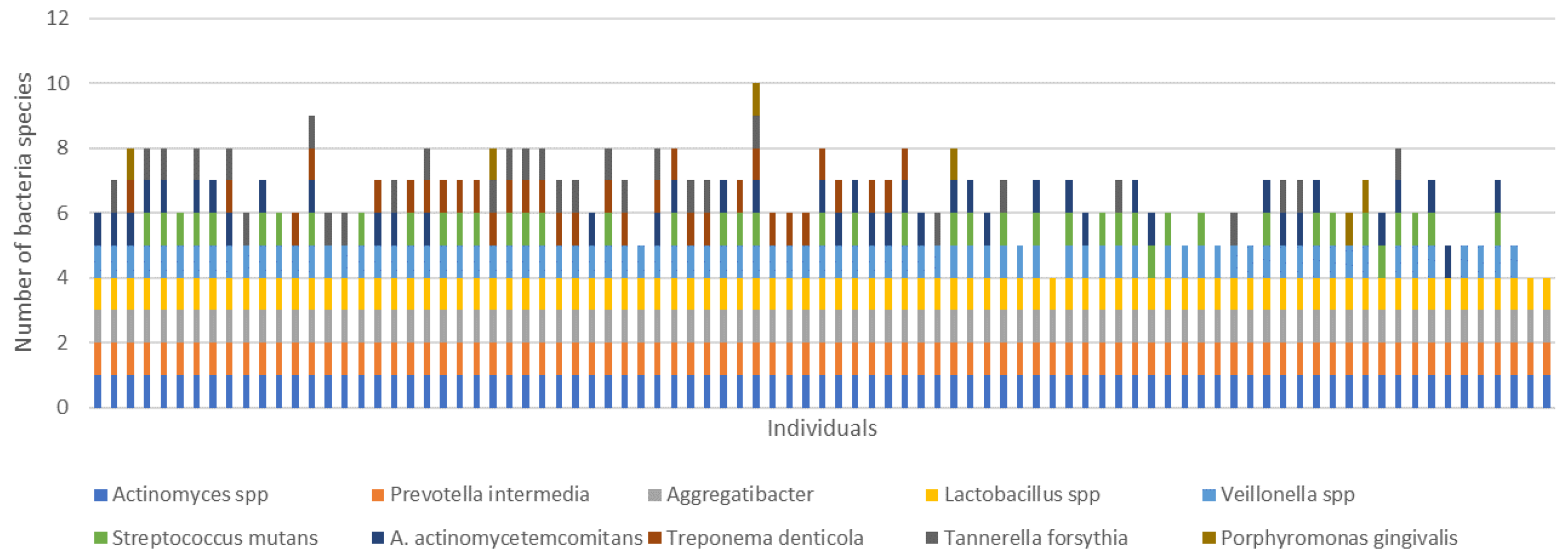
Data are presented as arithmetic mean with the relative standard deviation (SD), the medians and interquartile range [IQR], or as an absolute and relative frequency. Normal distribution of variables was assessed via the Kolmogorov–Smirnov test. Skewed variables were log-transformed unless deviations from the Gaussian distribution could not be corrected via transformation. Differences between patients stratified by sex and glycemic parameters were assessed via Student’s t-test, for Gaussian variables, and via the Mann–Whitney test, for skewed variables. A chi-square test was applied to detect differences in categorical variables. Correlations between variables were calculated by using Spearman’s rho. The relation between the presence of cariogenic bacteria (S. mutans and Veillonella spp.) and glycemic metabolic control parameters (i.e., % of HbA1c, TIR, TAR, GMI) was assessed using linear logistic regression analysis. Sex, age, bleeding during brushing, and professional hygiene frequency were used as covariates. Covariates included in multivariate regression models were selected as potential confounding factors based on their plausibility. Significance level for all tests was set at p < 0.05. All analyses were performed in R environment, STATA, and IBM SPSS Statistics 26 statistical package (SPSS, Chicago, IL, USA).

3. Results

A population of 89 children and adolescents with T1D (55 males, 61.8%) with a mean age of 12.6 ± 2.2 years was recruited. Table 1 showed the main anthropometric and metabolic characteristics and bacterial populations identified in the study sample stratified by sex and glucose control (i.e., HbA1c). Subjects’ characteristics are described according to glucometabolic control parameters (i.e., GMI, %TIR and %TAR) in Supplementary Tables S2–S4 are reported.
The bacterial distribution in our population is represented in Figure 1.
Actinomyces spp., A. actinomycetemcomitans, P. intermedia, and Lactobacillus spp. were found in all investigated samples, while 93.3% of the subjects were colonized by Veillonella spp. A. naeslundii, T. denticola, and T. forsythia were identified in 47.2%, 36.0%, and 33.7% of the cohort, respectively. S. mutans was found in about half of the analyzed sample (49.4%). Its distribution, according to glycemic control, in terms of TIR < 70%, TAR > 25%, and HbA1c > 7.5%, showed a significantly higher percentage (all p < 0.03). The difference by GMI was at the limit of statistical significance (p = 0.052). Similarly, the concomitant presence of Veillonella spp. and S. mutans, both known as cariogenic pathogens, was higher in subjects with poor glycemic control (all glycemic metrics but GMI). No differences in CFU/mL counts were found according to the glucose control parameters (Table 1 and Supplementary Tables S2–S4).
Respondents’ use of professional dental care and oral hygiene behaviors are shown in Table 2.
Slightly less than 40% of the children and adolescents (38.7%) reported bleeding episodes, an early sign of gingivitis, during brushing. According to oral hygiene habits, only 51.4% of subjects underwent professional oral hygiene at least once a year, while approximately 65% replaced toothbrushes or brush heads every two to three months. The simultaneous presence of T. forsythia, T. denticola, and P. gingivalis (i.e., the red complex) negatively correlated with virtuous oral health habits such as frequency of dental visits and professional oral hygiene (rho = −0.314; p = 0.006 and rho = −0.263; p = 0.023, respectively). In addition, S. mutans correlates with the above poor oral hygiene practices as supported by molecular and culture-based analysis (rho = −0.280; p = 0.021 and rho = −0. −0.301; p = 0.01, respectively).
The regression analysis confirmed that S. mutans and Veillonella spp. were associated with poor glycemic control: the combination of these two bacteria was associated with higher HbA1c (OR = 3.83, 95%CI (1.26;11.65)), higher TAR (OR = 6.48, 95% CI (1.6;26.2)), lower TIR (OR = 0.21, 95% CI (0.06;0.71)), and higher GMI (OR = 4.53, 95% CI (1.25;15.15)), adjusting for age, sex, bleeding during brushing, and professional hygiene frequency as covariates (all p < 0.02) (Table 3).

4. Discussion

Our study, in accordance with the literature [10,11], concurs in emphasizing the high prevalence of cariogenic and periodontopathogenic in children and adolescents with T1D and that their oral microbiota is characterized by the presence of Actinomyces spp., Lactobacillus spp., A. actinomycetemcomitans, and P. intermedia. In addition, in a high percentage of subjects, other opportunistic bacterial species associated with dental and periodontal disease have been found, including Veillonella spp., S. mutans, A. naeslundii, T. denticola, and T. forsythia, indicating evident oral microbiota dysbiosis. As regards pathogens belonging to the red complex (T. forsythia, T. denticola and P. gingivalis), known to be associated with chronic and severe PD, 80% of the subjects had at least one of them and 30% showed the co-presence of T. forsythia and T. denticola, despite the young age of the participants [41]. Similar frequencies were found in a study on adult patients with T1D and PD in which the combination of these two periodontal pathogens correlated with poor glycemic control [42,43]. Nonetheless, in our study, levels of these periodontopathogens did not differ according to glycemic control. However, the simultaneous presence of red complex periodontopathogens negatively correlated with virtuous oral hygiene habits such as frequency of toothbrush changes and professional oral hygiene, pointing out that these modifiable factors are fundamental determinants in the prevention of dysbiosis associated with the risk of PD [44].
The detection of A. actinomycetemcomitans in whole analyzed samples is in accordance with previous studies reporting a high presence of this microorganism in subjects with both PD and T1D or T2D [45,46,47]. Evidence clearly underlines its etiological role in localized aggressive juvenile PD [48].
S. mutans, along with colonization by Lactobacillus, is considered one of the key elements in the early development of caries and an important factor in the predisposition of future caries risk [49]. S. mutans leads to an alteration of the local environment by forming an acid and exopolysaccharide-rich milieu, thus creating a favorable niche for the growth of other acidogenic and aciduric species [50]. In a study by Gross et al., the presence of Veillonella spp. was associated with the future development of caries in a population of healthy children, suggesting a key role of this bacteria as an early indicator of a caries-predisposing oral environment [51]. In our study, Lactobacillus and Veillonella spp. have been found in 100% and 93.3% of subjects enrolled, respectively, suggesting that an early alteration in the composition of oral microbiota could increase the risk of developing caries in children and adolescents with T1D. Despite some controversial studies [21,22,52], most recent evidence supported the presence of higher levels of cariogenic bacteria (i.e., S. mutans, Veillonella spp. and Lactobacillus) in patients with diabetes, particularly in subjects with poor glycemic control, supporting an association between poor glycaemic control and dysbiosis status of the oral microbiota, which is associated with a higher risk of oral and dental diseases [21,22,53,54].
Accordingly, in our study children and adolescents with poor glycemic control had a significantly higher presence of cariogenic bacteria (i.e., S. mutans) than peers with a good metabolic control. Presence of S. mutans and Veillonella spp was significantly associated with indicators of suboptimal glycemic control, such as higher HbA1c and TAR, and lower TIR. Although our results support the idea that an adequate metabolic control may decelerate the proliferation of pathogenic oral bacteria, it remains unclear whether the association is causative or reactive and whether an intervention to manipulate the oral microbiota could be of clinical utility for improving diabetes control [55].
The positive correlation between diabetes and pro-cariogenic bacteria may also be related to environmental factors and behavioral aspects, particularly diet. Subjects with T1D have generally higher frequency of food intake than non-diabetic peers and more frequent use of simple and complex sugar intake to deal with hypoglycaemia episodes [56]. A diet rich in fermentable carbohydrates exposes these patients to prolonged acidic conditions. Furthermore, subjects with T1D have reduced salivary flow, increased viscosity, and reduced buffering and antimicrobial capacity of saliva [57,58]. All these factors favor the selection and proliferation of acidogenic and acid-tolerant bacterial species responsible for caries development. Brushing habits and frequent dental visits are considered the main methods to prevent oral diseases as early as during childhood, including gingivitis and dental caries. Our results indicate that the habit of brushing teeth after hypoglycaemia correction with simple sugars is infrequent (22%) and, in any case, this never occurs after overnight hypoglycaemia corrections.
Our study could provide significant insights into the interaction between glycemic control, oral hygiene habits, and oral microbiota composition in subjects with T1D. Although the underlying mechanism and the mutual interaction between oral microbiota composition and diabetes and glycaemic control need to be further investigated, the presence of periodontopathogens and cariogenic bacteria is certainly an early indicator of dental and periodontal disease risk that is associated with glycemic control and oral hygiene habits. Thus, oral health education and early diagnosis and treatment of PD should be recommended to T1D subjects as early as during childhood. In this regard, young patients with diabetes and their families should be educated on proper oral hygiene care to counteract the accumulation of bacterial biofilm for the prevention of caries and PD, also given the long-term impact on metabolic control. Appropriate patient education in hygiene maintenance combined with professional oral hygiene sessions with specific devices could positively influence the oral health of subjects, and in the case of T1D subjects, also have positive impacts on metabolic health in the short and long term. Subjecting patients to a monitoring protocol and professional oral hygiene sessions could be the key to the success in preventing oral complications [30]. Patients should be monitored regularly to assess the dental and periodontal health status, and sessions of professional oral hygiene should be scheduled periodically, particularly in patients with a tendency to accumulate plaque and tartar and with early signs of PD, such as gingivitis [59,60]. Probiotics and antibacterial substances for local and/or systemic use have been the subject of intense recent investigations [61] and may provide clinical benefits in the nonsurgical treatment of periodontal disease and prevention of complications, although these systems should be further studied and analyzed [62]. The use of paraprobiotics formulations resulted in a significant reduction in most clinical indices evaluated in comparison with conventional chlorhexidine treatments in adult patients with PD and significantly reduced, after 6 months of use, the percentage of pathological bacteria, including the “red complex” ones [63]. Their immunomodulatory role and their ability to maintain or restore the balance of the oral flora appear to be promising in the prevention and therapy of oral dysbiosis and may have long-term effects not only on the course of oral pathology, but also on metabolic control and the risk of complications.
The limitations of this study include: (1) the small sample size and qualitative microbial analysis; (2) the absence of healthy controls to compare prevalence of periodontopathogens; (3) the cross-sectional study design that is not suitable for inferring causality; and (4) the absence of oral health evaluation by a dental hygienist.
The strengths of our study are: (1) the young age of the sample which, lacking evident disease processes, allows for the detection of early changes in the composition of the oral microbiota and identification of potential targets for prevention and intervention; and (2) the use of CGM data recorded for a 12-week period that allows a detailed assessment of glucose metabolism.

5. Conclusions

Our study shows that children and adolescents with T1D have a characteristic composition of the oral microbiota with a high prevalence of cariogenic and periodontopathogens bacteria from an early age. The presence of cariogenic bacteria and periodontopathogens are associated with glycemic control parameters and oral hygiene habits. Therefore, it is crucial to pay attention to glycemic control and regular daily and professional oral hygiene to prevent the establishment of an oral microbiota predisposing to dental and periodontal pathology in subjects with T1D since a pediatric age. Given the bidirectional relationships between oral and metabolic health, prevention and treatment of dental and periodontal health should be part of the multidisciplinary care of T1D.

Supplementary Materials

The following supporting information can be downloaded at: https://www.mdpi.com/article/10.3390/microorganisms11030668/s1, Table S1: Primers and PCR conditions used in the study. See Refs. [39,40,64,65,66,67]; Table S2: Main clinical, biochemical and microbial characteristic of children and adolescents with type 1 diabetes according to glucose management indicator (GMI, cut-off = 7.5%); Table S3: Main clinical, biochemical and microbial characteristic of children and adolescents with type 1 diabetes according to percentage of time in target range (TIR, cut-off = 70%); Table S4: Main clinical, biochemical and microbial characteristic of children and adolescents with type 1 diabetes according to percentage of time above range (TAR, cut-off = 25%).

Author Contributions

Conceptualization, M.C. and F.O.; data curation, F.E. and I.U.; formal analysis, M.C. and C.Z.; investigation, F.O. and F.E.; methodology, A.M., C.Z. and F.E.; resources, G.D.G. and I.U.; software, N.Z.; supervision, C.S. and C.M.; validation, M.C.; Writing—original draft, M.C., A.M. and C.Z.; writing—review and editing, C.Z., F.O. and F.E. All authors have read and agreed to the published version of the manuscript.

Funding

The authors received no financial support for the research, authorship, and publication of this article.

Data Availability Statement

All data generated or analyzed during this study are included in this article. Further enquiries can be directed to the corresponding author.

Acknowledgments

We kindly thank the patients and their families who participated in the study.

Conflicts of Interest

The authors declare no conflict of interest.

References

  1. Atkinson, M.A.; Eisenbarth, G.S.; Michels, A.W. Type 1 diabetes. Lancet 2014, 383, 69–82. [Google Scholar] [CrossRef] [PubMed] [Green Version]
  2. Boulton, A.J.; Vileikyte, L.; Ragnarson-Tennvall, G.; Apelqvist, J. The global burden of diabetic foot disease. Lancet 2005, 366, 1719–1724. [Google Scholar] [CrossRef] [PubMed]
  3. Coresh, J.; Astor, B.C.; Greene, T.; Eknoyan, G.; Levey, A.S. Prevalence of chronic kidney disease and decreased kidney function in the adult US population: Third national health and nutrition examination survey. Am. J. Kidney Dis. 2003, 41, 1–12. [Google Scholar] [CrossRef]
  4. Yau, J.W.Y.; Rogers, S.L.; Kawasaki, R.; Lamoureux, E.L.; Kowalski, J.W.; Bek, T.; Chen, S.-J.; Dekker, J.M.; Fletcher, A.; Grauslund, J.; et al. Global Prevalence and Major Risk Factors of Diabetic Retinopathy. Diabetes Care 2012, 35, 556–564. [Google Scholar] [CrossRef] [PubMed] [Green Version]
  5. Einarson, T.R.; Acs, A.; Ludwig, C.; Panton, U.H. Prevalence of cardiovascular disease in type 2 diabetes: A systematic literature review of scientific evidence from across the world in 2007–2017. Cardiovasc. Diabetol. 2018, 17, 83. [Google Scholar] [CrossRef] [PubMed] [Green Version]
  6. Löe, H. Periodontal Disease: The sixth complication of diabetes mellitus. Diabetes Care 1993, 16, 329–334. [Google Scholar] [CrossRef]
  7. Pihlstrom, B.L.; Michalowicz, B.S.; Johnson, N.W. Periodontal diseases. Lancet 2005, 366, 1809–1820. [Google Scholar] [CrossRef] [Green Version]
  8. Tonetti, M.S.; Greenwell, H.; Kornman, K.S. Staging and grading of periodontitis: Framework and proposal of a new classification and case definition. J. Periodontol. 2018, 89, S159–S172. [Google Scholar] [CrossRef] [Green Version]
  9. Caton, J.G.; Armitage, G.; Berglundh, T.; Chapple, I.L.; Jepsen, S.; Kornman, K.S.; Mealey, B.L.; Papapanou, P.N.; Sanz, M.; Tonetti, M.S.; et al. A new classification scheme for periodontal and peri-implant diseases and conditions—Introduction and key changes from the 1999 classification. J. Periodontol. 2018, 89 (Suppl. S1), S1–S8. [Google Scholar] [CrossRef] [Green Version]
  10. Al-Khabbaz, A.K.; Al-Shammari, K.F.; Hasan, A.; Abdul-Rasoul, M. Periodontal Health of Children with Type 1 Diabetes Mellitus in Kuwait: A Case-Control Study. Med. Princ. Pract. 2012, 22, 144–149. [Google Scholar] [CrossRef]
  11. Carneiro, V.L.; Fraiz, F.C.; Ferreira, F.D.M.; Pintarelli, T.P.; Oliveira, A.C.B.; Boguszewski, M.C.D.S. The influence of glycemic control on the oral health of children and adolescents with diabetes mellitus type 1. Arq. Bras. Endocrinol. Metabol. 2015, 59, 535–540. [Google Scholar] [CrossRef] [Green Version]
  12. Salvi, G.E.; Carollo-Bittel, B.; Lang, N.P. Effects of diabetes mellitus on periodontal and peri-implant conditions: Update on associations and risks. J. Clin. Periodontol. 2008, 35, 398–409. [Google Scholar] [CrossRef] [PubMed]
  13. Stöhr, J.; Barbaresko, J.; Neuenschwander, M.; Schlesinger, S. Bidirectional association between periodontal disease and diabetes mellitus: A systematic review and meta-analysis of cohort studies. Sci. Rep. 2021, 11, 13686. [Google Scholar] [CrossRef] [PubMed]
  14. Nascimento, G.G.; Leite, F.R.M.; Vestergaard, P.; Scheutz, F.; López, R. Does diabetes increase the risk of periodontitis? A systematic review and meta-regression analysis of longitudinal prospective studies. Acta Diabetol. 2018, 55, 653–667. [Google Scholar] [CrossRef] [PubMed]
  15. Taylor, J.J.; Preshaw, P.M.; Lalla, E. A review of the evidence for pathogenic mechanisms that may link periodontitis and diabetes. J. Periodontol. 2013, 84 (Suppl. S4), S113–S134. [Google Scholar] [CrossRef] [PubMed]
  16. Pradhan, S.; Goel, K. Interrelationship between diabetes and periodontitis: A review. J. Nepal Med. Assoc. 2011, 51, 144–153. [Google Scholar] [CrossRef]
  17. Tetè, G.; D’orto, B.; Ferrante, L.; Polizzi, E.; Cattoni, F. Role of mast cells in oral inflammation. J. Biol. Regul. Homeost Agents. 2021, 35 (Suppl. S1), 65–70. [Google Scholar] [CrossRef]
  18. Paju, S.; Pussinen, P.; Suominen-Taipale, L.; Hyvönen, M.; Knuuttila, M.; Könönen, E. Detection of Multiple Pathogenic Species in Saliva Is Associated with Periodontal Infection in Adults. J. Clin. Microbiol. 2009, 47, 235–238. [Google Scholar] [CrossRef] [Green Version]
  19. Mineoka, T.; Awano, S.; Rikimaru, T.; Kurata, H.; Yoshida, A.; Ansai, T.; Takehara, T. Site-Specific Development of Periodontal Disease Is Associated with Increased Levels of Porphyromonas gingivalis, Treponema denticola, and Tannerella forsythia in Subgingival Plaque. J. Periodontol. 2008, 79, 670–676. [Google Scholar] [CrossRef]
  20. Deng, Z.-L.; Szafrański, S.P.; Jarek, M.; Bhuju, S.; Wagner-Döbler, I. Dysbiosis in chronic periodontitis: Key microbial players and interactions with the human host. Sci. Rep. 2017, 7, 3703. [Google Scholar] [CrossRef] [Green Version]
  21. Siudikiene, J.; Machiulskiene, V.; Nyvad, B.; Tenovuo, J.; Nedzelskiene, I. Dental Caries Increments and Related Factors in Children with Type 1 Diabetes Mellitus. Caries Res. 2008, 42, 354–362. [Google Scholar] [CrossRef] [PubMed]
  22. El-Tekeya, M.; El Tantawi, M.; Fetouh, H.; Mowafy, E.; Khedr, N.A. Caries risk indicators in children with type 1 diabetes mellitus in relation to metabolic control. Dent. Traumatol. 2012, 34, 510–516. [Google Scholar]
  23. Ferizi, L.; Dragidella, F.; Spahiu, L.; Begzati, A.; Kotori, V. The Influence of Type 1 Diabetes Mellitus on Dental Caries and Salivary Composition. Int. J. Dent. 2018, 2018, 5780916. [Google Scholar] [CrossRef] [PubMed] [Green Version]
  24. Becker, M.R.; Paster, B.J.; Leys, E.J.; Moeschberger, M.L.; Kenyon, S.G.; Galvin, J.L.; Boches, S.K.; Dewhirst, F.E.; Griffen, A.L. Molecular Analysis of Bacterial Species Associated with Childhood Caries. J. Clin. Microbiol. 2002, 40, 1001–1009. [Google Scholar] [CrossRef] [Green Version]
  25. Mashima, I.; Nakazawa, F. Interaction between Streptococcus spp. and Veillonella tobetsuensis in the Early Stages of Oral Biofilm Formation. J. Bacteriol. 2015, 197, 2104–2111. [Google Scholar] [CrossRef] [Green Version]
  26. Liu, S.; Wang, Y.; Zhao, L.; Sun, X.; Feng, Q. Microbiome succession with increasing age in three oral sites. Aging 2020, 12, 7874–7907. [Google Scholar] [CrossRef]
  27. Crielaard, W.; Zaura, E.; Schuller, A.A.; Huse, S.M.; Montijn, R.C.; Keijser, B.J.F. Exploring the oral microbiota of children at various developmental stages of their dentition in the relation to their oral health. BMC Med. Genom. 2011, 4, 22. [Google Scholar] [CrossRef] [Green Version]
  28. Burcham, Z.M.; Garneau, N.L.; Comstock, S.S.; Tucker, R.M.; Knight, R.; Metcalf, J.L.; Miranda, A.; Reinhart, B.; Meyers, D.; Woltkamp, D.; et al. Patterns of Oral Microbiota Diversity in Adults and Children: A Crowdsourced Population Study. Sci. Rep. 2020, 10, 2133. [Google Scholar] [CrossRef] [Green Version]
  29. Rodenburg, J.P.; Winkelhoff, A.J.; Winkel, E.G.; Goene, R.J.; Abbas, F.; Graaff, J. Occurrence of Bacteroides gingivalis, Bacteroides intermedius and Actinobacillus actinomycetemcomitans in severe periodontitis in relation to age and treatment history. J. Clin. Periodontol. 1990, 17, 392–399. [Google Scholar] [CrossRef]
  30. Cattoni, F.; Tetè, G.; D’orto, B.; Bergamaschi, A.; Polizzi, E.; Gastaldi, G. Comparison of hygiene levels in metal-ceramic and stratified zirconia in prosthetic rehabilitation on teeth and implants: A retrospective clinical study of a three-year follow-up. J. Biol. Regul. Homeost. Agents 2021, 35, 41–49. [Google Scholar] [CrossRef]
  31. Yang, Y.; Liu, S.; Wang, Y.; Wang, Z.; Ding, W.; Sun, X.; He, K.; Feng, Q.; Zhang, X. Changes of saliva microbiota in the onset and after the treatment of diabetes in patients with periodontitis. Aging 2020, 12, 13090–13114. [Google Scholar] [CrossRef]
  32. de Groot, P.F.; Belzer, C.; Aydin, Ö.; Levin, E.; Levels, J.H.; Aalvink, S.; Boot, F.; Holleman, F.; van Raalte, D.H.; Scheithauer, T.P.; et al. Distinct fecal and oral microbiota composition in human type 1 diabetes, an observational study. PLoS ONE 2017, 12, e0188475. [Google Scholar] [CrossRef] [Green Version]
  33. Jensen, E.D.; Selway, C.A.; Allen, G.; Bednarz, J.; Weyrich, L.S.; Gue, S.; Peña, A.S.; Couper, J. Early markers of periodontal disease and altered oral microbiota are associated with glycemic control in children with type 1 diabetes. Pediatr. Diabetes 2020, 22, 474–481. [Google Scholar] [CrossRef]
  34. Garn, S.M. Growth at adolescence. By J. M. Tanner. Pp. vii + 212. Blackwell Scientific Publications, Oxford. Publisher simultaneously by Charles C Thomas and the Ryerson Press. 1955. Am. J. Phys. Anthropol. 1956, 14, 120–122. [Google Scholar] [CrossRef]
  35. Maguolo, A.; Rioda, M.; Zusi, C.; Emiliani, F.; Olivieri, F.; Piona, C.; Marigliano, M.; Orsi, S.; Morandi, A.; Maffeis, C. Cardiovascular risk factors in children and adolescents with type 1 diabetes mellitus: The role of insulin resistance and associated genetic variants. Horm. Res. Paediatr. 2022. [Google Scholar] [CrossRef]
  36. De Onis, M.; Onyango, A.W.; Borghi, E.; Siyam, A.; Nishida, C.; Siekmann, J. Development of a WHO growth reference for school-aged children and adolescents. Bull. World Health Organ. 2007, 85, 660–667. [Google Scholar] [CrossRef] [PubMed]
  37. Flynn, J.T.; Kaelber, D.C.; Baker-Smith, C.M.; Blowey, D.; Carroll, A.E.; Daniels, S.R.; De Ferranti, S.D.; Dionne, J.M.; Falkner, B.; Flinn, S.K.; et al. Clinical Practice Guideline for Screening and Management of High Blood Pressure in Children and Adolescents. Pediatrics 2017, 140, e20171904. [Google Scholar] [CrossRef] [PubMed] [Green Version]
  38. Danne, T.; Nimri, R.; Battelino, T.; Bergenstal, R.M.; Close, K.L.; DeVries, J.H.; Garg, S.; Heinemann, L.; Hirsch, I.; Amiel, S.A.; et al. International Consensus on Use of Continuous Glucose Monitoring. Diabetes Care 2017, 40, 1631–1640. [Google Scholar] [CrossRef] [Green Version]
  39. Garcia, L.; Tercero, J.C.; Legido, B.; Ramos, J.A.; Alemany, J.; Sanz, M. Rapid detection of Actinobacillus actinomycetemcomitans, Prevotella intermedia and Porphyromona gingivalis by multiplex PCR. J. Periodontal Res. 1998, 33, 59–64. [Google Scholar] [CrossRef]
  40. Pardo, A.; Signoriello, A.; Signoretto, C.; Messina, E.; Carelli, M.; Tessari, M.; De Manna, N.D.; Rossetti, C.; Albanese, M.; Lombardo, G.; et al. Detection of Periodontal Pathogens in Oral Samples and Cardiac Specimens in Patients Undergoing Aortic Valve Replacement: A Pilot Study. J. Clin. Med. 2021, 10, 3874. [Google Scholar] [CrossRef]
  41. Sedghi, L.M.; Bacino, M.; Kapila, Y.L. Periodontal Disease: The Good, The Bad, and The Unknown. Front. Cell. Infect. Microbiol. 2021, 11, 1210. [Google Scholar] [CrossRef]
  42. Longo, P.L.; Dabdoub, S.; Kumar, P.; Artese, H.P.C.; Dib, S.A.; Romito, G.A.; Mayer, M.P.A. Glycaemic status affects the subgingival microbiome of diabetic patients. J. Clin. Periodontol. 2018, 45, 932–940. [Google Scholar] [CrossRef]
  43. Schara, R.; Skaleric, E.; Seme, K.; Skaleric, U. Prevalence of periodontal pathogens and metabolic control of type 1 diabetes patients. J. Int. Acad. Periodontol. 2013, 15, 29–34. [Google Scholar]
  44. Genco, R.J.; Borgnakke, W.S. Risk factors for periodontal disease. Periodontology 2000 2013, 62, 59–94. [Google Scholar] [CrossRef] [PubMed]
  45. Akherati, M.; Shafaei, E.; Salehiniya, H.; Abbaszadeh, H. Comparison of the frequency of periodontal pathogenic species of diabetics and non-diabetics and its relation to periodontitis severity, glycemic control and body mass index. Clin. Exp. Dent. Res. 2021, 7, 1080–1088. [Google Scholar] [CrossRef] [PubMed]
  46. Chakraborty, P.; Chowdhury, R.; Bhakta, A.; Mukhopahyay, P.; Ghosh, S. Microbiology of periodontal disease in adolescents with Type 1 diabetes. Diabetes Metab. Syndr. Clin. Res. Rev. 2021, 15, 102333. [Google Scholar] [CrossRef] [PubMed]
  47. Castrillon, C.A.; Hincapie, J.P.; Yepes, F.L.; Roldan, N.; Moreno, S.M.; Contreras, A.; Botero, J.E. Occurrence of red complex microorganisms and Aggregatibacter actinomycetemcomitans in patients with diabetes. J. Investig. Clin. Dent. 2013, 6, 25–31. [Google Scholar] [CrossRef]
  48. Raja, M. Aggregatibacter Actinomycetemcomitans—A Tooth Killer? J. Clin. Diagn. Res. 2014, 8, ZE13–ZE16. [Google Scholar] [CrossRef]
  49. Mitrakul, K.; Asvanund, Y.; Vongsavan, K. Prevalence of Five Biofilm-Related Oral Streptococci Species from Plaque. J. Clin. Pediatr. Dent. 2011, 36, 161–166. [Google Scholar] [CrossRef]
  50. Lemos, J.A.; Palmer, S.R.; Zeng, L.; Wen, Z.T.; Kajfasz, J.K.; Freires, I.A.; Abranches, J.; Brady, L.J. The Biology of Streptococcus mutans. Microbiol. Spectr. 2019, 7, 7. [Google Scholar] [CrossRef]
  51. Gross, E.L.; Beall, C.; Kutsch, S.R.; Firestone, N.D.; Leys, E.J.; Griffen, A.L. Beyond Streptococcus mutans: Dental Caries Onset Linked to Multiple Species by 16S rRNA Community Analysis. PLoS ONE 2012, 7, e47722. [Google Scholar] [CrossRef] [PubMed]
  52. Thorstensson, H. Periodontal disease in adult insulin-dependent diabetics. Swed. Dent. J. Suppl. 1995, 107, 1–68. [Google Scholar]
  53. Dusková, J.; Broukal, Z. Compensation criteria of basal disease in the prevention and treatment of periodontal disease in patients with diabetes mellitus. Prakt. Zubn. Lek. 1991, 39, 51–54. [Google Scholar]
  54. Lai, S.; Cagetti, M.G.; Cocco, F.; Cossellu, D.; Meloni, G.; Campus, G.; Lingström, P. Evaluation of the difference in caries experience in diabetic and non-diabetic children—A case control study. PLoS ONE 2017, 12, e0188451. [Google Scholar] [CrossRef] [PubMed]
  55. Yuan, X.; Wu, J.; Chen, R.; Chen, Z.; Su, Z.; Ni, J.; Zhang, M.; Sun, C.; Zhang, F.; Liu, Y.; et al. Characterization of the oral microbiome of children with type 1 diabetes in the acute and chronic phases. J. Oral Microbiol. 2022, 14, 2094048. [Google Scholar] [CrossRef] [PubMed]
  56. Nansel, T.R.; Haynie, D.; Lipsky, L.; Laffel, L.M.; Mehta, S.N. Multiple Indicators of Poor Diet Quality in Children and Adolescents with Type 1 Diabetes Are Associated with Higher Body Mass Index Percentile but not Glycemic Control. J. Acad. Nutr. Diet. 2012, 112, 1728–1735. [Google Scholar] [CrossRef] [Green Version]
  57. Zalewska, A.; Knaś, M.; Kuźmiuk, A.; Waszkiewicz, N.; Niczyporuk, M.; Waszkiel, D.; Zwierz, K. Salivary innate defense system in type 1 diabetes mellitus in children with mixed and permanent dentition. Acta Odontol. Scand. 2013, 71, 1493–1500. [Google Scholar] [CrossRef] [PubMed]
  58. Moreira, A.; Passos, I.; Sampaio, F.; Soares, M.; Oliveira, R. Flow rate, pH and calcium concentration of saliva of children and adolescents with type 1 diabetes mellitus. Braz. J. Med. Biol. Res. 2009, 42, 707–711. [Google Scholar] [CrossRef] [Green Version]
  59. Giuca, M.R.; Pasini, M.; Giuca, G.; Caruso, S.; Necozione, S.; Gatto, R. Investigation of periodontal status in type 1 diabetic adolescents. Eur. J. Paediatr. Dent. 2015, 16, 319. [Google Scholar]
  60. Sas, B. Anti-discoloration system: A new chlorhexidine mouthwash. J. Biol. Regul. Homeost. Agents 2021, 35, 113–118. [Google Scholar] [CrossRef]
  61. Chugh, P.; Dutt, R.; Sharma, A.; Bhagat, N.; Dhar, M.S. A critical appraisal of the effects of probiotics on oral health. J. Funct. Foods 2020, 70, 103985. [Google Scholar] [CrossRef]
  62. Butera, A.; Maiorani, C.; Gallo, S.; Pascadopoli, M.; Venugopal, A.; Marya, A.; Scribante, A. Evaluation of Adjuvant Systems in Non-Surgical Peri-Implant Treatment: A Literature Review. Healthcare 2022, 10, 886. [Google Scholar] [CrossRef]
  63. Butera, A.; Gallo, S.; Pascadopoli, M.; Maiorani, C.; Milone, A.; Alovisi, M.; Scribante, A. Paraprobiotics in Non-Surgical Periodontal Therapy: Clinical and Microbiological Aspects in a 6-Month Follow-Up Domiciliary Protocol for Oral Hygiene. Microorganisms 2022, 10, 337. [Google Scholar] [CrossRef] [PubMed]
  64. Xia, T.; Baumgartner, J.C. Occurrence of Actinomyces in Infections of Endodontic Origin. J. Endod. 2003, 29, 549–552. [Google Scholar] [CrossRef] [PubMed]
  65. Mashima, I.; Theodorea, C.F.; Thaweboon, B.; Thaweboon, S.; Nakazawa, F. Identification of Veillonella species in the tongue biofilm by using a novel one-step polymerase chain reaction method. PLoS ONE 2016, 11, e0157516. [Google Scholar] [CrossRef] [Green Version]
  66. Nakano, K.; Inaba, H.; Nomura, R.; Nemoto, H.; Takeda, M.; Yoshioka, H.; Matsue, H.; Takahashi, T.; Taniguchi, K.; Amano, A.; et al. Detection of cariogenic Streptococcus mutans in extirpated heart valve and atheromatous plaque specimens. J. Clin. Microbiol. 2006, 44, 3313–3317. [Google Scholar] [CrossRef] [Green Version]
  67. Byun, R.; Nadkarni, M.A.; Chhour, K.L.; Martin, F.E.; Jacques, N.A.; Hunter, N. Quantitative analysis of diverse Lactobacillus species present in advanced dental caries. J. Clin. Microbiol. 2004, 42, 3128–3136. [Google Scholar] [CrossRef] [Green Version]
Figure 1. Presence of bacterial species for each individual.
Microorganisms 11 00668 g001
Table 1. Main clinical, biochemical, and microbiota characteristics of children and adolescents with type 1 diabetes according to sex and glucose control (HbA1c, cut-off = 7.5%).
All (n = 89)Males (n = 55)Females (n = 34)p-ValueHbA1c ≤ 7.5% (n = 47)HbA1c > 7.5% (n = 42)p-Value
Female (%) 34 (38.2) 19 (40.4)15 (35.7)0.781
Male (%)55 (61.8) 28 (59.6)27 (64.3)
Age (years)12.56 ± 2.1712.60 ± 2.1812.50 ± 2.170.82412.55 ± 2.1112.58 ± 2.240.986
Diabetes duration (years)6.22 ± 2.976.08 ± 2.826.43 ± 3.230.5966.26 ± 3.006.17 ± 2.970.736
BMI (kg/m2)20.12 ± 3.0019.68 ± 3.0320.82 ± 2.830.08019.85 ± 3.0420.41 ± 2.940.342
HbA1c (%)7.54 ± 0.847.52 ± 0.877.57 ± 0.810.813
GMI (%)7.42 ± 0.717.42 ± 0.737.41 ± 0.680.9117.04 ± 0.467.87 ± 0.69<0.001
Pubertal status, n (%) 0.003 0.697
Prepubertal21 (23.6)15 (27.3)4 (11.8) 10 (21.3)9 (21.4)
Pubertal44 (49.4)32 (58.2)14 (41.2) 26 (55.3)20 (47.6)
Post-pubertal24 (27.0)8 (14.5)16 (47.1) 11 (23.4)13 (31.0)
Total Insulin (U/kg/die)0.88 ± 0.250.84 ± 0.230.94 ± 0.270.0640.88 ± 0.220.88 ± 0.270.818
Basal Insulin (U/kg/die)0.43 ± 0.150.41 ± 0.150.47 ± 0.140.0640.44 ± 0.150.42 ± 0.140.696
Prandial Insulin (U/kg/die)0.40 [0.29–0.55]0.38 [0.28–0.54]0.43 [0.33–0.58]0.2930.38 [0.28–0.54]0.42 [0.33–0.56]0.479
Time below range (%)3.90 ± 3.614.28 ± 3.753.27 ± 3.310.1774.04 ± 3.693.73 ± 3.550.690
Time in range (%)56.64 ± 15.1555.96 ± 14.8757.76 ± 15.760.59565.83 ± 11.0346.34 ± 12.28<0.001
Time above range (%)39.40 ± 16.2239.48 ± 16.0439.27 ± 16.750.95429.91 ± 11.9650.05 ± 13.60<0.001
Mean glycemia (sensor)172.1 ± 30.0171.9 ± 30.5172.5 ± 29.50.922156.1 ± 19.1191.5 ± 29.4<0.001
CV (%)37.89 ± 5.4938.64 ± 5.6936.59 ± 4.950.10436.65 ± 4.7239.48 ± 6.040.020
Blood agar, n = 82 (CFU × 108/mL) 10.4 ± 43.8 4.47 ± 4.1719.7 ± 69.60.0056.57 ± 5.4214.9 ± 64.30.161
Sabouraud agar, n = 81 (CFU × 104/mL) 0.35 ± 1.59 0.35 ± 1.490.36 ± 3.620.3470.56 ± 2.130.13 ± 0.450.890
Mannitol Salt agar, n = 82 (CFU × 108/mL) 0.10 ±0.89 1.84 ± 1.150.02 ± 0.0350.1330.19 ± 0.120.026 ± 0.160.860
Mitis Salivarius Agar, n = 82 (CFU × 107/mL) 10.2 ± 21.5 7.59 ± 14.214.3 ± 29.10.03411.9 ± 25.68.28 ± 1.550.262
MSB, N = 82 (CFU × 107/mL) 1.36 ± 3.24 0.78 ± 1.582.22 ± 4.650.0921.87 ± 4.160.82 ± 1.750.450
Veillonella spp., n (%)83 (93.3)51 (92.7)32 (94.1)0.58342 (89.4)41 (97.6)0.129
Actinomyces spp., n (%)89 (100)55 (100)34 (100)147 (100)42 (100)1
Actinomyces naeslundii, n (%)42 (47.2)29 (52.7)13 (38.2)0.13322 (46.8)20 (47.6)0.554
Treponema denticola, n (%)32 (36.0)17 (30.9)15 (44.1)0.15119 (40.4)13 (31.0)0.240
A. actinomycetemcomitans, n (%)89 (100)55 (100)34 (100)147 (100)42 (100)1
Prevotella intermedia, n (%)89 (100)55 (100)34 (100)147 (100)42 (100)1
Porphyromonas gingivalis, n (%)6 (6.7)2 (3.6)4 (11.8)0.1473 (6.4)3 (7.1)0.606
Tannerella forsythia, n (%)30 (33.7)19 (34.5)11 (32.4)0.51014 (29.8)13 (38.1)0.273
Streptococcus mutans, n (%)44 (49.4)31 (56.4)13 (38.2)0.07419 (40.4)25 (59.5)0.031
Lactobacillus spp., n (%)89 (100)55 (100)34 (100)147 (100)42 (100)1
Veillonella spp+ Streptococcus mutans., n (%)42 (47.2)29 (52.7)13 (38.2)0.13317 (36.2)25 (59.5)0.028
T. forsythia + T. denticola + P. gingivalis (at least 2 of them), n(%)16 (17.9)10 (18.1)6 (17.6)0.5928 (17.0)8 (19.1)0.510
Sample size, n = 89, unless otherwise indicated. Data are expressed as means ± SD, medians and interquartile range [IQR], or proportion (%). Differences between the two groups of individuals were tested using the unpaired Student’s for normally distributed variables, the Mann–Whitney U-test for non-normally distributed variables, or the chi-squared test for categorical variables, respectively. Abbreviations: BMI, Body mass index; CFU, colony-forming units; CV, coefficient of variation; GMI, glucose management indicator; MSB, mitis salivarius sucrose bacitracin.
Table 2. Oral hygiene habits questionnaire.
QuestionsFrequencyPercent
1. Dentistry frequencyNever1/741.35
Only if necessary29/7439.19
Once a year16/7421.62
Twice a year20/7427.03
More frequently8/7410.81
2. Professional hygiene frequency Never 12/7416.22
Only if necessary 24/7432.43
Once a year 22/7429.73
Twice a year 16/7421.62
3. Brushing frequencyOnce a day14/7518.67
Twice a day46/7561.33
More often15/7520.00
4. Brushing time Less than 1 min15/7320.55
From 1 to 2 min38/7352.05
More than 2 min20/7327.40
5. If I see blood when I brush my teeth Normal4/755.33
It rarely happens25/7533.33
It never happens46/7561.33
6. Brushing technique I don’t know16/6923.19
Horizontal movements16/6918.84
Horizontal and vertical movements97/6953.62
Others3/694.35
7. Types of toothbrushes Manual37/7549.33
Electric38/7550.67
8. Frequency of toothbrush/toothbrush head replace 2–3 months47/7265.28
6 months7/729.72
1 year2/722.78
Until is not working16/7222.22
9. Dental floss useNo61/7482.43
Yes13/7417.57
10. Dental brush use No70/7494.54
Yes4/745.41
11. Mouthwash use No43/7458.11
Yes31/7441.89
12. Do you brush your teeth when correcting hypoglycemia Never57/7378.08
Sometimes15/7320.55
Always during the day1/731.37
Table 3. Binary logistic regression analysis, where presence of Streptococcus mutans and Veillonella spp. is the response variable. The estimates were adjusted for gender, age class, professional hygiene frequency, and bleeding during brushing.
VariableORp-Value[95% CI]
Dependent Independent
Streptococcus mutans and Veionella spp.HbA1c3.830.0181.26;11.65
Gender0.400.0800.14;1.12
Age0.970.9520.35;2.69
Professional hygiene frequency1.100.8650.35;3.48
Bleeding during brushing1.560.4070.55;4.45
Pseudo R2 = 0.0969
GMI4.350.0211.25;15.15
Gender0.420.1010.15;1.19
Age1.290.6310.46;3.63
Professional hygiene frequency0.890.8350.31;2.58
Bleeding during brushing1.860.2540.64;5.40
Pseudo R2 = 0.0883
TAR6.480.0091.60;26.20
Gender0.320.0400.11;0.95
Age1.550.4160.54;4.44
Professional hygiene frequency0.840.7500.29;2.44
Bleeding during brushing1.790.2910.61;5.24
Pseudo R2 = 0.1153
TIR0.150.0070.04;0.60
Gender0.330.0480.11;0.99
Age1.500.4540.52;4.30
Professional hygiene frequency0.870.7980.30;2.53
Bleeding during brushing1.750.3090.60;5.12
Pseudo R2 = 0.1212
Disclaimer/Publisher’s Note: The statements, opinions and data contained in all publications are solely those of the individual author(s) and contributor(s) and not of MDPI and/or the editor(s). MDPI and/or the editor(s) disclaim responsibility for any injury to people or property resulting from any ideas, methods, instructions or products referred to in the content.

