Abstract
Recent studies have shown that Escherichia coli can survive in different environments, including soils, and they can maintain populations in sterile soil for a long period of time. This indicates that growth-supporting nutrients are available; however, when grown in non-sterile soils, populations decline, suggesting that other biological factors play a role in controlling E. coli populations in soil. Free-living protozoa can affect the bacterial population by grazing. We hypothesized that E. coli strains capable of surviving in non-sterile soil possess mechanisms to protect themselves from amoeba predation. We determined the grazing rate of E. coli pasture isolates by using Dictyostelium discoideum. Bacterial suspensions applied to lactose agar as lines were allowed to grow for 24 h, when 4 μL of D. discoideum culture was inoculated in the center of each bacterial line. Grazing distances were measured after 4 days. The genomes of five grazing-susceptible and five grazing-resistant isolates were sequenced and compared. Grazing distance varied among isolates, which indicated that some E. coli are more susceptible to grazing by protozoa than others. When presented with a choice between grazing-susceptible and grazing-resistant isolates, D. discoideum grazed only on the susceptible strain. Grazing susceptibility phenotype did not align with the phylogroup, with both B1 and E strains found in both grazing groups. They also did not align by core genome phylogeny. Whole genome comparisons revealed that the five most highly grazed strains had 389 shared genes not found in the five least grazed strains. Conversely, the five least grazed strains shared 130 unique genes. The results indicate that long-term persistence of E. coli in soil is due at least in part to resistance to grazing by soil amoeba.
1. Introduction
Escherichia coli is well known as a species of diverse pathogenic and benign strains associated with the gastrointestinal tract and other mammalian environments [1,2]. It is widely used as indicator of fecal pollution of water, sediments, and soils [3]. In addition to the mammalian gastrointestinal tract, it can be isolated from an array of environments including soil, freshwater sediments, water plants, and even beach sand [4,5,6]. While E. coli are thought to decline once introduced into the extra-host environment, some E. coli have adapted to a lifestyle outside of the gastrointestinal tract, such as capsulated strains found in reservoirs [7], or a population in alpine grassland soil [8]. This occurs even when there is no evidence for re-introduction. There is evidence for selection of E. coli after introduction to soil, for example, in cattle pastures, where some strains introduced from bovine feces establish populations, whereas others appear to decline [9]. Population maintenance of E. coli in soils appears to be influenced by soil conditions such as soil chemistry [10], but the factors underlying strain-specific population maintenance are not yet well understood.
Population maintenance of E. coli in an open environment is impacted by multiple factors, including the ability to grow within the niche, longevity or long-term stationary phase, resistance to stresses, fitness under competition with autochthonous microorganisms, and susceptibility to predation. We have demonstrated that a wide array of E. coli and other bacteria are able to grow using water-soluble nutrients available in soil [11,12]. Recently, we reported that diverse E. coli grown in liquid soil extract displayed long-term survival in the stationary phase (NandaKafle et al., submitted). In contrast to the GASP phenotype [13], these populations did not display a decline phase, with the entire population surviving in a culturable state. E. coli populations decline more in natural soils than in sterilized soils devoid of competitors [14,15]. They have been found to be susceptible to competition by autochthonous bacteria [16], but in other studies competition was reported to play a lesser role [16,17]. E. coli decay was minimal in outdoor microcosms that were exposed to natural UV radiation when the natural microbiota (predation and competition) was removed by disinfection [18]. This suggests that the natural microbiota play an important role in controlling E. coli population density in the environment. Taken together, some members of the species appear well attuned to maintain populations in soils in the face of challenges such as stress, competition, and predation.
The global terrestrial biomass is estimated to comprise 7 Gt carbon of bacteria, 0.5 of archaea, 12 of fungi and 1.6 of protists [19]. Bacterial ecology has focused much on interactions among bacteria, and to a lesser extent with fungi, but the role of protozoa in bacterial population dynamics is not well understood. Predation contributes to decreases in bacterial population density [20]. Predation requires three consecutive stages; recognition or sensing, internationalization or ingestion, and digestion [21]. Amoeba have evolved mechanisms to find, ingest, and digest bacteria [22]. Conversely, bacteria have evolved strategies to evade or resist predation by protozoa [23]. E. coli are reportedly susceptible to predation by protozoa [24], but some strains adapt phenotypically to resist predation [25]. E. coli has been found to be an excellent food source for Acanthamoeba polyphaga, A. castellanii, and H. vermiformis [26], and for Dictyostelium discoideum [17,24]. Various protozoa isolated from dairy wastewater have been reported to have different grazing effects on E. coli [27]. The bacterial elimination rate by natural protozoa varies depending on different bacterial characteristics such as cell size, cell wall composition, presence of virulence factors, and location [28,29,30,31]. There are various defense mechanisms that bacteria can use to either avoid or endure predation [23]. Surface properties contribute to grazing susceptibility. Curli-negative E. coli O157 were able to survive predation more than curli-decorated variants [32]. Some serotypes of Salmonella enterica are more resistant to predation by amoeba than others [33,34]. The swarming motility of Pantoea ananatis BRT175, as impacted by rhlA and rhlB, involved in glycolipid surfactant biosynthesis, impacts it susceptibility to grazing [35]. Virulence factors of E. coli have also been shown to provide protection against predation from bactivorous protozoa [31]. E. coli O157:H7 and ExPec, carrying virulence genes iroN, irp2, and fyuA, involved in iron uptake were more resistant to grazing by D. discoideum than commensal E. coli [28,36]. Some reports have, however, indicated that both commensal and fecal indicator E. coli are equally susceptible to predation [37,38]. Whereas stx-encoding prophages of E.coli O157:H7 were reported to provide protection against predation by grazing protozoa [31], neither Stx nor the products of other bacteriophage genes affected predation by Paramecium caudatum or Tetrahymena pyriformis when using E. coli O157:H7 (EDL933D) and its isogenic mutant [39]. Protozoan predation is thus an important factor in shaping the genotypic and phenotypic structure of planktonic and terrestrial bacterial communities [40,41].
We have previously reported that some E. coli introduced into pasture soil maintain populations, whereas others appear to decline [9]. Isolates from maintained and declining populations were able to grow in liquid pasture soil extract, and all displayed long-term survival in the stationary phase with no detectable decline phase (NandaKafle et al., unpublished). We hypothesized that E. coli strains that maintain populations in pasture soil are less susceptible to amoeboid grazing. Therefore, we determined the grazing susceptibility of 363 E. coli isolates obtained in a pasture study to D. discoideum. We find that E. coli vary widely in susceptibility to grazing.
2. Materials and Methods
2.1. Strains Evaluated
E. coli were previously isolated from an enclosed 12.14 ha pasture at Volga, SD, USA (GPS co-ordinates 44°22′17.70″ N 96°58′1.54″ W). The pasture hosted cattle for one month during July every year [9]. Samples were collected from soil cores taken during June 2013 before cattle were introduced (soil before grazing, SBG, 45 isolates), soil cores during grazing (120 isolates), run-off during grazing (163 isolates), and from fresh cattle feces (35 isolates). Isolates were stored at −80 °C in 50% glycerol. E. coli MG1655 (K12), 933D (O157:H7), and TW 10509 (clade I) were included as controls. The amoeba D. discoideum were obtained from Carolina Biological Supply.
2.2. Culturing Conditions
E. coli isolates were recovered from −80 °C glycerol stocks on LB agar overnight and then inoculated into modified HL5 medium, then incubated at 28 °C while shaking (180 rpm) overnight [28]. Modified HL5 medium contained 10 g L−1 protease peptone (in place of Thiotone E, discontinued), 10 g L−1 glucose, 5 g L−1 yeast extract, 0.35 g L−1 Na2HPO4 7H2O, 0.35 g L−1 KH2PO4, pH 6.5. Cells were washed once and re-suspended in HL5 medium, and the optical density was adjusted to A546 0.50. Amoeba were cultured in 50 mL modified HL5 medium at 24 °C in a shaking incubator overnight, and cells were washed once. Initially modified HL5 agar medium was used to pre-culture D. discoideum, but we could not detect a grazing effect, and no fruiting bodies were formed. Various alternative culture media were evaluated, including LB, R2A, and LA (lactose agar). D. discoideum cells developed fruiting bodies on LA medium (1 g L−1 lactose, 1 g L−1 proteose peptone, and 20 g L−1 agar), a condition when there is no availability of immediate food source for the amoeba.
2.3. Grazing Assay
All E. coli isolates (363) were evaluated for their susceptibility to grazing by using a quantitative assay as described by [42] with modifications. For this assay, D. discoideum was co-cultured with E. coli on LA plates. To test a particular isolate, 4 μL (A546 0.50) bacterial suspension was applied to plates as three parallel lines spread across the plate (Figure 1a), and incubated for 24 h at 22 °C. Four microliters of D. discoideum broth culture was inoculated at the center of each line (Figure 1a). All plates were incubated at 22 °C for 4 days in the dark. The proliferating (grazing) fronts advanced along the bacterial lines (Figure 1b). The distance of amoeba grazing was measured in millimeters. To determine the difference in grazing susceptibility among the four sample types, an ANOVA test was performed using the R program [43].
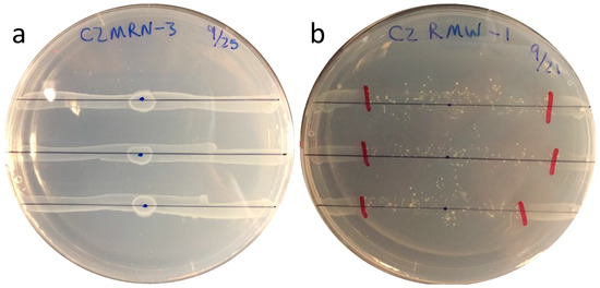
Figure 1.
Growth of E. coli lines on LA agar after 24 h at 25 °C, and with D. discoideum applied at center (a), and after a further 96 h incubation at 25 °C in the dark (b).
2.4. Grazing Preferences by Amoeba
For grazing preference determination, we chose two highly grazed (greatest grazing distance) and six least grazed strains (smallest grazing distance). Each susceptible strain was inoculated on an LA plate with a resistant strain. Four μL of culture of each was streaked on the plate as diverging straight lines touching each other at one end to create a V shape. After 24 h incubation, 4 μL of amoeba suspension was then placed at the base of the diverging bacterial lines. Plates were incubated at room temperature for 4 days and grazing distances were measured.
2.5. Genome Analysis of Most and Least Grazed Isolates
Genomic DNA was extracted from overnight LB agar cultures suspended in 10 mM phosphate buffer (pH 7.0) using the genomic DNA Quick Prep Kit (Zymo Research, Irvine, CA, USA), and all extracted DNA samples were quantified using a Nanodrop Spectrophotometer (ThermoFisher, Waltham, MA, USA) as well as a Qubit Fluorometer (ThermoFisher, Waltahma, MA, USA). The DNA samples were sent to Microbes NG, UK for sequencing (http://www.microbesng.com, accessed April 2016), which is supported by the BBSRC (grant number BB/L024209/1). The protocol used for sequencing is briefly explained; the Genomic DNA libraries were prepared using Nextera XT Library Prep Kit (Illumina, San Diego, CA, USA) following the manufacturer’s protocol with the following modifications: two nanograms of DNA instead of one were used as input, and PCR elongation time was increased to 1 min from 30 s. DNA quantification and library preparation were carried out on a Hamilton Microlab STAR automated liquid handling system. Pooled libraries were quantified using the Kapa Biosystems Library Quantification Kit for Illumina on a Roche light cycler 96 qPCR machine. Libraries were sequenced on the Illumina HiSeq using a 250 bp paired end protocol. Reads were adapter-trimmed using a Trimmomatic 0.30 with a sliding window quality cutoff of Q15 [44]. De novo assembly was performed on samples using SPAdes version 3.7 [45], and contigs were annotated using Prokka 1.11 [46].
Annotated genomes were uploaded to the EDGAR 3.0 platform for comparative genome analysis [47]. The core genome was determined on EDGAR 3.0 using 23 Escherichia genomes [47]. Two genomes representing clade I, four E. ruysiae, one E. marmotae, three E. albertii and one E. fergusonii genomes were used as an outgroup. The core genes were aligned using the MUSCLE plugin from the CLC Main Workbench 7.0 [48] (www.qiagenbioinformatics.com, accessed on 26 April 2023). The core genes were concatenated and partitioned using FASconCAT-G 1.02 and ProtTest 3.4, respectively [49,50]. ProtTest 3.4 was used to determine a model for each gene separately. A core maximum likelihood phylogenetic tree of the core genomes was drawn using RAxML with 100 bootstrap replicates [51]. The pangenome was determined using EDGAR 3.0. The core genes were removed to yield the accessory or non-core genes, and a UPGMA dendrogram constructed using PAST 3 (Paleontological Statistics Software Package for Education and Data Analysis) with the Jaccard similarity index [52].
2.6. Genes Associated with Grazing Susceptibility of E. coli Isolates
To identify factors contributing to grazing susceptibility, the genomes of five of the least grazed and five of the most grazed isolates were sequenced. These were designated least grazed group (LGG) and highly grazed group (HGG). Genes common to either LGG or HGG or common to both groups were identified using the EDGAR bioinformatics platform [53] and R program [43].
3. Results
3.1. Grazing Susceptibility
Grazing susceptibility of E. coli isolates from soil, run-off, soil before grazing (SBG), and bovine feces was determined by the grazing distances of D. discoideum introduced at the center of E. coli culture lines on lactose agar (Figure 1). Grazing distance after 96 h varied widely among the various E. coli; between 0 and 7 cm from the point of inoculation. This indicated that susceptibility of E. coli to grazing by D. discoideum was strain specific.
The distribution of grazing susceptibility varied significantly among the four sample sources (Figure 2). The SBG isolate group showed the lowest susceptibility to grazing by D. discoideum. The SBG isolates represented strains that were able to persist in soil over a full year [9]. The lower susceptibility to grazing of SBG isolates compared to soil, run-off, and feces isolates suggests that population maintenance in soil was due, at least in part, to persistence in the presence of grazing protozoa.
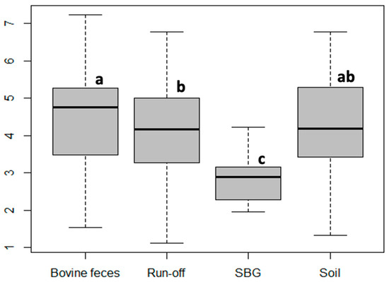
Figure 2.
Box and whisker plot depicting the grazing distances of D. discoideum on E. coli isolates from different environmental sources. Sample groups with the same letter were not significantly different as determined by ANOVA.
3.2. Grazing Preferences by D. discoideum
When D. discoideum was grown in the presence of two E. coli isolates of different grazing susceptibility (LGG or HGG), it preferred HGG over LGG strains. The grazing was initiated first on the highly susceptible isolates, where it grazed a longer distance; later, it grazed on the least susceptible isolates (Figure 3). Our results indicate that D. discoideum grazes on both isolates, but displays a preference for strains that are highly susceptible to grazing.
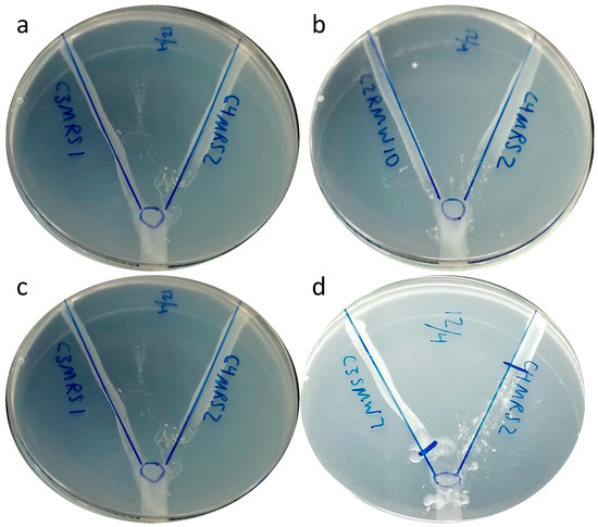
Figure 3.
Grazing preference of D. discoideum between grazing-susceptible (right side) and grazing-resistant strains (left side) of E. coli. Examples shown include the isolate pairs C3MRS1 and C4MRS2 (a), C2RMW10 and C4MRS2 (b), C3MRS1 and C4MRS2 (c) and C3SMW7 and C4MRS2 (d).
3.3. Presence of Virulence Genes and Grazing Susceptibility
To determine if there is any relationship between the presence of virulence genes and the grazing susceptibility of E. coli, the presence of six virulence genes was correlated with grazing distance. We had previously determined the presence of stx1, stx2, eaeA, hlyA, ST, and LT in each isolate by PCR (data not shown). There was no significant correlation found between grazing susceptibility and virulence gene prevalence (R2 = 0.1597) (Supplementary Figure S1).
3.4. Genomes of Grazing-Susceptible versus -Resistant Isolates
Genome comparisons were conducted to determine whether predation resistance in the LGG might be due to differences in genotype or phylogeny. The five highly and least grazed isolates were all shown to be true E. coli by core genome phylogeny (Figure 4). The average genome size for the least grazed group (LGG) was 4852 genes and the highly grazed group (HGG) had 5100 genes. Yet, the grazing susceptibility phenotype did not align with phylogeny or phylogroup (Figure 4). The five highly and least grazed isolate groups both had members of phylogroups B1 and E. Likewise, highly and least grazed isolates did not separate by non-core or accessory genome content, with isolates occurring among each other on the tree (Figure 5). Collectively, genome content overall did not align with grazing susceptibility, suggesting that grazing susceptibility is not based on the phylogeny of isolates.
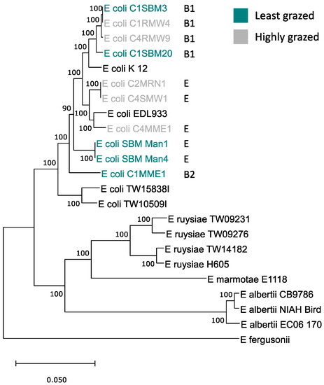
Figure 4.
Maximum likelihood phylogenetic tree of the core genomes of five highly and least grazed E. coli isolates compared to other strains of E. coli and other species of the genus. Letters identify the respective phylogroups of isolates.
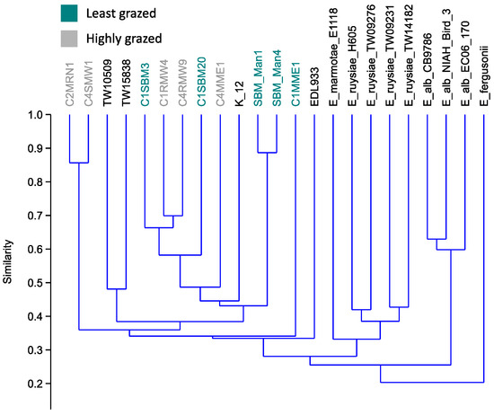
Figure 5.
UPGMA dendrogram of accessory or non-core genes of the highly and least grazed E. coli isolates, compared to other strains of E. coli and other species of the genus.
To determine whether there were genes common to either group of isolates, we looked for uniquely shared genes. The highly grazed group had 389 genes specific to their group, more than double the 130 unique genes shared by the LGG. These unique genes were grouped based on their function (Figure 6) and are listed in Table 1.
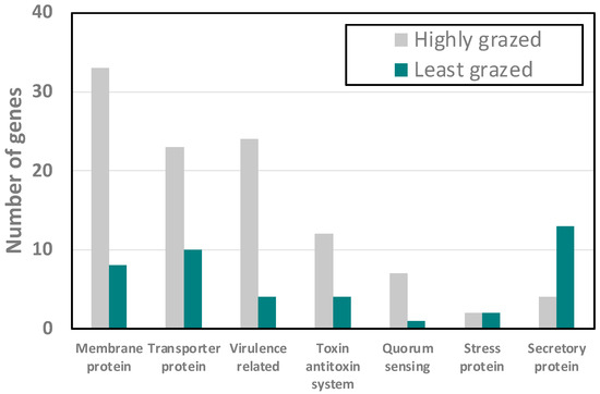
Figure 6.
Number of genes uniquely common to the five highly grazed versus five least grazed E. coli associated with grazing susceptibility.

Table 1.
Genes uniquely common to the five most grazed (highly grazed group) versus five least grazed (least grazed group) E. coli associated with grazing susceptibility.
The number of unique-membrane-related genes in the highly grazed group was 33 and in the LGG it was only 8, suggesting a substantial difference in their membrane composition. These included multiple outer membrane proteins which could act as specific surface molecules for recognition. The HGG contained 23 transporter genes compared to the LGG, with only 10. Surprisingly, we found that there were only 4 secretory-protein-related genes in the HGG, and they were related to the type III secretory system, whereas in the LGG there were 10 secretory system type-II-protein-related genes present. This points to differences in effectors excreted directly into potential eukaryotic cells (type III) by the LGG, and effectors secreted outside the cell in the HGG. The HGG also possessed many fimbial and flagellar genes, and small toxic proteins and hemolysin genes. The LGG had only three fimbrial and invasion-related genes, suggesting that the HGG could possibly contribute more virulence genes compared to the LGG. There were also a high number of toxin–antitoxin system genes in the HGG compared to the LGG. Quorum sensing molecules such as autoinducer-2-related genes were more abundant in the HGG compared to the LGG, where only one autoinducer 2-binding protein gene, lsrB, was present. Our results suggested that the LGG and HGG strains are phenotypically and genotypically different from each other in surface properties, proteins excreted, and signaling molecules.
4. Discussion
E. coli isolates from the cattle pasture showed different susceptibilities to protozoan predation. The majority of isolates from SBG samples, considered as environmental [9], showed significantly higher resistance to grazing compared to soil, run-off, and bovine feces isolates. This indicates that E. coli maintaining long-term populations in soil either lack traits that render the HGG susceptible or display traits warding off the grazing amoeba. Lesser susceptibility of the LGG to grazing was supported when D. discoideum was presented with a choice between pairs of LGG and HGG strains. The grazers consistently selected the HGG strain in each pair (Figure 3). Adiba et al. [28] have shown that D. discoideum was able to survive and phagocytize E. coli strains not harboring virulence genes involved in iron capture (iroN, fyuA, irp), not resistant to bile, serum, or lactoferrin, or that do not belong to phylogroup B2.
In our study, interestingly, we also found that isolates belonging to the B2 phylogroup showed resistance to protozoan grazing, although there were very few B2 isolates in our collection (in total, 368 isolates and only 9 B2 isolates). The highest grazing distance was 7.2 cm and the range of grazing distance for isolates belonging to phylogroup B2 was 0–3.1 cm. It has been shown that E.coli strains that harbor virulence genes are able to survive and replicate in common environmental protozoa such as E. coli O157 [31,54], or extra-intestinal pathogenic E. coli [28]. To determine the correlation between the presence of virulence genes and grazing resistance, we detected the presence and absence of six virulence genes in all isolates. We did not find any correlation between the presence of virulence genes and grazing resistance of E. coli. We also measured the grazing distance of E. coli O157:H7 strains and did not find any significant resistance by the strain. Our result is consistent with Schmidt, Shringi, and Besser [39], who reported that P. caudatum consistently reduced both E. coli O157:H7 (EDL933D) and non-Shiga-toxin-cattle-commensal E.coli populations by 1–3 log CFU when grown together in broth culture over three days at an ambient laboratory temperature.
If virulence genes are not the major factor for E. coli to be resistant to predation, then what are the traits responsible for their ability to evade grazing instead? To find out the difference between the least grazed isolates (resistant isolates) and the highly grazed group (susceptible isolates), we chose five isolates from the least grazed group (denoted as LGG) and five isolates from the highly grazed group (HGG) and sequenced these to compare their genome data. We found that the two groups shared a core genome consisting of 3414 genes, while each group also has some unique genes they do not share. The core genomes did not group into HGG versus LGG, indicating that grazing susceptibility was not due to variations among sequences of core genes, but rather due to the presence or absence of specific genes. The HGG had 389 shared genes not occurring in any of the LGG sequenced, and the LGG had 130 unique shared genes. It was interesting to see that the HGG has a higher abundance of membrane protein, transporter protein, fimbrial protein, flagellar protein, toxin–antitoxin-system-related protein, and autoinducer-2. However, the LGG has a high number of secretory-system-II proteins compared to the HGG, which has fewer secretory system III proteins.
A recent study by Snyder, et al. [55] found that mutant strains of E. coli that are resistant to D. discoideum phagocytosis possess several genes related to flagella, oxidoreductase, and acid resistance. These genes may have the potential to develop a mechanism to resist D. discoideum predation, which contributes to the selection and maintenance of bacterial virulence factors against mammalian hosts. Salmonella enterica subsp. Typhimurium inhibits the D. discoideum starvation response through the type III secretion system, thereby preventing sporulation [56]. The type-III secretion system in the HGG may also play a role in secreting substrate that may allow the starvation response of D. discoideum. Type II secretion systems occur in both pathogenic and non-pathogenic E. coli, and the output T2S secretory proteins can be a diverse group of toxins, degradative enzymes, and other effector proteins. This system is clearly used by bacteria for environmental survival and virulence [57]. This report suggests that the T2S system may play an important role in the LGG ‘s ability to resist predation. We also found autoinducer-2 related genes in the HGG, which are part of quorum-sensing system that allows communication with many different bacterial species [58]. It has also been reported that functional quorum sensing is important for the interaction of Vibrio cholera and the amoeba A. castellanii. Upon being phagocytized by the amoeba, V. cholera can resist intracellular killing [59]. The presence of autoinducer 2 in HGG indicates that the cells interact with D. discoideum to phagocytose. It may be possible that the cells are not completely killed, but form a symbiotic association with amoeba of farmer clones that carry bacteria through their social stages or dispersal stages, and can be identified by the presence of bacteria in their sorus [60]. It will be interesting to investigate the presence of E. coli cells in the sorus of D. discoideum that has grazed on HGG isolates.
Our study did not yield any detailed information about the association of genes specific to E. coli survival from protozoan predation. The presence of genes unique to thee HGG and LGG may play a role in the grazing susceptibility or grazing resistance of bacteria. To determine the role of these genes of protozoan predation, more investigation is needed. Our results, of a characterization of amoeba grazing on distinct E. coli isolates and a correlation between the presence of virulence genes and grazing resistance, deviate from previous reports [28,36]. These inconsistencies could be attributed to differences in amoeba clones, plating methods, nutrient conditions, and the laboratory atmosphere. In our study, we found that the plating medium clearly affects the growth of amoeba clones on distinct E. coli populations.
Population dynamics of E. coli in the environment have been studied widely from the perspectives of nutrient requirements, stationary phase physiology and stress response, and competition with other bacteria. In contrast, the role of amoeba in affecting population numbers through grazing has been little studied. Our results indicate that grazing by amoeba has a substantial effect on population densities of diverse E. coli in soils and sediments.
5. Conclusions
In conclusion, our study clearly depicts that there is a difference in the grazing susceptibility of E. coli isolates. The environmental E. coli that survived in the pasture without the presence of grazing animals were also significantly more resistant to grazing by D. discoideum. The highly grazed group contained a much larger number of genes encoding surface-related functions, such as membrane proteins and exporters than did the least grazed group. The results indicate that the long-term persistence of E. coli in soil is due, at least in part, to resistance to grazing by soil amoeba.
Supplementary Materials
The following supporting information can be downloaded at: https://www.mdpi.com/article/10.3390/microorganisms11061457/s1, Figure S1: Correlation between grazing distance and presence of six pathogenic genes stx1, stx2, eaeA, hlyA, ST and LT.
Author Contributions
Conceptualization, V.S.B.; methodology, G.N. and V.S.B.; validation, G.N. and V.S.B.; formal analysis, G.N., L.A.B., T.S. and V.S.B.; investigation, G.N., L.A.B. and T.S.; data curation, G.N.; writing—original draft preparation, G.N.; writing—review and editing, G.N., L.A.B., T.S. and V.S.B.; visualization, G.N. and T.S.; supervision, V.S.B.; funding acquisition, V.S.B. All authors have read and agreed to the published version of the manuscript.
Funding
This research was funded by the South Dakota Agricultural Experiment Station.
Data Availability Statement
Data available at http://edgar3.computational.bio accessed on 26 April 2023.
Conflicts of Interest
The authors declare no conflict of interest.
References
- Foster-Nyarko, E.; Pallen, M.J. The Microbial Ecology of Escherichia coli in the Vertebrate Gut. FEMS Microbiol. Rev. 2022, 46, fuac008. [Google Scholar] [CrossRef]
- Pokharel, P.; Dhakal, S.; Dozois, C.M. The Diversity of Escherichia coli Pathotypes and Vaccination Strategies against This Versatile Bacterial Pathogen. Microorganisms 2023, 11, 344. [Google Scholar] [CrossRef] [PubMed]
- Pachepsky, Y.A.; Shelton, D.R. Escherichia coli and Fecal coliforms in Freshwater and Estuarine Sediments. Crit. Rev. Environ. Sci. Technol. 2011, 41, 1067–1110. [Google Scholar] [CrossRef]
- Ishii, S.; Ksoll, W.B.; Hicks, R.E.; Sadowsky, M.J. Presence and Growth of Naturalized Escherichia coli in Temperate Soils from Lake Superior Watersheds. Appl. Environ. Microbiol. 2006, 72, 612–621. [Google Scholar] [CrossRef]
- Jang, J.; Hur, H.-G.; Sadowsky, M.J.; Byappanahalli, M.N.; Yan, T.; Ishii, S. Environmental Escherichia coli: Ecology and Public Health Implications—A Review. J. Appl. Microbiol. 2017, 123, 570–581. [Google Scholar] [CrossRef]
- van Elsas, J.D.; Semenov, A.V.; Costa, R.; Trevors, J.T. Survival of Escherichia coli in the Environment: Fundamental and Public Health Aspects. ISME J. 2011, 5, 173–183. [Google Scholar] [CrossRef]
- Power, M.L.; Littlefield-Wyer, J.; Gordon, D.M.; Veal, D.A.; Slade, M.B. Phenotypic and Genotypic Characterization of Encapsulated Escherichia coli Isolated from Blooms in Two Australian Lakes. Environ. Microbiol. 2005, 7, 631–640. [Google Scholar] [CrossRef] [PubMed]
- Texier, S.; Prigent-Combaret, C.; Gourdon, M.H.; Poirier, M.A.; Faivre, P.; Dorioz, J.M.; Poulenard, J.; Jocteur-Monrozier, L.; Moënne-Loccoz, Y.; Trevisan, D. Persistence of Culturable Escherichia coli Fecal Contaminants in Dairy Alpine Grassland Soils. J. Environ. Qual. 2008, 37, 2299–2310. [Google Scholar] [CrossRef] [PubMed]
- NandaKafle, G.; Seale, T.; Flint, T.; Nepal, M.; Venter, S.N.; Brözel, V.S. Distribution of Diverse Escherichia coli between Cattle and Pasture. Microbes Environ. 2017, 32, 226–233. [Google Scholar] [CrossRef] [PubMed]
- Dusek, N.; Hewitt, A.J.; Schmidt, K.N.; Bergholz, P.W. Landscape-Scale Factors Affecting the Prevalence of Escherichia coli in Surface Soil Include Land Cover Type, Edge Interactions, and Soil PH. Appl. Environ. Microbiol. 2018, 84, e02714-17. [Google Scholar] [CrossRef]
- NandaKafle, G.; Christie, A.A.; Vilain, S.; Brözel, V.S. Growth and Extended Survival of Escherichia coli O157: H7 in Soil Organic Matter. Front. Microbiol. 2018, 9, 762. [Google Scholar] [CrossRef] [PubMed]
- Liebeke, M.; Brözel, V.S.; Hecker, M.; Lalk, M. Chemical Characterization of Soil Extract as Growth Media for the Ecophysiological Study of Bacteria. Appl. Microbiol. Biotechnol. 2009, 83, 161–173. [Google Scholar] [CrossRef] [PubMed]
- Finkel, S.E. Long-Term Survival during Stationary Phase: Evolution and the GASP Phenotype. Nat. Rev. Microbiol. 2006, 4, 113–120. [Google Scholar] [CrossRef]
- Jiang, X.; Morgan, J.; Doyle, M.P. Fate of Escherichia coli O157:H7 in Manure-Amended Soil. Appl. Environ. Microbiol. 2002, 68, 2605–2609. [Google Scholar] [CrossRef]
- Semenov, A.V.; van Bruggen, A.H.C.; van Overbeek, L.; Termorshuizen, A.J.; Semenov, A.M. Influence of Temperature Fluctuations on Escherichia coli O157:H7 and Salmonella Enterica Serovar Typhimurium in Cow Manure. FEMS Microbiol. Ecol. 2007, 60, 419–428. [Google Scholar] [CrossRef]
- Feng, F.; Goto, D.; Yan, T. Effects of Autochthonous Microbial Community on the Die-off of Fecal Indicators in Tropical Beach Sand. FEMS Microbiol. Ecol. 2010, 74, 214–225. [Google Scholar] [CrossRef]
- Wanjugi, P.; Fox, G.A.; Harwood, V.J. The Interplay Between Predation, Competition, and Nutrient Levels Influences the Survival of Escherichia coli in Aquatic Environments. Microb. Ecol. 2016, 72, 526–537. [Google Scholar] [CrossRef]
- Korajkic, A.; Wanjugi, P.; Harwood, V.J. Indigenous Microbiota and Habitat Influence Escherichia coli Survival More than Sunlight in Simulated Aquatic Environments. Appl. Environ. Microbiol. 2013, 79, 5329–5337. [Google Scholar] [CrossRef] [PubMed]
- Bar-On, Y.M.; Phillips, R.; Milo, R. The Biomass Distribution on Earth. Proc. Natl. Acad. Sci. USA 2018, 115, 6506–6511. [Google Scholar] [CrossRef] [PubMed]
- Kurm, V.; van der Putten, W.H.; Weidner, S.; Geisen, S.; Snoek, B.L.; Bakx, T.; Hol, W.H.G. Competition and Predation as Possible Causes of Bacterial Rarity. Environ. Microbiol. 2019, 21, 1356–1368. [Google Scholar] [CrossRef]
- Pernthaler, J. Predation on Prokaryotes in the Water Column and Its Ecological Implications. Nat. Rev. Microbiol. 2005, 3, 537–546. [Google Scholar] [CrossRef] [PubMed]
- Shi, Y.; Queller, D.C.; Tian, Y.; Zhang, S.; Yan, Q.; He, Z.; He, Z.; Wu, C.; Wang, C.; Shu, L. The Ecology and Evolution of Amoeba-Bacterium Interactions. Appl. Environ. Microbiol. 2021, 87, e01866-20. [Google Scholar] [CrossRef]
- Matz, C.; Kjelleberg, S. Off the Hook—How Bacteria Survive Protozoan Grazing. Trends Microbiol. 2005, 13, 302–307. [Google Scholar] [CrossRef] [PubMed]
- Sato, K.; Taniyama, Y.; Yoshida, A.; Toyomasu, K.; Ryuda, N.; Ueno, D.; Someya, T. Protozoan Predation of Escherichia coli in Hydroponic Media of Leafy Vegetables. Soil Sci. Plant Nutr. 2019, 65, 234–242. [Google Scholar] [CrossRef]
- Kihara, K.; Mori, K.; Suzuki, S.; Hosoda, K.; Yamada, A.; Matsuyama, S.; Kashiwagi, A.; Yomo, T. Probabilistic Transition from Unstable Predator–Prey Interaction to Stable Coexistence of Dictyostelium Discoideum and Escherichia coli. Biosystems 2011, 103, 342–347. [Google Scholar] [CrossRef]
- Weekers, P.H.H.; Bodelier, P.L.E.; Wijen, J.P.H.; Vogels, G.D. Effects of Grazing by the Free-Living Soil Amoebae Acanthamoeba Castellanii, Acanthamoeba Polyphaga, and Hartmannella Vermiformis on Various Bacteria. Appl. Environ. Microbiol. 1993, 59, 2317–2319. [Google Scholar] [CrossRef]
- Ravva, S.V.; Sarreal, C.Z.; Mandrell, R.E. Identification of Protozoa in Dairy Lagoon Wastewater That Consume Escherichia coli O157:H7 Preferentially. PLoS ONE 2010, 5, e15671. [Google Scholar] [CrossRef]
- Adiba, S.; Nizak, C.; van Baalen, M.; Denamur, E.; Depaulis, F. From Grazing Resistance to Pathogenesis: The Coincidental Evolution of Virulence Factors. PLoS ONE 2010, 5, e11882. [Google Scholar] [CrossRef]
- González, J.M.; Iriberri, J.; Egea, L.; Barcina, I. Differential Rates of Digestion of Bacteria by Freshwater and Marine Phagotrophic Protozoa. Appl. Environ. Microbiol. 1990, 56, 1851–1857. [Google Scholar] [CrossRef]
- Iriberri, J.; Azúa, I.; Labirua-Iturburu, A.; Artolozaga, I.; Barcina, I. Differential Elimination of Enteric Bacteria by Protists in a Freshwater System. J. Appl. Bacteriol. 1994, 77, 476–483. [Google Scholar] [CrossRef]
- Meltz Steinberg, K.; Levin, B.R. Grazing Protozoa and the Evolution of the Escherichia coli O157:H7 Shiga Toxin-Encoding Prophage. Proc. R. Soc. B Biol. Sci. 2007, 274, 1921–1929. [Google Scholar] [CrossRef] [PubMed]
- Ravva, S.V.; Sarreal, C.Z.; Mandrell, R.E. Strain Differences in Fitness of Escherichia coli O157:H7 to Resist Protozoan Predation and Survival in Soil. PLoS ONE 2014, 9, e102412. [Google Scholar] [CrossRef] [PubMed]
- Tezcan-Merdol, D.; Ljungström, M.; Winiecka-Krusnell, J.; Linder, E.; Engstrand, L.; Rhen, M. Uptake and Replication of Salmonella Enterica in Acanthamoeba Rhysodes. Appl. Environ. Microbiol. 2004, 70, 3706–3714. [Google Scholar] [CrossRef]
- Wildschutte, H.; Wolfe, D.M.; Tamewitz, A.; Lawrence, J.G. Protozoan Predation, Diversifying Selection, and the Evolution of Antigenic Diversity in Salmonella. Proc. Natl. Acad. Sci. USA 2004, 101, 10644–10649. [Google Scholar] [CrossRef]
- Smith, D.D.N.; Nickzad, A.; Déziel, E.; Stavrinides, J. A Novel Glycolipid Biosurfactant Confers Grazing Resistance upon Pantoea Ananatis BRT175 against the Social Amoeba Dictyostelium Discoideum. mSphere 2016, 1, e00075-15. [Google Scholar] [CrossRef]
- Jenkins, M.B.; Fisher, D.S.; Endale, D.M.; Adams, P. Comparative Die-off of Escherichia coli 0157:H7 and Fecal Indicator Bacteria in Pond Water. Environ. Sci. Technol. 2011, 45, 1853–1858. [Google Scholar] [CrossRef]
- Artz, R.R.E.; Killham, K. Survival of Escherichia coli O157:H7 in Private Drinking Water Wells: Influences of Protozoan Grazing and Elevated Copper Concentrations. FEMS Microbiol. Lett. 2002, 216, 117–122. [Google Scholar] [CrossRef]
- Avery, L.M.; Williams, A.P.; Killham, K.; Jones, D.L. Survival of Escherichia coli O157:H7 in Waters from Lakes, Rivers, Puddles and Animal-Drinking Troughs. Sci. Total Environ. 2008, 389, 378–385. [Google Scholar] [CrossRef]
- Schmidt, C.E.; Shringi, S.; Besser, T.E. Protozoan Predation of Escherichia coli O157:H7 Is Unaffected by the Carriage of Shiga Toxin-Encoding Bacteriophages. PLoS ONE 2016, 11, e0147270. [Google Scholar] [CrossRef]
- Hahn, M.W.; Höfle, M.G. Grazing of Protozoa and Its Effect on Populations of Aquatic Bacteria. FEMS Microbiol. Ecol. 2001, 35, 113–121. [Google Scholar] [CrossRef]
- Jürgens, K.; Matz, C. Predation as a Shaping Force for the Phenotypic and Genotypic Composition of Planktonic Bacteria. Antonie Van Leeuwenhoek 2002, 81, 413–434. [Google Scholar] [CrossRef]
- Moore, D.M.; Withers, T.R.; Yu, H.; Trzyna, W. A Simple Plating Assay Using Acanthamoeba to Evaluate the Effect of Environmental Stressors on Growing and Dividing Cells. Proc. West Va. Acad. Sci. 2016, 88. [Google Scholar] [CrossRef]
- R: The R Project for Statistical Computing. Available online: https://www.r-project.org/ (accessed on 26 April 2023).
- Bolger, A.M.; Lohse, M.; Usadel, B. Trimmomatic: A Flexible Trimmer for Illumina Sequence Data. Bioinformatics 2014, 30, 2114–2120. [Google Scholar] [CrossRef]
- Bankevich, A.; Nurk, S.; Antipov, D.; Gurevich, A.A.; Dvorkin, M.; Kulikov, A.S.; Lesin, V.M.; Nikolenko, S.I.; Pham, S.; Prjibelski, A.D.; et al. SPAdes: A New Genome Assembly Algorithm and Its Applications to Single-Cell Sequencing. J. Comput. Biol. 2012, 19, 455–477. [Google Scholar] [CrossRef]
- Seemann, T. Prokka: Rapid Prokaryotic Genome Annotation. Bioinformatics 2014, 30, 2068–2069. [Google Scholar] [CrossRef]
- Dieckmann, M.A.; Beyvers, S.; Nkouamedjo-Fankep, R.C.; Hanel, P.H.G.; Jelonek, L.; Blom, J.; Goesmann, A. EDGAR3.0: Comparative Genomics and Phylogenomics on a Scalable Infrastructure. Nucleic Acids Res. 2021, 49, W185–W192. [Google Scholar] [CrossRef]
- Edgar, R.C. MUSCLE: Multiple Sequence Alignment with High Accuracy and High Throughput. Nucleic Acids Res. 2004, 32, 1792–1797. [Google Scholar] [CrossRef]
- Darriba, D.; Taboada, G.L.; Doallo, R.; Posada, D. ProtTest-HPC: Fast Selection of Best-Fit Models of Protein Evolution. In Proceedings of the Euro-Par 2010 Parallel Processing Workshops, Iscia, Italy, 31 August–3 September 2010; Guarracino, M.R., Vivien, F., Träff, J.L., Cannatoro, M., Danelutto, M., Hast, A., Perla, F., Knüpfer, A., Di Martino, B., Alexander, M., Eds.; Springer: Berlin/Heidelberg, Germany, 2011; pp. 177–184. [Google Scholar]
- Kück, P.; Longo, G.C. FASconCAT-G: Extensive Functions for Multiple Sequence Alignment Preparations Concerning Phylogenetic Studies. Front. Zool. 2014, 11, 81. [Google Scholar] [CrossRef]
- Stamatakis, A. RAxML-VI-HPC: Maximum Likelihood-Based Phylogenetic Analyses with Thousands of Taxa and Mixed Models. Bioinformatics 2006, 22, 2688–2690. [Google Scholar] [CrossRef]
- Hammer, O.; Harper, D.A.T.; Ryan, P.D. PAST: Paleontological Statistics Software Package for Education and Data Analysis. Palaeontol. Electron. 2001, 4, 178kb. [Google Scholar]
- Blom, J.; Kreis, J.; Spänig, S.; Juhre, T.; Bertelli, C.; Ernst, C.; Goesmann, A. EDGAR 2.0: An Enhanced Software Platform for Comparative Gene Content Analyses. Nucleic Acids Res. 2016, 44, W22–W28. [Google Scholar] [CrossRef]
- Barker, J.; Humphrey, T.J.; Brown, M.W.R. Survival of Escherichia coli 0157 in a Soil Protozoan: Implications for Disease. FEMS Microbiol. Lett. 1999, 173, 291–295. [Google Scholar] [CrossRef]
- Snyder, M.; Weichseldorfer, M.; Lane, J.; Kazmierski, R.; Bains, H.; White, T.; Parker, D.; Aklkharouf, N.; Hemm, M. Identification of Virulence and Virulence Regulatory Factors in Escherichia coli Using Dictyostelium Discoideum as a Model Phagocyte. J. Immunol. 2017, 198 (Suppl. S1), 131.19. [Google Scholar] [CrossRef]
- Sillo, A.; Matthias, J.; Konertz, R.; Bozzaro, S.; Eichinger, L. Salmonella Typhimurium Is Pathogenic for Dictyostelium Cells and Subverts the Starvation Response. Cell. Microbiol. 2011, 13, 1793–1811. [Google Scholar] [CrossRef]
- Cianciotto, N.P.; White, R.C. Expanding Role of Type II Secretion in Bacterial Pathogenesis and Beyond. Infect. Immun. 2017, 85, e00014-17. [Google Scholar] [CrossRef]
- Federle, M.J. Autoinducer-2-Based Chemical Communication in Bacteria: Complexities of Interspecies Signaling. In Bacterial Sensing and Signaling; Karger Publishers: Basel, Switzerland, 2009; Volume 16, pp. 18–32. [Google Scholar]
- Van der Henst, C.; Scrignari, T.; Maclachlan, C.; Blokesch, M. An Intracellular Replication Niche for Vibrio Cholerae in the Amoeba Acanthamoeba Castellanii. ISME J. 2016, 10, 897–910. [Google Scholar] [CrossRef]
- Brock, D.A.; Douglas, T.E.; Queller, D.C.; Strassmann, J.E. Primitive Agriculture in a Social Amoeba. Nature 2011, 469, 393–396. [Google Scholar] [CrossRef]
Disclaimer/Publisher’s Note: The statements, opinions and data contained in all publications are solely those of the individual author(s) and contributor(s) and not of MDPI and/or the editor(s). MDPI and/or the editor(s) disclaim responsibility for any injury to people or property resulting from any ideas, methods, instructions or products referred to in the content. |
© 2023 by the authors. Licensee MDPI, Basel, Switzerland. This article is an open access article distributed under the terms and conditions of the Creative Commons Attribution (CC BY) license (https://creativecommons.org/licenses/by/4.0/).