Article

Biofilm-Forming Capacity and Drug Resistance of Different Gardnerella Subgroups Associated with Bacterial Vaginosis

1
Department of Obstetrics and Gynecology, Peking University First Hospital, Beijing 100034, China
2
College of Food Science and Nutritional Engineering, China Agricultural University, Beijing 100083, China
*
Author to whom correspondence should be addressed.
Microorganisms 2023, 11(9), 2186; https://doi.org/10.3390/microorganisms11092186
Submission received: 29 May 2023 / Revised: 10 August 2023 / Accepted: 29 August 2023 / Published: 30 August 2023
(This article belongs to the Section Biofilm)

Abstract

:
Bacterial vaginosis (BV) is the most common infection of the lower reproductive tract among women of reproductive age. Recurrent infections and antibiotic resistance associated with biofilms remain significant challenges for BV treatment. Gardnerella species are commonly found in women with and without BV, indicating that genetic differences among Gardnerella isolates may distinguish pathogenic from commensal subgroups. This study isolated 11 Gardnerella strains from vaginal samples obtained from women with BV before or after treatment. The biofilm formation ability of each strain was examined by crystal violet staining. Eight strains were selected using phylogenetic analysis of the cpn60 sequences and classified as subgroups A (6/8), B (1/8), and D (1/8). The biofilm formation ability and antibiotic resistance profile of these strains was compared among the subgroups. Subgroup D had the strongest biofilm formation ability. Six of the planktonic strains exhibited resistance to the first-line BV drug, metronidazole, and one to clindamycin. Moreover, biofilm formation in vitro increased strain resistance to clindamycin. Two strains with strong biofilm ability, S20 and S23, and two with weak biofilm ability, S24 and S25, were selected for comparative genomic analysis. S20 and S23 were found to contain four key genes associated with biofilm formation and more genes involved in carbohydrate synthesis and metabolism than S24 and S25. Identifying differences in the expression of virulence factors between Gardnerella subgroups could inform the development of novel treatments for BV.

1. Introduction

Bacterial vaginosis (BV) is a common lower reproductive tract infection in women of reproductive age, affecting up to 58% of women worldwide, with a disproportionately high impact on those living in sub-Saharan Africa [1]. BV is characterized by a sharp decline in Lactobacillus counts, altered polymicrobial interactions, and an increase in the concentration of facultative or obligate anaerobic microbes [2,3,4]. Current Centers for Disease Control and Prevention (CDC) guidelines recommend metronidazole as a first-line treatment [5]. The clinical cure rates of different clinical treatments and routes of administration vary from 46.75% to 96.20% [6]. However, BV recurrence rates within 12 months of treatment remain above 70% [7]. The hallmark of BV is the presence of a highly structured polymicrobial biofilm on the vaginal epithelium that is primarily composed of Gardnerella spp. and other anaerobic species [8]. The overgrowth of vaginal biofilms seeded by initial Gardnerella spp. colonization results in recurrent symptomatic BV that has limited responsiveness to classical antibiotics [9].
The role of Gardnerella spp. in reproductive health remains incompletely understood. Some studies indicate that every Gardnerella spp. has the potential to cause BV, while others suggest that certain Gardnerella strains are genetically driven toward a more pathogenic phenotype [10]. While both BV- and non-BV-associated Gardnerella isolates contain virulence factors, BV-associated Gardnerella isolates appear to have a higher capacity for adherence and increased cytotoxicity [11,12]. Advanced molecular approaches have identified several novel Gardnerella species with distinct virulence potential [13]. Gardnerella vaginalis was the only recognized species for four decades; however, an amended description of Gardnerella spp. and descriptions of three new species, G. leopoldii, G. piotii, and G. swidsinskii, have been proposed [14]. Using PCR amplification and sequencing of the ‘universal target’ region of the 60-kDa chaperonin (cpn60) gene, Gardnerella spp. has been divided into four subgroups (A, B, C, and D) [15,16]. Phylogenetic and in vitro investigations suggest differences in their ecological and virulence properties [17].
Gardnerella spp. possess several virulence factors that can contribute to their pathogenic phenotype, including a hemolysin, mucus-degrading sialidases, resistance factors, and those involved in biofilm formation [18,19]. These factors have been extensively studied, though an understanding of the virulence potential of each subgroup remains insufficient [12,20,21,22]. The current study used the cpn60 sequence to compare biofilm formation, resistance, and the expression of biofilm-forming genes among various cpn60 subgroups. Functional analysis of the virulence potential of Gardnerella spp. using in vitro assays suggested that the most virulent Gardnerella spp. are responsible for poor BV treatment outcomes [11,21].
This study examined virulence factors from 11 Gardnerella strains isolated from vaginal samples. The biofilm-forming capacity and resistance of Gardnerella isolates were compared among cpn60 subgroups using an in vitro model. While the number of experimental strains was limited, the findings lay a foundation for future studies of BV pathogenesis and Gardnerella spp. virulence factors. The research of more efficacious BV treatments is clearly needed, particularly those that disrupt biofilms and target the most pathogenic bacteria.

2. Materials and Methods

2.1. Collection of Vaginal Specimens and Ethical Approval

A total of 11 women with BV who were patients in the Department of Gynecology at Peking University First Hospital were enrolled in the study and signed informed consent. The women included eight patients with BV (Nugent score of 7–10), two healthy women (Nugent score of 0–3), and one patient with intermediate BV (Nugent score of 4–6). The Ethics Committee of Peking University First Hospital in Beijing approved this study (V2.0/2020.04.20), which was performed in accordance with the Declaration of Helsinki. Eligible women with BV met at least three of four Amsel criteria and had a Nugent score of 7–10 [23,24].
All subjects were asked to provide two vaginal samples, one pre-treatment sample for strain isolation, and another to evaluate the efficacy of metronidazole treatment. The vaginal swabs were obtained from a standard anatomical site (lateral vaginal wall). One vaginal swab was sent to the microbiology laboratory for Gram staining while the second was placed in 1 mL of PBS and stored at −80 °C for genomic DNA extraction. The BV patients were treated with a standard 5-day metronidazole course (0.75% metronidazole gel vaginal administration from NiMeiXin, Tongfang Pharmaceutical Group Co., Ltd., Beijing, China, 5 g once daily) [5].

2.2. Culture Conditions and Strain Isolation

The vaginal swabs were inoculated onto a goat blood agar medium plate using the three-line method and cultured at 37 °C under anaerobic conditions (5% CO2, 95% nitrogen) for 48–72 h. Suspected colonies were purified onto blood agar medium for 48–72 h. After several purification cultures, 11 Gardnerella strains were completely isolated from BV patient vaginal samples.
Sheep blood agar plate: double anti-chocolate agar medium (22 g) was dissolved in 500 mL distilled water and mixed in a 500 mL triangular bottle. Under high pressure, the solution was cooled to 50 °C in a water bath, and 25 mL of sheep blood was added, mixed, and poured into a disposable Petri dish.
Supplemented brain–heart infusion broth (sBHI) medium: 18.5 g BHI basal medium, 1.5 g soluble starch, and 1.5 g glucose were added to 500 mL distilled water and autoclaved at 121 °C.

2.3. Strain Identification

After several purification cultures, single colonies were selected from the blood agar plates and added to 20 μL sterile ddH2O. The colonies were mixed with a pipetting gun, treated in a microwave at high temperature for 3 min, and incubated in an ice bath for 2 min. The resulting mixed system was a suspension of lysed bacteria, which was used as a colony PCR template. To identify the isolates, the 16S rDNA sequence primers, 16S rDNA-F (AGAGTTTGATCCTGGCTCAG) and 16S rDNA-R (TACGGTTACCTTGTTACGACTT), were used. The isolates were confirmed by comparing the 16S rDNA sequences with the GenBank library using the BLAST program (https://blast.ncbi.nlm.nih.gov (accessed on 10 June 2021)). The PCR amplification procedure included 30 cycles of pre-denaturation at 98 °C for 3 min, denaturation at 98 °C for 30 s, annealing at 52.8 for 30 s, and extension at 72 °C for 50 s, followed by a final extension at 72 °C for 10 min. PCR amplification products were detected using electrophoresis on a 1% agarose gel. Qualified products were sequenced by Beijing Qingke Xinye Biotechnology Co., Ltd. (Beijing, China), and the sequencing results were submitted to the online BLAST database for comparison.

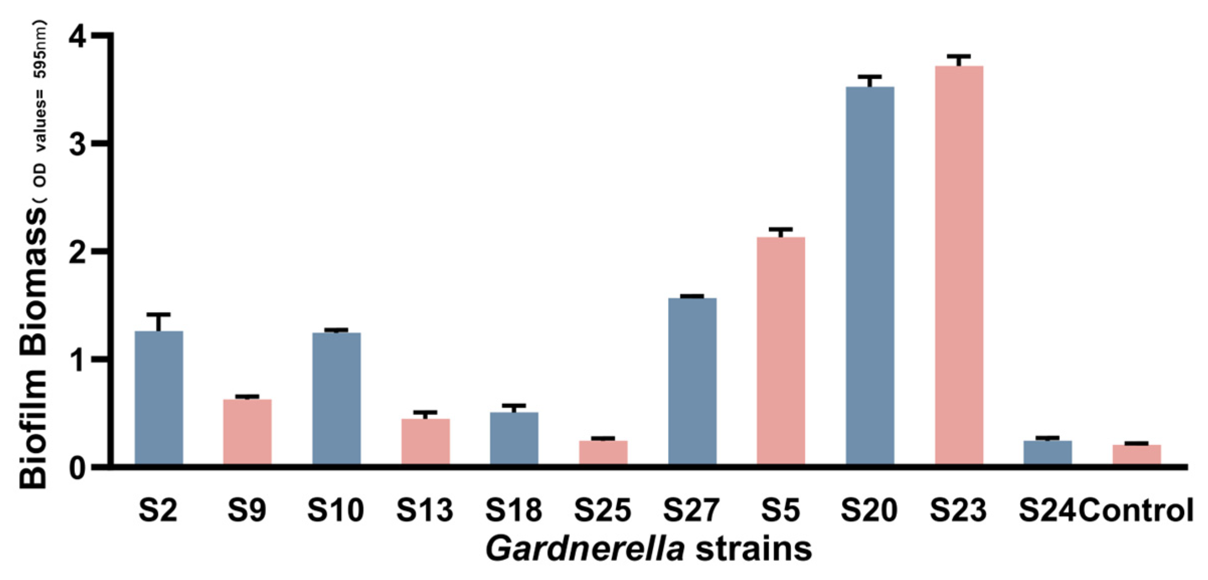
2.4. In Vitro Biofilm Formation and Quantification at Different Stages

The clinically isolated Gardnerella strains were adjusted to a turbidity of 0.5 using a turbidimeter and inoculated into a 96-well plate (200 μL/well). The solution (2 μL) was inoculated into 198 μL of sBHI broth and incubated anaerobically at 37 °C in 96-well plates for 48 h. The bacterial solution was discarded, and the planktonic bacteria were removed by slowly rinsing three times with phosphate-buffered saline (PBS). The wells were air-dried for 60 min, and the biofilms were stained with 200 μL 0.4% (w/v) crystal violet for 30 min. The stains were removed by light washing with 200 μL PBS and air drying for 5 min. Crystal violet was dissolved in 200 μL of a 33% (v/v) acetic acid solution. The absorbance of crystal violet at 595 nm was measured using a microplate reader (Infinite M200 PRO, TECAN). Three biofilm formation experiments were performed for each strain and the average value of the three experiments was recorded. The liquid sBHI medium without bacteria was used as the control group. The OD cut-off value (ODc) was defined as three standard deviations (SD) above the mean OD of the negative control: ODc = average OD of negative control + (3 × SD of negative control), OD ≤ ODc = non biofilm producer; ODc < OD ≤ 2 × ODc = weak biofilm producer; 2 × ODc < OD ≤ 4 × ODc = moderate biofilm producer; 4 × ODc < OD = strong biofilm producer [25].
The activated strain was inoculated into sBHI broth at a 1% concentration. The OD600 value was measured every 2 h until it was stable. This method was used to describe the 24 h growth curve of Gardnerella spp. in liquid media. The concentration of the bacterial solution was adjusted to a turbidity of 0.5, and 2 μL of the solution was inoculated into 198 μL of sBHI broth. The solution was anaerobically cultured at 37 °C in a 96-well plate for 24 h, 48 h, 72 h, and 96 h, and the amount of biofilm was determined by crystal violet staining.

2.5. cpn60 Sequencing of Gardnerella Strains and Detection of Virulence Factors

Three samples with unqualified DNA samples were excluded, allowing only eight strains to be sequenced for cpn60. The DNA of the strains was used as the PCR template, and the primers, cpn60 F (5′-ATGGCAAAGATTATTGCCTATGAAG-3′) and cpn60 R (5′-GCATTCTGTAGAGCAGAACGAGT-3′), were used to amplify and type the cpn60 isolates. The PCR amplification procedure and electrophoresis used the same methods described above. The results were observed using a gel imager, and the amplified samples were sent for sequencing and the detection of virulence factors (Tsingke Biotechnology Co., Ltd., Beijing, China). Sequences of the virulence factors are shown in Supplementary Table S1.
There has been little success in connecting the genotypic with phenotypic characteristics, or in identifying any genotype or phenotype with clinical significance. HMPREF0424_0103 is the vaginolysin (VLY) gene, which is cytotoxic, destroys the integrity of vaginal epithelial cells, and is considered to be associated with the pathogenicity of Gardnerella spp. [21]. HMPREF0424_1109 is the sialidase gene, which hydrolyzes sialic acid in the mucosa and promotes the destruction of the protective mucous layer of vaginal epithelium. This process may also promote the adhesion of BV-associated species on the vaginal epithelium, leading to the biofilm formation [26]. HMPREF0424_0125 encodes the assembly of TadE/G-like family proteins and is considered to play an important role in its adhesion to vaginal epithelial cells [27]. HMPREF0424_0821 encodes a glycosyltransferase that is required for the biosynthesis of extracellular polysaccharides and is closely related to the formation of biofilms [28].

2.6. Minimal Inhibitory Concentration Assays

The minimum inhibitory concentration (MIC) for metronidazole and clindamycin was defined as the lowest antibiotic concentration that produced a marked reduction or inhibition in growth. Using the Clinical Laboratory Standards Institute (CLSI) guidelines, the sensitivity and resistance of eight Gardnerella strains to metronidazole and clindamycin were determined in the planktonic cells and biofilms using the broth microdilution method [29].
The final concentrations of metronidazole and clindamycin for planktonic bacteria were 0.125–256 μg/mL and 0.0625–128 μg/mL, respectively. The Gardnerella strains were anaerobically incubated on blood plates for 48–72 h. Single colonies of the Gardnerella strains were moved from the blood plates to sBHI and the concentration of the bacterial suspension was adjusted to 108 CFU/mL (OD595 = 0.5) (Thermo Fisher Scientific, Waltham, MA, USA). A 100 μL solution composed of 2 × 10⁶ CFU/mL was mixed with different concentrations of the drug and bacterial suspension at a 1:1 ratio, inoculated on a 96-well plate, and incubated for 48–72 h. The preparation and inoculation process was completed within 15 min. According to the CLSI, the two antibiotics have the following critical values: metronidazole (sensitivity: MIC ≤ 8 μg/mL; intermediate: MIC = 16 μg/mL; drug-resistant: MIC ≥ 32 μg/mL) and clindamycin (sensitivity: MIC ≤ 2 μg/mL; intermediate: MIC = 4 μg/mL; resistant: MIC ≥ 8 μg/mL). The sBHI sample with bacterial growth and no antibiotic was used as the control group.

2.7. Statistical Analysis

SPSS19.0 software was used for statistical analyses. The Mann–Whitney test was used to analyze the differences in the biofilm formation ability of the cpn60 subgroups. The MIC of Gardnerella strains to metronidazole and clindamycin before and after biofilm formation was assessed using a T-test. p ≤ 0.05 indicated statistical significance.

3. Results

3.1. Clinical Characteristics of the Gardnerella Strains

The demographic and clinical characteristics of the 11 Gardnerella strains were generally well-balanced at baseline (Table 1). The strains were isolated from BV, BV intermediate, and healthy vaginal samples, even after metronidazole treatment, indicating that metronidazole is unable to completely eliminate Gardnerella spp. in the vagina. While evidence indicates that BV can be cured, clinical symptoms can persist for some patients.

3.2. cpn60 Typing of Gardnerella Strains and Biofilm Formation

The phylogenetic tree divided Gardnerella into three subgroup, A, B, and D. Six (75%) strains belonged to subgroup A, one (12.5%) belonged to subgroup B, one (12.5%) belonged to subgroup D, and none belonged to subgroup C. The biofilm formation ability of the strains was compared (Figure 1 and Table 2) and ranked from strong to weak as follows: S23, S20, S5, S27, S2, S10, S9, S18, S13, S24, and S25 (p < 0.005). The subgroups were ranked from strong to weak by their ability to form biofilms. Subgroup D had the strongest ability to form biofilms, followed by subgroups B and A (Table 2).

3.3. Growth Curves of the Gardnerella Strains

The strains with the strongest biofilm-forming ability, S23 and S20, and those with the weakest ability, S24 and S25, were selected to determine the 24-h growth curves (Figure 2). Gardnerella strains reached a stable period after 24 h. The OD value of S23 was highest at 24 h and reached 1.5. Meanwhile, the OD values of S24 and S25 were 1.25 and 1.3, respectively (Figure 1). S20 had the lowest growth rate but strong biofilm-forming ability (Figure 1), suggesting that the ability to form biofilms is not related to the growth activity of Gardnerella isolates.

3.4. Quantification of Gardnerella Biofilm Formation at Different Times

The biofilm formation of S20, S23, S24, and S25 was measured at 24 h, 48 h, 72 h and 96 h (Figure 3). S23 had the fastest ability to form biofilms. The amount of biofilm created by each strain reached its highest value by 48 h and did not increase at 72 h and 96 h. These findings indicate that Gardnerella spp. biofilms reach a steady state in vitro at 48 h and do not spontaneously dissolve during continuous cultivation.

3.5. The MIC of Gardnerella spp. Planktonic Cells and Biofilms in Response to Metronidazole and Clindamycin

The resistance of Gardnerella spp. to metronidazole and clindamycin before and after biofilm formation is shown in Table 3. Planktonic Gardnerella spp. isolates had a significantly higher susceptibility rate to clindamycin than metronidazole (87.5% vs. 25%, respectively) and a lower resistance rate (12.5% vs. 75%, respectively). These findings indicated that clindamycin is a better choice to eliminate Gardnerella spp. than metronidazole. Gardnerella biofilms had a higher MIC in response to metronidazole than planktonic isolates. Meanwhile, Gardnerella biofilms had a higher resistance to clindamycin than planktonic Gardnerella. The resistance of the cpn60 subgroups did not differ in response to metronidazole and clindamycin treatment for planktonic Gardnerella spp. (p >0.05).

3.6. Genomic Extraction of Gardnerella Isolates and Detection of Virulence Factor-Related Genes

The basic characteristics of the four Gardnerella isolate genomes are shown in Table 4. The genome ranged in size from 1.54 to 1.75 Mb. The GC content of the Gardnerella isolates was 41–43%, and each strain contained approximately 1300 genes. The sequences of plasmids, self-replicating DNA molecules that exist in prokaryotic cells independent of nuclear DNA, did not differ between the four strains. Non-coding RNAs (ncRNAs) are non-protein-coding RNAs, including tRNA, rRNA, snRNA, and siRNA, that are transcribed from the genome and function as direct RNA molecules. All four strains had the same amount of tRNA (n = 45). The CRISPR sequence was detected in S20 and S23 but not in S24 or S25 (Table 4). The KEGG functions of the four strains were primarily related to metabolism and genetic information processing (Table 4). There were significantly fewer genes involved in metabolism in S24 and S25 than in S20 and S23. The reduced involvement of genes involved in carbohydrate synthesis and metabolism may explain why S24 and S25 have less ability to form biofilms (Table 4). Meanwhile, S20 and S23, which have a strong ability to form biofilms, contain HMPREF0424_0103, HMPREF0424_1109, HMPREF0424_0125, and HMPREF0424_0821 (Table 4).

4. Discussion

Clue cells, vaginal squamous epithelial cells coated with Gardnerella spp. and other anaerobic bacteria, result from biofilms [30,31,32]. Interestingly, the current study found that women with clue cell-negative BV were more easily cured of the disease. While clue cells are used to confirm dysbiosis associated with BV, more than 60 years have passed since their first description and no consensus exists about how best to define them [33]. In addition, clue cell taxon indifferent imaging does not allow for exact analysis of the microbial layer adjacent to vaginal epithelial cells [34]. The simplistic view of BV as a dysbiosis, characterized by microscopic reference methods, has failed to inform the development of effective treatments [35].
BV is associated with a highly structured polymicrobial biofilm on the vaginal epithelium [36,37]. These biofilms protect BV-associated bacteria against antibiotics and promote disease recurrence [38]. The formation of Gardnerella spp. -dominated vaginal biofilms is a critical pathogenic agent in BV [39]. Biofilm formation is a continuous process of adhesion, coaggregation, maturation, and dispersion [38]. After biofilm maturation, it becomes difficult for the immune system to effectively clear the infection and standard antibiotics fail to completely eliminate the bacteria [40]. This explains why the rate of recurrence is >50% [41]. Consistent with these findings, our study found that natural dissipation of the biofilms did not occur after maturation in vitro. The biofilms were cultured in vivo for 48 h to reach a stable stage.
Gardnerella spp. is divided into four subgroups (A, B, C, and D) based on cpn60 barcode sequences [42,43] or classified into clades 1, 2, 3, and 4 by qPCR sequencing [44]. Subgroups A, B, C, and D correspond to clades 4, 2, 1, and 3, respectively [45]. Previous studies have confirmed that clades 4, 1, and 3 (Subgroups A, C, and D) are more often associated with BV [46,47]. Subgroup B, or clade 2, is more abundant in women with an intermediate Nugent score [45,46,47]. These studies highlight the potential clinical significance of Gardnerella spp. subgroups.
No differences were detected in the bacterial resistance of each subgroup; however, clindamycin was more effective than metronidazole in treating BV patients. Gardnerella clade 3 (subgroup D) and clade 4 (subgroup A) strains were previously shown to have 100% metronidazole resistance, while clade 1 (subgroup C) and clade 2 (subgroup B) had 35% and 7.1% metronidazole resistance, respectively [43]. The formation of biofilm is an important factor involved in Gardnerella spp. resistance [48,49]. BV women are often infected with multiple subtypes of Gardnerella spp. [50,51,52] and antibiotics typically eliminate sensitive Gardnerella subtypes but allow drug-resistant subtypes to survive, promoting treatment failure and disease recurrence [53,54].
RNA sequencing has shown that Gardnerella strains that form biofilms often have decreased metabolic activity, allowing for biofilm persistence. Cas genes are highly upregulated in treatment-resistant strains [55]. These genes also protect against phages and may be involved in DNA repair, mitigating the bactericidal effect of DNA-damaging agents, such as metronidazole [55]. The use of CRISPR-engineered phages for the treatment of dysbiosis may provide a deeper understanding of the human microbiome and inform the development of novel treatment options [56]. Environmental pressures or ecological disturbances of the vaginal niche may influence biofilm formation and the development of BV more than the Gardnerella spp. genotype alone [57]. However, it is important to note that the expression of biofilm-forming genes can promote biofilm formation in vitro.
In conclusion, multiple factors may contribute to the failure of current BV treatments. Subsequent studies using a larger sample size are needed to explore the relationship between BV treatment and strain pathogenicity.

5. Limitations

This study had several limitations. First, the number of isolated strains was insufficient. Three samples with unqualified DNA samples were excluded, allowing only eight strains to be sequenced for cpn60. Second, microtiter plate assays do not accurately represent in vivo conditions and growth media does not contain all factors associated with an in vivo infection [58,59]. Finally, this study only focused on the biofilm of a single Gardnerella species. Biofilms in the female vagina are usually composed of many species that interact synergistically to evade treatment, helping to explain the clinically high rates of BV recurrence [39].

Supplementary Materials

The following supporting information can be downloaded at: https://www.mdpi.com/article/10.3390/microorganisms11092186/s1, Table S1: Primer of virulence factor related genes in biofilm formation.

Author Contributions

B.X. designed and performed research. H.Q. drafted and revised the manuscript. Y.L. and H.Q. conducted laboratory experiments. Z.Z. analyzed and interpreted data. All authors have read and agreed to the published version of the manuscript.

Funding

This work was supported by the Peking University First Hospital Interdisciplinary clinical research program (NO.2022CR46), the National Natural Science Foundation of China (NO. 81971342) and the National Key Research and Development Program of China (2021YFC2301000).

Data Availability Statement

All data presented in this study are available on request by contacting the corresponding author.

Acknowledgments

We would like to thank all the participants for their support and co-operation. We would also like to thank all the researchers, clinicians, and technicians involved in our study for their participation and technical support.

Conflicts of Interest

The authors declare that the research was conducted in the absence of any commercial or financial relationships that could be construed as a potential conflict of interest.

References

  1. Kenyon, C.; Colebunders, R.; Crucitti, T. The global epidemiology of bacterial vaginosis: A systematic review. Am. J. Obstet. Gynecol. 2013, 209, 505–523. [Google Scholar] [CrossRef] [PubMed]
  2. Ravel, J.; Moreno, I.; Simón, C. Bacterial vaginosis and its association with infertility, endometritis, and pelvic inflammatory disease. Am. J. Obstet. Gynecol. 2021, 224, 251–257. [Google Scholar] [CrossRef] [PubMed]
  3. Zheng, N.; Guo, R.; Wang, J.; Zhou, W.; Ling, Z. Contribution of Lactobacillus iners to Vaginal Health and Diseases: A Systematic Review. Front. Cell. Infect. Microbiol. 2021, 11, 792787. [Google Scholar] [CrossRef]
  4. Pacha-Herrera, D.; Erazo-Garcia, M.P.; Cueva, D.F.; Orellana, M.; Borja-Serrano, P.; Arboleda, C.; Tejera, E.; Machado, A. Clustering Analysis of the Multi-Microbial Consortium by Lactobacillus Species Against Vaginal Dysbiosis among Ecuadorian Women. Front. Cell. Infect. Microbiol. 2022, 12, 863208. [Google Scholar] [CrossRef] [PubMed]
  5. Workowski, K.A.; Bachmann, L.H.; Chan, P.A.; Johnston, C.M.; Muzny, C.A.; Park, I.; Reno, H.; Zenilman, J.M.; Bolan, G.A. Sexually Transmitted Infections Treatment Guidelines, 2021. MMWR Recomm. Rep. 2021, 70, 1–187. [Google Scholar] [CrossRef]
  6. Muñoz-Barreno, A.; Cabezas-Mera, F.; Tejera, E.; Machado, A. Comparative Effectiveness of Treatments for Bacterial Vaginosis: A Network Meta-Analysis. Antibiotics 2021, 10, 978. [Google Scholar] [CrossRef]
  7. Bostwick, D.G.; Woody, J.; Hunt, C.; Budd, W. Antimicrobial resistance genes and modelling of treatment failure in bacterial vaginosis: Clinical study of 289 symptomatic women. J. Med. Microbiol. 2016, 65, 377–386. [Google Scholar] [CrossRef]
  8. Rosca, A.S.; Castro, J.; França, Â.; Vaneechoutte, M.; Cerca, N. Gardnerella Vaginalis Dominates Multi-Species Biofilms in both Pre-Conditioned and Competitive In Vitro Biofilm Formation Models. Microb. Ecol. 2021, 84, 1278–1287. [Google Scholar] [CrossRef]
  9. Machado, A.; Cerca, N. Influence of Biofilm Formation by Gardnerella vaginalis and Other Anaerobes on Bacterial Vaginosis. J. Infect. Dis. 2015, 212, 1856–1861. [Google Scholar] [CrossRef]
  10. Muzny, C.A.; Taylor, C.M.; Swords, W.E.; Tamhane, A.; Chattopadhyay, D.; Cerca, N.; Schwebke, J.R. An Updated Conceptual Model on the Pathogenesis of Bacterial Vaginosis. J. Infect. Dis. 2019, 220, 1399–1405. [Google Scholar] [CrossRef]
  11. Harwich, M.D., Jr.; Alves, J.M.; Buck, G.A.; Strauss, J.F., 3rd; Patterson, J.L.; Oki, A.T.; Girerd, P.H.; Jefferson, K.K. Drawing the line between commensal and pathogenic Gardnerella vaginalis through genome analysis and virulence studies. BMC Genom. 2010, 11, 375. [Google Scholar] [CrossRef] [PubMed]
  12. Castro, J.; Alves, P.; Sousa, C.; Cereija, T.; França, Â.; Jefferson, K.K.; Cerca, N. Using an in-vitro biofilm model to assess the virulence potential of bacterial vaginosis or non-bacterial vaginosis Gardnerella vaginalis isolates. Sci. Rep. 2015, 5, 11640. [Google Scholar] [CrossRef] [PubMed]
  13. Shvartsman, E.; Hill, J.E.; Sandstrom, P.; MacDonald, K.S. Gardnerella Revisited: Species Heterogeneity, Virulence Factors, Mucosal Immune Responses, and Contributions to Bacterial Vaginosis. Infect. Immun. 2023, 91, e0039022. [Google Scholar] [CrossRef] [PubMed]
  14. Vaneechoutte, M.; Guschin, A.; Van Simaey, L.; Gansemans, Y.; Van Nieuwerburgh, F.; Cools, P. Emended description of Gardnerella vaginalis and description of Gardnerella leopoldii sp. nov., Gardnerella piotii sp. nov. and Gardnerella swidsinskii sp. nov., with delineation of 13 genomic species within the genus Gardnerella. Int. J. Syst. Evol. Microbiol. 2019, 69, 679–687. [Google Scholar] [CrossRef] [PubMed]
  15. Hill, J.E.; Goh, S.H.; Money, D.M.; Doyle, M.; Li, A.; Crosby, W.L.; Links, M.; Leung, A.; Chan, D.; Hemmingsen, S.M. Characterization of vaginal microflora of healthy, nonpregnant women by chaperonin-60 sequence-based methods. Am. J. Obstet. Gynecol. 2005, 193, 682–692. [Google Scholar] [CrossRef]
  16. Schellenberg, J.J.; Links, M.G.; Hill, J.E.; Dumonceaux, T.J.; Kimani, J.; Jaoko, W.; Wachihi, C.; Mungai, J.N.; Peters, G.A.; Tyler, S.; et al. Molecular definition of vaginal microbiota in East African commercial sex workers. Appl. Environ. Microbiol. 2011, 77, 4066–4074. [Google Scholar] [CrossRef]
  17. Shvartsman, E.; Perciani, C.T.; Richmond, M.E.I.; Russell, J.N.H.; Tough, R.H.; Vancuren, S.J.; Hill, J.E.; Kavi, I.; Jaoko, W.; McKinnon, L.R.; et al. Gardnerella subgroup dominant microbiomes are associated with divergent cervicovaginal immune responses in a longitudinal cohort of Kenyan women. Front. Immunol. 2022, 13, 974195. [Google Scholar] [CrossRef]
  18. Mehta, S.D.; Nannini, D.R.; Otieno, F.; Green, S.J.; Agingu, W.; Landay, A.; Zheng, Y.; Hou, L. Host Genetic Factors Associated with Vaginal Microbiome Composition in Kenyan Women. mSystems 2020, 5, e00502-20. [Google Scholar] [CrossRef]
  19. Qin, H.; Xiao, B. Research Progress on the Correlation Between Gardnerella Typing and Bacterial Vaginosis. Front. Cell. Infect. Microbiol. 2022, 12, 858155. [Google Scholar] [CrossRef]
  20. Aroutcheva, A.A.; Simoes, J.A.; Behbakht, K.; Faro, S. Gardnerella vaginalis isolated from patients with bacterial vaginosis and from patients with healthy vaginal ecosystems. Clin. Infect. Dis. 2001, 33, 1022–1027. [Google Scholar] [CrossRef]
  21. Castro, J.; Martins, A.P.; Rodrigues, M.E.; Cerca, N. Lactobacillus crispatus represses vaginolysin expression by BV associated Gardnerella vaginalis and reduces cell cytotoxicity. Anaerobe 2018, 50, 60–63. [Google Scholar] [CrossRef]
  22. Castro, J.; Jefferson, K.K.; Cerca, N. Genetic Heterogeneity and Taxonomic Diversity among Gardnerella Species. Trends Microbiol. 2020, 28, 202–211. [Google Scholar] [CrossRef] [PubMed]
  23. Amsel, R.; Totten, P.A.; Spiegel, C.A.; Chen, K.C.; Eschenbach, D.; Holmes, K.K. Nonspecific vaginitis. Diagnostic criteria and microbial and epidemiologic associations. Am. J. Med. 1983, 74, 14–22. [Google Scholar] [CrossRef] [PubMed]
  24. Nugent, R.P.; Krohn, M.A.; Hillier, S.L. Reliability of diagnosing bacterial vaginosis is improved by a standardized method of gram stain interpretation. J. Clin. Microbiol. 1991, 29, 297–301. [Google Scholar] [CrossRef] [PubMed]
  25. Stepanovic, S.; Vukovic, D.; Dakic, I.; Savic, B.; Svabic-Vlahovic, M. A modified microtiter-plate test for quantification of staphylococcal biofilm formation. J. Microbiol. Methods 2000, 40, 175–179. [Google Scholar] [CrossRef]
  26. Hardy, L.; Jespers, V.; Van den Bulck, M.; Buyze, J.; Mwambarangwe, L.; Musengamana, V.; Vaneechoutte, M.; Crucitti, T. The presence of the putative Gardnerella vaginalis sialidase A gene in vaginal specimens is associated with bacterial vaginosis biofilm. PLoS ONE 2017, 12, e0172522. [Google Scholar] [CrossRef]
  27. Gelber, S.E.; Aguilar, J.L.; Lewis, K.L.; Ratner, A.J. Functional and phylogenetic characterization of Vaginolysin, the human-specific cytolysin from Gardnerella vaginalis. J. Bacteriol. 2008, 190, 3896–3903. [Google Scholar] [CrossRef]
  28. Nowak, R.G.; Randis, T.M.; Desai, P.; He, X.; Robinson, C.K.; Rath, J.M.; Glover, E.D.; Ratner, A.J.; Ravel, J.; Brotman, R.M. Higher Levels of a Cytotoxic Protein, Vaginolysin, in Lactobacillus-Deficient Community State Types at the Vaginal Mucosa. Sex. Transm. Dis. 2018, 45, e14–e17. [Google Scholar] [CrossRef]
  29. Brook, I.; Wexler, H.M.; Goldstein, E.J. Antianaerobic antimicrobials: Spectrum and susceptibility testing. Clin. Microbiol. Rev. 2013, 26, 526–546. [Google Scholar] [CrossRef]
  30. Swidsinski, A.; Mendling, W.; Loening-Baucke, V.; Ladhoff, A.; Swidsinski, S.; Hale, L.P.; Lochs, H. Adherent biofilms in bacterial vaginosis. Obstet. Gynecol. 2005, 106, 1013–1023. [Google Scholar] [CrossRef]
  31. Rosca, A.S.; Castro, J.; Sousa, L.G.V.; Cerca, N. Gardnerella and vaginal health: The truth is out there. FEMS Microbiol. Rev. 2020, 44, 73–105. [Google Scholar] [CrossRef] [PubMed]
  32. Vestby, L.K.; Grønseth, T.; Simm, R.; Nesse, L.L. Bacterial Biofilm and its Role in the Pathogenesis of Disease. Antibiotics 2020, 9, 59. [Google Scholar] [CrossRef] [PubMed]
  33. Redelinghuys, M.J.; Geldenhuys, J.; Jung, H.; Kock, M.M. Bacterial Vaginosis: Current Diagnostic Avenues and Future Opportunities. Front. Cell. Infect. Microbiol. 2020, 10, 354. [Google Scholar] [CrossRef] [PubMed]
  34. Martínez-Figueroa, C.; Estrada-Moreno, A.K.; Vences-Velázquez, A.; Cortés-Sarabia, K. One-Step Staining Method for the Identification of Clue Cells and Bacterial Morphotypes Associated with Bacterial Vaginosis. Microbiol. Spectr. 2022, 10, e0192721. [Google Scholar] [CrossRef]
  35. Swidsinski, A.; Loening-Baucke, V.; Swidsinski, S.; Sobel, J.D.; Dörffel, Y.; Guschin, A. Clue Cells and Pseudo Clue Cells in Different Morphotypes of Bacterial Vaginosis. Front. Cell. Infect. Microbiol. 2022, 12, 905739. [Google Scholar] [CrossRef]
  36. Khan, S.; Voordouw, M.J.; Hill, J.E. Competition Among Gardnerella Subgroups from the Human Vaginal Microbiome. Front. Cell. Infect. Microbiol. 2019, 9, 374. [Google Scholar] [CrossRef] [PubMed]
  37. Castro, J.; Rosca, A.S.; Muzny, C.A.; Cerca, N. Atopobium vaginae and Prevotella bivia Are Able to Incorporate and Influence Gene Expression in a Pre-Formed Gardnerella vaginalis Biofilm. Pathogens 2021, 10, 247. [Google Scholar] [CrossRef]
  38. Jung, H.S.; Ehlers, M.M.; Lombaard, H.; Redelinghuys, M.J.; Kock, M.M. Etiology of bacterial vaginosis and polymicrobial biofilm formation. Crit. Rev. Microbiol. 2017, 43, 651–667. [Google Scholar] [CrossRef]
  39. Castro, J.; Machado, D.; Cerca, N. Unveiling the role of Gardnerella vaginalis in polymicrobial Bacterial Vaginosis biofilms: The impact of other vaginal pathogens living as neighbors. ISME J. 2019, 13, 1306–1317. [Google Scholar] [CrossRef]
  40. Patterson, J.L.; Girerd, P.H.; Karjane, N.W.; Jefferson, K.K. Effect of biofilm phenotype on resistance of Gardnerella vaginalis to hydrogen peroxide and lactic acid. Am. J. Obstet. Gynecol. 2007, 197, e171–e177. [Google Scholar] [CrossRef]
  41. Swidsinski, S.; Moll, W.M.; Swidsinski, A. Bacterial Vaginosis—Vaginal Polymicrobial Biofilms and Dysbiosis. Dtsch. Arztebl. Int. 2023, 20, 347. [Google Scholar] [CrossRef]
  42. Jayaprakash, P.T.; Schellenberg, J.J.; Hill, J.E. Resolution and characterization of distinct cpn60-based subgroups of Gardnerella vaginalis in the vaginal microbiota. PLoS ONE 2012, 7, e43009. [Google Scholar] [CrossRef] [PubMed]
  43. Schellenberg, J.J.; Paramel Jayaprakash, T.; Withana Gamage, N.; Patterson, M.H.; Vaneechoutte, M.; Hill, J.E. Gardnerella vaginalis Subgroups Defined by cpn60 Sequencing and Sialidase Activity in Isolates from Canada, Belgium and Kenya. PLoS ONE 2016, 11, e0146510. [Google Scholar] [CrossRef] [PubMed]
  44. Ahmed, A.; Earl, J.; Retchless, A.; Hillier, S.L.; Rabe, L.K.; Cherpes, T.L.; Powell, E.; Janto, B.; Eutsey, R.; Hiller, N.L.; et al. Comparative genomic analyses of 17 clinical isolates of Gardnerella vaginalis provide evidence of multiple genetically isolated clades consistent with subspeciation into genovars. J. Bacteriol. 2012, 194, 3922–3937. [Google Scholar] [CrossRef] [PubMed]
  45. Balashov, S.V.; Mordechai, E.; Adelson, M.E.; Gygax, S.E. Identification, quantification and subtyping of Gardnerella vaginalis in noncultured clinical vaginal samples by quantitative PCR. J. Med. Microbiol. 2014, 63, 162–175. [Google Scholar] [CrossRef]
  46. Albert, A.Y.; Chaban, B.; Wagner, E.C.; Schellenberg, J.J.; Links, M.G.; van Schalkwyk, J.; Reid, G.; Hemmingsen, S.M.; Hill, J.E.; Money, D. A Study of the Vaginal Microbiome in Healthy Canadian Women Utilizing cpn60-Based Molecular Profiling Reveals Distinct Gardnerella Subgroup Community State Types. PLoS ONE 2015, 10, e0135620. [Google Scholar] [CrossRef]
  47. Hilbert, D.W.; Schuyler, J.A.; Adelson, M.E.; Mordechai, E.; Sobel, J.D.; Gygax, S.E. Gardnerella vaginalis population dynamics in bacterial vaginosis. Eur. J. Clin. Microbiol. Infect. Dis. 2017, 36, 1269–1278. [Google Scholar] [CrossRef]
  48. Swidsinski, A.; Mendling, W.; Loening-Baucke, V.; Swidsinski, S.; Dörffel, Y.; Scholze, J.; Lochs, H.; Verstraelen, H. An adherent Gardnerella vaginalis biofilm persists on the vaginal epithelium after standard therapy with oral metronidazole. Am. J. Obstet. Gynecol. 2008, 198, e91–e96. [Google Scholar] [CrossRef]
  49. Rosca, A.S.; Castro, J.; Sousa, L.G.V.; França, A.; Vaneechoutte, M.; Cerca, N. In vitro interactions within a biofilm containing three species found in bacterial vaginosis (BV) support the higher antimicrobial tolerance associated with BV recurrence. J. Antimicrob. Chemother. 2022, 77, 2183–2190. [Google Scholar] [CrossRef]
  50. Janulaitiene, M.; Paliulyte, V.; Grinceviciene, S.; Zakareviciene, J.; Vladisauskiene, A.; Marcinkute, A.; Pleckaityte, M. Prevalence and distribution of Gardnerella vaginalis subgroups in women with and without bacterial vaginosis. BMC Infect. Dis. 2017, 17, 394. [Google Scholar] [CrossRef]
  51. Vodstrcil, L.A.; Twin, J.; Garland, S.M.; Fairley, C.K.; Hocking, J.S.; Law, M.G.; Plummer, E.L.; Fethers, K.A.; Chow, E.P.; Tabrizi, S.N.; et al. The influence of sexual activity on the vaginal microbiota and Gardnerella vaginalis clade diversity in young women. PLoS ONE 2017, 12, e0171856. [Google Scholar] [CrossRef]
  52. Hill, J.E.; Albert, A.Y.K. Resolution and Cooccurrence Patterns of Gardnerella leopoldii, G. swidsinskii, G. piotii, and G. vaginalis within the Vaginal Microbiome. Infect. Immun. 2019, 87, 10–128. [Google Scholar] [CrossRef] [PubMed]
  53. Verstraelen, H.; Swidsinski, A. The biofilm in bacterial vaginosis: Implications for epidemiology, diagnosis and treatment. Curr. Opin. Infect. Dis. 2013, 26, 86–89. [Google Scholar] [CrossRef] [PubMed]
  54. Machado, D.; Castro, J.; Palmeira-de-Oliveira, A.; Martinez-de-Oliveira, J.; Cerca, N. Bacterial Vaginosis Biofilms: Challenges to Current Therapies and Emerging Solutions. Front. Microbiol. 2015, 6, 1528. [Google Scholar] [CrossRef] [PubMed]
  55. Deng, Z.L.; Gottschick, C.; Bhuju, S.; Masur, C.; Abels, C.; Wagner-Döbler, I. Metatranscriptome Analysis of the Vaginal Microbiota Reveals Potential Mechanisms for Protection against Metronidazole in Bacterial Vaginosis. mSphere 2018, 3, 10–128. [Google Scholar] [CrossRef]
  56. Belizário, J.E.; Napolitano, M. Human microbiomes and their roles in dysbiosis, common diseases, and novel therapeutic approaches. Front. Microbiol. 2015, 6, 1050. [Google Scholar] [CrossRef]
  57. Verstraelen, H.; Swidsinski, A. The biofilm in bacterial vaginosis: Implications for epidemiology, diagnosis and treatment: 2018 update. Curr. Opin. Infect. Dis. 2019, 32, 38–42. [Google Scholar] [CrossRef]
  58. Castro, J.; Cerca, N. BV and non-BV associated Gardnerella vaginalis establish similar synergistic interactions with other BV-associated microorganisms in dual-species biofilms. Anaerobe 2015, 36, 56–59. [Google Scholar] [CrossRef]
  59. Azeredo, J.; Azevedo, N.F.; Briandet, R.; Cerca, N.; Coenye, T.; Costa, A.R.; Desvaux, M.; Di Bonaventura, G.; Hébraud, M.; Jaglic, Z.; et al. Critical review on biofilm methods. Crit. Rev. Microbiol. 2017, 43, 313–351. [Google Scholar] [CrossRef]
Figure 1. Biofilm formation ability of 11 Gardnerella strains. Biomass quantification of Gardnerella strains using the crystal violet method.
Microorganisms 11 02186 g001
Figure 2. Growth curve of the four Gardnerella strains. The strains are ranked from fast to slow growth: S20, S23, S24, and S25.
Microorganisms 11 02186 g002
Figure 3. The amount of biofilm produced by the four Gardnerella strains over time. The amount of biofilm produced by the four Gardnerella strains reached a maximum at 48 h and 96 h.
Microorganisms 11 02186 g003
Table 1. Clinical and phenotypic characteristics of the Gardnerella strains used for cpn60 sequencing.
Clinical IsolateAgeSymptomsTreatment
BT/AT
Clue Cells
BT/AT
Nugent * ScoresDiagnosisTreatment
Outcomes
S244Abnormal dischargeBT+/+8BVUncured
S539Abnormal discharge, odorBT−/−10BVCure
S926Abnormal dischargeBT−/−8BVCure
S1042Abnormal dischargeBT−/−8BVCure
S1338Abnormal discharge, itchingBT+/−7BVCure
S1831Abnormal dischargeBT+/+7BVUncured
S2537Abnormal dischargeBT+/+8BVUncured
S2749Abnormal dischargeBT+/−8BVUncured
S2041None AT+/−4Intermediate
S2336Abnormal dischargeAT−/−1Nomal
S2424None AT−/−0Nomal
* A Nugent score of 0–3 is consistent with a Lactobacillus-predominant vaginal microbiota, a score of 4–6 correlates with intermediate microbiota (the emergence of Gardnerella spp.), and a score of 7–10 correlates with BV. After treatment, a patient with a Nugent score of 0–3 is considered cured and a patient with a Nugent score of 4–10 is considered uncured. +, cue cell positive; −, cue cell negative. BT, before treatment; AT after treatment. −, without result. Samples from after treatment and known treatment outcome.
Table 2. Gardnerella spp. subgroups created by cpn60 sequencing and biofilm formation ability.
cpn60 TypingClincal IsolateBiofilm
(Mean ± SD) (OD595)
Mean in Subgroup
(Mean ± SD) (OD595)
Subgroup AS21.262 ± 0.152 (S)0.7223 ± 0.4293
S90.627 ± 0.029 (M)
S101.244 ± 0.026 (S)
S130.449 ± 0.058 (W)
S180.507 ± 0.065 (M)
S250.245 ± 0.023 (W)
Subgroup BS271.567 ± 0.019 (S)
Subgroup DS52.132 ± 0.072 (S)
N, no biofilm producer; W, weak biofilm producer; M, moderate biofilm producer; S, strong biofilm producer. Mann–Whitney test: Subgroup A vs. B, p = 0.003; Subgroup A vs. D, p < 0.001; Subgroup B vs. D, p < 0.001. −, without result. The mean could not be calculated.
Table 3. Susceptibility of Gardnerella strains to metronidazole and clindamycin before and after biofilm formation.
SubgroupStrainsBiofilmMetronidazole (μg/mL)
MIC
Clindamycin (μg/mL)
MIC
Planktonic Cells24 h Biofilm48 h BiofilmPlanktonic Cells24 h Biofilm48 h Biofilm
Subgroup AS2S<0.12511<0.0625<0.06250.125
S9M128>256>256<0.0625>128>128
S10S641282560.25>128>128
S13W128>256>256<0.0625>128>128
S18M12832>25664>128>128
S25W<0.12511<0.0625<0.06250.125
Subgroup BS27S128256256<0.06250.1250.125
Subgroup DS5S6464128<0.0625<0.06250.125
Using CLSI guidelines, metronidazole and clindamycin have the following critical values: metronidazole (sensitivity: MIC ≤ 8 μg/mL; intermediate: MIC = 16 μg/mL; drug-resistant: MIC ≥ 32 μg/mL) and clindamycin (sensitivity: MIC ≤ 2 μg/mL; intermediate: MIC = 4 μg/mL; resistant: MIC ≥ 8 μg/mL). T-test: Biofilm formation did not differ significantly between the planktonic state of subgroups A vs. B/D after treatment with metronidazole (p = 0.576); subgroups B vs. D were not statistically different. Biofilm formation after 24 h and 48 h was similar for subgroup A vs. B/D (p = 0.076 and p = 0.175, respectively). Biofilm formation did not differ significantly between the planktonic state of subgroups A vs. B/D after treatment with clindamycin (p = 0.363); subgroups B vs. D were not statistically significant. Biofilm formation after 24 h and 48 h differed for subgroups A vs. B/D (p = 0.025 for both). The MIC of Gardnerella spp. in the planktonic state was similar for metronidazole and clindamycin (p = 0.286). After 24 h and 48 h, the MIC of Gardnerella spp. biofilms were similar in response to metronidazole and clindamycin (p = 0.437 and p = 0.576, respectively).
Table 4. Characteristics of the four Gardnerella strains.
StrainsS20S23S24S25
Size(bp)1,686,0961,685,5051,549,4191,582,566
CDS1316135912901286
PlasmidNONONONO
Island6666
GC %41.3141.8142.5542.74
tRNA45454545
rRNA6666
CRISPER1100
Metabolism376414281259
Genetic Information Processing276214270208
Environmental Information Processing8280114106
Cellular Processes5858102110
Organismal Systems28193625
Human Diseases31306244
HMPREF0424_0103++
HMPREF0424_1109++
HMPREF0424_0125++
HMPREF0424_0821++
+, gene positive; −, gene negative.
Disclaimer/Publisher’s Note: The statements, opinions and data contained in all publications are solely those of the individual author(s) and contributor(s) and not of MDPI and/or the editor(s). MDPI and/or the editor(s) disclaim responsibility for any injury to people or property resulting from any ideas, methods, instructions or products referred to in the content.

