Article

Biogenic Sulfide-Mediated Iron Reduction at Low pH †

1
Department of Biology, California State University Channel Islands, Camarillo, CA 93012, USA
2
Cambrian BioPharma, Inc., New York, NY 10003, USA
3
Department of Chemistry, California State University Channel Islands, Camarillo, CA 93012, USA
4
Department of Microbiology, University of Massachusetts Amherst, Amherst, MA 01003, USA
*
Author to whom correspondence should be addressed.
All work performed while at Department of Microbiology, University of Massachusetts Amherst, Amherst, MA, USA.
Microorganisms 2024, 12(10), 1939; https://doi.org/10.3390/microorganisms12101939
Submission received: 10 August 2024 / Revised: 17 September 2024 / Accepted: 19 September 2024 / Published: 25 September 2024
(This article belongs to the Section Environmental Microbiology)

Abstract

:
Acid mine drainage (AMD) pollutes natural waters, but some impacted systems show natural attenuation. We sought to identify the biogeochemical mechanisms responsible for the natural attenuation of AMD. We hypothesized that biogenic sulfide-mediated iron reduction is one mechanism and tested this in an experimental model system. We found sulfate reduction occurred under acidic conditions and identified a suite of sulfate-reducing bacteria (SRB) belonging to the groups Desulfotomaculum, Desulfobacter, Desulfovibrio, and Desulfobulbus. Iron reduction was not detected in microcosms when iron-reducing bacteria or SRB were selectively inhibited. SRB also did not reduce iron enzymatically. Rather, the biogenic sulfide produced by SRB was found to be responsible for the reduction of iron at low pH. Addition of organic substrates and nutrients stimulated iron reduction and increased the pH. X-ray diffraction and an electron microprobe analysis revealed that the polycrystalline, black precipitate from SRB bioactive samples exhibited a greater diversity of iron chalcogenide minerals with reduced iron oxidation states, and minerals incorporating multiple metals compared to abiotic controls. The implication of this study is that iron reduction mediated by biogenic sulfide may be more significant than previously thought in acidic environments. This study not only describes an additional mechanism by which SRB attenuate AMD, which has practical implications for AMD-impacted sites, but also provides a link between the biogeochemical cycling of iron and sulfur.

1. Introduction

Acid mine drainage (AMD) is a form of environmental contamination detrimental to aquatic life and water quality. It is produced from mining activities that expose sulfide-rich minerals such as pyrite to chemical and biological oxidation. Oxidation of pyrite produces sulfuric acid, which leads to the dissolution of metals such as iron [1]. The resulting acidic water with higher concentrations of dissolved metals is unsuitable for aquatic and wildlife, which necessitates the investigation of remediation strategies for AMD.
Our study site, Davis Mine in Rowe, Massachusetts, is an AMD site. However, unlike in other AMD systems, here, the environmental contamination is naturally attenuated within 200 m downstream of the mineshaft where it originates. Surface and groundwater sampled during a two-year period revealed that the contamination is contained within a low-drainage area in contact with waste rock piles and groundwater [2]. These characteristics make this area a model study site to explore the mechanisms that have led to the natural attenuation of AMD. Understanding how this natural attenuation occurs may offer strategies for actively remediating AMD.
Microbial communities play a crucial role in the biogeochemical mechanisms that contribute to the natural attenuation of AMD [3]. At Davis Mine, evidence of biological processes that could lead to the attenuation of AMD has been found at Davis Mine. For example, if sulfate reduction is occurring, then the pH of the water should increase while the oxidation–reduction potential and the quantity of dissolved metals decrease [4,5,6,7]. In fact, the concentration of dissolved metals gradually decreases downstream toward the periphery of the contaminated site where near-neutral pH and lower sulfate concentrations in the groundwater have been measured [8]. SRB reduce sulfate to sulfide. Under acidic conditions, this sulfide-ion forms hydrogen sulfide. At Davis Mine, we observed that sampled groundwater from wells smelled of hydrogen sulfide. Additionally, when sulfide binds with dissolved metals to form an often black precipitate, the sulfide is either transported or settled out of the water column. As a result, the co-precipitation of the metal sulfide further alleviates the environmental problem—high concentrations of dissolved metals [9,10,11,12]. Illustrating this process, at the subsurface of Davis Mine, we found black precipitates suspected to be iron sulfide in the subsurface layers of Davis Mine. These field observations support the idea that sulfate-reducing bacteria are contributing to the attenuation of AMD at Davis Mine.
Laboratory microcosms with sediment and groundwater of the site showed similar effects of a biologically influenced AMD attenuation as the effluent stream. When the microcosms were amended with organic carbon such as algae extract or glycerol, or when the incubation temperature was raised, the effects mentioned above were enhanced [13]. Both field and laboratory microcosms suggested a biological component to the attenuation of AMD, and this study investigated the role of SRB in this attenuation.
The specific mechanisms in which SRB play a role in attenuating AMD at this site had yet to be determined. For example, it is known that SRB can reduce sulfate to sulfide. However, some SRB also enzymatically reduce iron, which could also contribute to AMD attenuation [14,15,16,17]. Another possible attenuation mechanism is the nonenzymatic reduction of iron via sulfide produced by SRB. It has been previously shown that sulfide will reduce iron; however, this has not been demonstrated under natural in situ acidic conditions like our study site [18,19,20,21].
In this study, we tested for the following scenarios: If SRB can enzymatically reduce iron, then (i) stimulating SRB will result in an increase in Fe2+ and (ii) Fe2+ will still be produced even when iron-reducing bacteria are inhibited, and SRB are inhibited from reducing sulfate. If SRB do not enzymatically reduce iron, then (i) the Fe2+ will be produced abiotically through reaction with biogenic sulfide; (ii) increasing the rate of sulfate reduction will result in an increase in Fe2+ production; and (iii) iron sulfide precipitate will form.

2. Materials and Methods

2.1. Study Site Description

Established in 1882, Davis Mine in Rowe, Massachusetts was once the largest working pyrite mine in the state (Figure 1). After its collapse in 1909, the mine filled with groundwater that exited continuously from the old mine shafts and flowed over roughly 3 hectares of waste rock tailings piles, generating AMD. Today, it is a single AMD effluent stream that runs through Davis Mine and joins Davis Mine brook [22]. The mine area exhibits a patchiness of regions that display higher acidity and concentrations of sulfate and dissolved metals intermixed with regions of reduced acidity and lower concentrations of sulfate and dissolved metals [8].

2.2. Sample Collection

At Davis Mine, sediment samples were collected within an area where AMD was attenuated that was approximately 25 m from a well that had AMD and approximately 200 m from the mine shaft where the AMD effluent stream originated (42°40′51.3″ N 72°51′50.3″ W). This location was chosen because it was the most attenuated of all of the wells studied in our group in this area [2,8,13]. Sediment cores were taken in July 2007 at a depth of 40 to 56 cm below the surface. At this depth, indications of sulfate reduction such as black precipitates with an accompanying smell of hydrogen sulfide were observed. Sediment cores of 15 cm in length and 5.0 cm in diameter were collected using a sediment corer with a slide hammer attachment. Groundwater was collected from an adjacent borehole well at approximately 0.5 m from the extracted sediment cores of an AMD-attenuated area. Immediately after sampling, the sediment cores were kept in N2 (g)-filled mason jars and on ice during the one-hour transport back to the laboratory.

2.3. Microcosms Preparation and Analysis

A slurry consisting of a 1:1 ratio of sediment to autoclaved groundwater was prepared in an Ar (g)-filled anoxic chamber. Each microcosm was filled with 10 mL of sediment slurry and 90 mL of autoclaved groundwater, and with the remaining headspace filled with Ar (g). Crimp sealed microcosms were incubated in the dark without agitation at a constant temperature of 20 °C. Three types of amended microcosms were constructed. Carbon- and nutrient-amended microcosms, which are glycerol (G), nitrogen (N), and phosphate (P), denoted GNP-amended for 84 mg/L of glycerol, 30 mg/L of ammonium sulfate, (NH4)2SO4, and 31 mg/L KH2PO4, were created as previously described [13]. A set of microcosms was amended with 5 mM molybdate to selectively inhibit SRB, and another set was amended with 20 mM nitrate to competitively inhibit both SRB and FeRB.
To account for any changes due to abiogenic geochemical processes, abiotic controls were created by autoclaving microcosms 3 times on consecutive days to inactivate any biological agents including spores, essentially making these microcosms sterile.
To account for the effect of sulfide on Fe2+ production in an AMD system, as well as extent of natural precipitation or background redox reactions through the interaction of sulfide, we created a set of sterile microcosms (abiotic controls) that also contained 0.1 mM and 1.0 mM sodium sulfide.
To evaluate the composition of the black precipitates formed in the presence of SRB without soil contamination, soil-free Davis Mine water samples filtered using 0.45 µm pore size (Millipore, Billerica, MA, USA) were used for mineral and elemental analysis. Two of the soil-free samples were prepared following the abiotic control microcosms autoclaving procedure. To each of these, elemental abiotic control samples were spiked with sodium sulfide to a final concentration of 5 g/L of Na2S, which was added along with either 5 g/L Fe2+ chloride or 5 g/L Fe3+ chloride. A third elemental control sample of the bioactive mine water was also spiked with sodium sulfide at 5 g/L of Na2S. All three samples produced black precipitates shortly after the addition of sulfide and iron salts but were allowed to rest without agitation at a constant temperature of 20 °C. All precipitates were concentrated by centrifugation at 50,000× g for 30 min, then collected by filtration onto nitrate cellulose membrane filters with 0.8 µm pore size (Millipore, Billerica, MA, USA) in an anoxic chamber, where the filter was left to dry overnight at room temperature. Samples were stored under N2 (g) until analysis by X-ray diffraction (XRD) and electron microprobe analysis.
Homogenous microcosm samples were extracted aseptically using sterile syringes on the day of inoculation and periodically during the course of the incubation period. The pH and oxidation–reduction potential (ORP) values were measured using a pH/ORP meter (Thermo Orion, Waltham, MA, USA). Ferrous ion concentration was measured spectrophotometrically (Thermo Spectronic Genesys 5, Pittsburg, PA, USA) following the ferrozine assay [23,24]. Dissolved organic carbon (DOC) was measured using a Shimadzu TOC-VCPN analyzer with ASI-V autosampler (Shimadzu Corporation, Kyoto, Japan).

2.4. Characterization of Precipitate

Powder X-ray diffraction was performed with a mortar and pestle, fine-powdered samples using a PANalytical X’Pert Pro diffractometer using the X’Celerator Module (PANalytical, Westborough, MA, USA), and copper tube (PW3373/10 Cu LFF DK184016) with theta–theta goniometer (240 mm radius) generating Cu Kα radiation (λ = 0.154 nm). The samples were mounted on a zero-background silicon sample holder at room temperature and step-scanned using 45 kV and 40 mA at a 2θ range of 5° to 75° with 0.017° 2θ/s steps. Scans were made using a divergence slit size of 0.0290°, a specimen length of 10.00 mm, and a receiving slit size of 0.100 mm. Diffraction patterns of all samples were processed and overlaid using X’Pert HighScore (https://www.malvernpanalytical.com/en/products/category/software/x-ray-diffraction-software/highscore) and X’Pert Data View (https://www.malvernpanalytical.com/en/products/category/software/x-ray-diffraction-software/data-collector) (PANalytical, Westborough, MA, USA) [25].
Elemental composition analysis was performed on specimens using a CAMECA Ultrachron SX50 electron microprobe with SXRayN50 software (https://www.cameca.com/service/software/peaksight) and PAP matrix correction (Cameca, Mahwah, NJ, USA). This instrument has four wavelength-dispersive spectrometers with P10 β low-proportional counters: two counters at approximately 1 bar of pressure and two counters at 3 bars of pressure. For the analysis of YLγ, ThMα, PbMα, and UMβ, we used TAP (low P), PET (low P), PET (high P), and PET (high P), respectively. Samples of eight to ten randomly selected grains of precipitate were mounted on a glass slide as a standard petrographic thin section embedded in epoxy resin. To prevent absorption of current fluctuation and beam damage, a carbon coat (250 Å thickness) was applied to the outside of each mount [26].
The groundwater used in all samples was collected from a single site at the Davis Mine as labeled in Figure 1, and was found to contain pH 5.3 ± 0.2; ORP 253 ± 33; total ionic iron 46 ± 10 ppm, Mn2+ 2.3 ± 0.1 ppm, Zn2+ 1.4 ± 0.6 ppm, Cu2+ 83 ± 50 ppb, Al3+ 4.0 ± 2.4 ppb, Pb2+ 30 ± 4.3 ppb, Na+ 2.6 ± 0.1 ppm, Mg2+ 18 ± 1 ppm, K+ 85 ± 32 ppm, Ca2+ 117 ± 6 ppm, Si2+ 13 ± 1 ppm, Cl 9.3 ± 1.2 ppm, F 0 ± 0 ppb, Br 0 ± 0 ppb, NO3 32 ± 55 ppb, PO4 0 ± 0 ppb, and SO42− 608 ± 52 ppm. Cations were determined via ICP-OES on a Spectro® M120 sequential instrument (Kleve, Germany). Anions were determined on a Lachat® IC5000 ion chromatograph (Milwaukee, WI, USA) [2].

2.5. Sulfate Reduction Rate (SRR)

To determine the sulfate reduction rates, the unamended microcosms, GNP-amended microcosms, and abiotic controls after 20 days of incubation were injected with 3.0 µL of radiolabeled sodium sulfate, 35SO42− (Amersham Biosciences, Piscataway, NJ, USA) and incubated in the dark at 18 °C. Sulfate reduction was stopped by adding 20% (w/v) zinc acetate to the sample and kept at −20 °C. The aqueous and the sediment fractions of the microcosms were separated, and the sediment washed to remove radio-labeled sulfate that was not taken up by SRB. The washed sediment was added to a modified one-step distillation to which 20 mL of dimethylformamide, 8 mL of 6 N HCl, and 16 mL of 1 M CrCl2 was injected anoxically. The mixture was bubbled with N2 (g) for 2 h. The liberated sulfide was precipitated as ZnS in the zinc acetate solution trap. The radioactivity was measured on a scintillation counter as counts per minute (cpm). The rate of sulfate reduction was determined by comparing the activity of the total reduced sulfur to the activity of the total radiolabeled sodium sulfate [27].

2.6. Quantification of Sulfate-Reducing Bacteria (SRB)

After DNA extraction using the UltraClean Soil DNA isolation kit (MO BIO Laboratories, Solana Beach, CA, USA), the quantities of SRB in unamended microcosm and GNP-amended microcosms were determined in triplicate by quantitative PCR of the dsr gene using primers DSR1F and DSRQ2R [28]. Each reaction contained 10 µL 2X SYBR Green master mix (Stratagene, La Jolla, CA, USA), 0.3 µL of 1:500 Rox (Strategene, La Jolla, CA, USA), 2.4 µL of each primer to a final concentration of 250 nM, 3.3 µL of H2O, and 1.6 µL of template. The same concentration of template for each sample was used as determined with a spectrophotometer (Nanodrop1000, Wilmington, DE, USA). Using a qPCR thermocycler MX3005P (Stratagene, La Jolla, CA, USA), the reactions were run as follows: 40 cycles of 95 °C for 30 s, 55 °C for 1 min, 72 °C for 1 min with an initial denaturation step at 95 °C for 10 min, and a final extension at 55 °C for 30 s. Fluorescence was measured at the end of each cycle. Amplicons of dsr were diluted and used as standards for qPCR to determine the quantity of dsr in each microcosm. Three standard curves with R2 > 0.999 and efficiency above 83% were constructed.
Reaction mixtures for PCR amplification of 16S rRNA genes of six specific SRB groups were prepared with 1X buffer; 0.25 mM each of dNTP, 0.08 U/10 µL Taq polymerase (all from Promega, Madison, WI, USA), 2.0 mM MgCl2, and 400 ng/ µL BSA (Sigma, St. Louis, MO, USA); 0.5 µM each of primer sets DFM140 and DFM842, DSB127 and DSB1273, DBB121 and DBB1237, DBM169 and DBM1273, DCC305 and DCC1165, and DSV230 and DSV838 (Integrated DNA Technologies, Coralville, IA, USA); and 3.76 ng/µL DNA template in a final volume of 30 µL [29]. Amplification of the SRB group-specific 16S rRNA genes were performed in a PTC-200 Peltier thermal cycler (MJ Research, Waltham, MA, USA). Reactions were carried out in 30 cycles as follows for the amplification of the SRB group-specific 16S rRNA genes: 94 °C for 30 s, 55 °C for 30 s, 72 °C for 90 s with an initial denaturation step at 94 °C for 5 min, and a final extension at 72 °C 5 min with the exception of the annealing temperatures as follows: DFM set at 58 °C, DBB set at 66 °C, DBM set at 64 °C, DCC set at 65 °C, and DSV set at 61 °C [29]. Amplicons were purified prior to ligation using QIAquick PCR purification kit (Qiagen, Valencia, CA, USA). PCR products were confirmed by agarose gel electrophoresis and measuring the concentration using a nanodrop spectrophotometer (ND1000, Wilmington, DE, USA).

2.7. DAPI Staining for Total Cell Count

For a total direct cell count based on the nucleic acid stain 4′,6-diamidino-2-phenylindole (DAPI), the samples were fixed in a 3:1 volume of 4% paraformaldehyde and incubated overnight at 4 °C following manufacturer’s protocol (Thermo Fisher Scientific, Waltham, MA, USA). Samples were then concentrated by centrifugation, washed in 1X phosphate saline buffer (0.9 g of NaCl, sodium phosphate buffer 15 mM, pH 7.2), and concentrated again by centrifugation at 16,000× g for 10 min. The sediment slurry was sonicated for 30 s with Tween-20 (0.01% final concentration) and diluted 1:150 with phosphate saline buffer. The diluted sample was filtered through an MCE, Fisherbrand membrane (0.2 μm pore size, 25 mm in diameter, Fisher Scientific, Pittsburg, PA, USA). DAPI in an antifade reagent was added directly to the membrane (ProLong Gold, Invitrogen, Carlsbad, CA, USA) and allowed to cure overnight at room temperature. The membranes were viewed under an epifluorescence microscope (Nikon Eclipse E400, Tokyo, Japan) with a Hamamatsu digital camera (Shizuoka, Japan) and 30 fields per sample were counted.

3. Results

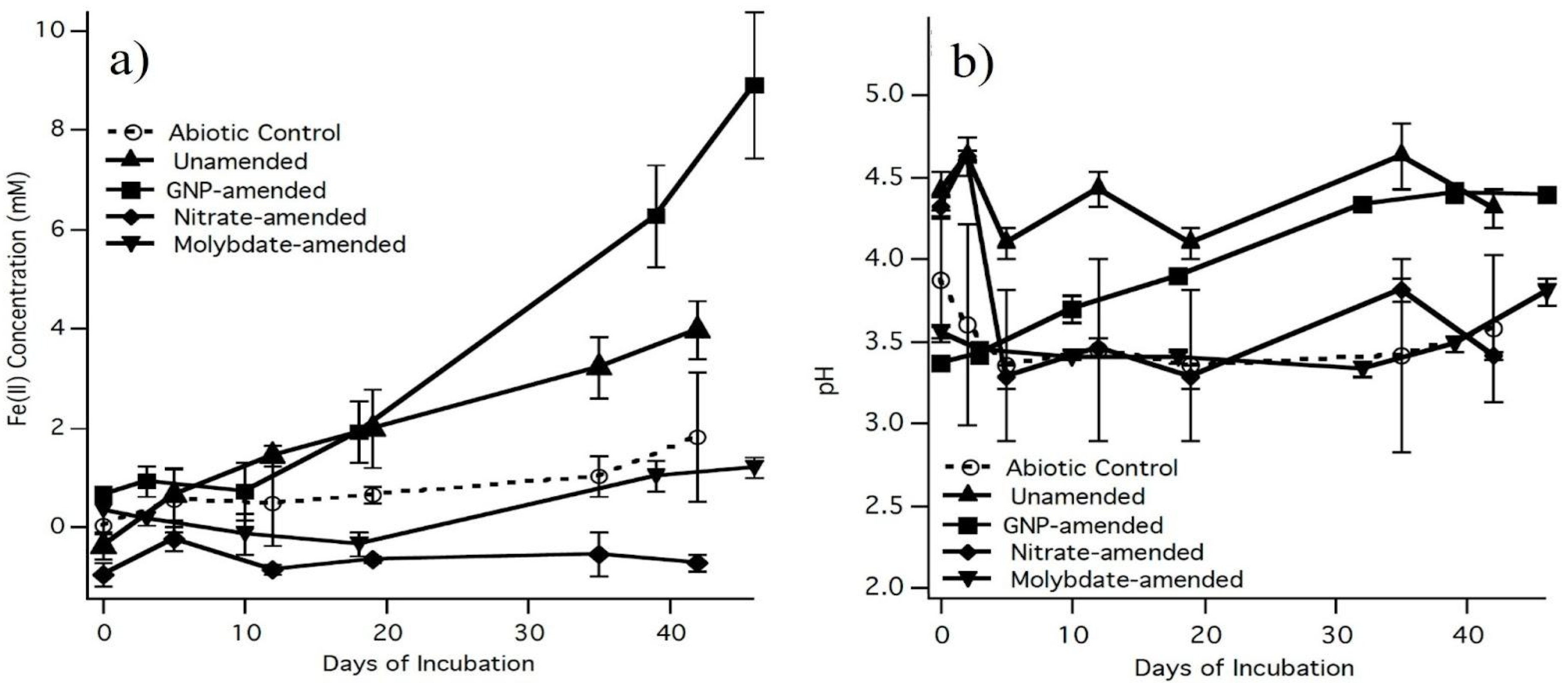
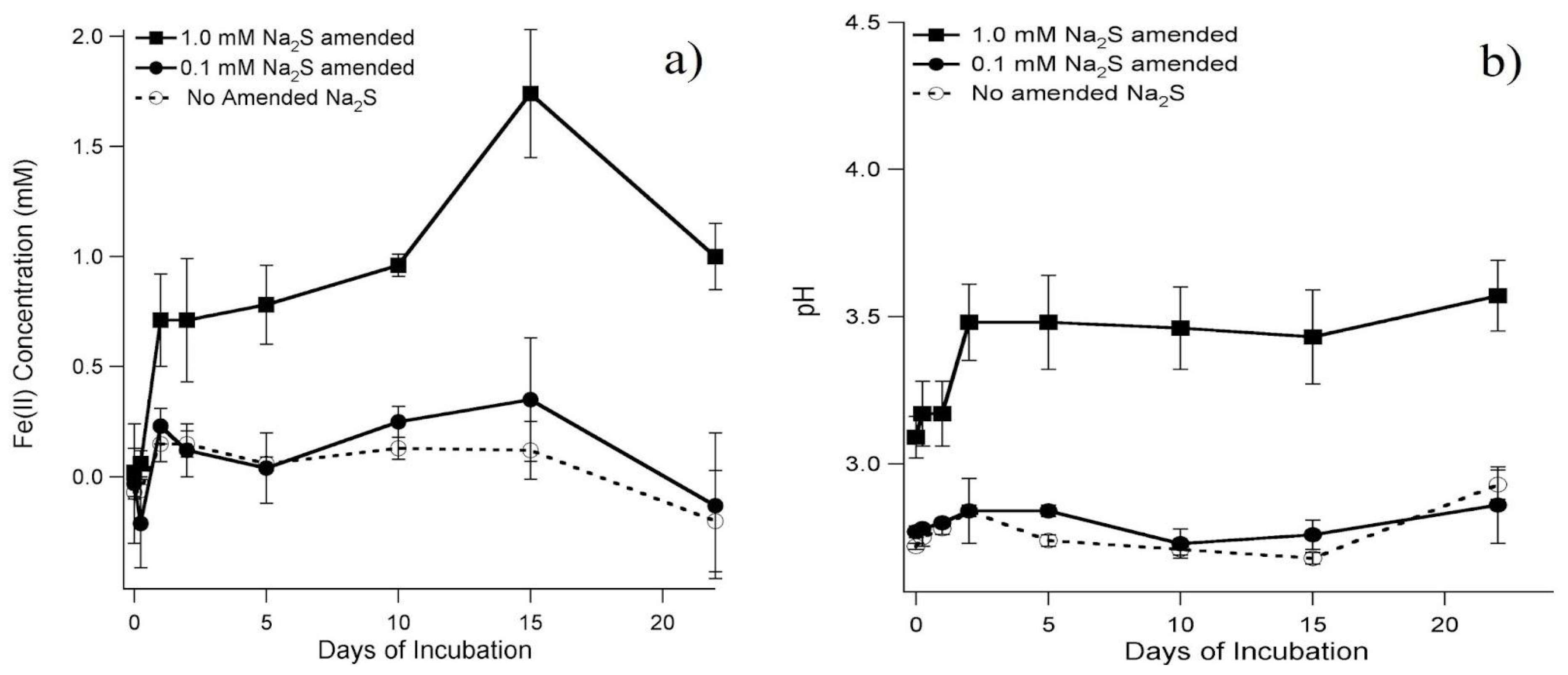
3.1. Inhibition and Stimulation of Sulfate-Reducing Bacteria (SRB) in Microcosms

To determine the effect of sulfate-reducing bacteria (SRB) on the production of Fe2+, the microcosms were amended to either stimulate or inhibit the SRB (Figure 2a). The GNP-stimulated SRB (0.67 ± 0.12 mM, day 1, 8.89 ± 1.46 mM, day 46, n = 3) showed a significant increase (T 95%) in Fe2+ production over the unamended microcosms (0.50 ± 0.46 mM, day 1, 2.35 ± 0.42 mM, day 46, n = 3) over a 46-day incubation. In contrast, no detectable Fe2+ was produced in microcosms that were amended with molybdate, a selective inhibitor for SRB, or nitrate, a competitive electron acceptor. The abiotic control exhibited an increase in Fe2+ from 1.32 ± 0.19 mM to 2.50 ± 0.44 mM, (n = 3) throughout the course of the incubation.
An increase in pH suggests that AMD attenuation occurred and therefore was monitored throughout the incubation period as well (Figure 2b). The pH of both the GNP-stimulated and unamended microcosms exhibited a significant increase (T95%) in pH from 3.37 ± 0.05 mM to 4.39 ± 0.03 mM, and from 3.06 ± 0.06 mM to 3.81 ± 0.10 mM (n = 3), respectively, throughout the course of the incubation. The pH of the abiotic control was comparable to the molybdate-amended microcosms with a pH average of 3.5, with no significant change during incubation.
Another indication that SRB were active was the development of black precipitates. Black precipitates developed in unamended microcosms and in GNP-amended microcosms. Also, while sampling these microcosms the odor of hydrogen sulfide, an indication of sulfate reduction was noticed.

3.2. Sulfate Reduction Rates

The sulfate reduction rates of a second set of unamended microcosms, GNP-amended microcosms, and abiotic controls were determined (Table 1). The sulfate reduction rates of the unamended microcosms had a mean of 19 ± 2 nmol/(cm3d). The sulfate reduction rates of the GNP-amended microcosms were 22 ± 2 nmol/(cm3d). At 95% confidence level using the T-test, there was no significant difference in the sulfate reduction rates of both sets of microcosms. The rates for the corresponding controls were negligible at 0.31 and 0.29 nmol/(cm3d), respectively.
In addition to sulfate reduction rates, we also monitored the pH, ORP, DOC, sulfate, and Fe2+. The increase in pH over time was higher and significantly different in the GNP, unamended, and ground water samples compared to the abiotic control and corresponded to the decrease in ORP. The pH of the abiotic controls remained relatively constant; however, the ORP increased, indicating oxidation. The pH and ORP of the pore water extracted from the sediment core that was used to construct the microcosms were like the pH and ORP of the microcosms at the end of the incubation. Negative ORP values indicated that reducing conditions were achieved. The greatest decrease was seen in the GNP-amended microcosms, followed by the unamended microcosms and the abiotic controls. The average sulfate concentrations for the unamended and GNP-amended microcosms were both higher than their respective abiotic controls. All ORP values were statistically unique to 95% confidence.

3.3. Abundance and Diversity of SRB

Enumeration of SRB was based on the quantification of the functional gene dsr, which encodes the dissimilatory sulfite reductase [28]. At the start of the incubation (t = 0), both the unamended and GNP-amended microcosms had similar counts of SRB, but by the end of the incubation period, the number of SRB in the GNP-amended microcosms had more than doubled the number of SRB seen in the unamended microcosms (Table 2).
Based on the DAPI counts of the total cell number, the percentage of SRB in the total population increased over time. The relative abundance of SRB in the unamended microcosms did not show a statistically significant change during the incubation period, where the GNP-amended did show a significant difference to a 95% confidence. With the SRB group-specific primers, we detected more groups of SRB in the GNP microcosms than the unamended microcosms (Table 3). SRB groups Desulfotomaculum, Desulfovibrio, and Desulfobulbus were found in both GNP microcosms than the unamended microcosms with the addition of Desulfobacter in the GNP microcosms.

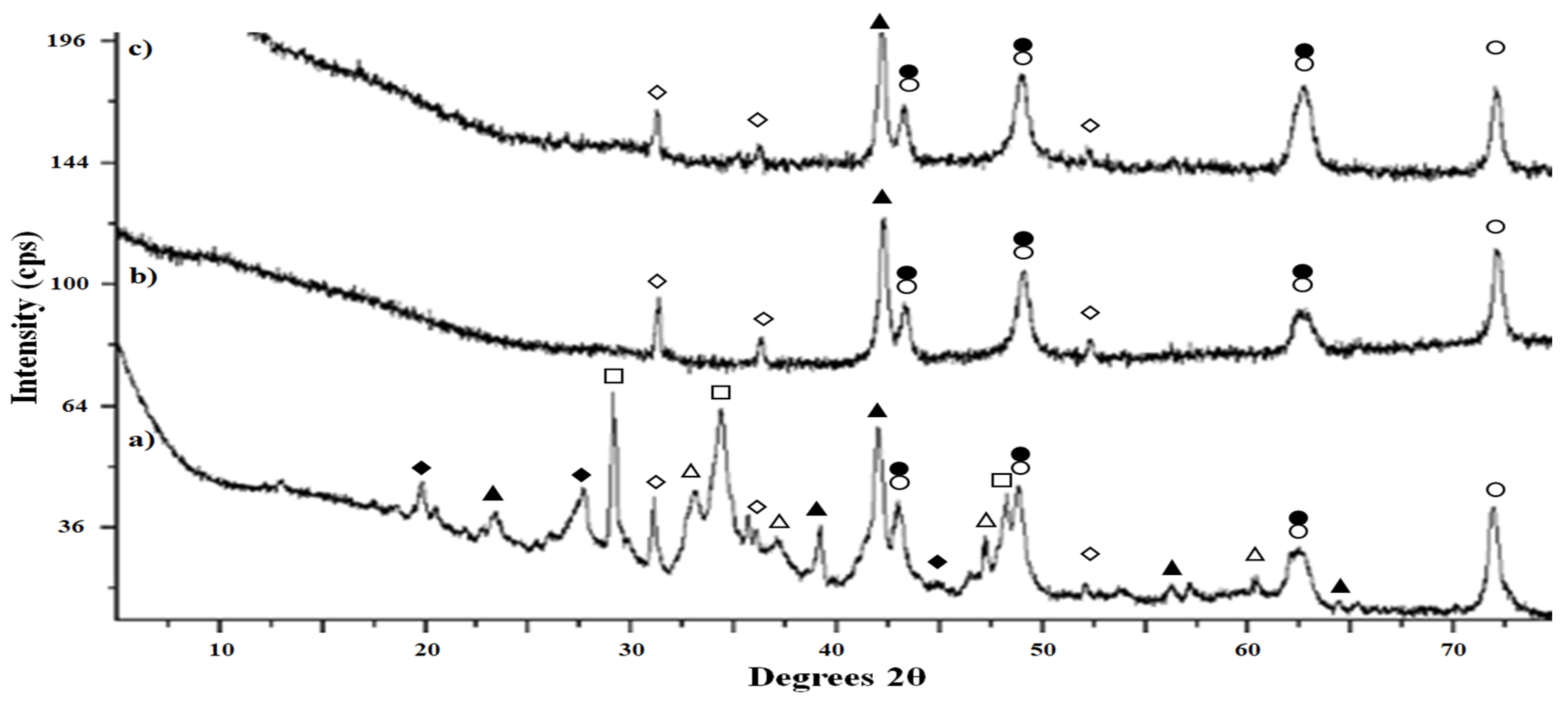
3.4. Origin and Characterization of Precipitate

Black precipitates developed immediately in the abiotic controls after the addition of sulfide, which accompanied an increase in Fe2+ concentration and pH (Figure 3a,b). The amount of Fe2+ produced corresponded to the amount of sulfide added: the average concentration of triplicate samples of Fe2+ generated in the presence of 0.1 mM and 1.0 mM sodium sulfide was 0.14 ± 0.2 mM and 0.98 ± 0.4 mM, respectively (Figure 3a). Similarly, the change in pH value also corresponded to the amount of sulfide added (Figure 3b). During the incubation, the pH of the 0.1 mM and 1.0 mM sodium sulfide-amended microcosms was approximately 2.8 and 3.4, respectively. Therefore, Fe2+ production and increase in pH were correlated.
The black precipitates that developed in the abiotic soil-free samples and the GNP-amended microcosms were analyzed using X-ray diffraction to determine the structure and identity of the precipitates (Figure 4) as well as an electron microprobe to determine the elemental composition (Table 4). The precipitates from the three soil-free samples were fine-grained powders that exhibited diffractograms with broad peak widths indicative of previously reported samples generated under similar conditions [30]. Peak widths are directly related to crystallite size, with precipitate formation being observed.
Shortly after initiation in addition to co-precipitation of several minerals, broader peaks are to be expected [31]. Though the large deviation in the GNP sample excludes statistical comparison, the soil-free abiotic samples are significantly similar and significantly different from the Davis Mine biotic sample to 95% confidence in iron concentration.
All three samples exhibit the same major peaks, which predominantly correspond to higher valent iron chalcogenide minerals such as hematite, magnetite, and greigite. However, the bioactive sample exhibits an additional range of minerals that are noteworthy for having a smaller average oxidation state of iron near or equal to two (troilite and pyrite). In addition, several of the additional peaks in the bioactive sample indicate minerals that adsorb or incorporate other metals from the groundwater (pseudomorphic pyrite and chalcopyrite), as shown in Figure 4 [32,33]. Metals such as copper and zinc were found in the groundwater, as listed previously. Though they do not seem to exist in high enough concentrations to contribute to the abiotic background precipitation, they do appear in the more complex mineralization in the bioactive sample. Average iron valence drops with bioactive samples as well as more mineralized precipitation.
Electron microprobe images of representative grains of the microcosm and soil-free abiotic controls are shown in Figure 5. The black precipitates of the microcosm appeared fine-grained and film-like (Figure 5a), compared to the more granular abiotic samples (Figure 5b,c). This trend is echoed in the elemental analysis with the tightly correlated compositions and smaller deviations than either bioactive sample (Table 4).

4. Discussion

Acid mine drainage (AMD) is an environmental contamination caused by mining activities that destroys aquatic ecosystems [1]. Yet, there are sites where the AMD is naturally attenuated [7,13,34,35]. Understanding how AMD is attenuated may offer strategies for actively remediating AMD. Previous studies on the natural attenuation of our AMD study site suggests that SRB are key [2,8,13,25]. SRB have been used in AMD remediation in other studies because they reduce sulfate [10,11,25,27,28,29,30,34,35,36,37,38,39]. However, in this study, we determined that SRB, and not FeRB, are also responsible for iron reduction, which contributes to remediation by co-precipitation of metal contaminants.

4.1. Iron Reduction Mediated by Sulfate-Reducing Bacteria

Fe2+ ions are generated through enzymatic or abiotic reactions. For example, iron-reducing bacteria (FeRB) and some sulfate-reducing bacteria (SRB) can enzymatically reduce iron, while sulfide as the only environmentally relevant compound can reduce iron abiotically [14,16,18,19,21,40,41,42,43,44]. In a bioreactor study that tested a pH range of 6.0–7.5, sulfate and iron reduction occurred at varying ratios that depended on the pH and the acetate concentration. The researchers of that study found that iron reduction occurred to a greater extent if sulfate reduction was occurring [45]. In this study, we determined the contribution of SRB versus FeRB to the production of Fe2+ ions by selectively inhibiting these bacterial groups.
SRB are metabolically inhibited by molybdate, a sulfate analog, while FeRB are not affected [16,46]. Nitrate is a more energetically favorable terminal electron acceptor and therefore was used to competitively inhibit the reduction of both iron and sulfate [47,48,49]. Although some SRB are able to reduce nitrate to nitrite, no increase of Fe2+ ion concentrations would be due to this metabolism [50]. In addition, nitrite may also inhibit SRB [51,52]. In our microcosms amended with nitrate, no Fe2+ ions were produced. In molybdate-amended microcosms, the Fe2+ ion concentration was similar to the nitrate-amended microcosms, which had both SRB and FeRB inhibited. Therefore, it is the acidotolerant SRB that are involved in the production of Fe2+ ions.
We recognize the potential role of aerobic iron oxidizers, which could also reduce iron. However, our experimental controls were designed to address this possibility. We defined direct iron reduction as abiotic processes or those not influenced by microbial activity. The absence of significant changes in Fe2+ concentration in abiotic and inhibited microcosms supports that direct iron reduction was not a major factor.
Although we did not specifically target iron-oxidizing microorganisms that can shift to iron reduction, we observed no Fe2+ production in molybdate- or nitrate-amended microcosms, where SRB or iron-reducing bacteria were inhibited. This lack of Fe2+ production indicates that if aerobic iron oxidizers were present, their contribution to iron reduction was below the detection level of our assays. Comparison between microcosms with inhibited SRB and unamended microcosms further confirmed that the observed iron reduction is primarily mediated by SRB.
Previous studies related to AMD remediation have shown that the addition of certain organic carbon stimulated microbial activity [37,38,53,54,55,56]. In earlier investigations and in this study, the addition of GNP as an organic carbon and micronutrient source increased the rate of iron reduction [13]. The choice in organic substrate was based on previous microcosms testing acetate, ethanol, pyruvate, lactate, and glycerol [57] as well as readily available, complex, organic substrates found at Davis Mine such as wood chips, algae [13], and cow manure to test for a potential, large-scale, on-site remediation [58]. An increase in the generation of Fe2+ ions as compared to unamended microcosms and abiotic controls indicates that iron reduction is biologically mediated under acidic conditions. We chose to amend the microcosms with a low concentration of glycerol because acidophilic microbes do not tolerate high concentrations of organic carbon [39,59,60]. In a comparison to a study of two AMD-treating bioreactors containing acidophilic sulfidogenic consortia, glycerol was used and consumed at a pH of 2.5 [61]. The results of the inhibition and stimulation experiment indicate that SRB mediate iron reduction.

4.2. Abundance and Diversity of SRB

SRB are present in greater abundance and activity in acidic environments than previously thought [62]. In our microcosms, the number of DAPI-stained cells remained constant from beginning to end of the incubation period. However, the number of SRB cells detected by qPCR of the dsr gene increased from 104/mL to 106/mL. As a comparison, in an iron-dominated, mining-impacted freshwater sediment, the abundance of cultivable SRB using the most probable number was within the range of 104 to 106 cultivable SRB g−1 wet weight sediment [63].
In this study, we tested for the presence of six major phylogenetic groups of SRB based on Daly 2000 [29]. Recently, 13 phylum-level lineages that were not known to have dsr genes brought the total to 20 distinct phylum-level lineages currently. However, delta-proteobacteria has the largest number with 34 genomes reported [64]. Therefore, in the context of this study, the six major groups that we investigated were of the delta-proteobacteria and Gram-positive for Desulfotomaculum.
The detected SRB groups depended on the length of the incubation and the type of amendments made to the microcosms. For example, using SRB group-specific primers, we detected Desulfotomaculum, Desulfovibrio, and Desulfobulbus in the unamended microcosms. However, using SRB group-specific primers for DesulfococcusDesulfonemaDesulfosarcina and Desulfobacterium, these SRB groups were not detected. For the GNP-amended microcosms, the same groups in the unamended microcosms were detected with the addition of Desulfobacter, which suggests low abundance at our site. Desulfobacter was only detected after 40 days of incubation and with the addition of GNP. In another AMD study, the authors also did not detect Desulfovibrio and Desulfosporosinus in their sediments but abundantly in their microcosms that had been amended with hydrogen [65]. Desulfotomaculum have been found in low pH environments and Desulfovibrio, Desulfobulbus, and Desulfobacter have been found in other AMD sites [66,67]. Desulfosporosinus was detected in a study that compared two acidophilic sulfidogenic consortia in bioreactors that treated AMD as well as in acidic mine pit lakes [59,66]. In a mildly acidic AMD site, the dominant clone sequence, representing 65% of the community, was related to SRB, Desulfosarcina variabilis [68].
Sequences related to SRB have also been found in other natural acidic environments such as volcanic-impacted water bodies and hydrothermal springs [12,56,69,70,71]. Fortin et al. recovered SRB in acidic and oxidizing conditions, while Senko et al. isolated an acidotolerant SRB able to reduce Fe(III)(hydr)oxide better under acidic conditions of pH 4.4 than at neutral pH 7.1 [42,59]. The genera that accounted for 100% of the SRB-like populations in a study that tested five types of sulfate-reducing passive bioreactors were Desulfosporosinus, Clostridium, Ruminococcus, Desulfovibrio, and Desulfobulbus [72]. Therefore, SRB are ubiquitous in nature and engineered systems like bioreactors.

4.3. Origin and Characterization of the Sulfide Precipitate

Despite the acidic nature of AMD sites, the formation of black FeS precipitates has previously been observed where sulfate reduction or SRB were detected [12]. In fact, this phenomenon has been used in a bioreactor treating acid rock drainage where zero-valent iron as an electron donor was added to produce metal sulfide precipitates [6]. Based on powder X-ray diffraction and electron microprobe elemental analysis, the polycrystalline, black precipitate from both bioactive and abiotic samples show that the presence of free ions in solution can lead to some precipitation or higher valence iron chalcogenide minerals. However, the diversity of minerals, minerals with reduced iron oxidation states, and minerals incorporating multiple metals were significantly increased in the bioactive samples. Black FeS precipitates have been detected when Na2S was added to liquid medium or to sulfate-reducing bacterial cultures in controlled environments [20,30]. For example, Gramp et al. detected iron sulfide minerals, greigite, mackinawite, and pyrite forming in media containing SRB [30]. At our study site, iron minerals such as goethite, pyrrhotite, chalcopyrite, pyrite, and magnetite were previously detected [2]. In aqueous samples from our microcosms, we observed that the production of Fe2+ ions was directly correlated with increasing sulfide concentration, which agrees with other studies [9,20,73,74]. Previous studies have shown that sulfide reduces iron oxy(hydr)oxides minerals such as lepidocrocite, goethite, magnetite, and hematite [7,41]. Although not a mineral, in another study that used SRB-immobilized particles to treat simulated AMD, “sulfate green rust”, another type of iron sulfate precipitate, developed in their system [75]. More examples demonstrating the iron sulfide sorption of other metals include mercury [76] and copper [77], as seen in the bioactive sample in Figure 4.
Another implication of this study is the improved sequestration of iron and other dissolved metals in the presence of SRB. Dissolved metals may precipitate as metal sulfides. For example, galena is formed through sulfide co-precipitation produced by SRB [56]. Similarly, iron can precipitate as a ferrous sulfide [9,54,59]. Another example is the sulfidation of arsenic on ferrihydrite to arsenic trisulfide in groundwater. Additionally, Fe2+ and sulfide could form mackinawite, onto which arsenate would adsorb [78]. In a study of mine-impacted freshwater, the scanning electron microscopy analysis showed that zinc sulfide formed as the dissolved zinc concentration decreased in anaerobic batch tests containing SRB [12]. This would have implications on the removal of dissolved metals, which contributes to the attenuation of AMD [7].
Although the rate of sulfate reduction is low compared to other sulfidic environments [79], our study found evidence of sulfate reduction comparable to other acidic environments [62]. However, higher rates of sulfate reduction have been previously found [80]. One explanation for the difference in the rates of sulfate reduction is that the SRB at our site may be reducing a different terminal electron acceptor [14,16,42,43,44]. Another explanation is that since AMD sites are high in iron concentration, sulfide may exist too briefly, as it quickly reacts with iron to reduce it, and thus escape our measurements based on evolving sulfide compounds.

4.4. Coupling Sulfur and Iron Cycles

Iron and sulfur are abundant elements on earth and are particularly plentiful in AMD sites. Sulfate reduction was thought to occur after the pool of iron or organic substrate was depleted because oxidized iron is more energetically favorable as the terminal electron acceptor at neutral pH [48,49]. However, AMD sites have high concentrations of dissolved iron and sulfur; therefore, there may not be a significant preference or progression of terminal electron acceptors utilized.
Research on iron reduction under acidic conditions is less extensive compared to neutral pH environments [1,12,79,81,82]. Even fewer studies on iron reduction by sulfide under low pH exist. For example, in a study that tested the influences of pH and acetate on the ratio of iron and sulfate reduction, the pH of the bioreactors were 6.0–7.5, a pH range higher than normally found in AMD-impacted sites [45]. This indicates that the cycling of iron influenced by sulfide in acidic systems might be more significant than previously understood, given the higher concentrations of dissolved Fe3+ ions in these environments compared to marine or subsurface settings.
In our study, we observed that Fe3+ interacts with sulfide through several mechanisms. Ferric iron can be precipitated by sulfide, with greigite (Fe2Fe3S4)—a mineral containing ferric and ferrous iron along with sulfur—being detected in our XRD analysis. This mineral was found in both bioactive and abiotic samples, indicating that greigite can form in the presence of sulfide regardless or microbial activity. Additionally, our results show that ferric iron is reduced by sulfide, as evidenced by the date presented in Figure 2 and Figure 3. However, the formation of ferric sulfide (Fe2S3) is not detected in the XRD data, aligning with its known solubility in acidic media.
While we did not specifically examine microbial reduction of ferric iron by organisms such as Acidithiobacillus or Ferroplasma, the presence of greigite in both bioactive and abiotic samples suggests that this mineral can form independent of microbial activity. The comparison between bioactive and abiotic samples further reveals that additional mineral species with lower average oxidation states are produced bioactive conditions, suggesting that microbial processes may enhance iron reduction.
The reactivity of dissolved Fe3+ ions with sulfide in acidic environments, coupled with the regeneration of sulfate or elemental sulfur through reactions with Fe3+ ions or microbial respiration, highlights the complex interactions between sulfur and iron cycles in AMD sites [4,18,83]. This underscores the substantial role of sulfide in the iron cycle, particularly under acidic conditions.

5. Conclusions

In this study, we described an additional mechanism by which SRB promotes attenuation of AMD by reducing iron via biogenic sulfide and provide additional information on the biogeochemical cycling of iron and sulfur. The implication of this study is that iron reduction mediated by biogenic sulfide may be more significant than previously thought in acidic environments.

Author Contributions

Conceptualization, C.A.B. and K.N.; methodology, C.A.B. and B.V.V.; software, C.A.B. and B.V.V.; formal analysis, C.A.B., B.M. and B.V.V.; investigation, C.A.B., B.M. and B.V.V.; resources, K.N.; data curation, C.A.B., B.M. and B.V.V.; writing—original draft preparation, C.A.B., B.M. and B.V.V.; writing—review and editing, C.A.B., B.M., B.V.V. and K.N.; visualization, C.A.B. and B.V.V.; supervision, C.A.B. and K.N.; project administration, K.N.; funding acquisition, K.N. All authors have read and agreed to the published version of the manuscript.

Funding

This research was funded by the U.S. National Science Foundation, CHE-0221791 and the Ford Foundation directly supporting C.A.B.

Data Availability Statement

All data generated or analyzed during this study are included in this published manuscript.

Acknowledgments

We would like to thank Matt Coggon, Wim de Jeu, Michael Jercinovic, Joy Ward, Jessica Bloom, and Richard Yuretich for technical assistance. We are grateful to Kyunghwa Baek, Teresa Conneely, George Hamoui, Anna Gilmour, Karen Miller, and Heather Reed for stimulating discussion.

Conflicts of Interest

Author Brendan Murphy was employed by the company Cambrian BioPharma, Inc. The remaining authors declare that the research was conducted in the absence of any commercial or financial relationships that could be construed as a potential conflict of interest.

References

  1. Johnson, D.B.; Hallberg, K.B. The microbiology of acid mine waters. Res. Microbiol. 2003, 154, 466–473. [Google Scholar] [CrossRef] [PubMed]
  2. Bloom, J.; Yuretich, R.F.; Gál, N.E. Environmental consequences of acid mine-drainage from Davis pyrite mine, Rowe, Massachusetts. Northeast. Geol. Environ. Sci. 2007, 29, 108–121. [Google Scholar]
  3. Aguinaga, O.E.; Wakelin, J.F.T.; White, K.N.; Dean, A.P.; Pittman, J.K. The association of microbial activity with Fe, S and trace element distribution in sediment cores within a natural wetland polluted by acid mine drainage. Chemosphere 2019, 231, 432–441. [Google Scholar] [CrossRef] [PubMed]
  4. Benner, S.G.; Gould, W.D.; Blowes, D.W. Microbial populations associated with the generation and treatment of acid mine drainage. Chem. Geol. 2000, 169, 435–448. [Google Scholar] [CrossRef]
  5. Gould, W.; Kapoor, A. The microbiology of acid mine drainage. In Environmental Aspects of Mine Wastes; Jambor, J.L., Blowes, D.W., Ritchie, A.I.M., Eds.; Mineralogical Association of Canada: Ottawa, ON, Canada, 2003; pp. 203–226. [Google Scholar]
  6. Ayala-Parra, P.; Sierra-Alvarez, R.; Field, J.A. Treatment of acid rock drainage using a sulfate-reducing bioreactor with zero-valent iron. J. Hazard. Mater. 2016, 308, 97–105. [Google Scholar] [CrossRef]
  7. Dore, E.; Fancello, D.; Rigonat, N.; Medas, D.; Cidu, R.; Da Pelo, S.; Frau, F.; Lattanzi, P.; Marras, P.A.; Meneghini, C.; et al. Natural attenuation can lead to environmental resilience in mine environment. Appl. Geochem. 2020, 117, 104597. [Google Scholar] [CrossRef]
  8. Ergas, S.J.; Harrison, J.; Bloom, J.; Forloney, K.; Ahlfeld, D.P.; Nüsslein, K.; Yuretich, R.F. Natural attenuation of acid mine drainage by acidophilic and acidotolerant Fe(III)- and sulfate-reducing bacteria. remediation of hazardous waste in the subsurface: Bridging flask and field studies. In American Chemical Society Symposium Series; Clark, C., II, Lindner, A., Eds.; ACS Publications: Washington, DC, USA, 2006; pp. 105–127. [Google Scholar]
  9. Neal, A.L.; Techkarnjanarauk, K.; Dohnalkova, A.; McCready, D.; Peyton, B.M.; Geesey, G. Iron sulfides and sulfur species produced at hematite surfaces in the presence of sulfate-reducing bacteria. Geochim. Cosmochim. Acta 2001, 65, 223–235. [Google Scholar] [CrossRef]
  10. Neculita, C.M.; Zagury, G.J.; Bussière, B. Passive treatment of acid mine drainage in bioreactors using sulfate-reducing bacteria: Critical review and research needs. J. Environ. Qual. 2007, 36, 1–16. [Google Scholar] [CrossRef]
  11. Church, C.D.; Wilkin, R.T.; Alpers, C.N.; Rye, R.O.; McCleskey, R.B. Microbial sulfate reduction and metal attenuation in pH 4 acid mine water. Geochem. Trans. 2007, 8, 10. [Google Scholar] [CrossRef]
  12. Paganin, P.; Alisi, C.; Dore, E.; Fancello, D.; Marras, P.A.; Medas, D.; Montereali, R.; Naitza, S.; Rigonat, N.; Sporacati, A.R.; et al. Microbial diversity of bacteria involved in biomineralization processes in mine-impacted freshwaters. Front. Microbiol. 2021, 12, 778199. [Google Scholar] [CrossRef]
  13. Becerra, C.A.; López-Luna, E.L.; Ergas, S.J.; Nüsslein, K. Microcosm-based study of the attenuation of an acid mine drainage-impacted site through biological sulfate and iron reduction. Geomicrobiol. J. 2009, 26, 9–20. [Google Scholar] [CrossRef]
  14. Coleman, M.L.; Hedrick, D.B.; Lovley, D.R.; White, D.C.; Pye, K. Reduction of Fe(III) in sediments by sulphate-reducing bacteria. Nature 1993, 361, 436–438. [Google Scholar] [CrossRef]
  15. Dalla Vecchia, E.; Suvorova, E.I.; Maillard, J.; Bernier-Latmani, R. Fe(III) reduction during pyruvate fermentation by Desulfotomaculum reducens strain MI-1. Geobiology 2013, 12, 48–61. [Google Scholar] [CrossRef] [PubMed]
  16. Lovley, D.R.; Roden, E.E.; Phillips, E.J.P.; Woodward, J.C. Enzymatic iron and uranium reduction by sulfate-reducing bacteria. Mar. Geol. 1993, 113, 41–53. [Google Scholar] [CrossRef]
  17. Li, Y.L.; Vali, H.; Sears, S.K.; Yang, J.; Deng, B.; Zhang, C.L. Iron reduction and alteration of nontronite NAu-2 by a sulfate-reducing bacterium. Geochim. Cosmochim. Acta 2004, 68, 3251–3260. [Google Scholar] [CrossRef]
  18. dos Santos Afonso, M.; Stumm, W. Reductive dissolution of iron(III) (hydr)oxide by hydrogen sulfide. Langmuir 1992, 8, 1671–1675. [Google Scholar] [CrossRef]
  19. Canfield, D.E.; Raiswell, R.; Bottrell, S. The reactivity of sedimentary iron minerals toward sulfide. Am. J. Sci. 1992, 292, 659–683. [Google Scholar] [CrossRef]
  20. Li, Y.L.; Valli, J.; Yang, J.; Phelps, T.J.; Zhang, C.L. Reduction of iron oxides by a sulfate-reducing bacterium and biogenic H2S. Geomicrobiol. J. 2006, 23, 103–117. [Google Scholar] [CrossRef]
  21. Roberts, M.; Srivastava, P.; Webster, G.; Weightman, A.J.; Sapsford, D.J. Biostimulation of jarosite and iron oxide-bearing mine waste enhances subsequent metal recovery. J. Hazard. Mater. 2023, 445, 130498. [Google Scholar] [CrossRef]
  22. Brown, P.W.; Williams, I.N. The History of Rowe, Massachusetts, 4th ed.; Rowe Historical Society: Rowe, MA, USA, 2006. [Google Scholar]
  23. Stookey, L.L. Ferrozine—A new spectrophotometric reagent for iron. Anal. Chem. 1970, 42, 779–781. [Google Scholar] [CrossRef]
  24. Lovley, D.R.; Phillips, E.J.P. Organic matter mineralization with the reduction of ferric iron in anaerobic sediments. Appl. Environ. Microbiol. 1996, 51, 683–689. [Google Scholar] [CrossRef] [PubMed]
  25. Coggon, M.; Becerra, C.A.; Nüsslein, K.; Miller, K.; Yuretich, R.; Ergas, S.J. Bioavailability of jarosite for stimulating acid mine drainage attenuation. Geochim. Cosmochim. Acta 2012, 78, 65–76. [Google Scholar] [CrossRef]
  26. Jercinovic, M.J.; Williams, M.L. Analytical perils (and progress) in electron microprobe trace element analysis applied to geochronology: Background acquisition, interferences, and beam irradiation effects. Am. Mineral. 2005, 90, 526–546. [Google Scholar] [CrossRef]
  27. Kallmeyer, J.; Ferdelman, T.G.; Weber, A.; Fossing, H.; Jorgensen, B.B. A cold chromium distillation procedure for radiolabeled applied to sulfate reduction measurements. Limnol. Oceanogr. Methods 2004, 2, 171–180. [Google Scholar] [CrossRef]
  28. Chin, K.J.; Sharma, M.I.; Russel, L.A.; O’Neill, K.R.; Lovely, D.R. Quantifying expression of a dissimilatory bi(sulfite) reductase gene in petroleum-contaminated marine harbor sediments. Microb. Ecol. 2008, 55, 489–499. [Google Scholar] [CrossRef]
  29. Daly, K.; Sharp, R.J.; McCarthy, A.J. Development of oligonucleotide probes and PCR primers for detecting phylogenetic subgroups of sulfate-reducing bacteria. Microbiology 2000, 146, 1693–1705. [Google Scholar] [CrossRef]
  30. Gramp, J.P.; Bigham, J.M.; Jones, F.S.; Tuovinen, O.H. Formation of Fe-sulfides in cultures of sulfate-reducing bacteria. J. Hazard. Mater. 2010, 175, 1062–1067. [Google Scholar] [CrossRef]
  31. Guinier, A. X-ray diffraction. In Crystals, Imperfect Crystals, and Amorphous Bodies; Dover Publications: New York, NY, USA, 1994. [Google Scholar]
  32. Lafuente, B.; Downs, R.T.; Yang, H.; Stone, N. The power of databases: The RRUFF project. In Highlights in Mineralogical Crystallography; Armbruster, T., Danisi, R.M., Eds.; W. De Gruyter: Berlin, Germany, 2015; pp. 1–30. [Google Scholar]
  33. The RRUFF ™ Project. RRUFF ID’s: Hematite R110013.9, Magnetite R061111.9, Greigite R120103, Troilite R070242.9, (Pseudomorphic) Pyrite R080030 and R070692.9, Pyrite R100166.9, Chalcopyrite R050559.1. Available online: https://rruff.info/ (accessed on 8 August 2024).
  34. Sakala, E.; Fourie, F.; Gomo, M.; Madzivire, G. Natural attenuation of acid mine drainage by various rocks in the Witbank, Ermelo and Highveld Coalfields, South Africa. Nat. Resour. Res. 2020, 30, 557–570. [Google Scholar] [CrossRef]
  35. Tum, S.; Toda, K.; Matsui, T.; Kikuchi, R.; Kong, S.; Meas, P.; Ear, U.; Ohtomo, Y.; Otake, T.; Sato, T. Seasonal effects of natural attenuation on drainage contamination from artisanal gold mining, Cambodia: Implication for passive treatment. Sci. Total Environ. 2022, 806, 150398. [Google Scholar] [CrossRef]
  36. Lovley, D.R.; Phillips, E.J.P. Rapid assay for microbially reducible ferric iron in aquatic sediments. Appl. Environ. Microb. 1987, 53, 1536–1540. [Google Scholar] [CrossRef]
  37. Coetser, S.E.; Pulles, W.; Heath, R.G.M.; Cloete, T.E. Chemical characteristics of organic electron donors for sulfate reduction for potential use in acid mine drainage treatment. Biodegradation 2006, 17, 67–77. [Google Scholar] [CrossRef] [PubMed]
  38. Bilgin, A.A.; Harrington, J.M. Enhancement of bacterial iron and sulfate respiration for in situ bioremediation of acid mine drainage sites: A case study. Miner. Metall. Proc. 2007, 24, 139–144. [Google Scholar] [CrossRef]
  39. Gyure, R.A.; Konopka, A.; Brooks, A.; Doemel, W. Microbial sulfate reduction in acidic (pH 3) strip-mine lakes. FEMS Microbiol. Ecol. 1990, 73, 193–201. [Google Scholar] [CrossRef]
  40. Lovley, D.R.; Phillips, E.J.P.; Lonergan, D.J. Enzymatic versus nonenzymatic mechanisms for Fe(III) reduction in aquatic sediments. Environ. Sci. Technol. 1991, 25, 1062–1067. [Google Scholar] [CrossRef]
  41. Poulton, S.W.; Krom, M.D.; Raiswell, R. A revised scheme for the reactivity of iron (oxyhydr)oxide minerals towards dissolved sulfide. Geochim. Cosmochim. Acta 2004, 68, 3703–3715. [Google Scholar] [CrossRef]
  42. Senko, J.M.; Zhang, G.; McDonough, J.T.; Bruns, M.A.; Burgos, W.D. Metal reduction at low pH by Desulfosporosinus species: Implications for the biological treatment of acidic mine drainage. Geomicrobiol. J. 2009, 26, 71–82. [Google Scholar] [CrossRef]
  43. Tebo, B.M.; Obraztsova, A.Y. Sulfate-reducing bacterium grows with Cr(VI), U(VI), Mn(IV), and Fe(III) as electron acceptors. FEMS Microbiol. Ecol. 1998, 162, 193–198. [Google Scholar] [CrossRef]
  44. Weber, K.A.; Achenbach, L.A.; Coates, J.D. Microorganisms pumping iron: Anaerobic microbial iron oxidation and reduction. Nature 2006, 4, 752–764. [Google Scholar] [CrossRef]
  45. Paper, J.M.; Flynn, T.M.; Boyanov, M.I.; Kemner, K.M.; Haller, B.R.; Crank, K.; Lower, A.M.; Jin, Q.; Kirk, M.F. Influences of pH and substrate supply on the ratio of iron to sulfate reduction. Geobiology 2021, 19, 405–420. [Google Scholar] [CrossRef]
  46. Stögbauer, A.; Koydon, S.; Berner, Z.; Winter, J.; Stüben, D. Effect of molybdate and cell growth on S-isotope fractionation during bacterial sulfate reduction. Geomicrobiol. J. 2004, 21, 207–219. [Google Scholar] [CrossRef]
  47. DiChristina, T.J. Effects of nitrate and nitrite on dissimilatory iron reduction by Shewanella putrefaciens 200. J. Bacteriol. 1992, 174, 1891–1896. [Google Scholar] [CrossRef] [PubMed]
  48. Finneran, K.T.; Housewright, M.E.; Lovley, D.R. Multiple influences of nitrate on uranium solubility during bioremediation of uranium-contaminated subsurface sediments. Environ. Microbiol. 2002, 4, 510–516. [Google Scholar] [CrossRef] [PubMed]
  49. Akob, D.M.; Mills, H.J.; Gihring, T.M.; Kerkhof, L.; Stucki, J.W.; Anastacio, A.S.; Chin, K.J.; Küsel, K.; Palumbo, A.V.; Watson, D.B.; et al. Functional diversity and electron donor dependence of microbial populations capable of U(VI) reduction in radionuclide-contaminated subsurface sediments. Appl. Environ. Microb. 2008, 74, 3159–3170. [Google Scholar] [CrossRef] [PubMed]
  50. Moura, J.J.G.; Gonzalez, P.; Moura, I.; Faque, G. Dissimilatory nitrate and nitrite ammonification by sulphate-reducing eubacteria. In Sulphate-Reducing Bacteria: Environmental and Engineered Systems; Barton, L.L., Hamilton, W.A., Eds.; Cambridge University Press: Cambridge, UK, 2007; pp. 241–264. [Google Scholar]
  51. Greene, E.A.; Brunelle, V.; Jenneman, G.E.; Voordouw, G. Synergistic inhibition of microbial sulfide production by combinations of the metabolic inhibitor nitrite and biocides. Appl. Environ. Microb. 2006, 72, 7897–7901. [Google Scholar] [CrossRef]
  52. Mangalo, M.; Einsiedl, F.; Meckenstock, R.U.; Stichler, W. Influence of the enzyme dissimilatory sulfite reductase on stable isotope fractionation during sulfate reduction. Geochim. Cosmochim. Acta 2008, 72, 1513–1520. [Google Scholar] [CrossRef]
  53. Vile, M.A.; Wieder, R.K. Alkalinity generation by Fe(III) reduction versus sulfate reduction in wetlands constructed for acid mine drainage treatment. Water Air Soil. Poll. 1993, 69, 425–441. [Google Scholar] [CrossRef]
  54. Küsel, K.; Dorsch, T. Effect of supplemental electron donors on the microbial reduction of Fe(III), sulfate, and CO2 in coal mining-impacted freshwater lake sediments. Microb. Ecol. 2000, 40, 238–249. [Google Scholar] [CrossRef]
  55. Gibert, O.; de Pablo, J.; Cortina, J.L.; Ayora, C. Chemical characterization of natural organic substrates for biological mitigation of acid mine drainage. Water Res. 2004, 38, 4186–4196. [Google Scholar] [CrossRef]
  56. Karnachuk, O.V.; Kurochkina, S.Y.; Tuovinen, O.H. Growth of sulfate-reducing bacteria with solid-phase electron acceptors. Appl. Microbiol. Biot. 2002, 58, 482–486. [Google Scholar] [CrossRef]
  57. Harrison, J. Ferric Iron and Sulfate Reduction in the Attenuation of Acid Mine Drainage: A Microcosm Study. Master’s Thesis, University of Massachusetts, Amherst, MA, USA, 2005. [Google Scholar]
  58. Gilmore, A. Attenuation of Acid Mine Drainage Enhanced by Organic Carbon and Limestone Addition: A Process Characterization. Master’s Thesis, University of Massachusetts, Amherst, MA, USA, 2011. [Google Scholar]
  59. Fortin, D.; Davis, B.; Beveridge, T.J. Role of Thiobacillus and sulfate-reducing bacteria in iron biocycling in oxic and acidic mine tailings. FEMS Microbiol. Ecol. 1996, 21, 11–24. [Google Scholar] [CrossRef]
  60. Johnson, D.B. Minireview: Biodiversity and ecology of acidophilic microorganisms. FEMS Microbiol. Ecol. 1998, 27, 307–317. [Google Scholar] [CrossRef]
  61. Frederico, T.D.; Nancucheo, I.; Santos, W.C.B.; Oliveira, R.R.M.; Buzzi, D.C.; Pires, E.S.; Silva, P.M.P.; Lucheta, A.R.; Alves, J.O.; de Oliveira, G.C.; et al. Comparison of two acidophilic sulfidogenic consortia for the treatment of acidic mine water. Front. Bioeng. Biotechnol. 2022, 29, 1048412. [Google Scholar] [CrossRef] [PubMed]
  62. Koschorreck, M.; Wedt-Potthoff, K.; Geller, W. Microbial sulfate reduction at low pH in sediments of an acidic lake in Argentina. Environ. Sci. Technol. 2003, 37, 1159–1162. [Google Scholar] [CrossRef] [PubMed]
  63. Ramamoorthy, S.; Piotrowski, J.S.; Langner, H.W.; Holben, W.E. Ecology of sulfate-reducing bacteria in an iron-dominated, mining-impacted freshwater sediment. J. Environ. Qual. 2009, 38, 675–684. [Google Scholar] [CrossRef]
  64. Anantharaman, K.; Hausmann, B.; Jungbluth, S.P.; Kantor, R.S.; Lavy, A.; Warren, L.A.; Rappe, M.S.; Pester, M.; Loy, A.; Thomas, B.C.; et al. Expanded diversity of microbial groups that shape the dissimilatory sulfur cycle. ISME J. 2018, 12, 1715–1728. [Google Scholar] [CrossRef]
  65. Valdez-Nunez, L.F.; Ayala-Munoz, D.; Sanchez-Espana, J.; Sanchez-Andrea, I. Microbial communities in Peruvian acid mine drainages: Low-abundance sulfate-reducing bacteria with high metabolic activity. Geomicrobiol. J. 2022, 39, 867–883. [Google Scholar] [CrossRef]
  66. Rambabu, K.; Banat, F.; Pham, Q.M.; Ho, S.H.; Ren, N.Q.; Show, P.L. Biological remediation of acid mine drainage: A review of past trends and current outlook. Environ. Sci. Ecotechnol. 2020, 2, 100024. [Google Scholar] [CrossRef]
  67. Sánchez-Andrea, I.; Sanz, J.L.; Bijmans, M.F.M.; Stams, A.J.M. Sulfate reduction at low pH to remediate acid mine drainage. J. Hazard. Mater. 2014, 269, 98–109. [Google Scholar] [CrossRef]
  68. Bruneel, O.; Duran, R.; Koffie, K.; Casiot, C.; Fourçans, A.; Elbaz-Poulichet, F.; Personne, J.-C. Microbial diversity in a pyrite-rich tailings impoundment (Carnoulès, France). Geomicrobiol. J. 2005, 22, 249–257. [Google Scholar] [CrossRef]
  69. Praharaj, T.; Fortin, D. Indicators of microbial sulfate reduction in acidic sulfide-rich mine tailings. Geomicrobiol. J. 2004, 21, 457–467. [Google Scholar] [CrossRef]
  70. Roychoudhury, A.N. Sulfate respiration in extreme environments: A kinetic study. Geomicrobiol. J. 2004, 21, 33–43. [Google Scholar] [CrossRef]
  71. Rowe, O.F.; Sánchez-España, J.; Hallberg, K.B.; Johnson, D.B. Microbial communities and geochemical dynamics in an extremely acidic, metal-rich stream at an abandoned sulfide mine (Huelva, Spain) underpinned by two functional primary production systems. Environ. Microbiol. 2007, 9, 1761–1771. [Google Scholar] [CrossRef] [PubMed]
  72. Sato, Y.; Hamai, T.; Hori, T.; Aoyagi, T.; Inaba, T.; Kobayashi, M.; Have, H.; Sakata, T. Desulfosporosinus spp. were the most predominant sulfate-reducing bacteria in pilot- and laboratory-scale passive bioreactors for acid mine drainage treatment. Appl. Microbiol. Biotech. 2019, 103, 7783–7793. [Google Scholar] [CrossRef] [PubMed]
  73. van der Maas, P.; Peng, S.; Klapwijk, B.; Lens, P. Enzymatic versus nonenzymatic conversions during the reduction of EDTA-chelated Fe(III) in BioDeNOx reactors. Environ. Sci. Technol. 2005, 39, 2616–2623. [Google Scholar] [CrossRef] [PubMed]
  74. Diez-Ercilla, M.; Sánchez-España, J.; Yusta, I.; Wendt-Potthoff, K.; Koschorreck, M. Formation of biogenic sulphides in the water column of an acidic pit lake: Biogeochemical controls and effects on trace metal dynamics. Biogeochemistry 2014, 121, 519–536. [Google Scholar] [CrossRef]
  75. An, W.; Hu, X.; Chen, H.; Wang, Q.; Zheng, Y.; Wang, J.; Di, J. Experimental study on the treatment of AMD by SRB immobilized particles containing “active iron” system. PLoS ONE 2023, 18, 20161. [Google Scholar] [CrossRef]
  76. Wang, M.; Li, Y.; Zhao, D.; Zhuang, L.; Yang, G.; Gong, Y. Immobilization of mercury by iron sulfide nanoparticles alters mercury speciation an dmicrobial methylation in contaminated groundwater. Chem. Eng. J. 2020, 381, 122664. [Google Scholar] [CrossRef]
  77. Osorio, H.; Mangold, S.; Denis, Y.; Nancucheo, I.; Esparza, M.; Johnson, D.B.; Bonnefoy, V.; Dopson, M.; Holmes, D.S. Anaerobic sulfur metabolism coupled to dissimilatory iron reduction in the extremophile Acidithiobacillus ferrooxidans. Appl. Environ. Microbiol. 2013, 79, 2172–2181. [Google Scholar] [CrossRef]
  78. Zhang, Y.; Xie, X.; Sun, S.; Wang, Y. Arsenic transformation and redistribution in groundwater induced by the complex geochemical cycling of iron and sulfur. Sci. Total Environ. 2023, 894, 164941. [Google Scholar] [CrossRef]
  79. Jorgensen, B.B.; Bak, F. Pathways and microbiology of thiosulfate transformations and sulfate reduction in a marine sediment (Kattegat, Denmark). Appl. Environ. Microbiol. 1991, 57, 847–856. [Google Scholar] [CrossRef]
  80. van der Graff, C.M.; Sanchez-Espana, J.; Yusta, I.; Ilin, A.; Shetty, S.A.; Bale, N.J.; Villanueva, L.; Stams, A.J.M.; Sanchez-Andrea, I. Biosulfidogenesis mediates natural attenuation in acidic mine pit lakes. Microorganisms 2020, 8, 1275. [Google Scholar] [CrossRef] [PubMed]
  81. Straub, K.L.; Benz, M.; Schink, B. Iron metabolism in anoxic environments at near neutral pH. FEMS Microbiol. Ecol. 2001, 34, 181–186. [Google Scholar] [CrossRef] [PubMed]
  82. Blöthe, M.; Akob, D.M.; Kostka, J.E.; Göschel, K.; Drake, H.L.; Küsel, K. pH gradient-induced heterogeneity of Fe(III)-reducing microorganisms in coal mining-associated lake sediments. Appl. Environ. Microbiol. 2008, 74, 1019–1029. [Google Scholar] [CrossRef] [PubMed]
  83. Blodau, C. A review of acidity generation and consumption in acidic coal mine lakes and their watersheds. Sci. Total Environ. 2006, 369, 307–332. [Google Scholar] [CrossRef]
Figure 1. Davis Mine in Rowe, Massachusetts (black star) (a) effluent stream (blue triangle) from the Davis Mine shaft; (b) the left fork of the map shows the effluent stream from the mine shaft (gray square) past Well 14 (red circle) where all groundwater used in this study was collected, to the junction with (c) the Davis Mine Brook (yellow diamond). 42°40′51.3″ N 72°51′50.3″ W.
Microorganisms 12 01939 g001
Figure 2. (a) Development of ferrous ion and (b) pH development of unamended (triangle), GNP-amended (square), nitrate-amended (diamond), and molybdate-amended (upside-down triangle) microcosms and the abiotic control (open circle) during the 42 days of incubation. Error bars indicate standard deviation of the mean of triplicate samples.
Microorganisms 12 01939 g002
Figure 3. (a) Abiotic development of ferrous ion and (b) pH in abiotic microcosms with 1.0 mM Na2S (square), 0.1 mM Na2S (solid circle), or without Na2S (empty circle, dashed line). Microcosms were incubated for 22 days at a constant temperature of 20 °C. Error bars indicate standard deviation of the mean of triplicate samples.
Microorganisms 12 01939 g003
Figure 4. X-ray diffraction patterns of the black precipitates formed from in the soil-free Davis Mine water samples amended with 5 g/L Na2S. From the bottom to top: (a) bioactive, (b) abiotic with 5 g/L FeCl3, (c) abiotic with 5 g/L FeCl2. All samples display peaks consistent with hematite (open circles), magnetite (solid circles), and greigite (open diamonds), while the bioactive shows a marked increase lower valence minerals including troilite (solid diamonds), pyrite (open triangles), pseudomorphic pyrite (solid triangles), and chalcopyrite (open squares).
Microorganisms 12 01939 g004
Figure 5. Electron microprobe imaging of black precipitates from (a) GNP-amended microcosms, (b) soil-free mine water FeCl2 + Na2S control, and (c) soil-free mine water FeCl3 + Na2S control.
Microorganisms 12 01939 g005
Table 1. Sulfate reduction rate (SRR) experiment. All microcosms were in triplicates except for the abiotic controls, which were in duplicate. NA is not available and BDL is below detection level.
MicrocosmsSRR (nmol/(cm3/d))pHORP
(mV)
DOC (mg/L)Sulfate Conc.
(mM)
Fe2+
(mM)
Unamended19 ± 25.6 ± 0.143 ± 58.4 ± 0.5 5.1 ± 0.31.6 ± 0.1
Glycerol + N + P22 ± 25.5 ± 0.3−25 ± 913.6 ± 0.94.5 ± 0.11.0 ± 0.1
Abiotic control0.3 ± 0.013.0 ± 0.1375 ± 712.8 ± 0.35NABDL
GroundwaterNA5.3 ± 0.2235 ± 32NA9.5 ± 0.8BDL
Table 2. Quantitation of SRB by qPCR of the dsr gene and direct cell counts of unamended and GNP-amended microcosms.
MicrocosmsSRB/mL (Initial)SRB/mL (Final)Cells/mL (Initial)Cells/mL (Final)
Unamended2.5 ± 0.8 × 1042.75 ± 0.01 × 1061.52 × 1071.89 × 107
Glycerol + N + P2.2 ± 0.7 × 1046.59 ± 0.06 × 1061.83 × 1072.84 × 107
Table 3. Detection of six specific SRB groups in unamended and GNP-amended microcosms based on group-specific amplification of the 16S rRNA gene. Detected (+), not detected (−).
SRB GroupUnamended MicrocosmsGlycerol + N + P Amended Microcosms
Desulfotomaculum (dfm)++
Desulfobacter (dsb)+
Desulfococcus–Desulfonema–Desulfosarcina (dcc)
Desulfovibrio (dsv)++
Desulfobulbus (dbb)++
Desulfobacterium (dbm)
Table 4. Elemental composition of black precipitates from unamended microcosms, Davis Mine on-site black precipitate, and iron chloride controls. Composition was determined by electron microprobe analysis and is presented as relative weight of Fe and S in percent weight (n = 10).
Precipitate SamplesFeS
GNP Microcosms55.2 ± 18.039.0 ± 11.8
Davis Mine 64.9 ± 4.835.1 ± 4.8
FeCl2 + Na2S 47.5 ± 1.937.1 ± 1.4
FeCl3 + Na2S 45.5 ± 0.737.4 ± 1.0
Disclaimer/Publisher’s Note: The statements, opinions and data contained in all publications are solely those of the individual author(s) and contributor(s) and not of MDPI and/or the editor(s). MDPI and/or the editor(s) disclaim responsibility for any injury to people or property resulting from any ideas, methods, instructions or products referred to in the content.

