Article

Antibiotic Susceptibility Profiles of Bacterial Isolates Recovered from Abscesses in Cattle and Sheep at a Slaughterhouse in Algeria

1
Centre Hospitalo-Universitaire Ben Badis, Service de Microbiologie, Constantine 25000, Algeria
2
Institut des Sciences Vétérinaires, Université des Frères Mentouri Constantine 1, Constantine 25000, Algeria
3
Team ReSIST, UMR1184, INSERM, School of Medicine, OI Healthi, Université Paris-Saclay, CEA, 94270 Le Kremlin-Bicêtre, France
4
Bacteriology-Hygiene Unit, Bicêtre Hospital, APHP Paris-Saclay, 94270 Le Kremlin-Bicêtre, France
5
Department of Hormonal Biochemistry, Hôpital de Bicêtre, Assistance Publique-Hôpitaux de Paris, 75610 Le Kremlin-Bicêtre, France
6
Faculté de Médecine, Université Salah Boubnider Constantine 3, Constantine 25000, Algeria
7
French National Reference Center for Antibiotic Resistance: Carbapenemase-Producing Enterobacterales, 94270 Le Kremlin-Bicêtre, France
*
Author to whom correspondence should be addressed.
Microorganisms 2024, 12(3), 524; https://doi.org/10.3390/microorganisms12030524
Submission received: 6 February 2024 / Revised: 22 February 2024 / Accepted: 3 March 2024 / Published: 5 March 2024

Abstract

:
Abscesses represent the most prominent emerging problem in the red meat industry, leading to great economic constraints and public health hazards. Data on etiological agents present in these purulent lesions in Algeria are very scarce. The aim of this study was to identify the bacteria responsible for these abscesses and to determine their antibiotic susceptibility profiles. A total of 123 samples of abscesses from 100 slaughtered sheep and 23 slaughtered cattle were cultured in several media. A total of 114 bacterial isolates were cultured from 103 abscesses. Bacteria were identified using MALDI–TOF, and antibiotic susceptibility was determined by the disk diffusion method on Mueller–Hinton agar. A total of 73.6% (n = 84) corresponded to Enterobacterales, of which four were multidrug-resistant (MDR). These isolates, together with Staphylococcus aureus, coagulase negative Staphylococci, and seven randomly chosen susceptible Escherichia coli isolates, were further characterized using WGS. Resistome analysis of the four MDR Enterobacterales isolates revealed the presence of OXA-48 carbapenemase in two Klebsiella pneumoniae ST985 and one E. coli ST10 isolates and a CTX-M-15 ESBL in one E. coli isolate ST1706. Two coagulase-negative Staphylococci isolates were found to carry the mecA gene. WGS showed the presence of different resistance genes and virulence genes. Our study revealed 5% of MDR Enterobacterales (including ESBLs and carbapenemases) identified from abscesses, thus urging the need for abscess monitoring in slaughterhouses.

1. Introduction

An abscess can be defined as an accumulation of pus surrounded by fibrous tissue. They can occur anywhere in the body where pyogenic bacteria can establish and multiply. Among the common causes of abscesses in cattle and sheep are the following:
Injuries to the feet, such as puncture wounds, bruising, and abscesses, which can result from excessive wear of moist feet on rough and abrasive flooring and poor needle practice, leading to abscesses at injection sites. Caseous lymphadenitis (CL) in sheep and goats is caused by Corynebacterium pseudotuberculosis and results in abscesses of peripheral and internal lymph nodes. In cattle, skin abscesses may occur at vaccination sites when vaccination is performed under suboptimal conditions, while liver abscesses are often associated with acidosis in fattening animals [1].
Abscesses are responsible for tremendous economic losses at the farm with a decline in the market value of animals, through a decrease in animal reproductive and productive efficiency, and variable mortality rates. Thus, during the inspection process of production in slaughterhouses, if the carcass presents a problem that could compromise food safety, a total or partial condemnation may occur. This condemnation represents financial losses for the production lines [2,3,4]. In addition, subcutaneous abscesses may be ruptured during the skinning process, and the bacteria responsible for the infection (zoonotic bacteria) may contaminate the surface of the carcass [5,6,7], which represents a public health hazard to slaughterhouse workers and meat inspectors as well as consumers [8].
Many species of zoonotic bacteria may be involved in the etiology of abscesses [6], including Staphylococcus spp., Streptococcus spp., Corynebacterium spp., Pasteurella spp., Pseudomonas aeruginosa, Escherichia coli, and other Gram-negative rods [9,10]. In many instances, samples showed more than one species of bacteria isolated from a single abscess [11].
Local antibiotics (including infusion into the abscess) tend to be more effective than systemic antibiotics. Many antibiotics cannot easily penetrate the capsule of an abscess and/or may not be effective in the abscess environment due to changes in pH and other factors in the pus [12]. In addition, antibiotic choices are limited in food animals, and strict adherence to withdrawal times is required to protect food safety. Most wound infections contain multiple bacterial species and should be cultured to determine optimal antibiotic therapy if needed. Therefore, identification of the specific agents involved in the abscess is necessary to adequately implement effective prophylactic or treatment strategies that could lead to the reduction of microbiological pressure at the farm level and eliminate risk factors [13,14].
In Algeria, a few studies were carried out on the epidemiological aspect of the abscesses, but none of these studies addressed the bacteriological aspects and the implication of micro-organisms in relation to their zoonotic potential as well as their antibiotic-resistance profiles and their molecular mechanisms of multidrug resistance [13,15]. The present work was designed to analyze the characteristics of bacteria growing from abscesses in slaughtered cattle and sheep at abattoirs in the Constantine region, northeast of Algeria.

2. Materials and Methods

2.1. Origin of Isolates

The study was conducted during the period from February 2019 to March 2020 in the largest slaughterhouse in the Constantine region, northeast of Algeria. During the study period, 677 cattle and 978 sheep (N = 1 655) were slaughtered and routinely inspected. The population of both cattle and sheep was predominantly male (561 vs. 116 in cattle and 975 vs. 3 in sheep, for males and females, respectively). A total of 123 abscess lesions were recorded (n = 23, 1.39%) and (n = 100, 6.04%) in cattle and sheep, respectively.
An information sheet was systematically established for each slaughtered animal, including sex, age, and weight status. The localization of examined abscesses in sheep were crural region, liver, lung, lymph node, udder, neckline, peritoneum, precrural region, prescapular region, sternum, cutaneous, and testicles, while in cattle: lung, liver, peritoneum, lymph node, and pericardium.
Intact abscesses from slaughtered cattle and sheep were excised individually, placed in sterile plastic bags, labeled, placed in ice-filled coolers, and transported to the microbiology laboratory of the University hospital Ibn Badis of Constantine, Algeria, for further characterization.

2.2. Microbiological Methods

The abscesses were opened by grasping the surface of the abscesses with a hot spatula and incising the capsule with a sterile scalpel. Observations were made of the abscess size and the consistency, color, and odor of the exudate. A loopful of the material contained in the abscess was streaked directly onto different culture medias including Trypticase Soy Agar, Columbia agar supplemented with 5% of sheep blood, Mac Conkey agar, and mannitol salt agar (BioMérieux, Marcy L’Etoile, France). The incubation was made in aerobic conditions at 37 °C for 24 h. Growing bacterial colonies were subcultured separately on the appropriate media to obtain pure cultures (after another incubation at 37 °C for 24 h).
Bacterial identification was carried out through biochemical galleries (API, bioMérieux), at the Microbiology laboratory of the University hospital Ibn Badis, and subsequently confirmed using MALDI–TOF: matrix-assisted laser desorption ionization–time of flight (Biotyper, Bruker, Hannover, Germany) at the University hospital Bicêtre, Le Kremlin-Bicêtre, France.

2.3. Antimicrobial Susceptibility Testing and MIC Determination

All isolates were submitted to susceptibility testing against antimicrobial agents using the Kirby–Bauer disc diffusion assay on Muller–Hinton agar, and the results were interpreted according to the European Committee on Antimicrobial Susceptibility Testing guidelines CA-SFM (Comité de l’antibiogramme de la Société Française de Microbiologie) as updated in 2022 (http://www.eucast.org (accessed on 24 January 2024)).
The antibiotic disks (bioMérieux) for bacteria belonging to Enterobacterales order were as follows: amoxicillin-clavulanic acid (AMC, 20/10 μg), ampicillin (AMP, 10 μg), amoxicillin (AMX, 25 µg), piperacillin (PIP, 100 µg), ticarcillin (TIC, 75 µg), Temocillin (TEM, 30 µg), cefazolin (CZ, 30 µg), cefoxitin (FOX, 30 µg), cefotaxime (CTX, 30 µg), ceftazidime (CAZ, 30 µg), aztreonam (ATM, 30 µg), imipenem (IPM, 10 µg), ertapenem (ETP, 30 µg), ciprofloxacin (CIP, 5 μg), nalidixic acid (NA, 30 μg), gentamicin (GM, 10 μg), kanamycin (K, 30 µg), amikacin (AN, 30 μg), tetracycline (TET, 30 μg), chloramphenicol (C, 30 µg), fosfomycin (FOS200, 200 µg), and trimethoprim/sulfamethoxazole (SXT, 1.25 + 23.7 µg).
Aeromonas spp. isolates were tested towards ceftazidime (CAZ, 30 µg), cefepime (FEP, 30 µg), aztreonam (ATM, 30 µg), ciprofloxacin (CIP, 5 μg), levofloxacin (LEV, 5 μg), and trimethoprim/sulfamethoxazole (SXT, 1.25 + 23.7 µg).
Gram-positive cocci were tested against penicillin (P, 10 Units), oxacillin (OX, 1 μg), cefoxitin (FOX, 30 µg), gentamicin (GM, 10 μg), kanamycin (K, 30), Tobramycin (TOB, 10 µg), erythromycin (ERY, 15 μg), clindamycin (CM, 2 μg), chloramphenicol (C, 30 μg), tetracycline (TE, 30 μg), Tigecycline (TGC 15 µg), ciprofloxacin (CIP, 5 μg), ofloxacin (OFX, 5 μg), Levofloxacin (LEV, 5 µg), Quinupristin/dalfopristin (QD, 15 μg), Linezolid (LNZ, 30 µg), trimethoprim/sulfamethoxazole (SXT, 1.25 + 23.7 µg), Rifampin (RA, 5 µg), fusidic Acid (FA, 10 μg), and Nitrofurantoin (F, 300 µg).
Finally, rod-shaped Gram-positive isolates were tested against meropenem (MER, 10 µg), imipenem (IPM, 10 µg), ciprofloxacin (CIP, 5 μg), erythromycin (ERY, 15 μg), clindamycin (CM, 2 μg), vancomycin (VAN, 30 µg), and Linezolid (LNZ, 30 µg).
Gram-negative isolates resistant to expanded spectrum cephalosporins and/or to carbapenems were further characterized by broth microdilution method using customized Sensititre plates (Thermo Fisher Scientific, Les Ulis, France). The minimum inhibitory concentrations (MICs) of beta-Lactams, ciprofloxacin, levofloxacin, and tobramycin were determined and interpreted using the EUCAST guidelines. The detection of a carbapenem-hydrolysis was carried out using the homemade Carba NP, and the presence of one of the 5 main carbapenemases was confirmed by the NG-Test CARBA 5 Lateral Flow ImmunoAssay (NG Biotech, Guipry, France) as previously described [16].

2.4. Whole-Genome Sequencing and Bioinformatic Analysis

Total DNA of twenty bacterial isolates were extracted using the PureLink™ Genomic DNA Mini-Kit (ThermoFisher Scientific, Les Ulis, France) following the manufacturer’s instructions and stored at −20 °C. DNA libraries were prepared using the NEBNext Ultra II FS DNA Library Prep Kit for Illumina (New England Biolabs, Evry, France) according to the manufacturer’s instructions, and run on a NextSeq 500 sequencer (Illumina, Évry-Courcouronnes, France) to generate paired-end 150-bp reads, as previously described [16]. Raw WGS data were assembled de novo using the CLC genomics 10.2 program (Qiagen, Les Ulis, France), and the genomes were analyzed online using software available at the center for genomic epidemiology-CGE (https://cge.food.dtu.dk/ (accessed on 24 January 2024)). The latter included MLST 2.0 software to determine the sequence types (ST), ResFinder 4.1 to determine the acquired resistome, PlasmidFinder 2.1, to identify known plasmid replicon types and VirulenceFinder 2.0. for the presence of potential virulence genes [16]. Additionally, the virulence factor database (VFDB) (http://www.mgc.ac.cn/VFs/main.htm (accessed on 20 December 2023)) was also used to search for virulence factors. Reference plasmid sequences were retrieved from the NCBI database (https://www.ncbi.nlm.nih.gov (accessed on 24 January 2024)). Contigs carrying carbapenemase genes were mapped to reference plasmids, using CLC genomics 10.2 program (Qiagen). Mutations in the quinolone-resistance-determining region (QRDR) of gyrA and parC were also analyzed.

2.5. Statistical Analysis

Collected data were statistically treated using Microsoft Excel 2019 and STATA (version 11.1). Pearson’s chi-square test and Fisher’s exact test, as appropriate, were applied to analyze the categorical variables. A two-tailed p-value < 0.05 was considered statistically significant.

2.6. Nucleotide Sequence Accession Number

The whole-genome sequences generated in the study have been submitted to the Genbank nucleotide sequence database under Bioproject PRJNA948715.

3. Results

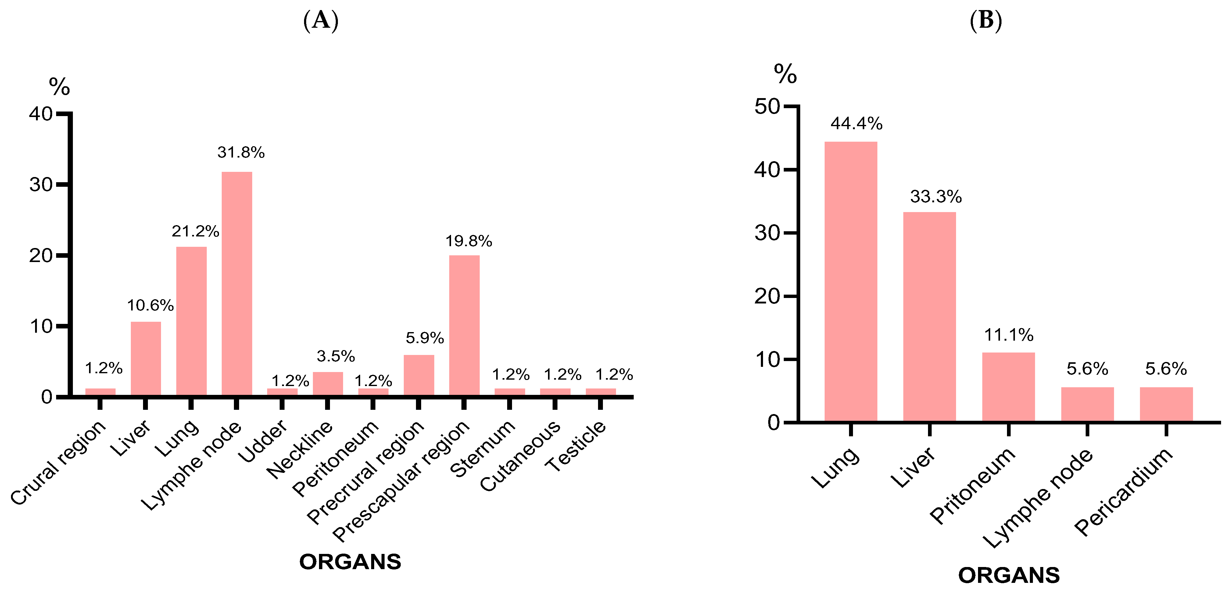
3.1. Abscess Characteristics

During the study period, 677 cattle and 978 sheep (N = 1655) were slaughtered and routinely inspected. The population of both cattle and sheep was predominantly male (561 vs. 116 in cattle and 975 vs. 3 in sheep, for males and females, respectively). A total of 123 abscess lesions were recorded (n = 23, 1.39%) and (n = 100, 6.04%) in cattle and sheep, respectively. A total of 123 abscess lesions were recorded from the 1655 slaughtered animals, giving an overall prevalence of 7.43%. This prevalence was significantly lower in cattle (n = 23, 1.39%) than in sheep (n = 100, 6.04%) (p < 0.001).
In sheep, 97 (97%) and 3 (3%) of the abscesses were recorded from males and females, respectively, while, in cattle, abscesses recorded from males and females were 19 (82.61%) and 4 (17.39%), respectively. The abscesses were mostly recorded from young animals ((N = 115, 93.5%); sheep (n = 97, 78.87%), and cattle (n = 18, 14.63%)) (Table 1).
Abscesses in sheep were more frequently located in lymph nodes (n = 27, 31.8%), prescapular region (n = 17, 19.8%), and lung (n = 28, 21.2%) (Figure 1A). In cattle, abscesses were frequently located in the lungs (n = 8, 44.4%) and liver (n = 6, 33.3%) (Figure 1B).
Finally, the characteristics of abscesses vary according to the bacterial species involved, as illustrated in Table 2.

3.2. Bacterial Isolates

Of the 123 abscesses analyzed, 103 gave a positive culture (18 in cattle and 85 in sheep), of which 114 bacterial isolates were identified (91 from sheep and 23 from cattle) and further characterized. Bacterial identification revealed Enterobacterales (Escherichia coli, Klebsiella pneumoniae, Klebsiella oxytoca, Morganella morganii, Citrobacter brackii, Citrobacter freundii, Enterobacter spp., Serratia marcescens, Proteus vulgaris, Lelliottia spp.) (n = 84, 73.6%), Staphylococcus spp. (n = 15, 13.1%), Aeromonas spp. (n = 13, 11.4%), and Bacillus spp. (n = 2, 1.9%) (Table 3). Escherichia coli represented 61.3% of the isolated bacteria, and 83% of the Enterobacterales.
In the 11 abscesses in which 2 species had been recovered (6 in sheep and 5 in cattle), the different combinations were as follows: E. coli/Aeromonas spp. (n = 5), E. coli/S. aureus (n = 5), and E. coli/Morganella morganii (n = 1).

3.3. Antibiotic Susceptibility Testing

Different rates of resistance were observed for the Enterobacterales isolates, with resistance to amoxicillin, ticarcillin, ampicillin, amoxicillin-clavulanic acid, and tetracycline being the highest rates recorded (Figure 2).
Three isolates with reduced susceptibility to carbapenems were identified. Two K. pneumoniae isolates (O103B2 and O103B1) had MICs of 2 µg/mL, 1 µg/mL, and 0.5 µg/mL for ertapenem, imipenem, and meropenem, respectively, and one E. coli isolate (O103A10) displayed MICs of 0.5 µg/mL for both ertapenem and imipenem, and 0.12 µg/mL for meropenem (Table 3). These isolates presented additionally high MICs for temocillin 512 µg/mL and 256 µg/mL, for E. coli O103A10 and K. pneumoniae O103B2 and O103B1, respectively (Table 4). These isolates gave positive results using the Carba NP, suggesting the likely presence of a carbapenem-hydrolyzing enzyme. The NG-Test CARBA 5 confirmed the presence of an OXA-48-like carbapenemase.
E. coli O103A9 displayed resistance to expanded-spectrum cephalosporins (ESCs) and exhibited a synergy image in the DD-test, indicating the presence of extended-spectrum beta-lactamases (ESBLs).
The Aeromonas spp. were susceptible to all the antibiotics tested except for two that were resistant to cotrimoxazole.
Two coagulase-negative isolates (S. epidermidis and S. pasteuri) were considered methicillin-resistant as they were resistant to oxacillin and cefoxitin. High resistance rates to tetracycline were observed (Figure 2). The two isolates of Bacillus spp. were pan-susceptible to all the antibiotics tested.

3.4. Resistome, MLST, Plasmidome, and O-Serogroups

Twenty strains were further investigated by WGS. Gram-positive isolates were chosen based on their resistance profile such as mecA-positive Staphylococci or their likelihood to carry virulence factors such as S. aureus isolates. Enterobacterales were chosen based on their resistance profile, such as expanded spectrum cephalosporin- or carbapenem-resistance on randomly chosen multisusceptible Escherichia coli isolates to obtain an overview of their STs or virulence factors likely involved in abscess formation.
The two K. pneumoniae isolates O103B1 and O103B2 harbored, in addition to blaOXA-48, several β-lactam resistance genes, including the ESBL blaCTX-M-15, blaOXA-1, blaSHV-187, and blaTEM-1B genes. Additionally, these isolates carried several other resistance genes to different antibiotic classes (Table 5).
The E. coli isolate O103A10 carries the carbapenemase blaOXA-48 gene, as well as tet(B) and mph(B) genes that are responsible for tetracycline and macrolide resistance, respectively (Table 5).
Furthermore, the remaining E. coli strain O103A9 harbored the ESBL blaCTX-M-15 and blaTEM-1B genes, along with aminoglycoside resistance genes aph(6)-Id and aph(3)-Ib, qnrS1 for quinolones, and sul2 for sulphonamides (Table 5). In silico MLST typing assigned the two E. coli O103A9 isolate to ST1706 and ST10 for O103A10, while the two K. pneumoniae isolates belonged to the same ST985. Several replicon-types were identified in the four MDR Enterobacterales (Table 5). The CTX-M-15-producing E. coli O103A9 harbored an IncY plasmid, while E. coli O103A10 carried several plasmids (Table 5), including Col156, IncFIA, IncFIB, and IncFII, but no IncL plasmid known to carry blaOXA-48 gene. A careful analysis of the contig carrying the blaOXA-48 gene suggested a chromosomal location, as the E. coli chromosomal genes are present on both sides of blaOXA-48 gene (Figure 3). In addition, electroporation experiments with plasmids extracted from E. coli O103A10 failed to transfer any plasmid carrying blaOXA-48 gene to E. coli Top10.
The two carbapenemase-producing K. pneumoniae isolates O103B1 and O103B2 also carried several plasmids, including an IncL plasmid, likely carrying blaOXA-48 gene (Table 5). Mapping the reads against the prototypical OXA-48 plasmid revealed 100% sequence coverage, suggesting the presence of the entire plasmid in these isolates [17,18].
The resistome of seven randomly chosen E. coli isolates revealed few resistance genes. Five isolates produced tetA and tetB genes, together with aminoglycoside resistance genes (aadA1, aadA2b, aph(3′)-Ia, aph(3″)-Ib, aph(6)-Id) in two of the seven E. coli isolates. Furthermore, two E. coli isolates expressed a sulfonamide resistance gene (sul2 and/or sul3). These E. coli isolates belonged to seven different STs: ST88, ST101, ST224, ST155, ST223, ST206. Only three plasmid replicon types were detected in the investigated E. coli isolates; these are IncFII, IncFIA, and IncFIB. Additionally, O-serogroups that could potentially pose public health concerns were also identified, including O8, O23, O37, O42, O116, O123, O144, and O153 (Table 5).
The S. epidermidis strain harbored several antibiotic resistance genes, including mecA (which encodes PLP2A conferring resistance to methicillin), blaZ (which encodes a narrow spectrum penicillinase), ant(6) and aph(3′) (which encode resistance to aminoglycosides), and fosB, fusB, and tet(K) (which encode resistance to fosfomycin, fusidic acid, and tetracycline, respectively). The S. pasteuri strain harbored only mecA and blaZ genes. Four isolates of S. aureus (O104F5, O104F7, O104F9, and O104F10) carried the blaZ gene, while the tet(K) and erm(T) genes were detected only in isolates O104F4 and O104F10, respectively (Table 6).
S. epidermidis strain belonged to ST61, and the six S. aureus isolates belonged to five different STs: ST700 for two isolates, ST522 for two isolates and ST398 from sheep, and ST97 and another novel allele from cattle.

3.5. Virulome

As these bacterial isolates were responsible for purulent infections, the search for virulence genes was of the utmost importance (Table 6 and Table 7). The ESBL-producing E. coli strain O103A9 was found to harbor only three known virulence genes: ompT, terC, and yehD, which encode an outer membrane protease, a tellurite resistance protein C, and a fimbrial protein, respectively. The E. coli strain-producing OXA-48 carried three genes as well, including terC, csgA, and traT, which encode a tellurite resistance protein C, a curli fimbriae subunit A, and a serum-resistance-associated protein, respectively.
Among the two K. pneumoniae isolates, O103B2 harbored 82 virulence genes, while O103B1 had 79 genes. Both strains shared common virulence genes such as type 1 fimbriae (fimABCDEFGHIK) and type 3 fimbriae operon (mrkABCDFHIJ), enterobactin gene clusters (entABCDEFS and fepABCDG), Type IV pili (pilW), the T6SS-II (impAFGHJ) operon, the stbABCD operon, and the siderophore iroAN cluster. Additionally, both strains were found to contain the integrative and conjugative element (ICEKp) containing the yersiniabactin gene cluster (ybtAEPQSTUX), irp1, irp2, and fyuA genes. In addition to these genes, other genes were found (Table 7).
The virulome of the seven susceptible E. coli isolates was also investigated, and the most common factors found were terC, fimH, nlpI, lpfA, fyuA, hlyF, iutA, and cvaC (Table 7).
The WGS analysis of S. aureus isolates revealed that all but one (O104F6) carried the hemolysin-encoding genes hlgA, hlgB, and hlgC. Leucocidin genes lukD and lukE were identified in five strains. The serine protease-encoding genes splA and splB were detected in all six strains, while splE was detected in only four strains. Enterotoxin genes sec and sel were detected in strains O104F7 and O104F9, and the immune evasion gene sak was detected in three isolates (O104F4, O104F6, and O104F8), while the scn gene was detected in two isolates (O104F8 and O104F10) (Table 6).

4. Discussion

This study revealed a concerning rate of multidrug resistance in bacteria isolated from abscesses in one slaughterhouse in Algeria. In low- to middle-income countries, the intensification of farming is on the rise, primarily driven by a scarcity of available land and the continuous growth of human populations. This intensified farming demands greater use of antibiotics to combat infectious diseases, subsequently fostering the transfer of antibiotic resistance genes (ARGs) between microbes, resulting in multidrug-resistant bacteria in livestock but also in domestic animals and quick spread of resistant micro-organisms, which have serious consequences on animal health, productivity, and food production that pose both economic and human health problems [19,20]. Livestock animals are at risk of infectious diseases, especially with pyogenic organisms that cause abscesses at various sites of the body, and consequently have an impact on productivity, fertility, and in general to livestock health [2,21,22]. Infected carcasses at slaughterhouses can be partially or completely discarded [23].
In this study, we reported 123 abscesses, including 100 from sheep and 23 from cattle. Abscesses occur commonly in sheep as ovines are more susceptible to developing them than other ruminants. It cannot be determined solely based on clinical examination and generally relies on postmortem inspection of carcasses in slaughterhouses [24,25]. The analysis of bacterial isolates from pus showed the predominance of Enterobacterales isolates, particularly E. coli, followed by Staphylococcus spp. Enterobacterales, and E. coli especially have been found to be major players in the formation of abscesses in animals [5,26,27], although several studies have isolated S. aureus from abscesses as the quintessential suppurative pathogen in large proportions [13,14,28,29]. These bacterial species isolated are zoonotic pathogens, which places public health at risk.
The presence of ESBLs, and in particular CTX-M-15, in animals has previously been linked to the human sector before it was also detected in animals and the environment [30]. Additionally, ceftiofur, a third-generation cephalosporin, is the main cephalosporin used in veterinary medicine due to its effectiveness in treating bacterial infections in food-producing animals; for this reason, these antibiotics could provide selection pressure that favors co-selection of plasmids carrying mobile genes (transposon, integron, cassette gene) that result in carbapenem-resistant (CR) strains [31,32]. Here, we identified a CTX-M-15 producing E. coli ST1706, an ST that has previously been described in Japan from different pig farms [33].
The presence of OXA-48-carbapenemase producers is very worrying, as carbapenems remain the last-resort therapy for treating human infections caused by MDR Gram-negative and Gram-positive bacteria [31]. However, the clinical use of these antibiotics is presently at risk due to the global proliferation of β-lactamases (BLs) with the ability to degrade them, and the increase in the worldwide emergence of carbapenem-resistant organisms (CROs), which constitute a critical growing public health threat [34]. In livestock or veterinary fields, carbapenems are not licensed and have no legal indication, so their use is prohibited [32,35,36]. Nevertheless, many studies conducted in Algeria have reported CROs in livestock, companion animals, and birds [37,38,39].
In our study, we isolated, for the first time, OXA-48 carbapenemase-producing Enterobacterales from abscesses of farm animals. OXA-48 is the most common carbapenemase in Enterobacterales and one of the most frequently isolated around the Mediterranean rim [40]. It is most frequently detected in K. pneumoniae and E. coli but can also occur in other Enterobacterales species [40]. Both K. pneumoniae isolates belong to ST985, an ST type that has been isolated from various sources and geographic locations. MDR K. pneumoniae ST985 isolates carrying up to 16 different resistance genes, including blaCTX-M-55 gene, were isolated from rectal swab samples of dairy cows from Quetta in Pakistan [41]. In Austria, blaCTX-M-15-producing K. pneumoniae ST985 were isolated from river water samples that were identical to clinical isolates from Austrian hospitals [42]. Finally, blaCTX-M-15-producing K. pneumoniae ST985 have been involved in an outbreak in Israel in a neonatal intensive care unit [43]. The simultaneous presence of blaOXA-48 and blaCTX-M-15 genes in K. pneumoniae strains ST985 is of particular concern, as this ST seem to be responsible for human and animal infections, and as the combination of these two β-lactamases lead to the resistance to almost all β-lactams. Extended-spectrum β-lactamase (ESBL) and carbapenemase genes are often associated with an MDR phenotype [44], as illustrated in our study by the presence of different resistance to different classes of antibiotics.
The BlaOXA-48 gene is usually located on transferable Inc L plasmids [45]; however, there have also been reports of chromosomally located blaOXA-48 genes in some STs, such as ST38 CPE [46,47,48]. Here, we report a chromosomal localization of the blaOXA-48 gene in E. coli ST 10. Chromosomal insertion of resistance genes is believed to favor the stability of resistance genes in the absence of selective pressure [49,50].
Methicillin-resistant staphylococci are among the emerging pathogens that now constitute a threat to human and animal health. Due to its rapid development of antibiotic resistance in clinical settings, methicillin-resistant S. aureus (MRSA) is regarded as one of major life-threatening pathogens. The recent isolation of MRSA strains in several animals is thought to be one of the main factors in the spread of infection and disease in both humans and animals [51,52]. In our study, we identified a methicillin-susceptible S. aureus ST398, an ST spreading worldwide in animals and humans and often referred to as livestock-associated MRSA (LS-MRSA) [53,54].
This resistance, as our study shows, concerned coagulase-negative Staphylococci (CoNS: S. epidermidis and S. pasteuri) isolated from abscesses and harbored the mecA gene, indicating methicillin resistance. Methicillin-resistant coagulase-negative staphylococci have been less studied, but their importance as pathogens is increasing. For a long time, S. aureus was thought to be the predominant pathogenic Staphylococci species. However, recent investigations have shown the increasing role of CoNS in causing antibiotic-resistant infections [55,56]. In a previous study conducted in Algeria, which focused on unpasteurized cow’s milk, three isolated CoNS were resistant to methicillin, and all were mecA-positive [57]. A few studies carried out on CoNS in livestock have revealed that food-producing animals constitute a large reservoir of multiresistant CoNS [58].
Virulence factors of Enterobacterales or Staphylocci are based on their ability to adhere to host cells, produce toxins, and resist host immune defenses [59]. The identification of specific virulence genes is crucial for understanding the pathogenic potential and for the development of preventive strategies, effective vaccines, and novel therapeutics [60]. For Enterobacterales, important virulence factors, including adhesins, fimbriae, intimin, capsules, iron metabolism, siderophores, heme/hemoglobin transport proteins, and cell invasion, were identified. Furthermore, a study conducted in Western Algeria in 2017 to determine the prevalence of carbapenemase-producing Enterobacteriaceae (CPE) in chicken meat revealed that carbapenemase-producing K. pneumoniae isolates harbored several virulence factors, such as fimH type 1 fimbriae virulence gene, ureA, involved in the hydrolysis of urea to ammonia, mrkD, encoding a type 3 fimbriae that promotes biofilm development, uge, that codes for a UDP galacturonate 4-epimerase, and wabG, encoding the biosynthesis of the core lipopolysaccharide [61]. The relationship between antimicrobial resistance and virulence factors in bacteria is complex. There are common characteristics shared between virulence and resistance, such as the involvement of efflux pumps, porins, and cell wall alterations. Additionally, some studies have found a significant association between certain virulence genes and antimicrobial resistance, suggesting that acquiring resistance to some antibiotics may impact the expression of virulence factors [62,63,64]. In contrast to other studies, KPC-producing Enterobacterales, such as K. pneumoniae, typically exhibit lower levels of virulence compared to non-carbapenemase-producing strains. Overall, the presence of carbapenemase genes does not necessarily imply a reduction in virulence, although there might be trade-offs between virulence and resistance capabilities in certain strains [65].

5. Conclusions

The presence of blaESBL, blaOXA-48, and mecA genes in animal abscesses highlights the importance of monitoring the use of antimicrobial in animals. Preventing the spread of multidrug-resistant bacteria in livestock animals should, therefore, be a priority for public health, which can be achieved through the reduction in and proper use of antimicrobial agents in animal husbandry and in humans, and also acting at the farm level, improving hygiene and biosecurity measures, based primarily on the elimination of risk factors and vaccination of small ruminants.

Author Contributions

Conceptualization, C.Y., C.B., and T.N.; methodology, C.Y. and T.N.; validation, C.Y., S.O., and D.G.; formal analysis, C.Y.; investigation, C.Y., A.P., and D.D.; resources, C.Y. and T.N.; data curation, T.N.; writing—original draft preparation, C.Y. and T.N.; writing—review and editing, all; supervision, S.O. and T.N.; funding acquisition, T.N. All authors have read and agreed to the published version of the manuscript.

Funding

This work was supported by the Assistance Publique—Hôpitaux de Paris (AP-HP), the University Paris-Saclay, the Institut National de la Santé et de la Recherche Médicale (INSERM), the Seq2Diag projects supported by grants from the French National Research Agency [ANR-20-PAMR-0010], and by the short-term Ph.D. fellowship of the Algerian ministry of research for CY’s stay in France.

Data Availability Statement

Raw data are available upon request, and WGS sequences have been deposited at NCBI.

Acknowledgments

We are grateful to the slaughterhouse workers for their commitment and continuous support all along this project. Belabed Kadour médecin Chef du service de Microbiologie CHU de Constantine.

Conflicts of Interest

The authors declare no conflicts of interest.

References

  1. Misk, T.N.; El-Sherry, T.; Misk, N.A. Retrospective study on body surface abscesses in farm animals. Assiut Vet. Med. J. 2020, 66, 47–61. [Google Scholar] [CrossRef]
  2. Mariappan, S.; Mydeen, S.N.M.; Samuel, S.J.R.; Jegathambigai, J.; Rajan, K.E.; Sudhakar, S. Bacillus sp. Causing Abscessation in Sheep and Goat Population. Curr. Sci. 2012, 103, 921–925. [Google Scholar]
  3. Awad, A.; Awadin, W.F. Pathological and bacteriological studies on some recorded internal abscesses in slaughtered cattle. Assiut Vet. Med. J. 2016, 63, 52–61. [Google Scholar]
  4. Nagati, S.F.; El Shafii, S.S.A.; Abd El Mawgoud, S.R.A. Effectiveness of Disinfectants on Environmental Multidrug Resistance Contaminants Causing Skin Abscess in Farm Animals. Am. J. Anim. Vet. Sci. 2021, 16, 128–138. [Google Scholar] [CrossRef]
  5. Tavassoli, M.; Imani, A.; Yousefnia, M.; Tukmechi, A.; Tajik, H. Bacteria associated with subcutaneous abscesses of cattle caused by Hypoderma spp. larvae in north of Iran. Vet. Res. Forum 2010, 1, 123–127. [Google Scholar]
  6. AL-Tuffyli, Y.K.; Shekhan, M.I. Clinical and Bacteriological Study of Subcutaneous Abscesses Caused by Gram Positive Bacteria in Cow and Sheep in Al-Qadissiyia Province. Al-Qadisiyah J. Vet. Med. Sci. 2012, 11, 17–24. [Google Scholar]
  7. Ninios, T.; Lundén, J.; Korkeala, H.; Fredriksson-Ahomaa, M. Meat Inspection and Control in the Slaughter House, 1st ed.; John Wiley & Sons: Chichester, UK, 2014; pp. 154–157. [Google Scholar]
  8. Aljameel, M.A.; Halima, M.O.; ElTigani-Asil, E.A.; El-Eragi, A.M. Bacteriological and histopathological studies on pulmonary abscesses in camels (Camelus dromedarius) slaughtered at Nyala Slaughterhouse, South Darfur State, Sudan. J. Vet. Med. Anim. Prod. 2013, 4, 26–38. [Google Scholar]
  9. Herenda, D.; Chambers, P.G.; Ettriqui, A.; Seneviratna, P.; Da Silva, T.J.P. Manuel on Meat Inspection for Developing Countries; FAO: Rome, Italy, 1994; pp. 58–60. [Google Scholar]
  10. Buba, D.M.; Gurumyen, G.Y.; Oragwa, O.A.; Oziegbe, S.D.; Patrobas, M.N.; Dunka, H.I. Retrospective Analysis of Cutaneous Abscess in Cattle, Goats and Pigs Slaughtered at the Jos Abattoir, Nigeria. Sokoto J. Vet. Sci. 2020, 17, 44. [Google Scholar] [CrossRef]
  11. Al-nakeeb, N.K.; HameedAl-Fetly, D.R. Anaerobic Bacterial Isolation with Histopatholgical Exam of Liver Abscesses in Cattle, Sheep, and Camels in Al-Qadisiyah Province. Al-Qadisiyah J. Vet. Med. Sci. 2016, 15, 40–46. [Google Scholar]
  12. Wagner, C.; Sauermann, R.; Joukhadar, C. Principles of Antibiotic Penetration into Abscess Fluid. Pharmacology 2006, 78, 1–10. [Google Scholar] [CrossRef]
  13. Alloui, M.N.; Kaba, J.; Alloui, N. Prevalence and risk factors of caseous lymphadenitis in sheep and goats of Batna area (Algeria). Res. Opin. Anim. Vet. Sci. 2011, 1, 162–164. [Google Scholar]
  14. Ferreira, A.; Monteiro, J.M.; Vieira-Pinto, M. The Importance of Subcutaneous Abscess Infection by Pasteurella spp. and Staphylococcus aureus as a Cause of Meat Condemnation in Slaughtered Commercial Rabbits. World Rabbit. Sci. 2014, 22, 311–317. [Google Scholar] [CrossRef]
  15. Saadi, C. Les Motifs de Saisie des Viandes Rouges et Abats les Plus Fréquents au Niveau de L’abattoir de Frère Ben Aissa Biskra. Master’s Thesis, University Mohamed Khider Biskra, Biskra, Algeria, 2018. [Google Scholar]
  16. Perez-Palacios, P.; Girlich, D.; Soraa, N.; Lamrani, A.; Maoulainine, F.M.R.; Bennaoui, F.; Amri, H.; El Idrissi, N.S.S.; Bouskraoui, M.; Birer, A.; et al. Multidrug-Resistant Enterobacterales Responsible for Septicaemia in a Neonatal Intensive Care Unit in Morocco. J. Glob. Antimicrob. Resist. 2023, 33, 208–217. [Google Scholar] [CrossRef] [PubMed]
  17. Aubert, D.; Naas, T.; Héritier, C.; Poirel, L.; Nordmann, P. Functional Characterization of IS1999, an IS4 Family Element Involved in Mobilization and Expression of β-Lactam Resistance Genes. J. Bacteriol. 2006, 188, 6506–6514. [Google Scholar] [CrossRef] [PubMed]
  18. Bonnin, R.A.; Nordmann, P.; Carattoli, A.; Poirel, L. Comparative Genomics of IncL/M-Type Plasmids: Evolution by Acquisition of Resistance Genes and Insertion Sequences. Antimicrob. Agents Chemother. 2013, 57, 674–676. [Google Scholar] [CrossRef]
  19. Ferriol-González, C.; Domingo-Calap, P. Phage Therapy in Livestock and Companion Animals. Antibiotics 2021, 10, 559. [Google Scholar] [CrossRef] [PubMed]
  20. Wang, H.; Qi, J.F.; Qin, R.; Ding, K.; Graham, D.W.; Zhu, Y.G. Intensified Livestock Farming Increases Antibiotic Resistance Genotypes and Phenotypes in Animal Feces. Commun. Earth Environ. 2023, 4, 123. [Google Scholar] [CrossRef]
  21. Hatem, M.E.; Arab, R.H.; Ata, S. Nagwa Bacterial Abscessationin Sheep and Goat in Giza Governorate with Full Antibiogram Screening. Glob. Vet. 2013, 10, 372–381. [Google Scholar]
  22. Tamai, I.A.; Mohammadzadeh, A.; Mahmoodi, P.; Pakbin, B.; Salehi, T.Z. Antimicrobial Susceptibility, Virulence Genes and Genomic Characterization of Trueperella Pyogenes Isolated from Abscesses in Dairy Cattle. Res. Vet. Sci. 2023, 154, 29–36. [Google Scholar] [CrossRef]
  23. Rodrigues, R.M.; Martins, T.O.; Procópio, D.P. Economic Loss from the Main Causes of Whole Bovine Carcass Condemnation in Slaughterhouses Supervised by the Federal Inspection Service in São Paulo State from 2010 to 2019. Acta Sci. 2022, 44, e55220. [Google Scholar] [CrossRef]
  24. Al-Harbi, K.B. Prevalence and Etiology of Abscess Disease of Sheep and Goats at Qassim Region, Saudi Arabia. Vet. World 2011, 4, 495–499. [Google Scholar] [CrossRef]
  25. Omar Ali, A.S.; Mohammed, Z.M.A. Gross and Histopathological Findings on Lung Abscesses in Slaughtered Sheep in Libya. Eur. J. Vet. Med. 2023, 3, 1–4. [Google Scholar] [CrossRef]
  26. Mcvey, D.S.; Kennedy, M.; Chengappa, M.M. Veterinary Microbiology, 3rd ed.; Wiley-Blackwell: Ames, IA, USA, 2013; pp. 184–194. [Google Scholar]
  27. Madhav Mugale, N.; Balachandran, C.; Dillibabu, V.; Kirubharan, J.; Dhinakar Raj, G.; Sridhar, R.; Selvasubramaniam, S. Hepatic Abscess in Sheep and Goat Caused by O26 Escherichia Coli Serotype: An Emerging Pathogen. Indian Vet. J. 2015, 92, 76–79. [Google Scholar]
  28. Al-Anbagi, N.A. Isolaton and Identification some bacterial causes of lung abscesses sheep by chromogenic media. Basrah J. Vet. Res. 2016, 15, 360–370. [Google Scholar] [CrossRef]
  29. Didkowska, A.; Żmuda, P.; Kwiecień, E.; Rzewuska, M.; Klich, D.; Wędzina, M.K.; Witkowski, L.; Żychska, M.; Kaczmarkowska, A.; Orłowska, B.; et al. Microbiological Assessment of Sheep Lymph Nodes with Lymphadenitis Found during Post—Mortem Examination of Slaughtered Sheep: Implications for Veterinary—Sanitary Meat Control. Acta Vet. Scand. 2020, 62, 48. [Google Scholar] [CrossRef] [PubMed]
  30. Haenni, M.; Lupo, A.; Madec, J.-Y. Résistance Aux Carbapénèmes Chez Les Animaux En l’absence d’usage. Bull. Acad. Vet. Fr. 2018, 171, 4–8. [Google Scholar] [CrossRef]
  31. Das, S. The Crisis of Carbapenemase-Mediated Carbapenem Resistance across the Human–Animal–Environmental Interface in India. Infect. Dis. Now 2023, 53, 104628. [Google Scholar] [CrossRef]
  32. Ramírez-Castillo, F.Y.; Guerrero-Barrera, A.L.; Avelar-González, F.J. An Overview of Carbapenem-Resistant Organisms from Food-Producing Animals, Seafood, Aquaculture, Companion Animals, and Wildlife. Front. Vet. Sci. 2023, 10, 1158588. [Google Scholar] [CrossRef]
  33. Norizuki, C.; Kawamura, K.; Wachino, J.I.; Suzuki, M.; Nagano, N.; Kondo, T.; Arakawa, Y. Detection of Escherichia coli Producing CTX-M-1-Group Extended-Spectrum β-Lactamases from Pigs in Aichi Prefecture, Japan, between 2015 and 2016. Jpn. J. Infect. Dis. 2018, 71, 33–38. [Google Scholar] [CrossRef]
  34. Naas, T.; Dabos, L.; Bonnin, R.A. β-Lactamase Genes without Limits. Microorganisms 2023, 11, 1200. [Google Scholar] [CrossRef]
  35. Madec, J.Y.; Haenni, M.; Nordmann, P.; Poirel, L. Extended-Spectrum β-Lactamase/AmpC- and Carbapenemase-Producing Enterobacteriaceae in Animals: A Threat for Humans? Clin. Microbiol. Infect. 2017, 23, 826–833. [Google Scholar] [CrossRef] [PubMed]
  36. Uyanik, T.; Çadirci, Ö.; Gücükoğlu, A.; Can, C. Investigation of Major Carbapenemase Genes in ESBL-Producing Escherichia Coli and Klebsiella Pneumoniae Strains Isolated from Raw Milk in Black Sea Region of Turkey. Int. Dairy J. 2022, 128, 105315. [Google Scholar] [CrossRef]
  37. Yaici, L.; Haenni, M.; Saras, E.; Boudehouche, W.; Touati, A.; Madec, J.Y. Bla NDM-5-Carrying IncX3 Plasmid in Escherichia Coli ST1284 Isolated from Raw Milk Collected in a Dairy Farm in Algeria. J. Antimicrob. Chemother. 2016, 71, 2671–2672. [Google Scholar] [CrossRef] [PubMed]
  38. Loucif, L.; Chelaghma, W.; Bendjama, E.; Cherak, Z.; Khellaf, M.; Khemri, A.; Rolain, J.M. Detection of blaOXA-48 and mcr-1 Genes in Escherichia Coli Isolates from Pigeon (Columba livia) in Algeria. Microorganisms 2022, 10, 975. [Google Scholar] [CrossRef] [PubMed]
  39. Loucif, L.; Chelaghma, W.; Cherak, Z.; Bendjama, E.; Beroual, F.; Rolain, J.M. Detection of NDM-5 and MCR-1 Antibiotic Resistance Encoding Genes in Enterobacterales in Long-Distance Migratory Bird Species Ciconia ciconia, Algeria. Sci. Total Environ. 2022, 814, 152861. [Google Scholar] [CrossRef] [PubMed]
  40. Hamprecht, A.; Sommer, J.; Willmann, M.; Brender, C.; Stelzer, Y.; Krause, F.F.; Tsvetkov, T.; Wild, F.; Riedel-Christ, S.; Kutschenreuter, J.; et al. Pathogenicity of Clinical OXA-48 Isolates and Impact of the OXA-48 IncL Plasmid on Virulence and Bacterial Fitness. Front. Microbiol. 2019, 10, 2509. [Google Scholar] [CrossRef]
  41. Habib, S.; Gibbon, M.J.; Couto, N.; Kakar, K.; Habib, S.; Samad, A.; Munir, A.; Fatima, F.; Mohsin, M.; Feil, E.J. The Diversity, Resistance Profiles and Plasmid Content of Klebsiella spp. Recovered from Dairy Farms Located around Three Cities in Pakistan. Antibiotics 2023, 12, 539. [Google Scholar] [CrossRef]
  42. Frenk, S.; Rakovitsky, N.; Temkin, E.; Schechner, V.; Cohen, R.; Kloyzner, B.S.; Schwaber, M.J.; Solter, E.; Cohen, S.; Stepansky, S.; et al. Investigation of Outbreaks of Extended-Spectrum Beta-Lactamase-Producing Klebsiella Pneumoniae in Three Neonatal Intensive Care Units Using Whole Genome Sequencing. Antibiotics 2020, 9, 705. [Google Scholar] [CrossRef]
  43. Lepuschitz, S.; Schill, S.; Stoeger, A.; Pekard-Amenitsch, S.; Huhulescu, S.; Inreiter, N.; Hartl, R.; Kerschner, H.; Sorschag, S.; Springer, B.; et al. Whole genome sequencing reveals resemblance between ESBL-producing and carbapenem resistant Klebsiella pneumoniae isolates from Austrian rivers and clinical isolates from hospitals. Sci. Total Environ. 2019, 662, 227–235. [Google Scholar] [CrossRef]
  44. Nordmann, P.; Poirel, L. Strategies for Identification of Carbapenemase-Producing Enterobacteriaceae. J. Antimicrob. Chemother. 2013, 68, 487–489. [Google Scholar] [CrossRef]
  45. Poirel, L.; Bonnin, R.A.; Nordmann, P. Genetic Features of the Widespread Plasmid Coding for the Carbapenemase OXA-48. Antimicrob. Agents Chemother. 2012, 56, 559–562. [Google Scholar] [CrossRef]
  46. Hendrickx, A.P.A.; Landman, F.; de Haan, A.; Witteveen, S.; van Santen-Verheuvel, M.G.; Schouls, L.M. blaOXA-48-like Genome Architecture among Carbapenemase-Producing Escherichia coli and Klebsiella pneumoniae in The Netherlands. Microb. Genom. 2021, 7, 000512. [Google Scholar]
  47. Lee, C.R.; Lee, J.H.; Park, K.S.; Kim, Y.B.; Jeong, B.C.; Lee, S.H. Global Dissemination of Carbapenemase-Producing Klebsiella Pneumoniae: Epidemiology, Genetic Context, Treatment Options, and Detection Methods. Front. Microbiol. 2016, 7, 895. [Google Scholar] [CrossRef]
  48. Mairi, A.; Pantel, A.; Sotto, A.; Lavigne, J.P.; Touati, A. OXA-48-like Carbapenemases Producing Enterobacteriaceae in Different Niches. Eur. J. Clin. Microbiol. Infect. Dis. 2018, 37, 587–604. [Google Scholar] [CrossRef]
  49. Bonnet, R.; Marchandin, H.; Chanal, C.; Sirot, D.; Labia, R.; De Champs, C.; Jumas-Bilak, E.; Sirot, J. Chromosome-Encoded Class D β-Lactamase OXA-23 in Proteus Mirabilis. Antimicrob. Agents Chemother. 2002, 46, 2004–2006. [Google Scholar] [CrossRef]
  50. Beyrouthy, R.; Robin, F.; Delmas, J.; Gibold, L.; Dalmasso, G.; Dabboussi, F.; Hamzé, M.; Bonnet, R. IS1R-Mediated Plasticity of IncL/M Plasmids Leads to the Insertion of BlaOXA-48 into the Escherichia Coli Chromosome. Antimicrob. Agents Chemother. 2014, 58, 3785–3790. [Google Scholar] [CrossRef] [PubMed]
  51. Venugopal, N.; Mitra, S.; Tewari, R.; Ganaie, F.; Shome, R.; Rahman, H.; Shome, B.R. Molecular Detection and Typing of Methicillin-Resistant Staphylococcus Aureus and Methicillin-Resistant Coagulase-Negative Staphylococci Isolated from Cattle, Animal Handlers, and Their Environment from Karnataka, Southern Province of India. Vet. World 2019, 12, 1760–1768. [Google Scholar] [CrossRef]
  52. Zaher, H.A.; El Baz, S.; Alothaim, A.S.; Alsalamah, S.A.; Alghonaim, M.I.; Alawam, A.S.; Eraqi, M.M. Molecular Basis of Methicillin and Vancomycin Resistance in Staphylococcus Aureus from Cattle, Sheep Carcasses and Slaughterhouse Workers. Antibiotics 2023, 12, 205. [Google Scholar] [CrossRef] [PubMed]
  53. Silva, V.; Araújo, S.; Monteiro, A.; Eira, J.; Pereira, J.E.; Maltez, L.; Igrejas, G.; Lemsaddek, T.S.; Poeta, P. Staphylococcus aureus and MRSA in Livestock: Antimicrobial Resistance and Genetic Lineages. Microorganisms 2023, 11, 124. [Google Scholar] [CrossRef]
  54. Bouiller, K.; Bertrand, X.; Hocquet, D.; Chirouze, C. Human Infection of Methicillin-Susceptible Staphylococcus aureus CC398: A Review. Microorganisms 2020, 8, 1737. [Google Scholar] [CrossRef] [PubMed]
  55. Bonvegna, M.; Grego, E.; Sona, B.; Stella, M.C.; Nebbia, P.; Mannelli, A.; Tomassone, L. Occurrence of Methicillin-Resistant Coagulase-Negative Staphylococci (Mrcons) and Methicillin-Resistant Staphylococcus aureus (MRSA) from Pigs and Farm Environment in Northwestern Italy. Antibiotics 2021, 10, 676. [Google Scholar] [CrossRef] [PubMed]
  56. Chajęcka-Wierzchowska, W.; Gajewska, J.; Zadernowska, A.; Randazzo, C.L.; Caggia, C. A Comprehensive Study on Antibiotic Resistance among Coagulase-Negative Staphylococci (CoNS) Strains Isolated from Ready-to-Eat Food Served in Bars and Restaurants. Foods 2023, 12, 514. [Google Scholar] [CrossRef] [PubMed]
  57. Chenouf, N.S.; Mama, O.M.; Messaï, C.R.; Ruiz-Ripa, L.; Fernández-Fernández, R.; Carvalho, I.; Zitouni, A.; Hakem, A.; Torres, C. Detection of Methicillin-Resistant Coagulase-Negative Staphylococci and PVL/MecA Genes in Cefoxitin-Susceptible Staphylococcus Aureus (T044/ST80) from Unpasteurized Milk Sold in Stores in Djelfa, Algeria. J. Dairy Sci. 2021, 104, 2684–2692. [Google Scholar] [CrossRef]
  58. Bhargava, K.; Zhang, Y. Multidrug-Resistant Coagulase-Negative Staphylococci in Food Animals. J. Appl. Microbiol. 2012, 113, 1027–1036. [Google Scholar] [CrossRef] [PubMed]
  59. Bujňáková, D.; Puvača, N.; Ćirković, I. Virulence factors and antibiotic resistance of enterobacterales. Microorganisms 2022, 10, 1588. [Google Scholar] [CrossRef] [PubMed]
  60. Pakbin, B.; Brück, W.M.; Rossen, J.W. Virulence factors of enteric pathogenic Escherichia coli: A review. Int. J. Mol. Sci. 2021, 22, 9922. [Google Scholar] [CrossRef] [PubMed]
  61. Chaalal, N.; Touati, A.; Bakour, S.; Aissa, M.A.; Sotto, A.; Lavigne, J.-P.; Pantel, A. Spread of OXA-48 and NDM-1-Producing Klebsiella pneumoniae ST48 and ST101 in Chicken Meat in Western Algeria. Microb. Drug Resist. 2021, 27, 492–500. [Google Scholar] [CrossRef]
  62. Schroeder, M.; Brooks, B.D.; Brooks, A.E. The complex relationship between virulence and antibiotic resistance. Genes 2017, 8, 39. [Google Scholar] [CrossRef]
  63. Beceiro, A.; Tomás, M.; Bou, G. Antimicrobial resistance and virulence: A successful or deleterious association in the bacterial world? Clin. Microbiol. Rev. 2013, 26, 185–230. [Google Scholar] [CrossRef]
  64. Miranda-Estrada, L.I.; Ruíz-Rosas, M.; Molina-López, J.; Parra-Rojas, I.; González-Villalobos, E.; Castro-Alarcón, N. Relationship between virulence factors, resistance to antibiotics and phylogenetic groups of uropathogenic Escherichia coli in two locations in Mexico. Enfermedades Infecc. Microbiol. Clin. (Engl. Ed.) 2017, 35, 426–433. [Google Scholar] [CrossRef]
  65. Mendes, G.; Santos, M.L.; Ramalho, J.F.; Duarte, A.; Caneiras, C. Virulence factors in carbapenem-resistant hypervirulent Klebsiella pneumoniae. Front. Microbiol. 2023, 14, 1325077. [Google Scholar] [CrossRef]
Figure 1. Abscess seats location in sheep (A) and cattle (B).
Microorganisms 12 00524 g001
Figure 2. Susceptibility results of bacteria isolated from the abscesses: (A) Enterobacterales; (B) Staphylococci. Results were interpreted according to Eucast breakpoints 2022. (S: sensitive, I: intermediate, R: resistance). Amikacin (AN), amoxicillin (AMX), amoxicillin-clavulanic acid (AMC), ampicillin (AMP), aztreonam (ATM), cefazolin (CZ), cefotaxime (CTX), cefoxitin (FOX), ceftazidime (CAZ), chloramphenicol (C), ciprofloxacin (CIP), clindamycin (CM), ertapenem (ETP), erythromycin (ERY), fosfomycin (FOS200), fusidic Acid (FA), gentamicin (GM), imipenem (IPM), kanamycin (K), Levofloxacin (LEV), Linezolid (LNZ), nalidixic acid (NA), Nitrofurantoin (F), ofloxacin (OFX), Oxacillin (OX), penicillin (P), piperacillin (PIP), Quinupristin/dalfopristin (QD), Rifampin (RA), Temocillin (TEM), tetracycline (TE), ticarcillin (TIC), Tigecycline (TGC), Tobramycin (TOB), and trimethoprim/sulfamethoxazole (SXT). Disk loads are indicated in Section 2.
Microorganisms 12 00524 g002
Figure 3. Genetic environment of blaOXA-48 gene located on the chromosome in E. coli O103A10. BlaOXA-48 gene is indicated in red.
Microorganisms 12 00524 g003
Table 1. Frequency of abscesses in relation to age and sex in sheep and cattle.
SheepCattle
Age 12 Months > 12   M o n t h s 2 Years 2 5 Years > 5   Y e a r s
Male 97/18/1
Female/3/31
Table 2. Clinical presentations of abscesses according to the bacterial species.
Pus Characteristics Staphylococcus aureusStaphylococcus
Coagulase negative
Aeromonas spp.Bacillus spp.Enterobacterales
Bacteria
Consistency Grumbling viscous
Homogeneous viscous
Homogeneous viscous
Homogeneous fluid
Homogeneous viscous
Thick homogeneous
Homogeneous viscous
Thick homogeneous
Grumbling fluid
Grumbling viscous
Homogeneous fluid
Homogeneous viscous
Thick grumbling
Thick homogeneous
Viscous grumbling hemorrhagic
ColorWhite
Light yellow
Light yellow
Yellow
white
Green
Green
White
Yellow
Green
Yellow
Green
White
Yellow
OdorFadeFadeFadeFadeNauseating
Fade
Table 3. Bacterial species isolated from abscesses in cattle and sheep.
Bacterial SpeciesAnimal SpeciesTotalPercentage (%)
SheepCattle
Escherichia coli56147061.3%
Staphylococcus aureus5276.1%
Aeromonas veronii3365.2%
Aeromonas hydrophila1343.5%
Klebsiella pneumoniae3 32.6%
Morganella morganii2132.6%
Staphylococcus epidermidis3 32.6%
Klebsiella oxytoca2 21.7%
Citrobacter brackii1 10.9%
Citrobacter freundii1 10.9%
Enterobacter spp.1 10.9%
Serratia marcescens1 10.9%
Proteus vulgaris1 10.9%
Lelliottia spp.1 10.9%
Staphylococcus lentus1 10.9%
Staphylococcus cohnii1 10.9%
Staphylococcus simulans1 10.9%
Staphylococcus pasteuri1 10.9%
Staphylococcus vitulinus1 10.9%
Aeromonas bestiarum1 10.9%
Aeromonas salmonicida1 10.9%
Aeromonas eucrenophila1 10.9%
Bacillus cereus1 10.9%
Bacillus mojavensis1 10.9%
Total9123114100%
Table 4. MICs of Enterobacterales harboring blaOXA-48 gene.
Antimicrobial (s)K. pneumoniae O103B2 *K. pneumoniae O103B1 *E. coli O103A10 *
Amoxicillin >32 (R)>32 (R)>32 (R)
Amoxicillin + CLA >128 (R)>128 (R)>128 (R)
Ticarcillin >32 (R)>32 (R)<4 (S)
Piperacillin >32 (R)>32 (R)<4 (S)
Piperacillin-tazobactam >32 (R)>32 (R)<4 (S)
Temocillin256(R)256 (R)512 (R)
Tigecycline1 (R)1 (R)1 (R)
Ceftazidime 16 (R)16 (R)0.25 (S)
Ceftazidime/Avibactam0.25 (S)0.25 (S)0.25 (S)
Cefotaxime 8 (R)8 (R)0.5 (S)
Ceftolozane/Tazobactam16 (R)16 (R)0.5 (S)
Cefepime 8 (I)8 (I)0.5 (S)
Cefiderocol1 (S)1 (S)0.12 (S)
Aztreonam 16 (R)16 (R)0.12 (S)
Imipenem 1 (S)1 (S)0.5 (S)
Imipenem/Relebactam0.5 (S)0.5 (S)0.25 (S)
Meropenem0.5 (S)0.5 (S)0.12 (S)
Meropenem/Vaborbactam0.5 (S)0.5 (S)0.12 (S)
Ertapenem2 (R)2 (R)0.5 (S)
Ciprofloxacin2 (R)2 (R)0.12 (S)
Tobramycin8 (R)8 (R)1 (S)
Levofloxacin0.5 (S)1 (I)0.25 (S)
Colistin0.5 (S)0.5 (S)0.5 (S)
* Values are in micrograms per milliliter. S and R stand for susceptible and resistant.
Table 5. Resistance genes, O-serogroups, MLST, plasmid replicons, and virulence factors detected in E. coli and K. pneumoniae isolates as revealed with ResFinder-4.1, SerotypeFinder-2.0, MLST 2.0, PlasmidFinder 2.0, and VirulenceFinder-2.0 softwares available at CGE the center for genomic epidemiology. (https://cge.food.dtu.dk/ (accessed on 24 January 2024)) and virulence factor database (VFDB) (http://www.mgc.ac.cn/VFs/main.htm (accessed on 20 December 2023)).
Isolates 1Clinical
Features
MLSTSerotypeAntibiotic Resistance GenesPoint MutationsPlasmid Replicon
Beta-Lactam AminoglycosidesQuinolonesVarious
Animal 2Site 3Date of Isolation 4 blaCTX-M-15blaSHV-187blaTEM-1BblaTEM-1CblaOXA-48blaOXA-1aadA1aadA2baac(6′)-Ib-craph(3′)-Iaaph(6)-Idaph(3″)-IbqnrB1qnrS1sul2sul3dfrA14fosAtet(A)tet(B)catB3mph(B)sitABCDcmlA1 IncYColCol156IncFIAIncFIBIncFIIColRNAIIncFIB(K)IncFII(K)IncL
Ec O103A9SPR23/04ST1706O116:H8 acrR parC
Ec O103A10SLN12/03ST10O8:H10
Kp O103B1SPR28/05ST985
Kp O103B2SLN29/05ST985 acrR ompK36 ompK37
Ec O104G3CLi21/05ST88O8:H17
Ec O104G4SLi16/06ST101O153:H21
Ec O104G5SLi21/05ST224O42:H8 gyrA parE parC
Ec O104G6CPR09/03ST155O23:H21
Ec O104G7SL22/05ST223O:123:H21
Ec O104G8SPR06/03ST206O144:H5 parC
Ec O104H4CL28/05UnknownO37:H12
(1) Ec: E. coli; Kp: K. pneumoniae; (2) S: sheep; C: cattle; (3) PR: prescapular region; LN: lymph node; Li: liver; L: lung; (4) day/month/2019. Filled boxes indicated presence of a given allele.
Table 6. Resistance genes, MLST, and virulence factors detected in Staphylococci isolates, as revealed with ResFinder-4.1, MLST 2.0, PlasmidFinder 2.0, and VirulenceFinder-2.0 softwares available at CGE the center for genomic epidemiology. (https://cge.food.dtu.dk/ (accessed on 24 January 2024)).
Isolates 1Clinical
Features
MLSTAntibiotic Resistance GenesVirulence Factors
Beta-lactamsAminoglycosidesFosfomycinFusidic acidTetracyclineErythromycinAdherenceEnzymeImmune EvasionToxin
Animal 2Site 3Date of Isolation 4 blaZmecAant(6)aph(3′)fosBfusBtet(K)erm(T)ebpsdrCsdrGicaA,B,Chlgluk D,EsaksplsspAsspBsspCgehlipaurnuccapBscnhlbsecseaselcylR2
Sepi O104F3SPR23/04ST61
Spas O104G1SNK21/05
Sa O104F4SLN18/06ST522
Sa O104F5CLi21/04Unk
Sa O104F6SNK24/02ST522
Sa O104F7SLN06/03ST700
Sa O104F8CL29/05ST97
Sa O104F9SLN05/03ST700
Sa O104F10SLN05/05ST398
(1) Sa: S. aureus; Sepi: S. epidermidis; Spas: S. pasteuri (2) S: sheep; C: cattle; (3) PR: prescapular region; LN: lymph node; Li: liver; NK: neckline; L: lung; (4) day/month/2019. Filled boxes indicated presence of a given allele.
Table 7. Virulome of E. coli and K. pneumoniae isolates.
Virulence Factors
Bacteria 1 Animal 2Site 3MLSTVariousHemolysinType 3 FimbriaeType 1 FimbriaeType IV PiliEnt SiderophoreSalmochelinAerobactinYersiniabactinTwo-component Regulatory SystemT6SSStbFerrous iron TransportAcrAB
fimHF17 A,C,D,GompTterCcsgAtraTtraJciacvaCetsCgadlpfAmchFmchCnlpIpapCtiayeh A,B,C,DcmaespIihaisshraAslAcolE2- likehlyFmrkA-JfimA-KpilWentA-FentSfepA-DfepEfesiroEiroNiutAiucCirp1-2ybtA, E, Q, S, T, U, XfyuArcsArcsB23 genesstbA-Dsit AsitDsitCAcrABacrB
Ec O103A9SPRST1706
Ec O103A10SLNST10
Kp O103B1SPRST985
Kp O103B2SLNST985
Ec O104G3CLiST88
Ec O104G4SLiST101
Ec O104G5SLiST224
Ec O104G6CPRCST155
Ec O104G7SLST223
Ec O104G8SPRST206
Ec O104H4CLUnk
(1) Ec: E. coli; Kp: K. pneumoniae; (2) S: sheep; C: cattle; (3) PR: prescapular region; LN: lymph node; Li: liver; PRC: pericardium; L: lung. Filled boxes indicated presence of a given allele.
Disclaimer/Publisher’s Note: The statements, opinions and data contained in all publications are solely those of the individual author(s) and contributor(s) and not of MDPI and/or the editor(s). MDPI and/or the editor(s) disclaim responsibility for any injury to people or property resulting from any ideas, methods, instructions or products referred to in the content.

