Article

Development of a Real-Time Recombinase-Aided Amplification Method for the Rapid Detection of Streptococcus equi subsp. equi

1
State Key Laboratory for Animal Disease Control and Prevention, Harbin Veterinary Research Institute, Chinese Academy of Agricultural Sciences, Harbin 150069, China
2
College of Veterinary Medicine, Nanjing Agricultural University, Nanjing 210095, China
3
Institute of Western Agriculture, The Chinese Academy of Agricultural Sciences, Changji 831100, China
*
Authors to whom correspondence should be addressed.
These authors contributed equally to this work.
Microorganisms 2024, 12(4), 777; https://doi.org/10.3390/microorganisms12040777
Submission received: 23 February 2024 / Revised: 9 March 2024 / Accepted: 10 March 2024 / Published: 11 April 2024

Abstract

:
Streptococcus equi subspecies equi (S. equi) is the causative pathogen of strangles in horses, donkeys, and other equine animals. Strangles has spread globally and causes significant losses to the horse industry. In response to the urgent need for effective disease control, this study introduces a novel nucleic acid diagnostic method known as a real-time recombinase-assisted amplification (RAA) assay, developed based on the eqbE gene, for the rapid detection of S. equi nucleic acid. The real-time RAA method employs specifically designed probes and primers targeting the eqbE gene, enhancing the overall specificity and sensitivity of the detection. After efficiency optimization, this real-time RAA method can detect 10 or more copies of nucleic acid within 20 min. The method demonstrates high specificity for S. equi and does not cross-react with other clinically relevant pathogens. Real-time RAA diagnostic performance was evaluated using 98 nasal swab samples collected from horses and compared with the real-time PCR detection method. Results revealed that 64 and 65 samples tested positive for S. equi using real-time RAA and real-time PCR, respectively. The overall agreement between the two assays was 96.94% (95/98), with a kappa value of 0.931 (p < 0.001). Further linear regression analysis indicated a significant correlation in the detection results between the two methods (R2 = 0.9012, p < 0.0001), suggesting that the real-time RAA assay exhibits a detection performance comparable to that of real-time PCR. In conclusion, the real-time RAA assay developed here serves as a highly specific and reliable diagnostic tool for the detection of S. equi in equine samples, offering a potential alternative to real-time PCR methods. In conclusion, the real-time RAA nucleic acid diagnostic method, based on the eqbE gene, offers rapid and accurate diagnosis of S. equi, with the added advantage of minimal equipment requirements, thus contributing to the efficient detection of strangles in horses.

1. Introduction

Streptococcus equi subspecies equi (S. equi), commonly known as the causative agent of equine strangles, is a widespread pathogen that can cause severe upper respiratory tract infections in horses [1,2,3,4,5]. This bacterium is widely distributed in the natural environment and is characterized by clinical manifestations such as fever, and submandibular lymph node enlargement and rupture. In severe cases, it can lead to a fatal outcome resembling strangulation, hence the name “equine strangles” [2,6,7,8].
It is worth noting that sporadic cases have been reported where veterinarians or individuals in close contact with infected horses have also contracted the disease [9,10]. Since its discovery, S. equi has been rapidly disseminated worldwide [4,11,12,13,14,15,16,17,18], acquiring notable antibiotic resistance [19], and therefore has significant implications for human biosecurity [20]. Carriers of this pathogen may remain asymptomatic, resulting in an undetectable disease state [21,22]. These carriers can transmit the bacteria, harbored within the pharyngeal sac, to other equines through their surroundings, including gear such as tack and grooming equipment, as well as water and fodder [23]. This mode of transmission contributes to widespread strangles epidemics, which have substantial welfare and economic consequences on a global scale. Hence, it has become crucial to employ rapid, accurate, and efficient diagnostic techniques for the detection of equine strangles, as these play a pivotal role in its management, containment, and consequent mitigation of substantial economic losses.
Conventional approaches such as drug sensitivity tests and bacterial culture methods have inherent limitations [24,25]. Time constraints during specimen collection, and extended timelines for sample processing and culture, pose challenges and might yield erroneous results. Furthermore, the phenotypic resemblance of other subspecies of Streptococcus equi further complicates accurate identification.
To address these limitations, alternative diagnostic methods have been developed. Polymerase chain reaction (PCR) assays, which offer rapid and highly sensitive detection of S. equi DNA in clinical samples, have gained prominence [26,27]. PCR-based techniques provide results within a few hours, enabling timely diagnosis and appropriate management strategies. Additionally, real-time PCR assays allow for quantification of bacterial load, facilitating disease severity assessment and treatment monitoring [28,29,30,31,32].
However, this methodology has limitations as PCR requires intricate procedures, expertise, and costly instrumentation, potentially limiting its application in general organizations, non-specialist labs, and among farmers. Although past diagnostic assays for S. equi have played a vital role, they also have their limitations. Therefore, the development of a new assay with higher sensitivity and specificity for the prevention and control of S. equi is urgently required.
The recombinase-assisted amplification (RAA) assay is a reliable and innovative diagnostic technique. It utilizes three essential enzymes: recombinase, strand-switching DNA polymerase (for amplification and elongation), and DNA polymerase single-stranded DNA-binding proteins. Amplification products can be detected by adding specific fluorescent probes [33,34]. This method offers a significant advantage as it does not require specialized PCR thermocycling equipment and can be completed in 30 min at a consistently low temperature (37–42 °C). The technique is becoming increasingly important in the discovery of bacteria and viruses and has been extensively applied in research and disease management, showing potential for in situ detection [35,36].
Specifically, our aim is to develop a straightforward, sensitive, and effective diagnostic approach for the detection of S. equi. This will involve combining the design and screening of specific probes and primers with the RAA technique and comparing it with the real-time PCR method, which has shown strong diagnostic potential. Our primary focus is to deliver a reliable and efficient diagnostic technique that can be easily implemented in veterinary settings but that ensures the accurate detection of S. equi.

2. Materials and Methods

2.1. Strains of Pathogens, Samples, and Extraction of DNA

S. equi (HLJ2018), equine influenza virus (H3 subtype), equine anemia virus, equine herpesvirus type 1 and type 4, equine arteritis virus, and Escherichia coli were cultured from clinical samples and maintained at the laboratory of the Equine Infectious Diseases and Lentivirus Research Innovation Team, Harbin Veterinary Research Institute, Chinese Academy of Agricultural Sciences. Nasal samples were collected from suspected sick horses at various horse farms in Heilongjiang Province, China. Genomic DNA or RNA was extracted from the preservative liquid of the collected swabs using the TIANamp Bacteria Genomic DNA Extraction Kit Ver.3.0 (TIANGEN Biotechnology) following the manufacturer’s instructions.

2.2. Plasmid

The pUC57-S.equi-eqbE plasmid was constructed by cloning a partial-length eqbE gene (positions 1,226,841 to 1,227,157 of the Se4047 genome) obtained from strain HLJ2018 into the pUC57 vector, using a standard protocol. The resulting plasmid, pUC57-S.equi-eqbE, was then extracted and purified using a plasmid kit (TianGen Biotech Co., Ltd., Beijing, China) following the manufacturer’s instructions. The purity and concentration of the purified plasmid were assessed using a NanoDrop OneC Spectrophotometer (ThermoFisher, Waltham, MA, USA).

2.3. Real-Time RAA Primer and Probe

To ensure the specificity and sensitivity of the method, the conserved region within eqbE (approximately 300 bp, positions 1,226,874 to 1,227,127 of the Se4047 genome) was chosen as the target candidate for the real-time RAA assay (Figure 1A). The selection of the eqbE gene was based on its exclusive presence in S. equi, which allows for distinguishing between this and other subspecies or external bacteria.
According to the amplification guidelines, the probe for the real-time RAA technique should typically fall in the middle of the sequence and be between 46 and 52 bp in length. The 5′ end of the THF site should generally be at least 30 bp, and the 3′ end at least 15 bp. The fluorophore and quencher group can only be labeled on thymidine (T), and the interval between the fluorophore and quencher group should be 1–5 bp. A longer interval would result in a higher background signal, lower signal-to-noise ratio, and lower quenching efficiency. In this study, the probe (p1,226,969 to 1,227,017) was designed as an exo detector, with two conserved thymine residues (T1,227,047, T1,227,050) labeled with a FAM-dT fluorophore residue and a BHQ1-dT quencher residue, respectively (Figure 1B).
Our design process resulted in the generation of a series of candidate forward and reverse primers, and we were careful to ensure that the primers fell within the range of 30 to 35 base pairs. Primer Premier 5.0 software (Premier Biosoft International, Palo Alto, CA, USA) was used to facilitate accurate primer design.
Figure 1. Design of primers and probes for our eqbE real-time RAA. (A) Schematic representation of conserved regions of the eqbE gene as determined with multiple sequence alignment. The 14 full-length eqbE sequences from different strains were aligned using Geneious Prime software 9.0.2. The conserved region was selected as the final target for real-time RAA detection and is marked with a red box. The positions of the three designed primer pairs are also shown in (B), with the forward primer shown in dark green and the reverse primer in light green. Meanwhile, three forward primers and three reverse primers surrounding the 1,226,874 to 1,227,127 region of the Se4047 (Genbank IDFM204883.1) genome probe were designed and synthesized. These primers were used in a primer screening assay to select the best performing primer pair for the real-time RAA assay (Figure 1B) (Table 1), ensuring optimal amplification fluorescence signal.
Microorganisms 12 00777 g001

2.4. Real-Time RAA Assay

The real-time RAA Nucleic Acid Amplification Kit (Fluorescent Method) Instruction Manual (Jiangsu Qitian Gene biotechnology Co., Ltd., Wuxi, Jiangsu, China) was followed for the real-time RAA assay. The reaction mixture consisted of 25 μL buffer VI, real-time RAA master mix, 2.1 μL of each primer pair (10 μM), 0.6 μL of the probe (10 μM), and nuclease-free water to reach a final volume of 45 μL. The reaction mixture was carefully closed, vortexed briefly, and centrifuged. Then, 5 μL of magnesium acetate I was added to the tube cap, followed by the nucleic acid sample. The reaction mixture was immediately loaded into an instrument designated MA-688 (MOLARRAY, Suzhou, China) and incubated at 39 °C for 30 min with cycling every half minute. Real-time fluorescence signals were monitored, and samples showing exponential amplification curves higher than that of the negative control within 30 min were considered positive. The negative control sample, which utilized nuclease-free water as its template, underwent the same processing steps as the test sample to monitor and eliminate any potential contamination or false positive results. A flat amplification curve close to the baseline in the negative control is a prerequisite for experiment validity.

2.5. Real-Time PCR Assay

Previously reported eqbE [26] gene-based real-time PCR assays were performed using an MA-688 real-time quantitative fluorescence instrument (MOLARRAY, China). The reaction mixture (AceQ Universal U + Probe Master Mix V2) contained 10 µL of 2× AceQ Universal U + Probe Master Mix V2, 1 µL of eqbE primer F/R (10 µM), 0.5 µL of probe (10 µM), 5.5 µL of nuclease-free water, and 2 µL of the nucleic acid template. The thermal cycling conditions included an initial incubation of 5 min at 95 °C, followed by 2 min at 37 °C, and then 40 cycles of 95 °C for 10 s and 60 °C for 30 s. Samples that produced a cycle threshold (Ct) value of <36 were considered positive.

2.6. Analytical Specificity

To evaluate the specificity of the developed real-time RAA for the detection of genomic eqbE, templates were prepared from examples of clinically relevant bacteria and viruses from horses, including equine influenza virus (H3 subtype), equine anemia virus, equine herpesvirus type 1 and type 4, equine arteritis virus, and Escherichia coli. Templates were then tested using the real-time RAA under the reaction conditions described above.

2.7. Analytical Sensitivity

The number of pUC57-S.equi-eqbE plasmid copies contained in each microliter standard was calculated as previously described. Real-time RAA assays were performed using a 10-fold gradient dilution of pUC57-S.equi-eqbE at a concentration of 106 to 100 copies /µL as the template, with 1µL of template added to each reaction tube.

3. Results

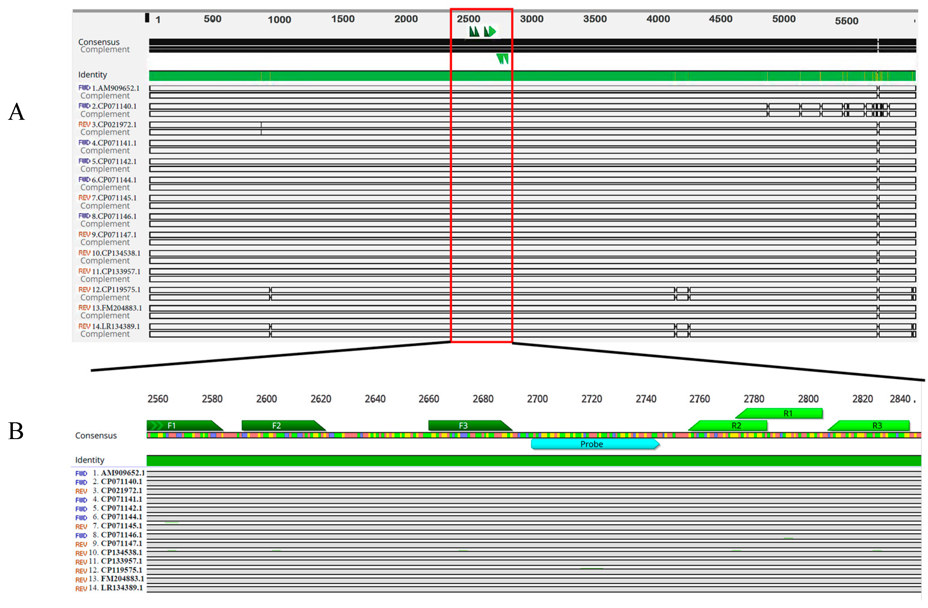
3.1. Design of the eqbE Real-Time RAA Primers and Probe

To evaluate the sensitivity and specificity of the real-time RAA method, we initially selected the eqbE gene from S. equi as the target. This gene has been reported exclusively from S. equi, and not from other subspecies, making it an appropriate diagnostic target.
To identify the most conserved regions of the eqbE gene, we aligned the available eqbE sequences (positions 1223642 to 1229713 of the Se4047 genome) from the NCBI database using the MAFFT Alignment program in Geneious Prime (Biomatters Ltd., Auckland, New Zealand). This alignment enabled us to identify a highly conserved region spanning nucleotide positions 1,226,841 to 1,227,157 (GenBank no. FM204883.1) (Figure 1A). This specific region, which is approximately 300 bp in length, was selected as the amplification target for the real-time RAA assay.
We had several considerations when designing the probe. Our aim was to position it at the midpoint of the target sequence, with a length ranging from 46 to 52 bp. We ultimately developed an exo detector probe (p1,226,969 to 1,227,017) as an exo detector, with two conserved thymine residues (T1,227,047, T1,227,050) labeled with an FAM-dT fluorophore residue and a BHQ1-dT quencher residue, respectively (see Figure 1B). These design considerations ensure robust and specific detection of the eqbE gene in S. equi, enhancing the sensitivity and specificity of the real-time RAA method.

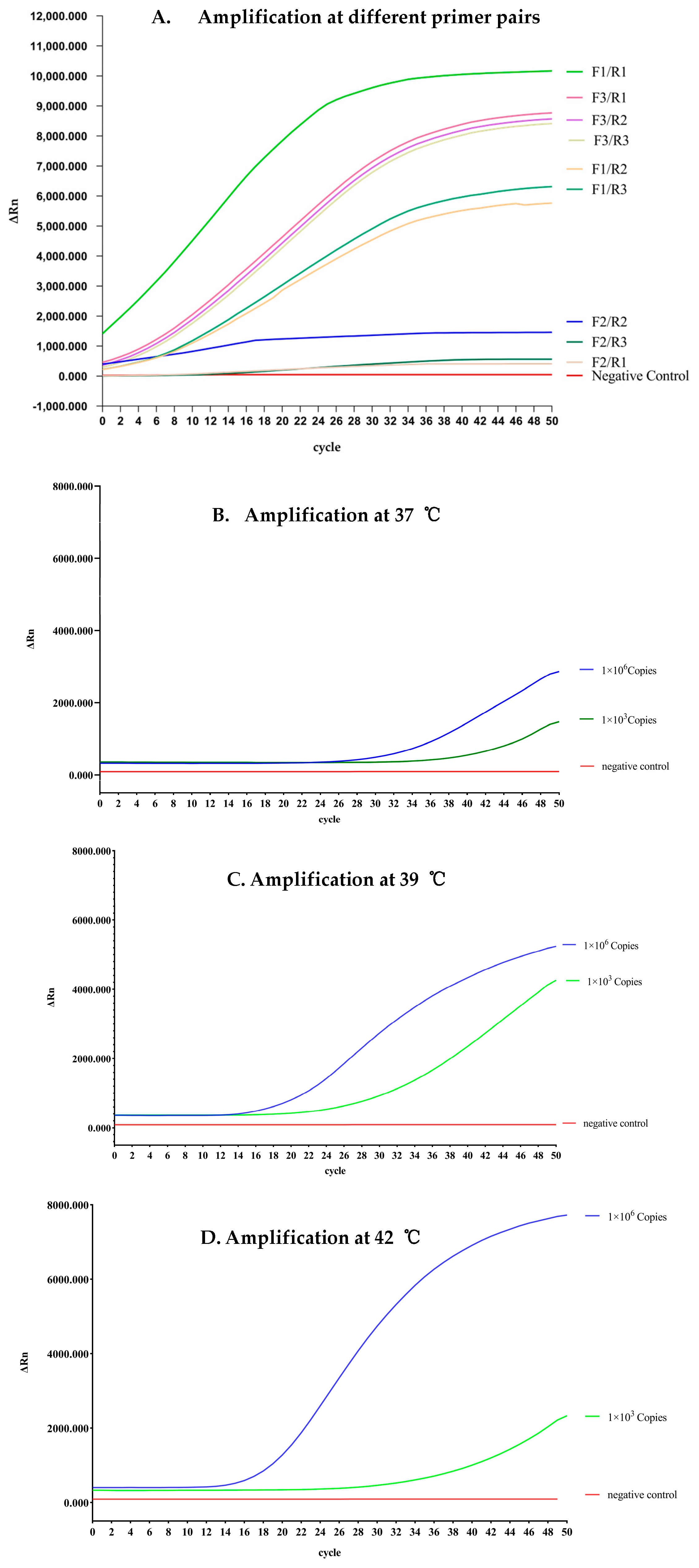
3.2. Reaction Optimization for Real-Time RAA Assays

We then adjusted the reaction temperature and duration to optimize the reaction system of our real-time RAA. Specifically, we employed a standard plasmid, pUC57-S.equi-eqbE, at 4.173 × 10⁸ copies/μL concentrations representing positive samples. By examining the amplification results, we found that the primer pair F1 and R2 was optimal, as it produced the shortest reaction time and the highest fluorescence signal (Figure 2A).
We conducted amplification experiments using a negative control (water), a strong positive control (1 × 10⁶ copies), and a weak positive control (1 × 10³ copies). The resulting products were then assessed using a fluorescence instrument. Each reaction was carried out at constant temperatures of 37 °C, 39 °C, and 42 °C, with a reaction program consisting of 50 cycles (25 min). The amplification curves were then analyzed, leading to the optimized conditions presented in Figure 2B, 2C, and 2D, respectively.
We observed that at a temperature of 39 °C, the amplification signal for the strongly positive sample exhibited clear visibility, while the amplification signal for the weakly positive sample also appeared rapidly, typically within 30 cycles. The strongly positive sample displayed a robust fluorescence curve, and, similarly, the amplification signal for the weakly positive sample was detected early on, usually around 30 cycles. These findings demonstrate that real-time RAA is capable of efficiently detecting samples containing 1 × 10³ copies/μL within a timeframe of 20 min. Based on these results, we have determined 39 °C as the optimal reaction temperature and recommend a reaction time of 20 min.

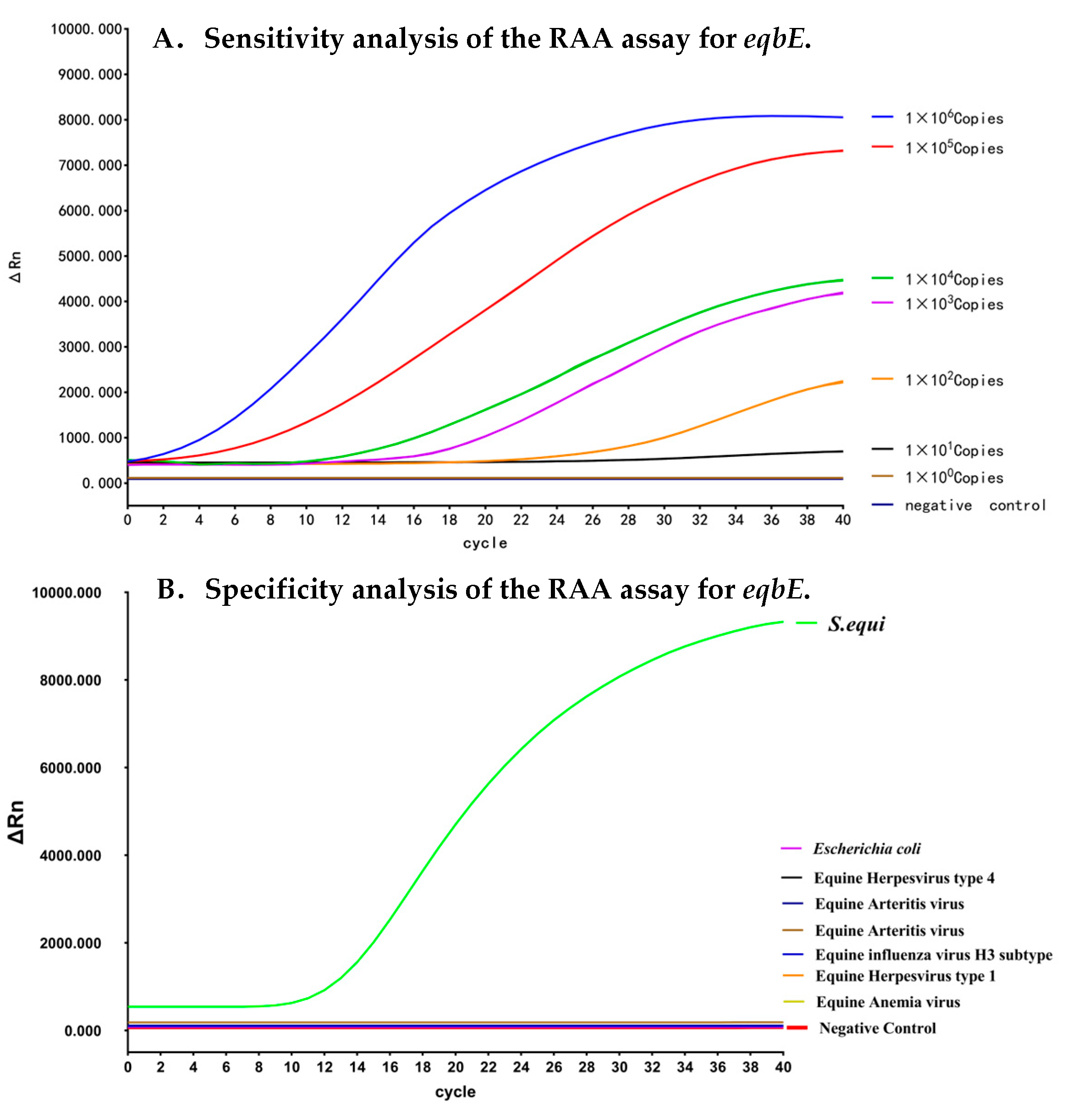
3.3. Analytical Sensitivity of the Real-Time RAA Assay

We evaluated the sensitivity of the real-time RAA detection method by using 10-fold serial dilutions of positive plasmids as the amplification template. The results are given in Figure 3A. Compared with the negative control, the reaction system containing positive plasmids exhibited clear and reproducible amplification signals. Additionally, higher concentrations of plasmid samples resulted in higher fluorescence signals after amplification. Even when the concentration of the positive plasmid was reduced to 10 copies per reaction, the amplified product fluorescence signal could still be detected. However, when the plasmid concentration was further reduced to one copy per reaction, the amplified product signal disappeared. Therefore, we determined the detection limit of the real-time RAA detection method to be 10 copies per reaction.

3.4. Analytical Specificity of the Real-Time RAA Assay

We next analyzed the specificity of the real-time RAA assay. We used nucleic acid samples from other common clinically relevant equine disease pathogens, including equine influenza virus (H3 subtype), equine anemia virus, equine herpesvirus type 1 and type 4, equine arteritis virus, and Escherichia coli, for comparison. As depicted in Figure 3B, only the samples from S. equi tested positive in our real-time RAA assay, and samples from other diseases all tested negative. This demonstrates that the real-time RAA assay developed in this study exhibits excellent analytical specificity for eqbE.

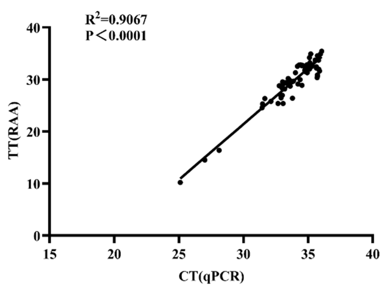
3.5. Diagnostic Performance of Real-Time RAA Using Clinical Nasal Swabs

In order to evaluate the effectiveness of the real-time RAA assay in a clinical setting, a total of 98 nasal swabs were collected from horses on farms with suspected cases of S. equi. These samples were subjected to both real-time RAA and real-time PCR testing. The results are summarized in Table 2.
When the real-time RAA assay was used, 66 samples tested positive for S. equi and 32 samples tested negative. The real-time PCR test identified 65 individuals as positive for S. equi, with 33 individuals testing negative. This yielded an impressive concordance rate of 96.94% (95 out of 98 samples) between the real-time PCR and real-time RAA detection methods.
Additional analysis using linear regression and Pearson correlation demonstrated a significant correlation between the real-time PCR and real-time RAA results, as reflected by an R2 value of 0.9067 (Figure 4) and a kappa value of 0.931. The real-time RAA assay is therefore a reliable tool for the detection of S. equi in the field.
The real-time RAA assay demonstrated excellent sensitivity and specificity, with a concordance rate of 96.94% compared with real-time PCR. This indicates that the real-time RAA assay is highly reliable in identifying the presence or absence of S. equi in clinical samples. The significant correlation and high kappa value (0.931) between the real-time RAA and real-time PCR results further reinforces its accuracy as a diagnostic tool in the detection of S. equi in clinical nasal swabs. Collectively, these findings highlight the potential of the real-time RAA assay to be employed in clinical settings for the simple, rapid, and accurate diagnosis of strangles, facilitating effective management and control of the disease in equine populations.

4. Discussion

Strangles, a prevalent equine disease worldwide, has caused substantial economic losses in the horse industry. Existing diagnostic methods for strangles have limitations in accuracy, speed, and cost-effectiveness. Traditional diagnostic approaches relying on symptom observation and laboratory cultures often lead to delayed diagnosis, susceptibility to interference from other illnesses, and reduced accuracy. Additionally, these methods tend to be time-consuming, delaying timely intervention, and exacerbating the severity of the disease. Moreover, their reliance on complex laboratory procedures and specialized skills restricts their application in the field or in resource-limited environments. The high cost of some laboratory techniques further limits their widespread use, particularly in areas with limited resources. There is therefore an urgent need for a new, simple, fast, accurate, and cost-effective diagnostic method for strangles to improve early detection and timely intervention. This study aimed to address these challenges by developing a novel diagnostic approach based on the design of primers and probes using real-time RAA technology.
Real-time RAA, an emerging nucleic acid detection method, has shown significant potential compared with traditional PCR technology in disease diagnosis. One of the major advantages of real-time RAA is the minimal equipment required, making it suitable for on-site field applications. In this study, we used real-time RAA technology and designed primers and probes to overcome the limitations of previous diagnostic methods. Following established guidelines, we ensured that the lengths of our primer pairs were 30–35 bases longer than conventional PCR primers. We then identified the optimal primer pairs and probes and further optimized reaction time and temperature. Our real-time RAA detection system achieved high sensitivity (10 copies per sample) and exhibited excellent specificity to S. equi. To validate the reliability of this method, we compared its diagnostic performance with that of conventional real-time PCR. The results demonstrated high consistency (95/98) between the two methods, validating the accuracy and reliability of real-time RAA for the detection of S. equi. Furthermore, our real-time RAA-based method offers the advantages of short reaction times (20 min), completing at approximately 39 °C under isothermal conditions (Figure 5), while real-time PCR requires longer amplification times. Despite the slightly higher cost associated with real-time RAA, its lower equipment requirements will save considerable capital expenditure, which is of particular importance for the monitoring of strangles in resource-scarce regions. The real-time RAA diagnostic method developed here showed consistently high sensitivity, even at low reaction temperatures. However, these low temperatures potentially elevate the risk of nucleic acid sample contamination. Therefore, rigorous handling of samples and stringent control of reaction conditions are imperative when employing the real-time RAA diagnostic method to ensure accuracy.
In conclusion, our study introduces a novel diagnostic method based on real-time RAA, showcasing flexibility and adaptability, compatible with portable isothermal fluorescence detection devices and existing real-time PCR instruments. This approach facilitates on-site sample testing, reducing reaction time and transportation costs. Our developed real-time RAA diagnostic method holds promise as a potent tool in strangles prevention and control, and should contribute significantly to the health and economics of the equine industry.

5. Conclusions

To summarize, this study has successfully devised a real-time RAA assay specifically targeting the eqbE gene for the swift detection of equine strangles; in this method, the RAA amplification reaction and signal detection only take 20 min. The entire detection process has been schematically illustrated (Figure 5). The assay showed remarkable sensitivity, with a detection limit of 10 copies/μL, and achieved an impressively high coincidence rate with the real-time PCR technique when applied to clinical samples. Importantly, the real-time RAA assay has the advantages of user-friendliness, efficiency, and compatibility with commonly employed instruments and equipment, making it an invaluable and practical tool for the accurate diagnosis and effective management of strangles.

Author Contributions

Conceptualization, X.W. and W.G.; methodology, validation, data curation R.S. and J.L.; formal analysis, resources, M.W. and X.G.; methodology, validation, data curation, writing—original draft, writing—review and editing, H.Z. All authors have read and agreed to the published version of the manuscript.

Funding

This study was supported by the National Key Research and Development Project of China (grants number 2020YFE0203400) and The Natural Science Foundation of Heilongjiang Province (grant number TD2022C006).

Data Availability Statement

Data are contained within the article.

Acknowledgments

We would like to express our gratitude to Cheng Du, Liuke Sun and Bingqian Zhou for their valuable insights on this work. Special thanks to Zixuan Du for her technical support.

Conflicts of Interest

The authors declare no conflicts of interest.

References

  1. Newton, J.R.; Wood, J.L.N.; Dunn, K.A.; DeBrauwere, M.N.; Chanter, N. Naturally occurring persistent and asymptomatic infection of the guttural pouches of horses with Streptococcus equi. Vet. Rec. 1997, 140, 84–90. [Google Scholar] [CrossRef]
  2. Neamat-Allah, A.N.; Damaty, H.M. Strangles in Arabian horses in Egypt: Clinical, epidemiological, hematological, and biochemical aspects. Vet. World 2016, 9, 820–826. [Google Scholar] [CrossRef] [PubMed]
  3. Waller, A. Streptococcus equi: Breaking its strangles-hold. Vet. Rec. 2018, 182, 316–318. [Google Scholar] [CrossRef] [PubMed]
  4. Ikhuoso, O.A.; Monroy, J.C.; Rivas-Caceres, R.R.; Cipriano-Salazar, M.; Barbabosa Pliego, A. Streptococcus equi in Equine: Diagnostic and Healthy Performance Impacts. J. Equine Vet. Sci. 2020, 85, 102870. [Google Scholar] [CrossRef] [PubMed]
  5. Rendle, D.; de Brauwere, N.; Hallowell, G.; Ivens, P.; McGlennon, A.; Newton, R.; White, J.; Waller, A. Streptococcus equi infections: Current best practice in the diagnosis and management of ‘strangles’. UK-Vet Equine 2021, 5, S3–S15. [Google Scholar] [CrossRef]
  6. Mallicote, M. Update on Streptococcus equi subsp equi infections. Vet. Clin. N. Am. Equine Pract. 2015, 31, 27–41. [Google Scholar] [CrossRef] [PubMed]
  7. Seyum, F.; Mahendra, P. Clinical and microbiological observations on strangles in donkeys. Haryana Vet. 2015, 54, 64–66. [Google Scholar]
  8. Tonpitak, W.; Sornklien, C.; Wutthiwithayaphong, S. Characterization of a Streptococcus equi ssp. equi Isolate from a Strangles Outbreak in Thailand. J. Equine Vet. Sci. 2016, 38, 30–32. [Google Scholar] [CrossRef]
  9. Torpiano, P.; Nestorova, N.; Vella, C. Streptococcus equi subsp. equi meningitis, septicemia and subdural empyema in a child. IDCases 2020, 21, e00808. [Google Scholar] [CrossRef]
  10. Bohlman, T.; Waddell, H.; Schumaker, B. A case of bacteremia and pneumonia caused by Streptococcus equi subspecies equi infection in a 70-year-old female following horse exposure in rural Wyoming. Ann. Clin. Microbiol. Antimicrob. 2023, 22, 65. [Google Scholar] [CrossRef]
  11. Judy, C.E.; Chaffin, M.K.; Cohen, N.D. Empyema of the guttural pouch (auditory tube diverticulum) in horses: 91 cases (1977–1997). J. Am. Vet. Med. Assoc. 1999, 215, 1666–1670. [Google Scholar] [CrossRef] [PubMed]
  12. Boyle, A.G.; Timoney, J.F.; Newton, J.R.; Hines, M.T.; Waller, A.S.; Buchanan, B.R. Streptococcus equi Infections in Horses: Guidelines for Treatment, Control, and Prevention of Strangles-Revised Consensus Statement. J. Vet. Intern. Med. 2018, 32, 633–647. [Google Scholar] [CrossRef] [PubMed]
  13. Liu, Y. Identification of a Novel Genotype of Streptococcus equi Subspecies equi in a Donkey Suffering from Strangles. Pak. Vet. J. 2019, 39, 609–611. [Google Scholar] [CrossRef]
  14. Pusterla, N.; Bowers, J.; Barnum, S.; Hall, J.A. Molecular detection of Streptococcus equi subspecies equi in face flies (Musca autumnalis) collected during a strangles outbreak on a Thoroughbred farm. Med. Vet. Entomol. 2020, 34, 120–122. [Google Scholar] [CrossRef] [PubMed]
  15. Robinson, C.; Waller, A.S.; Frykberg, L.; Flock, M.; Zachrisson, O.; Guss, B.; Flock, J.I. Intramuscular vaccination with Strangvac is safe and induces protection against equine strangles caused by Streptococcus equi. Vaccine 2020, 38, 4861–4868. [Google Scholar] [CrossRef]
  16. McGlennon, A.; Waller, A.; Verheyen, K.; Slater, J.; Grewar, J.; Aanensen, D.; Newton, R. Surveillance of strangles in UK horses between 2015 and 2019 based on laboratory detection of Streptococcus equi. Vet. Rec. 2021, 189, e948. [Google Scholar] [CrossRef]
  17. Jaramillo-Morales, C.; James, K.; Barnum, S.; Vaala, W.; Chappell, D.E.; Schneider, C.; Craig, B.; Bain, F.; Barnett, D.C.; Gaughan, E.; et al. Voluntary Biosurveillance of Streptococcus equi Subsp. equi in Nasal Secretions of 9409 Equids with Upper Airway Infection in the USA. Vet. Sci. 2023, 10, 78. [Google Scholar] [CrossRef]
  18. Rotinsulu, D.A.; Ewers, C.; Kerner, K.; Amrozi, A.; Soejoedono, R.D.; Semmler, T.; Bauerfeind, R. Molecular Features and Antimicrobial Susceptibilities of Streptococcus equi ssp. equi Isolates from Strangles Cases in Indonesia. Vet. Sci. 2023, 10, 49. [Google Scholar] [CrossRef]
  19. Arafa, A.A.; Hedia, R.H.; Ata, N.S.; Ibrahim, E.S. Vancomycin resistant Streptococcus equi subsp. equi isolated from equines suffering from respiratory manifestation in Egypt. Vet. World 2021, 14, 1808–1814. [Google Scholar] [CrossRef]
  20. Bell, D.S.H.; Goncalves, E. Diabetogenic effects of cardioprotective drugs. Diabetes Obes. Metab. 2021, 23, 877–885. [Google Scholar] [CrossRef]
  21. Pringle, J.; Venner, M.; Tscheschlok, L.; Bachi, L.; Riihimaki, M. Long term silent carriers of Streptococcus equi ssp. equi following strangles; carrier detection related to sampling site of collection and culture versus qPCR. Vet. J. 2019, 246, 66–70. [Google Scholar] [CrossRef]
  22. Durham, A.E.; Kemp-Symonds, J. Failure of serological testing for antigens A and C of Streptococcus equi subspecies equi to identify guttural pouch carriers. Equine Vet. J. 2021, 53, 38–43. [Google Scholar] [CrossRef]
  23. Duran, M.C.; Goehring, L.S. Equine strangles: An update on disease control and prevention. Austral J. Vet. Sci. 2021, 53, 23–31. [Google Scholar] [CrossRef]
  24. Boyle, A.G.; Stefanovski, D.; Rankin, S.C. Comparison of nasopharyngeal and guttural pouch specimens to determine the optimal sampling site to detect Streptococcus equi subsp equi carriers by DNA amplification. BMC Vet. Res. 2017, 13, 75. [Google Scholar] [CrossRef]
  25. Stöckle, S.D.; Winter, J.C.; Schöpe, S.S.; Gehlen, H. Possibilities and limitations of the prevention of strangles outbreaks on horse farms. Pferdeheilkunde 2019, 35, 258–264. [Google Scholar] [CrossRef]
  26. Lindahl, S.; Båverud, V.; Egenvall, A.; Aspán, A.; Pringle, J. Comparison of sampling sites and laboratory diagnostic tests for S. equi subsp. equi in horses from confirmed strangles outbreaks. J. Vet. Intern. Med. 2013, 27, 542–547. [Google Scholar] [CrossRef] [PubMed]
  27. Waller, A.S. New Perspectives for the Diagnosis, Control, Treatment, and Prevention of Strangles in Horses. Vet. Clin. N. Am.-Equine 2014, 30, 591–607. [Google Scholar] [CrossRef]
  28. Webb, K.; Barker, C.; Harrison, T.; Heather, Z.; Steward, K.F.; Robinson, C.; Newton, J.R.; Waller, A.S. Detection of Streptococcus equi subspecies equi using a triplex qPCR assay. Vet. J. 2013, 195, 300–304. [Google Scholar] [CrossRef]
  29. Harms, C.; Mapes, S.; Akana, N.; Coatti Rocha, D.; Pusterla, N. Detection of modified-live equine intranasal vaccine pathogens in adult horses using quantitative PCR. Vet. Rec. 2014, 175, 510. [Google Scholar] [CrossRef] [PubMed]
  30. Cordoni, G.; Williams, A.; Durham, A.; Florio, D.; Zanoni, R.G.; La Ragione, R.M. Rapid diagnosis of strangles (Streptococcus equi subspecies equi) using PCR. Res. Ve.t Sci. 2015, 102, 162–166. [Google Scholar] [CrossRef]
  31. Pusterla, N.; Leutenegger, C.M.; Barnum, S.M.; Byrne, B.A. Use of quantitative real-time PCR to determine viability of Streptococcus equi subspecies equi in respiratory secretions from horses with strangles. Equine Vet. J. 2018, 50, 697–700. [Google Scholar] [CrossRef] [PubMed]
  32. Riihimaki, M.; Aspan, A.; Ljung, H.; Pringle, J. Long term dynamics of a Streptococcus equi ssp equi outbreak, assessed by qPCR and culture and seM sequencing in silent carriers of strangles. Vet. Microbiol. 2018, 223, 107–112. [Google Scholar] [CrossRef] [PubMed]
  33. Zheng, Y.Z.; Chen, J.T.; Li, J.; Wu, X.J.; Wen, J.Z.; Liu, X.Z.; Lin, L.Y.; Liang, X.Y.; Huang, H.Y.; Zha, G.C.; et al. Reverse Transcription Recombinase-Aided Amplification Assay with Lateral Flow Dipstick Assay for Rapid Detection of 2019 Novel Coronavirus. Front. Cell Infect. Microbiol. 2021, 11, 613304. [Google Scholar] [CrossRef] [PubMed]
  34. Fan, G.; Zhang, R.; He, X.; Tian, F.; Nie, M.; Shen, X.; Ma, X. RAP: A Novel Approach to the Rapid and Highly Sensitive Detection of Respiratory Viruses. Front. Bioeng. Biotechnol. 2021, 9, 766411. [Google Scholar] [CrossRef]
  35. Liu, Y.Y.; Ren, W.C.; Xue, Z.T.; Miao, Y.D.; Wang, W.; Zhang, X.X.; Yao, C.; Shang, Y.Y.; Li, S.S.; Mi, F.L.; et al. Real-time recombinase-aided amplification assay for rapid amplification of the gene of s. Eur. J. Clin. Microbiol. 2023, 42, 963–972. [Google Scholar] [CrossRef]
  36. Cao, Y.H.; Fang, T.S.; Shen, J.L.; Zhang, G.D.; Guo, D.H.; Zhao, L.A.; Jiang, Y.; Zhi, S.; Zheng, L.; Lv, X.F.; et al. Development of Recombinase Aided Amplification (RAA)-Exo-Probe Assay for the Rapid Detection of Shiga Toxin-Producing Escherichia coli. J. AOAC Int. 2023, 106, 1246–1253. [Google Scholar] [CrossRef]
Figure 2. Optimization of real-time RAA reaction for the detection of eqbE. (A) Representative amplification curves from different primer pairs amplified using real-time RAA are shown. All nine candidate primer pairs (F1/R1-F3/R3) were screened using fixed amplification parameters. The F1/R1 combination produced the highest fluorescence signals in the fewest cycles. (BD) Optimization of reaction time and temperature. High and low concentrations of plasmids were each subjected to amplification at temperatures of (B) 37 °C, (C) 39 °C, and (D) 42 °C for 30 min. Representative amplification curves for each temperature are shown.
Microorganisms 12 00777 g002
Figure 3. Sensitivity and specificity analyses of the real-time RAA assay for eqbE. (A) Sensitivity analysis of the real-time RAA assay. Different amplification curves indicate the real-time RAA reaction at different concentrations of plasmids. Serial dilutions of the plasmid were added to the reaction, with dilutions ranging from 106 copies/reaction to 1 copy/reaction. (B) Analytical specificity of the real-time RAA assay. The real-time RAA reaction readings, post addition of genomic DNA templates, are illustrated using ion curves of distinct colors. Samples containing the equine influenza virus (H3 subtype), equine anemia virus, equine herpesvirus type 1 and type 4, equine arteritis virus, or Escherichia coli all tested negative, while only the sample containing S. equi DNA manifested an amplification curve.
Microorganisms 12 00777 g003
Figure 4. The correlation between real-time RAA and real-time PCR in diagnosing equine strangles in clinical samples. The data are based on 98 clinical swabs from horses on farms with suspected cases of S. equi. The Y-axis represents the threshold time (TT) for the real-time RAA and the X-axis shows the cycle threshold (Ct) for the real-time PCR. A linear regression analysis was carried out using GraphPad Prism software 8.0. The results of the two tests showed a strong positive correlation (R2 = 0.9012, p < 0.0001).
Microorganisms 12 00777 g004
Figure 5. Flowchart of real-time RAA detection. The process begins with the collection of a nasal swab, followed by extracting nucleic acid from the swab’s liquid (approximately 30 min). Subsequently, the reaction solution for real-time RAA is prepared, and nucleic acids from negative, positive, and test samples are added, respectively. Finally, the real-time RAA reaction is conducted on the portable instrument (20 min), and the results can be directly displayed on the screen of the portable detection instrument.
Microorganisms 12 00777 g005
Table 1. Sequences of primers and probes for real-time RAA and real-time PCR.
NameSequence (5′-3′)LocationReference
eqbE-F1CATCTATTTGGTCAAACCATTTGAATGTACCAAG1,226,818 to 1,226,852This study
eqbE-F2CCGAAAGATTGGATTTCCATTCCATATGGTAG1,226,859 to 1,226,890This study
eqbE-F3TGGTAGGATCTGCCCTAATTATGTTAAAGGTG1,226,830 to 1,226,861This study
eqbE-R1CTACCATTATCTCCAGTTCTATACCACCTCATC1226, 928 to 1,226,958This study
eqbE-R2TACCACCTCATCCCATCTTGTTCGAAGTAC1,226,947 to 1,226,979This study
eqbE-R3CCAAGAAACTCAATAATCCCATCATTCCATG1,226,982 to 1,226,912This study
ProbeTATCGGTGGAGTTGGTGTTGCTAAATGTTA/i6FAMdT//idSp/A/iBHQ1dT/GGTGACGAAGAATTA-3′C3 Spacer1,226,969 to 1,227,017This study
eqbE-FATGTAGCTATGGCAAATGTGGC1,223,599 to 1,223,620[26]
eqbE-RAACACCCTTAGGAACACCTG1,223,689 to 1,223,708[26]
eqbE-probeFAM-ATTGTTACTATGGCTGAAGGT-BHO11,223,966 to 1,223,987[26]
Table 2. Comparison of the performance of eqbE-based real-time RAA and real-time PCR for the detection of Streptococcus equi subspecies equi in clinical samples.
Method Real-Time PCR Kappap-Value
PositiveNegativeTotal
RAAPositive642660.9310.0001
Negative13132
Total653398
Disclaimer/Publisher’s Note: The statements, opinions and data contained in all publications are solely those of the individual author(s) and contributor(s) and not of MDPI and/or the editor(s). MDPI and/or the editor(s) disclaim responsibility for any injury to people or property resulting from any ideas, methods, instructions or products referred to in the content.

