
Effects of Graphene-Based Nanomaterials on Microorganisms and Soil Microbial Communities

Abstract
:1. Introduction
2. The Effects of GBNs on Bacteria and Fungus
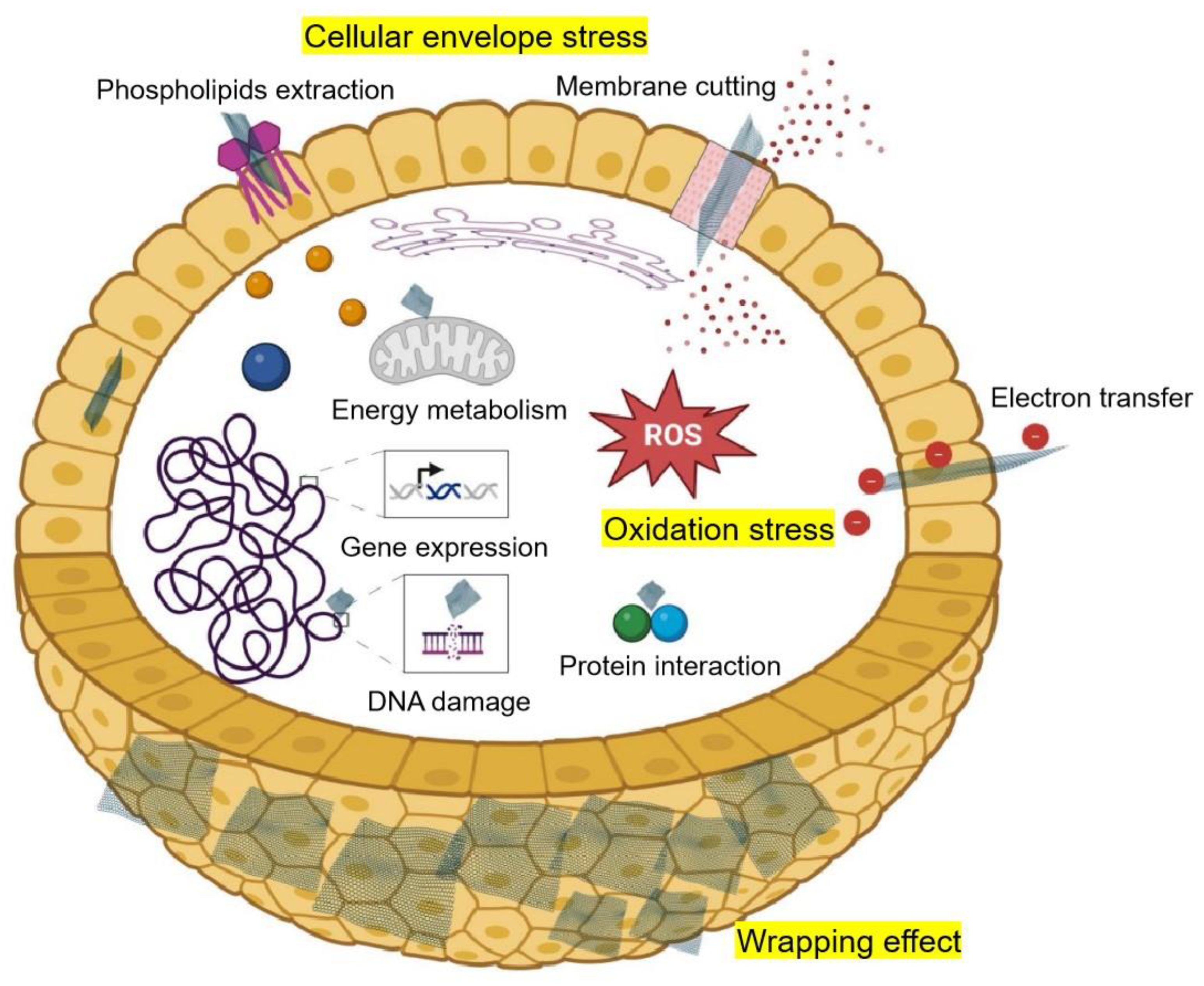
3. The Mechanism of GBNs’ Antimicrobial Activity
4. The Factors Influencing GBN Antimicrobial Activity
5. The Effects of GBNs on Soil Microbial Communities
6. Discussion and Conclusions
Author Contributions
Funding
Conflicts of Interest
References
- Shoemaker, W.R.; Locey, K.J.; Lennon, J.T. A macroecological theory of microbial biodiversity. Nat. Ecol. Evol. 2017, 1, 107. [Google Scholar] [CrossRef] [PubMed]
- Biswas, K.; Mohanta, Y. An overview of carbon-based nanomaterials and their derivatives for different sensing applications. In Nanoscale Matter and Principles for Sensing and Labeling Applications; Springer Nature Singapore: Singapore, 2024; pp. 305–325. [Google Scholar]
- Shahazi, R.; Majumdar, S.; Islam, M.A.; Mondal, J.; Rahman, M.; Alam, M.M. Carbon nanomaterials for biomedical applications: A comprehensive review. Nano Carbons 2024, 1, 448. [Google Scholar] [CrossRef]
- Novoselov, K.S.; Geim, A.K.; Morozov, S.; Jiang, D.; Zhang, Y.; Dubonos, S.V.; Grigorieva, I.; Firsov, A. Electric field effect in atomically thin carbon films. Science 2004, 306, 666–669. [Google Scholar] [CrossRef] [PubMed]
- Novoselov, K.S.; Fal’ko, V.I.; Colombo, L.; Gellert, P.R.; Schwab, M.G.; Kim, K. A roadmap for graphene. Nature 2012, 490, 192–200. [Google Scholar] [CrossRef] [PubMed]
- Yu, W.; Sisi, L.; Haiyan, Y.; Jie, L. Progress in the functional modification of graphene/graphene oxide: A review. RSC Adv. 2020, 10, 15328–15345. [Google Scholar] [CrossRef] [PubMed]
- Li, J.; Zeng, H.; Zeng, Z.; Zeng, Y.; Xie, T. Promising graphene-based nanomaterials and their biomedical applications and potential risks: A comprehensive review. ACS Biomater. Sci. Eng. 2021, 7, 5363–5396. [Google Scholar] [CrossRef] [PubMed]
- Mohan, V.B.; Lau, K.t.; Hui, D.; Bhattacharyya, D. Graphene-based materials and their composites: A review on production, applications and product limitations. Compos. Part B Eng. 2018, 142, 200–220. [Google Scholar] [CrossRef]
- Kononenko, O.; Brzhezinskaya, M.; Zotov, A.; Korepanov, V.; Levashov, V.; Matveev, V.; Roshchupkin, D. Influence of numerous Moiré superlattices on transport properties of twisted multilayer graphene. Carbon 2022, 194, 52–61. [Google Scholar] [CrossRef]
- Han, S.; Sun, J.; He, S.; Tang, M.; Chai, R. The application of graphene-based biomaterials in biomedicine. Am. J. Transl. Res. 2019, 11, 3246–3260. [Google Scholar]
- Chen, G.; Tan, Z.; Zhao, Y.; Ni, B.; Zhu, Y.; Lu, Y. Applications of graphene for energy storage and conversion. Sci. Sin. Chim. 2013, 43, 704. [Google Scholar] [CrossRef]
- Zhang, X.; Cao, H.; Wang, H.; Zhang, R.; Jia, H.; Huang, J.; Zhao, J.; Yao, J. Effects of graphene on morphology, microstructure and transcriptomic profiling of Pinus tabuliformis Carr. roots. PLoS ONE 2021, 16, e0253812. [Google Scholar] [CrossRef] [PubMed]
- Zhang, X.; Cao, H.; Zhao, J.; Wang, H.; Xing, B.; Chen, Z.; Li, X.; Zhang, J. Graphene oxide exhibited positive effects on the growth of Aloe vera L. Physiol. Mol. Biol. Plants 2021, 27, 815–824. [Google Scholar] [CrossRef] [PubMed]
- Pang, L.; Dai, C.; Bi, L.; Guo, Z.; Fan, J. Biosafety and antibacterial ability of graphene and graphene oxide in vitro and in vivo. Nanoscale Res. Lett. 2017, 12, 564. [Google Scholar] [CrossRef]
- Shankar, K.; Agarwal, S.; Mishra, S.; Bhatnagar, P.; Siddiqui, S.; Abrar, I. A review on antimicrobial mechanism and applications of graphene-based materials. Biomater. Adv. 2023, 150, 213440. [Google Scholar] [CrossRef]
- Gungordu Er, S.; Edirisinghe, M.; Tabish, T.A. Graphene-based nanocomposites as antibacterial, antiviral and antifungal agents. Adv. Healthc. Mater. 2023, 12, 2201523. [Google Scholar] [CrossRef]
- Zhang, X.; Kong, H.; Yang, G.; Zhu, D.; Luan, X.; He, P.; Wei, G. Graphene-based functional hybrid membranes for antimicrobial applications: A review. Appl. Sci. 2022, 12, 4834. [Google Scholar] [CrossRef]
- Yaragalla, S.; Bhavitha, K.; Athanassiou, A. A review on graphene based materials and their antimicrobial properties. Coatings 2021, 11, 1197. [Google Scholar] [CrossRef]
- Fatima, N.; Qazi, U.; Mansha, A.; Ahmad, I.; Javaid, R.; Abbas, Q.; Nadeem, N.; Rehan, Z.; Noreen, S.; Zahid, M. Recent developments for antimicrobial applications of graphene-based polymeric composites: A review. J. Ind. Eng. Chem. 2021, 100, 40–58. [Google Scholar] [CrossRef]
- Radhi, A.; Mohamad, D.; Abdul Rahman, F.; Abdullah, A.; Hasan, H. Mechanism and factors influence of graphene-based nanomaterials antimicrobial activities and application in dentistry. J. Mater. Res. Technol. 2021, 11, 1290–1307. [Google Scholar] [CrossRef]
- Chen, M.; Sun, Y.; Liang, J.; Zeng, G.; Li, Z.; Tang, L.; Zhu, Y.; Jiang, D.; Song, B. Understanding the influence of carbon nanomaterials on microbial communities. Environ. Int. 2019, 126, 690–698. [Google Scholar] [CrossRef]
- Braylé, P.; Pinelli, E.; Gauthier, L.; Mouchet, F.; Barret, M. Graphene-based nanomaterials and microbial communities: A review of their interactions, from ecotoxicology to bioprocess engineering perspectives. Environ. Sci. Nano 2022, 9, 3725–3741. [Google Scholar] [CrossRef]
- Sanchez Armengol, E.; Harmanci, M.; Laffleur, F. Current strategies to determine antifungal and antimicrobial activity of natural compounds. Microbiol. Res. 2021, 252, 126867. [Google Scholar] [CrossRef] [PubMed]
- Balouiri, M.; Sadiki, M.; Ibnsouda, S.K. Methods for in vitro evaluating antimicrobial activity: A review. J. Pharm. Anal. 2016, 6, 71–79. [Google Scholar] [CrossRef] [PubMed]
- Liu, S.; Hu, M.; Zeng, T.H.; Wu, R.; Jiang, R.; Wei, J.; Wang, L.; Kong, J.; Chen, Y. Lateral dimension-dependent antibacterial activity of graphene oxide sheets. Langmuir 2012, 28, 12364–12372. [Google Scholar] [CrossRef] [PubMed]
- Pulingam, T.; Thong, K.L.; Ali, M.; Nelson Appaturi, J.; Dinshaw, I.; Ong, Z.Y.; Leo, B.F. Graphene oxide exhibits differential mechanistic action towards Gram-positive and Gram-negative bacteria. Colloids Surf. B Biointerfaces 2019, 181, 6–15. [Google Scholar] [CrossRef] [PubMed]
- Liu, S.; Zeng, T.; Hofmann, M.; Burcombe, E.; Wei, J.; Jiang, R.; Kong, J.; Chen, Y. Antibacterial activity of graphite, graphite oxide, graphene oxide, and reduced graphene oxide: Membrane and oxidative stress. ACS Nano 2011, 5, 6971–6980. [Google Scholar] [CrossRef]
- Akhavan, O.; Ghaderi, E. Toxicity of graphene and graphene oxide nanowalls against bacteria. ACS Nano 2010, 4, 5731–5736. [Google Scholar] [CrossRef] [PubMed]
- Rodrigues, D.; Moreirinha, C.; Neves, A.; Freitas, S.; Sequeira, S.; Russo, S.; Craciun, M.; Almeida, A.; Alves, H. Conversion of antibacterial activity of graphene-coated textiles through surface polarity. Nano Sel. 2022, 4, 502–512. [Google Scholar] [CrossRef]
- Sengupta, I.; Bhattacharya, P.; Talukdar, M.; Neogi, S.; Pal, S.; Chakraborty, s. Bactericidal effect of graphene oxide and reduced graphene oxide: Influence of shape of bacteria. Colloid Interface Sci. Commun. 2018, 28, 60–68. [Google Scholar] [CrossRef]
- Gurunathan, S.; Han, J.W.; Dayem, A.A.; Eppakayala, V.; Kim, J.H. Oxidative stress-mediated antibacterial activity of graphene oxide and reduced graphene oxide in Pseudomonas aeruginosa. Int. J. Nanomed. 2012, 7, 5901–5914. [Google Scholar] [CrossRef]
- Ghanem, A.; Youssef, A.; Abdel Rehim, M. Hydrophobically modified graphene oxide as a barrier and antibacterial agent for polystyrene packaging. J. Mater. Sci. 2020, 55, 4685–4700. [Google Scholar] [CrossRef]
- Chen, J.; Li, S.; Luo, J.; Zhang, Y.; Ding, W. Graphene oxide induces toxicity and alters energy metabolism and gene expression in Ralstonia solanacearum. J. Nanosci. Nanotechnol. 2017, 17, 186–195. [Google Scholar] [CrossRef] [PubMed]
- Chen, J.; Peng, H.; Wang, X.; Shao, F.; Yuan, Z.; Han, H.Y. Graphene oxide exhibits broad-spectrum antimicrobial activity against bacterial phytopathogens and fungal conidia by intertwining and membrane perturbation. Nanoscale 2013, 6, 1879–1889. [Google Scholar] [CrossRef] [PubMed]
- Chen, J.; Wang, X.; Han, H.-Y. A new function of graphene oxide emerges: Inactivating phytopathogenic bacterium Xanthomonas oryzae pv. Oryzae. J. Nanopart. Res. 2013, 15, 1658. [Google Scholar] [CrossRef]
- Wang, X.; Cai, A.; Wen, X.; Jing, D.; Qi, H.; Yuan, H. Graphene oxide-Fe3O4 nanocomposites as high-performance antifungal agents against Plasmopara viticola. Sci. China Mater. 2017, 60, 9005. [Google Scholar] [CrossRef]
- Zhang, X.; Cao, H.; Wang, J.; Li, F.; Zhao, J. Graphene oxide exhibits antifungal activity against Bipolaris sorokiniana in vitro and in vivo. Microorganisms 2022, 10, 1994. [Google Scholar] [CrossRef] [PubMed]
- Farzanegan, A.; Roudbary, M.; Falahati, M.; Khoobi, M.; Gholibegloo, E.; Farahyar, S.; Karimi, P.; Khanmohammadi, M. Synthesis, characterization and antifungal activity of a novel formulated nanocomposite containing Indolicidin and Graphene oxide against disseminated candidiasis. J. Mycol. Med. 2018, 28, 628–636. [Google Scholar] [CrossRef] [PubMed]
- Wang, X.; Liu, X.; Chen, J.; Han, H.-Y.; Yuan, Z. Evaluation and mechanism of antifungal effects of carbon nanomaterials in controlling plant fungal pathogen. Carbon 2014, 68, 798–806. [Google Scholar] [CrossRef]
- Sawangphruk, M.; Srimuk, P.; Chiochan, P.; Sangsri, T.; Siwayaprahm, P. Synthesis and antifungal activity of reduced graphene oxide nanosheets. Carbon 2012, 50, 5156–5161. [Google Scholar] [CrossRef]
- Hao, Y.; Cao, X.; Ma, C.; Zhang, Z.; Zhao, N.; Ali, A.; Tianqi, H.; Xiang, Z.; Zhuang, J.; Wu, S.; et al. Potential applications and antifungal activities of engineered nanomaterials against gray mold disease agent Botrytis cinerea on rose petals. Front. Plant Sci. 2017, 8, 1332. [Google Scholar] [CrossRef]
- Zou, X.; Zhang, L.; Wang, Z.; Luo, Y. Mechanisms of the antimicrobial activities of graphene materials. J. Am. Chem. Soc. 2016, 138, 2064–2077. [Google Scholar] [CrossRef] [PubMed]
- He, J.; Zhu, X.; Qi, Z.; Wang, C.; Mao, X.; Zhu, C.; He, Z.; Li, M.; Tang, Z. Killing dental pathogens using antibacterial graphene oxide. ACS Appl. Mater. Interfaces 2015, 7, 5605–5611. [Google Scholar] [CrossRef]
- Tu, Y.; Lv, M.; Xiu, P.; Huynh, T.; Zhang, M.; Castelli, M.; Liu, Z.; Huang, Q.; Fan, C.; Fang, H.; et al. Destructive extraction of phospholipids from Escherichia coli membranes by graphene nanosheets. Nat. Nanotechnol. 2013, 8, 594–601. [Google Scholar] [CrossRef] [PubMed]
- Duan, G.; Zhang, Y.; Luan, B.; Weber, J.K.; Zhou, R.W.; Yang, Z.; Zhao, L.; Xu, J.; Luo, J.; Zhou, R. Graphene-induced pore formation on cell membranes. Sci. Rep. 2017, 7, 42767. [Google Scholar] [CrossRef]
- Yadav, N.; Dubey, A.; Shukla, S.; Saini, C.; Gupta, G.; Priyadarshini, R.; Lochab, B. Graphene oxide-coated surface: Inhibition of bacterial biofilm formation due to specific surface–interface interactions. ACS Omega 2017, 2, 3070–3082. [Google Scholar] [CrossRef]
- Guo, Z.; Xie, C.; Zhang, J.; Wang, G.; He, X.; Ma, Y.; Zhao, B. Toxicity and transformation of graphene oxide and reduced graphene oxide in bacteria biofilm. Sci. Total Environ. 2016, 580, 1300–1308. [Google Scholar] [CrossRef] [PubMed]
- Song, C.; Yang, C.-M.; Sun, X.F.; Xia, P.F.; Qin, J.; Guo, B.B.; Wang, S.G. Influences of graphene oxide on biofilm formation of Gram-negative and Gram-positive bacteria. Environ. Sci. Pollut. Res. 2018, 25, 2853–2860. [Google Scholar] [CrossRef]
- Li, J.; Zhu, H.; Zhang, M.; Zheng, X.; Di, Z.; Liu, X.; Wang, X. Antibacterial activity of large-area monolayer graphene film manipulated by charge transfer. Sci. Rep. 2014, 4, 4359. [Google Scholar] [CrossRef]
- Mejias Carpio, I.; Santos, C.; Wei, X.; Rodrigues, D. Toxicity of a polymer–graphene oxide composite against bacterial planktonic cells, biofilms, and mammalian cells. Nanoscale 2012, 4, 4746–4756. [Google Scholar] [CrossRef]
- Luan, B.; Huynh, T.; Zhao, L.; Ruhong, Z. Potential toxicity of graphene to cell functions via disrupting protein-protein interactions. ACS Nano 2014, 9, 663–669. [Google Scholar] [CrossRef]
- Chen, Y.W.; Su, Y.L.; Hu, S.H.; Chen, S.Y. Functionalized graphene nanocomposites for enhancing photothermal therapy in tumor treatment. Adv. Drug Deliver. Rev. 2016, 105, 190–204. [Google Scholar] [CrossRef] [PubMed]
- Chen, Y.; Gao, Y.; Chen, Y.; Liu, L.; Mo, A.; Peng, Q. Nanomaterials-based photothermal therapy and its potentials in antibacterial treatment. J. Control. Release 2020, 328, 251–262. [Google Scholar] [CrossRef] [PubMed]
- Kumar, R. Antimicrobial mechanisms and effectiveness of graphene and graphene-functionalized biomaterials. A scope review. Front. Bioeng. Biotechnol. 2020, 8, 465–486. [Google Scholar] [CrossRef]
- Mangadlao, J.; Santos, C.; Leon, A.; Rodrigues, D.; Advincula, R. On the antibacterial mechanism of graphene oxide (GO) Langmuir–Blodgett films. Chem. Commun. 2015, 51, 2886–2889. [Google Scholar] [CrossRef] [PubMed]
- Pham, V.; Truong, V.K.; Quinn, M.; Notley, S.; Guo, Y.; Baulin, V.; Al Kobaisi, M.; Crawford, R.; Ivanova, E. Graphene Induces Formation of Pores That Kill Spherical and Rod-Shaped Bacteria. ACS Nano 2015, 9, 8458–8467. [Google Scholar] [CrossRef] [PubMed]
- De Jesus, L.R.; Dennis, R.V.; Depner, S.W.; Jaye, C.; Fischer, D.A.; Banerjee, S. Inside and outside: X-ray absorption spectroscopy mapping of chemical domains in graphene oxide. J. Phys. Chem. Lett. 2010, 4, 3144–3151. [Google Scholar] [CrossRef] [PubMed]
- Akhavan, O.; Ghaderi, E. Escherichia coli bacteria reduce graphene oxide to bactericidal graphene in a self-limiting manner. Carbon 2012, 50, 1853–1860. [Google Scholar] [CrossRef]
- Arias, L.R.; Yang, L. Inactivation of bacterial pathogens by carbon nanotubes in suspensions. Langmuir 2009, 25, 3003–3012. [Google Scholar] [CrossRef] [PubMed]
- Lyon, D.Y.; Adams, L.K.; Falkner, J.C.; Alvarezt, P.J. Antibacterial activity of fullerene water suspensions: Effects of preparation method and particle size. Environ. Sci. Technol. 2006, 40, 4360–4366. [Google Scholar] [CrossRef]
- Karahan, H.E.; Wei, L.; Goh, K.; Liu, Z.; Birer, O.; Dehghani, F.; Xu, C.; Wei, J.; Chen, Y. Bacterial physiology is a key modulator of the antibacterial activity of graphene oxide. Nanoscale 2016, 8, 17181–17189. [Google Scholar] [CrossRef]
- Zhang, X.; Cao, H.; Wang, H.; Zhao, J.; Gao, K.; Qiao, J.; Li, J.; Ge, S. The effects of graphene-family nanomaterials on plant growth: A review. Nanomaterials 2022, 12, 936. [Google Scholar] [CrossRef] [PubMed]
- Hu, W.; Peng, C.; Luo, W.; Lv, M.; Li, X.; Li, D.; Huang, Q.; Fan, C. Graphene-based antibacterial paper. ACS Nano 2010, 4, 4317–4323. [Google Scholar] [CrossRef] [PubMed]
- Ruiz, O.N.; Fernando, K.A.S.; Wang, B.; Brown, N.A.; Luo, P.G.; McNamara, N.D.; Vangsness, M.; Sun, Y.P.; Bunker, C.E. Graphene oxide: A nonspecific enhancer of cellular growth. ACS Nano 2011, 5, 8100–8107. [Google Scholar] [CrossRef] [PubMed]
- Chung, H.; Kim, M.; Ko, K.; Kim, J.; Kwon, H.A.; Hong, I.; Park, N.; Lee, S.W.; Kim, W. Effects of graphene oxides on soil enzyme activity and microbial biomass. Sci. Total Environ. 2015, 514C, 307–313. [Google Scholar] [CrossRef] [PubMed]
- Kim, M.-J.; Ko, D.; Ko, K.; Kim, D.; Lee, J.; Woo, S.; Kim, W.; Chung, H. Effects of silver-graphene oxide nanocomposites on soil microbial communities. J. Hazard. Mater. 2017, 346, 93–102. [Google Scholar] [CrossRef] [PubMed]
- Du, J.; Zhou, Q.; Wu, J.; Li, G.; Li, G.; Wu, Y. Vegetation alleviate the negative effects of graphene oxide on benzo[a]pyrene dissipation and the associated soil bacterial community. Chemosphere 2020, 253, 126725. [Google Scholar] [CrossRef] [PubMed]
- Ge, Y.; Shen, C.; Wang, Y.; Sun, Y.Q.; Schimel, J.; Gardea-Torresdey, J.; Holden, P. Carbonaceous nanomaterials have higher effects on soybean rhizosphere prokaryotic communities during the reproductive growth phase than during vegetative growth. Environ. Sci. Technol. 2018, 52, 6636–6646. [Google Scholar] [CrossRef] [PubMed]
- Ren, W.; Ren, G.; Teng, Y.; Li, Z.; Li, L. Time-dependent effect of graphene on the structure, abundance, and function of the soil bacterial community. J. Hazard. Mater. 2015, 297, 286–294. [Google Scholar] [CrossRef] [PubMed]
- Forstner, C.; Orton, T.; Skarshewski, A.; Wang, P.; Dennis, P. Effects of graphene oxide and graphite on soil bacterial and fungal diversity. Sci. Total Environ. 2019, 671, 140–148. [Google Scholar] [CrossRef]
- Forstner, C.; Orton, T.; Wang, P.; Dennis, P. Effects of carbon nanotubes and derivatives of graphene oxide on soil bacterial diversity. Sci. Total Environ. 2019, 682, 356–363. [Google Scholar] [CrossRef]
- Rong, Y.; Wang, Y.; Guan, Y.; Ma, J.; Cai, Z.; Yang, G.; Zhao, X. Pyrosequencing reveals soil enzyme activities and bacterial communities impacted by graphene and its oxides. J. Agric. Food Chem. 2017, 65, 9191–9199. [Google Scholar] [CrossRef] [PubMed]
- Du, J.; Zhou, Q.; Wu, J.; Li, G.; Li, G.; Wu, Y. Soil bacterial communities respond differently to graphene oxide and reduced graphene oxide after 90 days of exposure. Soil Ecol. Lett. 2020, 2, 176–179. [Google Scholar] [CrossRef]
- Wang, Y.; Wang, F.; Ford, R.; Tang, W.; Zhou, M.; Ma, B.; Zhang, M. The influences of graphene oxide and nitrification inhibitor on vegetable growths and soil and endophytic bacterial communities: Double-edge sword effects and nitrate risk controls. Sci. Total Environ. 2023, 903, 166337. [Google Scholar] [CrossRef] [PubMed]
- Wu, F.; Jiao, S.; Hu, J.; Wu, X.; Wang, B.; Shen, G.; Yang, Y.; Tao, S.; Wang, X. Stronger impacts of long-term relative to short-term exposure to carbon nanomaterials on soil bacterial communities. J. Hazard. Mater. 2020, 410, 124550. [Google Scholar] [CrossRef] [PubMed]
- Du, J.; Zhou, Q. Graphene oxide regulates the bacterial community and exhibits property changes in soil. RSC Adv. 2015, 5, 27009–27017. [Google Scholar] [CrossRef]
- Ge, Y.; Priester, J.; Mortimer, M.; Chang, C.; Ji, Z.; Schimel, J.; Holden, P. Long-term effects of multiwalled carbon nanotubes and graphene on microbial communities in dry soil. Environ. Sci. Technol. 2016, 50, 3965–3974. [Google Scholar] [CrossRef] [PubMed]
- Song, J.; Duan, C.; Sang, Y.; Wu, S.; Ru, J.; Cui, X. Effects of graphene on bacterial community diversity and soil environments of haplic cambisols in northeast China. Forests 2018, 9, 677. [Google Scholar] [CrossRef]
- Xiong, T.; Yuan, X.Z.; Hou, W.; Leng, L.; Li, H.; Wu, Z.; Jiang, L.; Xu, R.; Zeng, G. Implication of graphene oxide in Cd-contaminated soil: A case study of bacterial communities. J. Environ. Manag. 2017, 205, 99–106. [Google Scholar] [CrossRef] [PubMed]
- Zhang, X.; Zhang, H.; Liu, D.; Sang, Y.; Wang, H.; Guo, J.; Song, J. Graphene increased the richness and diversity of bacterial community in Cd-polluted Haplic Cambisols in a time-dependent manner. J. Soils Sediments 2023, 23, 3485–3498. [Google Scholar] [CrossRef]
- Fang, J.; Weng, Y.; Li, B.; Liu, H.; Liu, L.; Tian, Z.; Du, S. Graphene oxide decreases the abundance of nitrogen cycling microbes and slows nitrogen transformation in soils. Chemosphere 2022, 309, 136642. [Google Scholar] [CrossRef]
- Yadav, S.; Singh Raman, A.P.; Meena, H.; Goswami, A.G.; Bhawna; Kumar, V.; Jain, P.; Kumar, G.; Sagar, M.; Rana, D.K. An update on graphene oxide: Applications and toxicity. ACS Omega 2022, 7, 35387–35445. [Google Scholar] [CrossRef] [PubMed]
- Ou, L.; Song, B.; Liang, H.; Liu, J.; Feng, X.; Deng, B.; Sun, T.; Shao, L. Toxicity of graphene-family nanoparticles: A general review of the origins and mechanisms. Part. Fibre Toxicol. 2016, 13, 57. [Google Scholar] [CrossRef] [PubMed]
- Lin, L.; Zhang, J.; Su, H.; Li, J.; Sun, L.; Wang, Z.; Xu, F.; Liu, C.; Lopatin, S.; Zhu, Y. Towards super-clean graphene. Nat. Commun. 2019, 10, 1912. [Google Scholar] [CrossRef] [PubMed]
- Liao, C.; Li, Y.; Tjong, S. Graphene nanomaterials: Synthesis, biocompatibility, and cytotoxicity. Int. J. Mol. Sci. 2018, 19, 3564. [Google Scholar] [CrossRef] [PubMed]
- Montes-Zavala, I.; Castrejón-González, E.O.; González-Calderón, J.A.; Rico-Ramírez, V. Colloidal stability of graphene in aqueous medium: A theoretical approach through molecular dynamics. J. Mol. Model. 2023, 29, 220. [Google Scholar] [CrossRef] [PubMed]
- Suter, J.L.; Coveney, P.V. Principles governing control of aggregation and dispersion of aqueous graphene oxide. Sci. Rep. 2021, 11, 22460. [Google Scholar] [CrossRef]
- Sun, T.; Gottschalk, F.; Hungerbühler, K.; Nowack, B. Comprehensive probabilistic modelling of environmental emissions of engineered nanomaterials. Environ. Pollut. 2013, 185C, 69–76. [Google Scholar] [CrossRef]


| Microbial Species | GBNs | Methods | Antimicrobial Performance | Ref. |
|---|---|---|---|---|
| E. coli (G−) | GO | Colony counting methods | GO exhibits lateral size-, time- and concentration-dependent antibacterial activity. | [25] |
| GO | Viability, time-kill and lactose dehydrogenase release assays | GO exhibits concentration- and time-dependent antibacterial activity. | [26] | |
| GO, rGO | Colony counting methods | GO and rGO have much higher bacterial inactivation percentages compared with those of Gt and GtO. | [27] | |
| GO, rGO | Drop-test and colony counting methods | E. coli is less sensitive than S. aureus to GO and rGO treatment; rGO is more toxic to bacteria than GO. | [28] | |
| FLG | Bioluminescent signal measure, disk diffusion method | FLG concentration-dependently decreases viable E. coli. | [29] | |
| S. aureus (G+) | GO | Viability, time-kill and lactose dehydrogenase release assays | GO exhibits concentration- and time-dependent antibacterial activity. | [26] |
| GO, rGO | Drop-test and colony counting methods | S. aureus is more sensitive than E. coli to GO and rGO treatment; rGO is more toxic to bacteria than GO. | [28] | |
| GO, rGO | Growth curve analysis | GO and rGO restrict S. aureus cell growth by 93.7% and 67.7%, respectively. | [30] | |
| FLG | Bioluminescent signal measure, disk diffusion method | Only high concentrations of FLG can lead to decreased viability of S. aureus. Both HNO3 and H2O2 in doped FLG-coated textile are capable of inhibiting growth of adjacent bacteria. | [29] | |
| E. faecalis (G+) | GO | Viability, time-kill and lactose dehydrogenase release assays | GO exhibits concentration- and time-dependent antibacterial activity. | [26] |
| P. aeruginosa (G−) | GO | Viability, time-kill and lactose dehydrogenase release assays | GO exhibits concentration- and time-dependent antibacterial activity. | [26] |
| GO, rGO | Colony-counting method | GO and rGO show concentration- and time-dependent antibacterial activity against P. aeruginosa cells. | [31] | |
| GO, rGO | Growth curve analysis | GO and rGO restrict P. aeruginosa cell growth by 48.6% and 93.3%, respectively. | [30] | |
| HGO | Diffusion plate method (solid nutrient agar) | Composite film containing 15 wt% HGO is able to develop clear inhibition zone compared to blank polystyrene film. | [32] | |
| R. solanacearum (G−) | GO | Growth curve analysis | GO suppresses growth of R. solanacearum at all test concentrations (ranging from 62.5 to 500 μg/mL) in less than 2 h of incubation time. | [33] |
| P. syringae (G−) | GO | Growth curve analysis | GO can significantly inhibit bacterial growth in concentration range 10 to 500 mg/mL. | [34] |
| X. campestris pv. undulosa (G−) | GO | Growth curve analysis | GO can significantly inhibit bacterial growth in concentration range 10 to 500 mg/mL. | [34] |
| Xoo (G−) | GO, rGO | Colony counting methods | GO and rGO delay growth of Xoo, primarily depending on concentration and type of buffer. GO is more toxic to bacteria than rGO. | [35] |
| B. cereus (G+) | HGO | Diffusion plate method (solid nutrient agar) | Composite film containing 15 wt% HGO able to develop clear inhibition zone compared to blank polystyrene film. | [32] |
| Microbial Species | GBNs | Methods | Antimicrobial Performance | Ref. |
|---|---|---|---|---|
| P. viticola | GO | Analyze protective effect, fungicidal effect, and curative effect of GO | GO represses germination of sporangia and inhibits development of P. viticola. | [36] |
| B. sorokiniana | GO | Observe mycelial and spore growth | GO inhibits spore germination and mycelial growth of B. sorokiniana concentration-dependently, and attenuates pathogenicity of pathogenic fungi in vivo. | [37] |
| C. albicans | GO | Determine MIC value using broth microdilution assay | MIC value and MFC of GO is 6.25 and 12.5 μg/mL, respectively. | [38] |
| HGO | Diffusion plate method (solid potato dextrose agar) | Composite film containing 15 wt% HGO able to develop clear inhibition zone compared to blank polystyrene film. | [32] | |
| F. poae | GO, rGO | Observe mycelial growth, mycelial biomass, and spore germination rate | GO and rGO produce no effect on mycelial growth rate, but decrease hyphae density of F. poae. | [39] |
| F. graminearum | GO | Calculate spore germination rate | GO inhibits spore germination and germ-tube elongation of F. graminearum dose-dependently. | [34] |
| GO, rGO | Observe mycelial growth, mycelial biomass, and spore germination rate | GO and rGO produce no effect on mycelial growth rate, but decrease hyphae density of F. graminearum. | [39] | |
| F. oxysporum | GO | Calculate spore germination rate | GO inhibits spore germination and germ-tube elongation of F. oxysporum dose-dependently. | [34] |
| rGO | Observe mycelial growth | rGO inhibits mycelial growth of F. oxysporum. | [40] | |
| A. niger | rGO | Observe mycelial growth | rGO inhibits mycelial growth of A. niger. | [40] |
| A. oryzae | rGO | Observe mycelial growth | rGO inhibits mycelial growth of A. oryzae. | [40] |
| B. cinerea | rGO | Measure mycelia diameter in in vitro conditions and measure colony area in whole cut flowers | rGO inhibits mycelial growth of B. cinerea significantly in concentrations of 100 and 200 mg/L, but not in 5 and 50 mg/L. | [41] |
| Soil Type | Parameter | GBNs | Concentration (mg/g) | Exposure Duration | Effects | Ref. |
|---|---|---|---|---|---|---|
| Urban soil | Total organic carbon | GO | 0.1, 0.5, 1 | 59 days | GO treatment did not significantly change the soil microbial biomass throughout the incubation period. | [65] |
| Urban soil | Total organic carbon, bacterial community | GO | 1 | 7, 14, 21 days | GN treatment did not significantly change the soil microbial biomass, richness, and diversity of soil microbial communities. | [66] |
| Farmland soil (benzo [a] pyrene-contaminated soil) | Bacterial community | GO | 0.1 | 90 days | GO had no significant effects on the richness and diversity of bacterial communities in benzo [a] pyrene -contaminated soil. | [67] |
| Urban soil | Bacterial community | GN | 10−4, 0.1, 1 | 20, 39 days | The soybean rhizosphere bacterial community can be significantly phylogenetically and functionally altered in response to GN, especially at the reproductive stage. | [68] |
| Farmland soil | Bacterial community | GN | 0.01, 0.1, 1 | 4, 21, 60 days | The biomass of the bacterial populations increased significantly after 4 days of GN treatment, but completely recovered after 21 days of treatment. | [69] |
| Farmland soil | Bacterial and fungal communities | GO | 10−12, 10−6, 10−3 | 7, 14, 30 days | The composition, but not the alpha diversity, of bacterial and fungal communities was significantly influenced by GO at all doses with the exception of the lowest dose on day 14. | [70] |
| Farmland soil | Bacterial community | GO, rGO, aGO | 10−12, 10−6, 10−3 | 7, 14, 30 days | The bacterial community composition, but not alpha diversity, was altered by all treatments except the 10−12 mg/g GO, 10−12 mg/g rGO, and 10−3 mg/g aGO treatments on day 14 only. | [71] |
| Farmland soil | Bacterial community | GN, GO | 0.1 | 10, 90 days | Both GN and GO treatments increased the abundance and diversity of soil microbial communities. | [72] |
| Farmland soil | Bacterial community | GO, rGO | 0.05 | 90 days | The rGO, but not GO, induced a lower bacterial richness than the control. However, GO induced larger changes in the community composition and functions than RGO. | [73] |
| Farmland soil | Bacterial community | GO | 0.05 | 26 days | GO application significantly decreased the InvSimpson index of the soil bacterial community and the ACE and Chao1 richness estimators of the endophytic bacterial community. | [74] |
| Farmland soil | Bacterial community | GN | 0.3 | 30, 360 days | The alpha diversity of soil bacterial communities was significantly increased with 30 days of GN exposure, and then significantly decreased after 360 days treatment. Compared to 30 days exposure, 360 days exposure more strongly altered the beta diversity of soil bacterial communities. | [75] |
| Farmland soil | Bacterial community | PGO | 5 | 90 days | PGO treatment increased the richness and diversity and altered the structure of soil bacterial communities. | [76] |
| Grassland soil | Bacterial and fungal communities | GN | 1 | 1 year | GN exposure reduced soil DNA and altered bacterial communities, but did not affect soil fungal community profiles. | [77] |
| Forest soil | Bacterial community | GN | 0.01, 0.1, 1 | 7, 15, 30, 60, 90 days | GN significantly increased the community richness and diversity index as well as the abundances of the bacterial community in a concentration and incubation time-dependent manner. | [78] |
| Mountain soil (Cd-contaminated soil) | Bacterial community | GO | 1, 2 | 60 days | GO increased the population of some bacteria at the genus level but decreased the diversity of bacterial communities in Cd-contaminated soil. | [79] |
| Forest farm soil (Cd-polluted Haplic Cambisols) | Bacterial community | GN | 0.01, 0.1, 1 | 15, 30, 45, 60 days | GN increased the richness of bacterial communities in Cd-contaminated soil. | [80] |
Disclaimer/Publisher’s Note: The statements, opinions and data contained in all publications are solely those of the individual author(s) and contributor(s) and not of MDPI and/or the editor(s). MDPI and/or the editor(s) disclaim responsibility for any injury to people or property resulting from any ideas, methods, instructions or products referred to in the content. |
© 2024 by the authors. Licensee MDPI, Basel, Switzerland. This article is an open access article distributed under the terms and conditions of the Creative Commons Attribution (CC BY) license (https://creativecommons.org/licenses/by/4.0/).
Share and Cite
Cao, H.; Zhang, X.; Wang, H.; Ding, B.; Ge, S.; Zhao, J. Effects of Graphene-Based Nanomaterials on Microorganisms and Soil Microbial Communities. Microorganisms 2024, 12, 814. https://doi.org/10.3390/microorganisms12040814
Cao H, Zhang X, Wang H, Ding B, Ge S, Zhao J. Effects of Graphene-Based Nanomaterials on Microorganisms and Soil Microbial Communities. Microorganisms. 2024; 12(4):814. https://doi.org/10.3390/microorganisms12040814
Chicago/Turabian StyleCao, Huifen, Xiao Zhang, Haiyan Wang, Baopeng Ding, Sai Ge, and Jianguo Zhao. 2024. "Effects of Graphene-Based Nanomaterials on Microorganisms and Soil Microbial Communities" Microorganisms 12, no. 4: 814. https://doi.org/10.3390/microorganisms12040814