Article

Molecular Analysis of Tick-Borne Bacterial Pathogens from Ticks Infesting Animal Hosts in Kyrgyzstan, 2021

1
Division of Vectors and Parasitic Diseases, Korea Disease Control and Prevention Agency (KDCA), 187 Osongsaenmyeong 2-ro, Osong-eup, Heungdeok-gu, Cheongju 28159, Republic of Korea
2
Faculty of Veterinary Medicine, Kyrgyz National Agrarian University Named after K. I. Skryabin, Bishkek 720005, Kyrgyzstan
*
Author to whom correspondence should be addressed.
Microorganisms 2024, 12(6), 1046; https://doi.org/10.3390/microorganisms12061046
Submission received: 25 April 2024 / Revised: 10 May 2024 / Accepted: 20 May 2024 / Published: 22 May 2024
(This article belongs to the Section Parasitology)

Abstract

:
This study investigated the prevalence of Anaplasma and Ehrlichia in 494 engorged ticks collected from various animal hosts, including cattle, horses, sheep, chickens, dogs, and cats, in six regions of northern Kyrgyzstan. Ten tick species, belonging to two families and six genera, were identified based on CO1, 16S rRNA, and ITS2 genes: Argas persicus (26.5%), Haemaphysalis punctata (18.0%), Dermacentor spp. (16.0%), Rhipicephalus annulatus (11.8%), R. turanicus (10.9%), D. marginatus (7.7%), Hyalomma scupense (4.5%), Hy. marginatum (3.8%), R. sangineus complex (0.6%), and Ornithodoros lahorensis (0.2%). PCR analysis revealed a 15.0% (74/494) overall infection rate of Anaplasma and Ehrlichia. Anaplasma species were found in six tick species and were identified as A. bovis (n = 44), Anaplasma spp. (n = 20), A. ovis (n = 5), and A. capra (n = 2). Ehrlichia species were found only in H. punctata (n = 5) and identified as E. chaffeensis (n = 1) and Ehrlichia spp. (n = 4). Additionally, two H. punctata were co-infected with Anaplasma and Ehrlichia. This is the first study to investigate tick-borne bacterial pathogens in ticks collected from animal hosts in Kyrgyzstan. Our findings contribute to a better understanding of the epidemiology and emergence of tick-borne infections in Kyrgyzstan.

1. Introduction

Kyrgyzstan is a landlocked country bordered by Kazakhstan, Tajikistan, Uzbekistan, and the Xinjiang Uygur Autonomous Region (XUAR, northwestern China). Over 85% of Kyrgyzstan’s agricultural land area comprises mountain pastures and hay fields, which are favorable for raising livestock [1,2]. As a traditional nomadic country, approximately 40% of the population of Kyrgyzstan is engaged in agriculture and animal husbandry, and the production of meat, milk, and wool by farming animals such as sheep, goats, and cows is economically vital [3,4].
Ticks are one of the major ectoparasitic arthropods affecting the health of domestic animals [5,6]. They may transmit a wide range of pathogens, including bacteria (Anaplasma, Ehrlichia [7], Rickettsia [8]), viruses (Dabie bandavirus [9] and Orthoflavivirus encephalitidis [10,11]), and protozoa (Babesia and Theileria [12,13]). When large numbers of ticks feed on animals, they can reduce milk and meat production, thereby decreasing the value of domestic animals. In addition, controlling and preventing ticks and tick-borne diseases are expensive, leading to significant financial losses [5,14]. The environmental conditions, such as climate change, affect tick populations and change their geographic distribution, which may lead to an increase in tick-borne diseases [15]. Therefore, the identification of vector ticks and tick-borne pathogens (TBPs) is crucial for the clinical diagnosis, treatment, and surveillance of tick-borne diseases.
Tick species are typically identified based on their morphological characteristics, which require well-trained technicians for accurate identification [16]. This process becomes more challenging when ticks are engorged with blood or are physically damaged [17]. To address this challenge, various molecular identification techniques have been employed using mitochondrial (cytochrome oxidase subunit 1; CO1 [18], 12S and 16S ribosomal RNA [19]) and nuclear markers (internal transcribed spacer 1 and 2, 18S, 28S [19,20]). In Kyrgyzstan, tick identification has been performed mainly through the microscopic examination of several ticks of the genera Argas, Dermacentor, Haemaphysalis, Hyalomma, Ixodes, and Rhipicephalus reported in Kyrgyzstan [21,22]. These ticks often act as vectors for various TBPs [5,23,24,25,26].
In addition, various TBPs have been detected in animal hosts, including cattle (Anaplasma centrale, A. phagocytophilum like-1, A. capra, A. marginale, Babesia bovis, B. bigemina, B. major, Theileria orientalis, and T. annulata) [22,27,28,29,30], sheep (A. phagocytophilum-like 1, A. ovis, and A. capra) [22,31], and dogs (B. vogeli, B. vulpes, and T. ovis) [22,32] in Kyrgyzstan. Although the molecular studies on tick-transmitted pathogens in the blood of several animals in Kyrgyzstan have been reported, no research has been conducted on tick-borne bacterial pathogens in tick-infesting animal hosts. Therefore, we aimed to investigate the distribution of tick species and detect Anaplasma and Ehrlichia species in ticks collected from northern Kyrgyzstan.

2. Materials & Methods

2.1. Study Area and Tick Collection

In 2021, 494 ticks were collected across six regions (Alamudun, Chuy, Issyk-Ata, Moskov, Sokuluk, and Panfilov) and two cities (Bishkek and Tokmok) in the Chuy Province in Kyrgyzstan (Figure 1). The Chuy province is located in the northern part of the country and is characterized by a humid continental climate. In the survey period between March and June in the collection area, the average temperature was 14.8 °C with a mean humidity of 63%. Partially or fully engorged ticks were collected from animal hosts such as cats, cattle, chickens, dogs, horses, and sheep. Ticks on cats, chickens, and dogs were mainly collected from animals living in residential houses and had not been given any specific anti-parasitic treatment. Additionally, ticks were collected from stray dogs that had been trapped, neutered, and sterilized at a veterinary clinic to control their population. Samples were randomly removed using tweezers from the entire surface of the animal hosts. The collected ticks were preserved in 70% ethanol, and information such as the location, host preference, number of ticks, and date of collection was recorded and transported to the laboratory for species identification and pathogen detection. Individual ticks were placed in 2.0 mL cryovials and then frozen at −80 °C until DNA extraction.

2.2. DNA Extraction

Genomic DNA was extracted using the Clear-S™ Quick DNA extraction kit (INVIRUSTECH, Gwangju, Republic of Korea) for each tick. Briefly, 600 µL of lysis buffer and 2.8 mm ceramic (zirconium oxide) beads were used to homogenize the ticks using a Precellys 24 tissue homogenizer (30 Hz for 2 min) (Bertin Instruments, Montigny-le-Bretonux, France). After centrifuging the tick lysate at 12,000× g for 10 min, the supernatant was used for DNA isolation, according to the manufacturer’s instructions. Isolated DNA was eluted in 50 µL of elution buffer and kept at −80 °C for subsequent analysis.

2.3. Molecular Identification of Tick Species and Tick-Borne Pathogens

The molecular identification of tick species was performed by amplifying the sequences of cytochrome oxidase subunit 1 (CO1), 16S rRNA, and the internal transcribed spacer 2 (ITS2) genes [18,33,34]. For the screening of Anaplasma and Ehrlichia using the 16S rRNA gene, the AE1-F/AE1-R primer set was first used for amplification, and then PCR products were used as templates for the nested PCR of Anaplasma spp. and Ehrlichia spp. using the EE3/EE4 and HE1/HE3 primer sets, respectively [35,36,37]. Conventional and nested PCRs were performed using an AccuPower® PCR PreMix (Bioneer, Seoul, Republic of Korea). For the primary PCR, the reaction mix consisted of 1 µL (10 pmol) of each primer, 13 µL of ddH2O, and 5 µL of total DNA extracted from the ticks in a total volume of 20 µL. For the nested PCR, 1 µL of the corresponding primary products was used as templates. All the reactions were carried out in a C1000 Touch Thermal Cycler (Bio-Rad Laboratories, Hercules, CA, USA), using the previously published primers and protocols listed in Table 1. The total genomic DNA of laboratory strains of A. phagocytophilum and E. chaffeensis provided by the Division of Bacterial Diseases and the Division of Zoonotic and Vector Borne Disease Research KDCA served as positive controls. The amplified products were visualized via capillary electrophoresis using an automated QIAxcel system (QIAGEN, Hilden, Germany).

2.4. Nucleotide Sequencing and Phylogenetic Analysis

All positive PCR products were sequenced using each primer set at BIOFACT (Daejeon, Republic of Korea). The generated sequences were compared to previously published sequences from the National Center for Biotechnology Information (NCBI, Bethesda, MD, USA). For phylogenetic analysis, the nucleotide sequences were aligned with each homologous sequence using CLUSTAL Omega (v. 1.2.1). Neighbor-joining phylogenetic trees were constructed using the Kimura 2-parameter distance model in MEGA 5.2 software. To assess the bootstrap values of the obtained tree, 1000 bootstrap replicates were performed.

2.5. Statistical Analysis

A 95% confidence interval (CI) for all estimates was calculated using https://www.medcalc.org/calc/rate_ci.php (MedCalc Software Ltd. Version 22.018; accessed on 23 January 2024).

3. Result

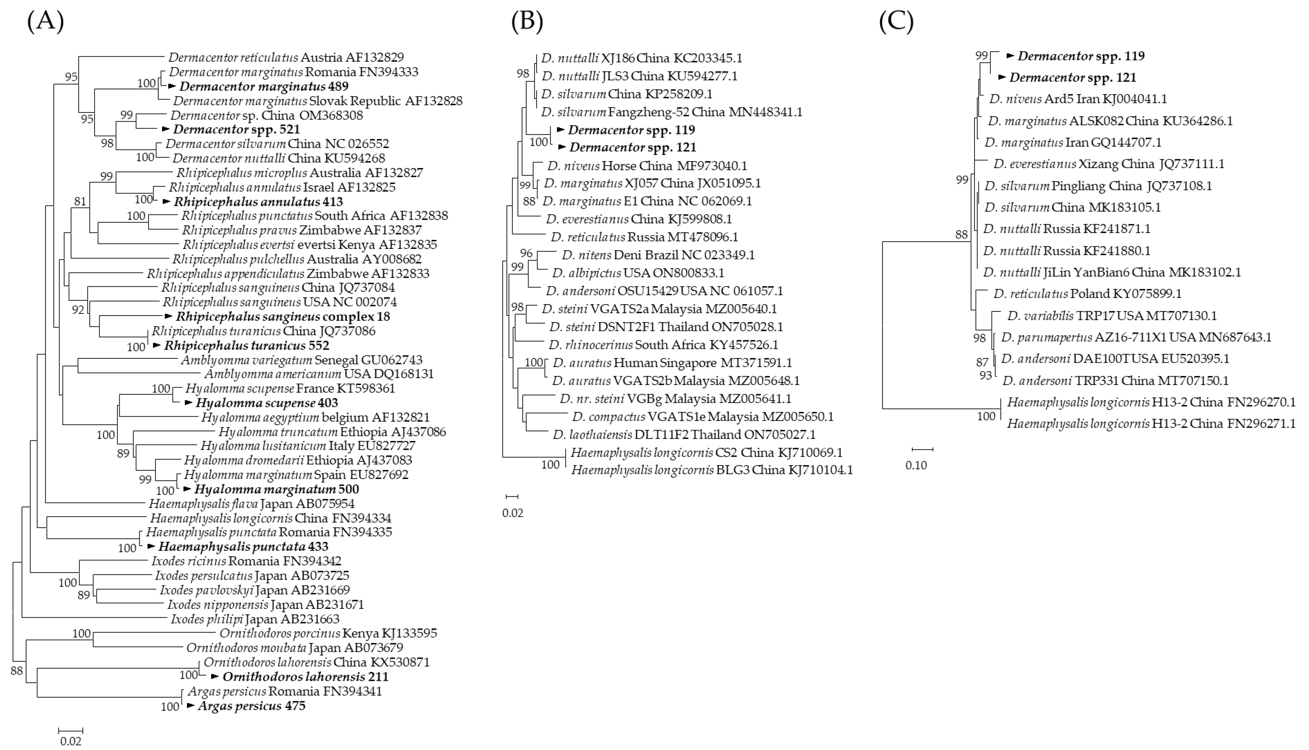
3.1. Molecular Identification of Tick Species

In northern Chuy Province, Kyrgyzstan, 494 ticks were collected and molecularly identified. Among these 494 ticks, 10 species belonging to six genera were represented. The most dominant tick species was Argas persicus (n = 131, 26.5%), followed by Haemaphysalis punctata (n = 89, 18.0%), Dermacentor spp. (n = 79, 16.0%), Rhipicephalus annulatus (n = 58, 11.8%), R. turanicus (n = 54, 10.9%), D. marginatus (n = 38, 7.7%), Hyalomma scupense (n = 22, 4.5%), Hy. marginatum (n = 19, 3.8%), R. sangineus complex (n = 3, 0.6%), and Ornithodoros lahorensis (n = 1, 0.2%) (Figure 1, Supplementary Table S1). The CO1 sequencing results confirmed that each tick sequence clustered at the species level (Figure 2A). However, the R. sangineus complex sequence identified in this study was placed in an independent cluster and was not grouped with other Rhipicephalus species sequences (Figure 2A), and some Dermacentor spp. were placed in different clusters with related reference sequences in the 16S rRNA (Figure 2B) and ITS2 (Figure 2C) analyses.

3.2. Detection of Tick-Borne Pathogens in Ticks

A total of 494 tick specimens were screened for the presence of Anaplasma and Ehrlichia using the 16S rRNA gene. Seventy-one Anaplasma were identified, of which the most common species were A. bovis (n = 44, 8.9%; 95% CI: 0.06–0.12) followed by Anaplasma spp. (n = 20, 4.0%, 95% CI: 0.02–0.06), A. ovis (n = 5, 1.0%; 95% CI: 0.10–0.17), and A. capra (n = 2, 0.4%; 95% CI: 0.00–0.01) (Table 2). In the case of Ehrlichia, only five H. punctata collected from cattle and horses were infected with E. chaffeensis (n = 1, 0.2%; 95% CI: 0.00–0.02) and Ehrlichia spp. (n = 4, 0.8%; 95% CI: 0.00–0.02). In addition, ticks positive for Ehrlichia spp. and E. chaffeensis were coinfected with A. capra and A. bovis, respectively (Table 2).
H. punctata (n = 40, 44.9%) was the most prevalent tick species infected with Anaplasma and Ehrlichia, followed by A. persicus (n = 19, 14.5%), R. annulatus (n = 7, 12.1%), R. turanicus (n = 6, 11.1%), Demacentor spp. (2.5%, n = 2), Hy. marginatum (n = 1, 5.3%), and H. scupense (n = 1, 4.5%) (Table 2). According to the host, the ticks detached from sheep (n = 28, 17.4%) had a high tick-borne pathogen detection rate, followed by those from cattle (n = 22, 18.0%), horses (n = 14, 19.7%), chickens (n = 10, 9.6%), and dogs (n = 5, 5.9%). No pathogens were detected in any ticks taken from cats (Table 2). Ticks collected from the Chuy region (n = 34, 26.8%) had the highest positivity rate of Anaplasma and Ehrlichia followed by Issyk-Ata (n = 19, 11.0%), Sokuluk (n = 6, 17.1%), Bishkek City (n = 4, 13.3%), Moskov (n = 4, 20.0%), Panfilov (n = 3, 9.7%), Tokmok City (n = 3, 9.7%), and Alamudun (n = 3, 6.3%) (Supplementary Table S2).
The representative sequences of different hosts for Anaplasma and Ehrlichia species obtained in this study were deposited in the NCBI database with the following accession numbers: A. bovis (OR234597–OR23463), A. capra (OR150332), A. ovis (OR150327–OR150329), Anaplasma spp. (OR150334, OR150337, OR150343, OR150349), E. chaffeensis (OR140774), and Ehrlichia spp. (OR140773, OR140775–OR140777).

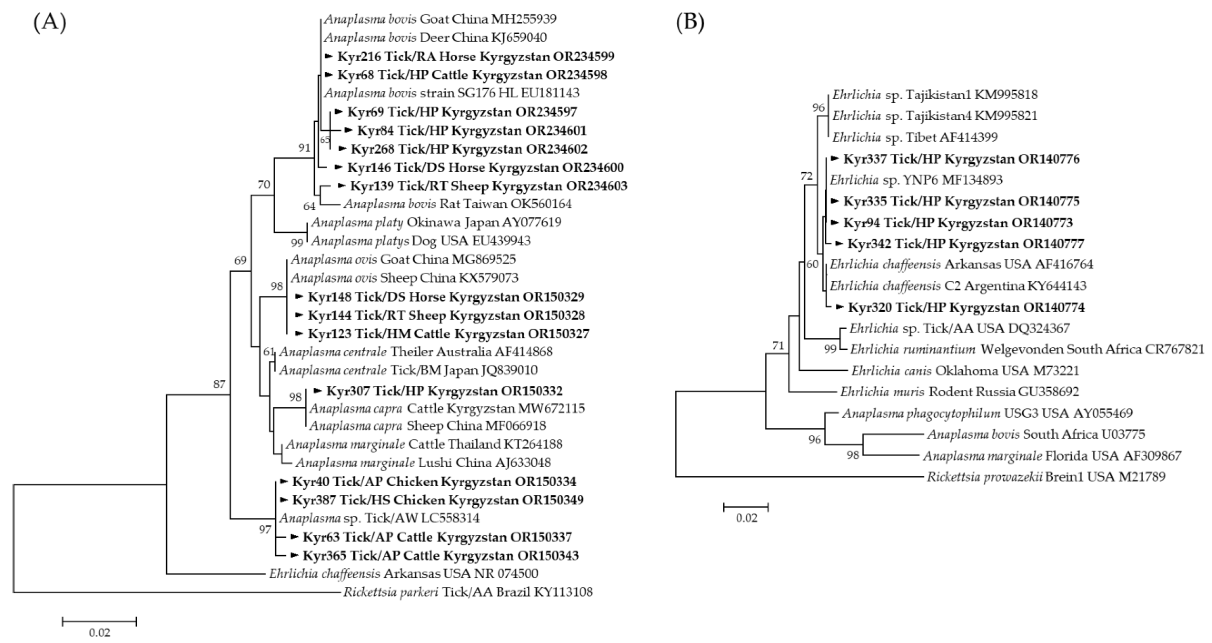
3.3. Molecular and Phylogenetic Analysis of Tick-Borne Pathogens

A total of 12 representative sequences for Anaplasma and 4 representative sequences for Ehrlichia were selected without duplicate sequences among those identified as positive for 71 Anaplasma and 5 Ehrlichia species, respectively (Table 2). Each pathogen was detected in two to three tick species. A. bovis sequences obtained from H. punctata and R. annulatus (OR234598) showed a 100% identity with A. bovis isolated from goats in China (MH255939). A. bovis sequences obtained from R. turanicus (OR234603) were similar to A. bovis isolated from rats in Taiwan (OK560164), and A. bovis sequences obtained from Demacentor spp. and H. punctata (OR234597 and OR23460-OR234602) revealed a 99.30–99.88% similarity with A. bovis isolated from H. longicornis in ROK (EU181143). A. ovis sequences obtained from Hy. marginatum, R. turanicus, and Dermacentor spp. (OR150327) exhibited a 100% identity with A. ovis detected in goats and sheep in China (MG869525 and KX579073, respectively.) A. capra sequences obtained from H. punctata (OR150332) shared 100% and 99.88% identities with A. capra isolated from cattle in Kyrgyzstan (MW672115) and sheep in China (MF066918), respectively. Anaplasma spp. sequences obtained from A. persicus and H. scupense (OR150334, OR150337, OR150343, OR150349) shared a 99.19–99.54% identity with that of Anaplasma spp. isolated from the soft tick, A. walkerae, in Zambia (LC558323). Phylogenetic analysis based on partial 16S rRNA sequences revealed that A. bovis, A. capra, A. ovis, and Anaplasma spp. detected in this study were included in the Anaplasma species (Figure 3A).
The results of the BLAST analysis showed that one positive sequence (OR140774) isolated from H. punctata revealed a 99.74% identity with E. chaffeensis obtained from the Arkansas strain in the USA (AF416764) and C2 strain in Argentina (KY644143). Ehrlichia spp. (OR140773–OR140777) revealed a 99.74–100% identity with Ehrlichia spp., which was deposited in GenBank (MF134893). Phylogenetic analyses demonstrated that the 16S rRNA sequences of E. chaffeensis and Ehrlichia spp. clustered with the previously documented sequences (Figure 3B).

4. Discussion

The present study investigated engorged ticks collected from animal hosts in six regions of Kyrgyzstan to identify their species and the prevalence of Anaplasma and Ehrlichia pathogens within them using molecular detection and phylogenetic analysis.
In accordance with previous studies, Fedorova et al. [21] and Aknazarov et al. [22] identified several tick species in Kyrgyzstan using morphological classification keys and confirmed Ixodes persulcatus, Haemaphysalis punctata, H. erinacei, Hyalomma anatolicum, Hy. Scupense, Hy. Marginatum, Dermacentor marginatus, Rhipicephalus pumilio, R. sanguineus, and R. turanicus. In our investigation, tick species were identified via molecular analysis, and Rhipicephalus annulatus and Ornithodoros lahorensis were newly identified.
The R. sangineus complex comprises several tick species, ranging from R. sanguineus to R. turanicus [38,39]. Owing to their close relationships and similar phenotypic features, the R. sanguineus complex is frequently misidentified as other species within the complex [40]. The taxonomic status of this group remains uncertain, with different classifications provided by various authors [41,42,43]. Although scanning electron microscopy (SEM) and transmission electron microscopy (TEM) have enabled the morphological distinction of Rhipicephalus species, genetic analysis remains the most reliable method for identifying the different species within the R. sanguineus complex [18,38,44]. In this study, three Rhipicephalus species sequences were placed between R. sanguineus and R. turanicus for phylogenetic analysis using CO1 and were determined as the R. sangineus complex.
For tick species’ identification, CO1 should be considered first; if CO1 does not provide reliable results, 16S rRNA, ITS2, or 12S rRNA can be used [19]. Our study found that the sequences of Dermacentor spp. formed a clade distinct from other validated Dermacentor species in the phylogenetic analysis using CO1. Additional phylogenetic analysis was conducted using 16S rRNA and ITS2 for cases in which CO1 was not analyzable.
Little research has been conducted on the prevalence of tick-borne pathogens among ticks infesting animal hosts in Kyrgyzstan. Sang et al. [45] detected five species of piroplasms, Babesia occultans, B. caballi, Theileria ovis, T. annulata, and T. equi, in ticks collected from domestic animals in Kazakhstan. In Tajikistan, Ehrlichia spp. (4/382 pools, 1.1%) and Theileria spp. (3/92 pools, 3.3%) were detected in Hyalomma anatolicum detached from domestic animals [46]. Several studies have investigated the bacterial TBPs harbored by animal hosts in Kyrgyzstan. In 2019, Aktas [27] performed the first molecular epidemiological study on bovine piroplasmosis, including B. major, T. orientalis, and T. annulata, using cattle blood samples. Ozubek [28] identified the genotypes of T. orientalis in the blood of cattle using Single-Strand Conformation Polymorphism (SSCP) analysis. In 2022, Anaplasma species (A. phagocytophilum like-1, A. ovis and A. capra) were detected in cattle [29] and sheep [31] in nine regions of Kyrgyzstan. Recently, Aknazarov [22] reported that blood harvested from sick animals in Kyrgyzstan carried six pathogens, including Anaplasma, Ehrlichia, Babesia, Theileria, Nuttalia, and Hemobartonella, using Giemsa staining in 2023.
Although several studies of tick-borne pathogens have been reported in animal hosts in Kyrgyzstan, no information is available on using molecular tools to characterize their prevalence in ticks. The only pathogen identified in ticks is the tick-borne encephalitis virus (TBEV), which causes severe neurological disorders and is found in Ixodes persulcatus in the northern region [10,11]. Our investigation revealed that seven of the ten tick species including H. punctata, A. persicus, R. annulatus, R. turanicus, Demacentor spp., Hy. marginatum, and H. scupense were PCR-positive for four Anaplasma species (A. bovis, A. capra, A. ovis, and Anaplasma spp.) and two Ehrlichia species (E. chaffeensis and Ehrlichia spp.). In the literature, Anaplasma and Ehrlichia have been confirmed in O. lahorensis [47], D. marginatus [47], and R. sangineus complex [48], but no positive ticks were found in this study.
Anaplasma and Ehrlichia species are known as important tick-borne pathogens of both medical and veterinary importance [49]. These pathogens have been documented in numerous geographical areas and various animal species around the world [23,24,25,26]. According to a previous study, A. bovis was identified in H. punctata from birds in Spain [50], A. ovis was found in R. turanicus, and R. annulatus was identified in goats in Tunisia [51]. Recently, a new Anaplasma species, A. capra, was reported in goats in China [52]. A. capra, which was detected in two H. punctata specimens in our study, showed a 100% identity (43% coverage) with an A. capra sequence isolated from cattle in Kyrgyzstan [29]. In the case of Ehrlichia species, E. chaffeensis was detected in Amblyomma parvum ticks in Argentina [53], and Ehrlichia spp. was detected in Hy. anatolicum, Tajikistan [46].
Aknazarov et al. [22] revealed through the blood smear examination of sick animals in Kyrgyzstan that six species infesting sick animals were carriers of six pathogens (anaplasmosis, ehrlichiosis, babesiosis, theileriosis, nuttaliosis, and hemobartonellosis) during an investigation in 2021 and 2022. Our study revealed that ticks collected from the animals investigated in 2021 were infected with four species of Anaplasma and two species of Ehrlichia. Since pathogens and diseases were identified in both studies (ticks and animals), we believe that the collected species serve as vectors for these two tick-borne diseases and that anaplasmosis and ehrlichiosis silently circulate within this territory in Kyrgyzstan. This study was conducted only on partially or fully engorged ticks collected from animal hosts. Therefore, a limitation of this study is the possibility that the identified TBPs may have originated from animal hosts rather than vectors. Nevertheless, blood-feeding vectors are known to be reliable indicators of the presence or absence of pathogens in an area [54]. Our results also found co-infection with two species of pathogens (E. chaffeensis with A. bovis, and Ehrlichia spp. with A. capra) from two H. punctata specimens, and further investigation is expected to discover more co-infections with various pathogens, such as Babesia and Theileria [27,30,32]. To our knowledge, this is the first study to investigate tick-borne bacterial pathogens in the ticks collected from animal hosts in Kyrgyzstan.
Most populations in Kyrgyzstan depend on livestock for their livelihood. Owing to their nomadic lifestyle and geographical distribution, diverse animal and human exchanges with neighboring countries may facilitate the spread of ticks and tick-borne diseases. The current study focused mainly on the characterization of TBPs in tick vectors in a few areas of northern Kyrgyzstan. Therefore, further studies for various tick species and hosts should be continuously investigated across several regions to monitor the prevalence of TBPs. This study contributes to a better understanding of the epidemiology of tick-borne infections and the possibility of tick-borne diseases in Kyrgyzstan.

Supplementary Materials

The following supporting information can be downloaded at: https://www.mdpi.com/article/10.3390/microorganisms12061046/s1, Table S1: Geographical distribution of tick species collected from animal hosts in Kyrgyzstan in 2021; Table S2: Infection rates of Anaplasma and Ehrlichia in Kyrgyzstan by region.

Author Contributions

Conceptualization, S.Y.K. and H.I.L.; formal analysis, Y.J.K. and J.Y.S.; investigation, Y.J.K., J.S.P., B.A. and N.A; resources, B.A. and N.A.; data curation, Y.J.K.; writing–original draft preparation, Y.J.K.; writing–review and editing, S.Y.K. and H.I.L.; funding acquisition, H.I.L. All authors have read and agreed to the published version of the manuscript.

Funding

This study was financially supported by the Korea Disease Control and Prevention Agency (grant no. 6300-6332-305).

Data Availability Statement

Data supporting the conclusions of this article are included within the article. The newly generated sequences were submitted to the GenBank database under the accession numbers OR150327–OR150329, OR150332, OR150334, OR150337, OR150343, OR150349, OR140773–OR140777, and OR234597–OR23463. The datasets used and/or analyzed during the present study are available from the corresponding author upon reasonable request.

Acknowledgments

We are sincerely grateful to the members of the Division of Bacterial Diseases and the Division of Zoonotic and Vector Borne Disease Research, Korea Disease Control and Prevention Agency (KDCA) for providing the positive control samples used in this study.

Conflicts of Interest

The authors declare no conflicts of interest.

References

  1. Zhumanova, M. A Study on Livestock and Land Management in Kyrgyzstan; FAO: Rome, Italy, 2011. [Google Scholar]
  2. Kydyshov, K.; Usenbaev, N.; Sharshenbekov, A.; Aitkuluev, N.; Abdyraev, M.; Chegirov, S.; Kazybaeva, J.; Brangsch, H.; Melzer, F.; Neubauer, H.; et al. Brucellosis in humans and animals in Kyrgyzstan. Microorganisms 2022, 10, 1293. [Google Scholar] [CrossRef] [PubMed]
  3. Yang, J.; Wang, Y.; Zhang, H.; Su, Y.; Wu, X.; Yan, S.; Yang, S. Impact of socio-economic and environmental factors on livestock production in Kyrgyzstan. Front. Environ. Sci. 2022, 10, 1049187. [Google Scholar] [CrossRef]
  4. Sagynbekova, L. Environment, rural livelihoods, and labor migration: A case study in central Kyrgyzstan. Mt. Res. Dev. 2017, 37, 456–463. [Google Scholar] [CrossRef]
  5. Desta, B. Review on the impact of ticks on livestock health and productivity. J. Biogr. Agric. Health 2016, 6, 1–7. [Google Scholar]
  6. Hurtado, O.J.B.; Giraldo-Ríos, C. Economic and health impact of the ticks in production animals. In Ticks and Tick-Borne Pathogens; Abubakar, M., Perera, P.K., Eds.; IntechOpen: London, UK, 2018. [Google Scholar] [CrossRef]
  7. Aktas, M.; Özübek, S.; Altay, K.; Ipek, N.D.; Balkaya, İ.; Utuk, A.E.; Kırbas, A.; Şimsek, S.; Dumanlı, N. Molecular detection of tick-borne rickettsial and protozoan pathogens in domestic dogs from Turkey. Parasites Vectors 2015, 8, 157. [Google Scholar] [CrossRef]
  8. Seo, J.Y.; Kim, Y.J.; Kim, S.Y.; Lee, H.I. Molecular detection of Anaplasma, Ehrlichia and Rickettsia pathogens in ticks collected from humans in the Republic of Korea, 2021. Pathogens 2023, 12, 802. [Google Scholar] [CrossRef] [PubMed]
  9. Liu, Q.; He, B.; Huang, S.Y.; Wei, F.; Zhu, X.Q. Severe fever with thrombocytopenia syndrome, an emerging tick-borne zoonosis. Lancet Infect. Dis. 2014, 14, 763–772. [Google Scholar] [CrossRef] [PubMed]
  10. Briggs, B.J.; Atkinson, B.; Czechowski, D.M.; Larsen, P.A.; Meeks, H.N.; Carrera, J.P.; Duplechin, R.M.; Hewson, R.; Junushov, A.T.; Gavrilova, O.N.; et al. Tick-borne encephalitis virus, Kyrgyzstan. Emerg. Infect. Dis. 2011, 17, 876–879. [Google Scholar] [CrossRef] [PubMed]
  11. Jung, H.; Choi, C.H.; Lee, M.; Kim, S.Y.; Aknazarov, B.; Nyrgaziev, R.; Atabekova, N.; Jetigenov, E.; Chung, Y.S.; Lee, H.I. Molecular detection and phylogenetic analysis of tick-borne encephalitis virus from ticks collected from cattle in Kyrgyzstan, 2023. Viruses 2024, 16, 107. [Google Scholar] [CrossRef]
  12. de la Fuente, J.; Estrada-Pena, A.; Venzal, J.M.; Kocan, K.M.; Sonenshine, D.E. Overview: Ticks as vectors of pathogens that cause disease in humans and animals. Front. Biosci. 2008, 13, 6938–6946. [Google Scholar] [CrossRef]
  13. de la Fuente, J.; Antunes, S.; Bonnet, S.; Cabezas-Cruz, A.; Domingos, A.G.; Estrada-Peña, A.; Johnson, N.; Kocan, K.M.; Mansfield, K.L.; Nijhof, A.M.; et al. Tick-pathogen interactions and vector competence: Identification of molecular drivers for tick-borne diseases. Front. Cell. Infect. Microbiol. 2017, 7, 114. [Google Scholar] [CrossRef] [PubMed]
  14. Singh, K.; Kumar, S.; Sharma, A.K.; Jacob, S.S.; RamVerma, M.; Singh, N.K.; Shakya, M.; Sankar, M.; Ghosh, S. Economic impact of predominant ticks and tick-borne diseases on Indian dairy production systems. Exp. Parasitol. 2022, 243, 108408. [Google Scholar] [CrossRef] [PubMed]
  15. Bouchard, C.; Dibernardo, A.; Koffi, J.; Wood, H.; Leighton, P.A.; Lindsay, L.R. N Increased risk of tick-borne diseases with climate and environmental changes. Can. Commun. Dis. Rep. 2019, 45, 83–89. [Google Scholar] [CrossRef] [PubMed]
  16. Hussain, S.; Hussain, A.; Ho, J.; Li, J.; George, D.; Rehman, A.; Zeb, J.; Sparagano, O. An epidemiological survey regarding ticks and tick-borne diseases among livestock owners in Punjab, Pakistan: A one health context. Pathogens 2021, 10, 361. [Google Scholar] [CrossRef] [PubMed]
  17. Paguem, A.; Manchang, K.; Kamtsap, P.; Renz, A.; Schaper, S.; Dobler, G.; Bakkes, D.K.; Chitimia-Dobler, L. Ticks and Rickettsiae associated with wild animals sold in bush meat markets in Cameroon. Pathogens 2023, 12, 348. [Google Scholar] [CrossRef] [PubMed]
  18. Folmer, O.; Black, M.; Hoeh, W.; Lutz, R.; Vrijenhoek, R. DNA primers for amplification of mitochondrial cytochrome c oxidase subunit I from diverse metazoan invertebrates. Mol. Mar. Biol. Biotechnol. 1994, 3, 294–299. [Google Scholar] [PubMed]
  19. Lv, J.; Wu, S.; Zhang, Y.; Chen, Y.; Feng, C.; Yuan, X.; Jia, G.; Deng, J.; Wang, C.; Wang, Q.; et al. Assessment of four DNA fragments (COI, 16S rDNA, ITS2, 12S rDNA) for species identification of the Ixodida (Acari: Ixodida). Parasites Vectors 2014, 7, 93. [Google Scholar] [CrossRef] [PubMed]
  20. Abouelhassan, E.M.; El-Gawady, H.M.; Abdel-Aal, A.A.; El-Gayar, A.K.; Esteve-Gassent, M.D. Comparison of some molecular markers for tick species identification. J. Arthropod Borne Dis. 2019, 13, 153–164. [Google Scholar] [CrossRef] [PubMed]
  21. Fedorova, S.Z. Ixodidae ticks in Bishkek. Med. Parazitol. 2005, 4, 34–37. [Google Scholar]
  22. Aknazarov, B.; Jetigenov, E.; Atabekova, N.; Suerkulov, U.; Abdumanap, N. Spread of arthropod-borne infections in Kyrgyzstan. In E3S Web of Conferences; EDP Sciences: Les Ulis, France, 2023. [Google Scholar] [CrossRef]
  23. Orkun, Ö.; Karaer, Z.; Çakmak, A.; Nalbantoğlu, S. Identification of tick-borne pathogens in ticks feeding on humans in Turkey. PLoS Negl. Trop. Dis. 2014, 8, e3067. [Google Scholar] [CrossRef]
  24. Perveen, N.; Muzaffar, S.B.; Al-Deeb, M.A. Ticks and tick-borne diseases of livestock in the Middle East and North Africa: A review. Insects 2021, 12, 83. [Google Scholar] [CrossRef] [PubMed]
  25. Wang, Q.; Pan, Y.S.; Jiang, B.G.; Ye, R.Z.; Chang, Q.C.; Shao, H.Z.; Cui, X.M.; Xu, D.L.; Li, L.F.; Wei, W.; et al. Prevalence of multiple tick-borne pathogens in various tick vectors in Northeastern China. Vector Borne Zoonotic Dis. 2021, 21, 162–171. [Google Scholar] [CrossRef] [PubMed]
  26. Jongejan, F.; Uilenberg, G. The global importance of ticks. Parasitology 2004, 129, S3–S14. [Google Scholar] [CrossRef] [PubMed]
  27. Aktaş, M.; Kısadere, İ.; Özübek, S.; Cihan, H.; Salıkov, R.; Cirak, V.Y. First molecular survey of piroplasm species in cattle from Kyrgyzstan. Parasitol. Res. 2019, 118, 2431–2435. [Google Scholar] [CrossRef] [PubMed]
  28. Ozubek, S.; Ulucesme, M.C.; Cirak, V.Y.; Aktas, M. Detection of Theileria orientalis genotypes from cattle in Kyrgyzstan. Pathogens 2022, 11, 1185. [Google Scholar] [CrossRef] [PubMed]
  29. Altay, K.; Erol, U.; Sahin, O.F.; Aytmirzakizi, A. First molecular detection of Anaplasma species in cattle from Kyrgyzstan; molecular identification of human pathogenic novel genotype Anaplasma capra and Anaplasma phagocytophilum related strain. Ticks Tick Borne Dis. 2022, 13, 101861. [Google Scholar] [CrossRef] [PubMed]
  30. Zhyldyz, A.; Aitakin, K.; Atabek, B.; Elmurat, J.; Rysbek, N.; Jailobek, O.; Ahedor, B.; Otgonsuren, D.; Mumbi, N.N.M.; Guswanto, A.; et al. An epidemiological survey of vector-borne pathogens infecting cattle in Kyrgyzstan. Parasitol. Int. 2023, 97, 102791. [Google Scholar] [CrossRef] [PubMed]
  31. Altay, K.; Erol, U.; Sahin, O.F.; Aytmirzakizi, A.; Temizel, E.M.; Aydin, M.F.; Dumanli, N.; Aktas, M. The detection and phylogenetic analysis of Anaplasma phagocytophilum-like 1, A. ovis and A. capra in sheep: A. capra divides into two genogroups. Vet. Res. Commun. 2022, 46, 1271–1279. [Google Scholar] [CrossRef] [PubMed]
  32. Altay, K.; Erol, U.; Sahin, O.F.; Aydin, M.F.; Aytmirzakizi, A.; Dumanli, N. First molecular evidence of Babesia vogeli, Babesia vulpes, and Theileria ovis in dogs from Kyrgyzstan. Pathogens 2023, 12, 1046. [Google Scholar] [CrossRef]
  33. Lv, J.; Wu, S.; Zhang, Y.; Zhang, T.; Feng, C.; Jia, G.; Lin, X. Development of a DNA barcoding system for the Ixodida (Acari: Ixodida). Mitochondrial DNA 2014, 25, 142–149. [Google Scholar] [CrossRef]
  34. Wang, F.; Wang, D.; Guo, G.; Hu, Y.; Wei, J.; Liu, J. Species delimitation of the Dermacentor ticks based on phylogenetic clustering and niche modeling. PeerJ 2019, 7, e6911. [Google Scholar] [CrossRef] [PubMed]
  35. Barlough, J.E.; Madigan, J.E.; DeRock, E.; Bigornia, L. Nested polymerase chain reaction for detection of Ehrlichia equi genomic DNA in horses and ticks (Ixodes pacificus). Vet. Parasitol. 1996, 63, 319–329. [Google Scholar] [CrossRef] [PubMed]
  36. Oh, J.Y.; Moon, B.; Bae, B.K.; Shin, E.; Ko, Y.H.; Kim, Y.; Park, Y.H.; Chae, J. Genetic identification and phylogenetic analysis of Anaplasma and Ehrlichia species in Haemaphysalis longicornis collected from Jeju Island, Korea. J. Bacteriol. Virol. 2009, 39, 257–267. [Google Scholar] [CrossRef]
  37. Lee, S.O.; Na, D.K.; Kim, C.M.; Li, Y.H.; Cho, Y.H.; Park, J.H.; Lee, J.H.; Eo, S.K.; Klein, T.A.; Chae, J.S. Identification and prevalence of Ehrlichia chaffeensis infection in Haemaphysalis longicornis ticks from Korea by PCR, sequencing and phylogenetic analysis based on 16S rRNA gene. J. Vet. Sci. 2005, 6, 151–155. [Google Scholar] [CrossRef]
  38. Coimbra-Dores, M.J.; Nunes, T.; Dias, D.; Rosa, F. Rhipicephalus sanguineus (Acari: Ixodidae) species complex: Morphometric and ultrastructural analyses. Exp. Appl. Acarol. 2016, 70, 455–468. [Google Scholar] [CrossRef] [PubMed]
  39. Nava, S.; Estrada-Peña, A.; Petney, T.; Beati, L.; Labruna, M.B.; Szabó, M.P.; Venzal, J.M.; Mastropaolo, M.; Mangold, A.J.; Guglielmone, A.A. The taxonomic status of Rhipicephalus sanguineus (Latreille, 1806). Vet. Parasitol. 2015, 208, 2–8. [Google Scholar] [CrossRef] [PubMed]
  40. Hekimoğlu, O.; Sağlam, İ.K.; Özer, N.; Estrada-Peña, A. New molecular data shed light on the global phylogeny and species limits of the Rhipicephalus sanguineus complex. Ticks Tick-Borne Dis. 2016, 7, 798–807. [Google Scholar] [CrossRef] [PubMed]
  41. Camicas, J.L.; Hervy, J.P.; Adam, F.; Morel, P.C. The Ticks of the World (Acarida, Ixodida): Nomenclature, Described Stages, Hosts, Distribution; Orstom Editions: Paris, Italy, 1998. [Google Scholar]
  42. Gray, J.; Dantas-Torres, F.; Estrada-Peña, A.; Levin, M. Systematics and ecology of the brown dog tick, Rhipicephalus sanguineus. Ticks Tick-Borne Dis. 2013, 4, 171–180. [Google Scholar] [CrossRef] [PubMed]
  43. Walker, J.B.; Keirans, J.E.; Horak, I.G. The Genus Rhipicephalus (Acari, Ixodidae): A Guide to the Brown Ticks of the World; Cambridge University Press: Cambridge, UK, 2000. [Google Scholar]
  44. Caetano, R.L.; Vizzoni, V.F.; Bitencourth, K.; Carriço, C.; Sato, T.P.; Pinto, Z.T.; De Oliveira, S.V.; Amorim, M.; Voloch, C.M.; Gazeta, G.S. Ultrastructural morphology and molecular analyses of tropical and temperate ‘species’ of Rhipicephalus sanguineus sensu lato (Acari: Ixodidae) in Brazil. J. Med. Entomol. 2017, 54, 1201–1212. [Google Scholar] [CrossRef] [PubMed]
  45. Sang, C.; Yang, M.; Xu, B.; Liu, G.; Yang, Y.; Kairullayev, K.; Bauyrzhan, O.; Hazihan, W.; Hornok, S.; Wang, Y. Tick distribution and detection of Babesia and Theileria species in Eastern and Southern Kazakhstan. Ticks Tick-Borne Dis. 2021, 12, 101817. [Google Scholar] [CrossRef]
  46. Kartashov, M.Y.; Kononova, Y.V.; Petrova, I.D.; Tupota, N.L.; Mikryukova, T.P.; Ternovoi, V.A.; Tishkova, F.H.; Loktev, V.B. Detection of Ehrlichia spp. and Theileria spp. in Hyalomma anatolicum ticks collected in Tajikistan. Vavilovskii Zhurnal Genet. Selektsii 2020, 24, 55–59. [Google Scholar] [CrossRef] [PubMed]
  47. Tajedin, L.; Bakhshi, H.; Faghihi, F.; Telmadarraiy, Z. High infection of Anaplasma and Ehrlichia spp. among tick species collected from different geographical locations of Iran. Asian Pac. J. Trop. Dis. 2016, 6, 787–792. [Google Scholar] [CrossRef]
  48. Bowman, D.D. Introduction to the alpha-proteobacteria: Wolbachia and Bartonella, Rickettsia, Brucella, Ehrlichia, and Anaplasma. Top. Companion Anim. Med. 2011, 26, 173–177. [Google Scholar] [CrossRef]
  49. Dumler, J.S. Anaplasma and Ehrlichia infection. Ann. N. Y. Acad. Sci. 2005, 1063, 361–373. [Google Scholar] [CrossRef] [PubMed]
  50. Palomar, A.M.; Portillo, A.; Santibáñez, P.; Mazuelas, D.; Roncero, L.; García-Álvarez, L.; Santibáñez, S.; Gutiérrez, Ó.; Oteo, J.A. Detection of tick-borne Anaplasma bovis, Anaplasma phagocytophilum and Anaplasma centrale in Spain. Med. Vet. Entomol. 2015, 29, 349–353. [Google Scholar] [CrossRef] [PubMed]
  51. Belkahia, H.; Ben Said, M.; Ghribi, R.; Selmi, R.; Ben Asker, A.; Yahiaoui, M.; Bousrih, M.; Daaloul-Jedidi, M.; Messadi, L. Molecular detection, genotyping and phylogeny of Anaplasma spp. in Rhipicephalus ticks from Tunisia. Acta Trop. 2019, 191, 38–49. [Google Scholar] [CrossRef] [PubMed]
  52. Li, H.; Zheng, Y.C.; Ma, L.; Jia, N.; Jiang, B.G.; Jiang, R.R.; Huo, Q.B.; Wang, Y.W.; Liu, H.B.; Chu, Y.L.; et al. Human infection with a novel tick-borne Anaplasma species in China: A surveillance study. Lancet Infect. Dis. 2015, 15, 663–670. [Google Scholar] [CrossRef] [PubMed]
  53. Tomassone, L.; Nuñez, P.; Gürtler, R.E.; Ceballos, L.A.; Orozco, M.M.; Kitron, U.D.; Farber, M. Molecular detection of Ehrlichia chaffeensis in Amblyomma parvum ticks, Argentina. Emerg. Infect. Dis. 2008, 14, 1953–1955. [Google Scholar] [CrossRef] [PubMed]
  54. Sparagano, O.A.E.; Allsopp, M.; Mank, R.; Rijpkema, S.; Figueroa, J.; Jongejan, F. Molecular detection of pathogen DNA in ticks (Acari: Ixodidae): A review. Exp. Appl. Acarol. 1999, 23, 929–960. [Google Scholar] [CrossRef]
Figure 1. A map of tick species’ composition in the different sampling regions of Chuy Province, Kyrgyzstan, in 2022. The different colors indicate different tick species. The sizes of the circles represent the number of collected ticks and the pie charts illustrate their relative proportions in each collection region.
Microorganisms 12 01046 g001
Figure 2. (A) Phylogenetic analysis based on CO1 (597 bp), (B) 16S rRNA genes (345 bp), and (C) ITS2 (655 bp). The sequences identified in this study are indicated in bold (▶). The phylogenetic tree was constructed using the neighbor-joining method (A) and maximum likelihood method (B,C) based on the Kimura 2-parameter mode. The numbers on the branches indicate bootstrap percentages based on 1000 replications. The cut-off value for the consensus tree was 60%. Over 60% of the total number were collected in Issyk-Ata (n = 172, 34.8%) and Chuy (n = 127, 25.7%) followed by Alamudun (n = 48, 9.7%), Sokuluk (n = 35, 7.1%), Panfilov (n = 31, 6.3%), Tokmok City (n = 31, 6.3%), Bishkek City (n = 30, 6.1%), and Moskov (n = 20, 4.0%) (Figure 1). Regarding tick infestation in host animals, 494 of partially or fully engorged ticks were collected from sheep (n = 161, 32.6%), followed by cattle (n = 122, 24.7%), chickens (n = 104, 21.1%), horses (n = 71, 14.4%), dogs (n = 34, 6.9%), and cats (n = 2, 0.4%) (Supplementary Table S1).
Microorganisms 12 01046 g002
Figure 3. Phylogenetic relationships for Anaplasma and Ehrlichia based on the partial nucleotide sequence of (A) Anaplasma 16S rRNA, (B) Ehrlichia 16S rRNA. The neighbor-joining method was used to construct a phylogenetic tree. The numbers at the nodes represent the proportion of bootstrap values for the branch point. The positive sequences for Anaplasma and Ehrlichia identified in this study are indicated in bold (▶). Scale bars indicate sequence distances. Reference strains of Anaplasma and Ehrlichia with the host, country of detection, and the National Center for Biotechnology Information accession numbers are also shown. The cut-off value for the consensus tree was 60%. (AP: A. persicus, DS: Dermacentor spp., HM: H. marginatum, HP: H. punctata, RA: R. annulatus, RT: R. turanicus).
Microorganisms 12 01046 g003
Table 1. Primer sets used for the identification of tick and Anaplasma and Ehrlichia species.
TargetPrimersSequence (5′ to 3′)Amplicon
Length (bp)
References
CO1LCO1490
HC02198
GGTCAACAAATCATAAAGATATTGG
TAAACTTCAGGGTGACCAAAAAATCA
710[18]
16S rRNA16S_F
16S_R2
TTAAATTGCTGTRGTATT
CAACATCGAGGTCGCAAWCYA
395[33]
ITS2F3/1
R1/1
GGGTCGATGAAGAACGCAGCCAGC
TTCAGGGGGTTGTCTCGCCTGATG
~1039[34]
Anaplasma
16S rRNA
AE1-F
AE1-R
1st PCRAAGCTTAACACATGCAAGTCGAA
AGTCACTGACCCAACCTTAAATG
1406[35]
EE3
EE4
2nd PCRGTCGAACGGATTATTCTTTATAGCTTGC
CCCTTCCGTTAAGAAGGATCTAATCTCC
926
Ehrlichia
16S rRNA
AE1-F
AE1-R
1st PCRAAGCTTAACACATGCAAGTCGAA
AGTCACTGACCCAACCTTAAATG
1406[36]
HE1
HE3
2nd PCRCAATTGCTTATAACCTTTTGGTTATAAAT
TATAGGTACCGTCATTATCTTCCCTAT
390[37]
Table 2. Infection rate of Anaplasma- and Ehrlichia-positive tick species collected from animal hosts in Kyrgyzstan in 2021.
HostTick SpeciesNumber of
Tested Ticks
Detected Pathogens in Ticks
A. bovisA. capraA. ovisAnaplasma spp.E. chaffeensisEhrlichia spp.Total (%)
CatRhipicephalus turanicus20000000
Subtotal20000000 (0)
CattleArgas persicus410009009
Dermacentor marginatus280000000
Dermacentor spp.120000000
Haemaphysalis punctata2110 (1 *)0001 (1 )112
Hyalomma marginatum90010001
Hyalomma scupense20000000
Rhipicephalus annulatus90000000
Subtotal122100191122 (18.0)
ChickenArgas persicus820009009
Haemaphysalis punctata20000000
Hyalomma scupense190001001
Ornithodoros lahorensis10000000
Subtotal104000100010 (9.6)
DogRhipicephalus annulatus40000000
Rhipicephalus sangineus complex10000000
Rhipicephalus turanicus292000002
Subtotal342000002 (5.9)
HorseDermacentor spp.61010002
Haemaphysalis punctata2011 (1 **)0003 (1 ††)5
Rhipicephalus annulatus457000007
Subtotal7191100314 (19.7)
SheepArgas persicus80001001
Dermacentor marginatus100000000
Dermacentor spp.610000000
Haemaphysalis punctata46221000023
Hyalomma marginatum100000000
Hyalomma scupense10000000
Rhipicephalus sangineus complex20000000
Rhipicephalus turanicus231030004
Subtotal161231310028 (17.4)
Total (%)Argas persicus131000190019 (14.5)
Dermacentor marginatus380000000 (0.0)
Dermacentor spp.791010002 (2.5)
Haemaphysalis punctata89332001440 (44.9)
Hyalomma marginatum190010001 (5.3)
Hyalomma scupense220001001 (4.5)
Ornithodoros lahorensis10000000 (0.0)
Rhipicephalus annulatus587000007 (12.1)
Rhipicephalus sangineus complex30000000 (0.0)
Rhipicephalus turanicus543030006 (11.1)
Total (%)49444 (8.9)
(CI 0.06–0.12)
2 (0.4)
(CI 0.0–0.01)
5 (1.0)
(CI 0.10–0.17)
20 (4.0)
(CI 0.02–0.06)
1 (0.2)
(CI 0.00–0.01)
4 (0.8)
(CI 0.00–0.02)
76 (15.3)
(CI 0.12–0.19)
* co-infection with E. chaffeensis; ** co-infection with Ehrlichia spp.; co-infection with A. bovis. †† co-infection with A. capra.
Disclaimer/Publisher’s Note: The statements, opinions and data contained in all publications are solely those of the individual author(s) and contributor(s) and not of MDPI and/or the editor(s). MDPI and/or the editor(s) disclaim responsibility for any injury to people or property resulting from any ideas, methods, instructions or products referred to in the content.

