Article

A Novel View on the Taxonomy of Sulfate-Reducing Bacterium ‘Desulfotomaculum salinum’ and a Description of a New Species Desulfofundulus salinus sp. nov.

by
Tamara N. Nazina
1,*,
Tatyana P. Tourova
1,
Denis S. Grouzdev
2,
Salimat K. Bidzhieva
1 and
Andrey B. Poltaraus
3
1
Winogradsky Institute of Microbiology, Research Center of Biotechnology, Russian Academy of Sciences, 119071 Moscow, Russia
2
SciBear OU, Tartu mnt 67/1-13b, 10115 Tallinn, Estonia
3
Engelhardt Institute of Molecular Biology, Russian Academy of Sciences, 119991 Moscow, Russia
*
Author to whom correspondence should be addressed.
Microorganisms 2024, 12(6), 1115; https://doi.org/10.3390/microorganisms12061115
Submission received: 8 May 2024 / Revised: 24 May 2024 / Accepted: 27 May 2024 / Published: 30 May 2024

Abstract

:
Two thermophilic spore-forming sulfate-reducing strains, 435T and 781, were isolated from oil and gas reservoirs in Western Siberia (Russia) about 50 years ago. Both strains were found to be neutrophilic, chemoorganotrophic, anaerobic bacteria, growing at 45–70 °C (optimum, 55–60 °C) and with 0–4.5% (w/v) NaCl (optimum, 0.5–1% NaCl). The major fatty acids were iso-C15:0, iso-C17:0, C16:0, and C18:0. In sulfate-reducing conditions, the strains utilized H2/CO2, formate, lactate, pyruvate, malate, fumarate, succinate, methanol, ethanol, propanol, butanol, butyrate, valerate, and palmitate. In 2005, based on phenotypic characteristics and a 16S rRNA gene sequence analysis, the strains were described as ‘Desulfotomaculum salinum’ sp. nov. However, this species was not validly published because the type strain was not deposited in two culture collections. In this study, a genomic analysis of strain 435T was carried out to determine its taxonomic affiliation. The genome size of strain 435T was 2.886 Mb with a 55.1% genomic G + C content. The average nucleotide identity and digital DNA–DNA hybridization values were highest between strain 435T and members of the genus Desulfofundulus, 78.7–93.3% and 25.0–52.2%, respectively; these values were below the species delineation cut-offs (<95–96% and <70%). The cumulative phenotypic and phylogenetic data indicate that two strains represent a novel species within the genus Desulfofundulus, for which the name Desulfofundulus salinus sp. nov. is proposed. The type strain is 435T (=VKM B-1492T = DSM 23196T). A genome analysis of strain 435T revealed the genes for dissimilatory sulfate reduction, autotrophic carbon fixation via the Wood–Ljungdahl pathway, hydrogen utilization, methanol and organic acids metabolism, and sporulation, which were confirmed by cultivation studies.

1. Introduction

O, be some other name!
What’s in a name! that which we call a rose
By any other name would smell as sweet…
William Shakespeare, Romeo and Juliet
For a long time, the genus Desulfotomaculum was the only known genus of spore-forming sulfate-reducing bacteria [1]. Application of marker genes and genomic approaches to a number of phenotypically similar spore-forming sulfidogens of the genus Desulfotomaculum led to the revision of this genus and establishment of new genera—Desulfosporosinus, Desulfallas, Desulfofundulus, Desulfofarcimen, and Desulfohalotomaculum [2,3].
Thermophilic sulfate-reducing spore-forming bacteria, strains 435T (VKM B-1492T, DSM 23196T) and 781 (VKM B-1379), were isolated about 50 years ago from high-temperature hydrocarbon deposits in Western Siberia (Russia) [4]. The new isolates were phenotypically close to the species Desulfotomaculum nigrificans [1]. However, the strains had halotolerant properties, and it was proposed to classify them as a subspecies Desulfotomaculum nigrificans subsp. salinus [4]. Morphology, ultrastructural organization, and physiology, including the ability to fix nitrogen of strains 435T and 781 were studied, and the results were reported in papers [5,6,7]. DNA-DNA hybridization and phylogenetic analysis of their 16S rRNA genes were performed, which made it possible to classify strains 435T and 781 to the new species ‘Desulfotomaculum salinum’ and to propose a description of the species [8]. However, at this time, the type strain 435T was deposited in only one collection of microorganisms (VKM B-1492T), and therefore the new species was not validly published. Later, strain 435T was deposited in the German Collection of Microorganisms and Cell Cultures (DSMZ) under the number DSM 23196.
Sequencing and preliminary comparative analysis of the genome of strain 435T [9] showed that it was most closely phylogenetically related to members of the genus Desulfofundulus. The overall genomic relatedness indices (OGRIs) of strain 435T, such as average nucleotide identity (ANI) and digital DNA-DNA hybridization (dDDH) were below the commonly used threshold of 95–96% and 70%, respectively, for species designation [10], and confirmed the independent species status of strain 435T within the genus Desulfofundulus. The name ‘Desulfofundulus salinum’ has been proposed for the new species.
The genus Desulfofundulus was proposed by Watanabe et al. [3,11] based on a comparative phylogenetic analysis of the 16S rRNA genes of members of the polyphyletic genus Desulfotomaculum. The main features of the bacteria of the genus Desulfofundulus are rod-shaped cells, motility, spore formation, obligate anaerobiosis, thermophily, and the use of sulfate and thiosulfate as electron acceptors. Desulfofundulus spp. were isolated from subsurface thermal mineral water, formation water of oil reservoirs, hot springs, methanogenic reactors, and others. The genus Desulfofundulus belongs to the family Peptococcaceae of the order Eubacteriales in the class Clostridia, phylum Bacillota. Assignment of the genus Desulfotomaculum with the type species Desulfotomaculum nigrificans to the family Desulfotomaculaceae of the order Eubacteriales in the class Clostridia has recently been proposed [12].
At the time of writing this article in March 2024, the genus Desulfofundulus included nine species of thermophilic spore-forming sulfate-reducing bacteria—D. kuznetsovii, D. australicus, D. luciae, D. solfataricus, D. thermoacetoxidans, D. thermobenzoicus, D. thermocisternus, and D. thermosubterraneus; the genus also includes ‘Desulfofundulus salinum’ as a not validly published species (List of Prokaryotic Names with Standing in Nomenclature (LPSN), https://lpsn.dsmz.de/genus/desulfofundulus, accessed on 5 March 2024) [13]. The type species of the genus is Desulfofundulus kuznetsovii [3,14,15].
The purpose of this work was to combine previously published information on the phenotypic characteristics of thermophilic spore-forming sulfate-reducing bacterial strains 435T and 781, to perform phylogenetic and genomic analyses, and to provide a formal description of the species Desulfofundulus salinus sp. nov.

2. Materials and Methods

2.1. Origin of Bacterial Strains

Strains 435T (VKM B-1492T, DSM 23196T) and 781 (VKM B-1379) were isolated from samples of reservoir fluids taken by E.P. Rozanova from high-temperature hydrocarbon deposits (Western Siberia, Russia), as previously described [4]. The strain 435T was isolated in 1973 by E.P. Rozanova from a mixture of condensation and reservoir water coming from well 435 of the Igrim gas field (63°06′2.12″ N, 64°19′54.15″ E), located in the Berezovsky district (Khanty-Mansiysk Autonomous Okrug, Yugra, Russia). The water came from terrigenous Jurassic sediments from a depth of 1600 to 1620 m. The reservoir temperature was 59 °C. The reservoir water belonged to the chloride–calcium type and contained 15.3 g of salt per liter. Strain 781 was isolated in 1974 by T.N. Nazina from a sample of reservoir water from production well 781 of the Ust-Balyk oil and gas field (61°05′ N, 72°18′33.33″ E), located in the Nefteyugansk district (Khanty-Mansiysk Autonomous Okrug, Yugra, Russia). The reservoir water came from the terrigenous strata of Cretaceous sediments from a depth of about 2100 m, belonged to the chloride–calcium type, and contained about 16.0 g of salts per liter. The reservoir temperature was 63–70 °C. Sulfates and hydrogen sulfide were absent in both studied water samples. Since the isolation, the strains have been stored in the Laboratory of Petroleum Microbiology, Winogradsky Institute of Microbiology, Research Center of Biotechnology, Russian Academy of Sciences.

2.2. Media Composition and Isolation

Postgate’s B, C, D, and N media were used to isolate the strains [16]; media were amended with 10–20% v/v formation water from the Ust-Balyk oil and gas field, which in subsequent experiments was replaced by 0.5–1.0% w/v NaCl. The strains were isolated by successive transfer from a liquid medium to a solid one and subsequent inoculation of single black colonies to a liquid medium. The strains were also cultivated in Widdel’s [17] medium brackish No. 1 containing the following (g/L distilled water): NaCl—5.0, MgCl2—0.6, KCl—0.3, CaCl2—0.1, NH4Cl—0.3, KH2PO4—0.2, Na2SO4—2.8, NaHCO3—1.5, and sodium lactate—3.5; the medium was reduced by Na2S·9H2O—0.3 and Na2S2O4—0.05. Trace elements [18] were added to the medium. The bacteria were cultured in Hungate’s tubes, and the gas phase was represented by molecular nitrogen or argon.
For comparative studies, Desulfofundulus (formerly Desulfotomaculum) kuznetsovii strain 17T was used which, since its isolation, has also been stored in the collection of the Laboratory of Petroleum Microbiology, Winogradsky Institute of Microbiology, Research Center of Biotechnology, Russian Academy of Sciences [3,14,15]. Strain 17T was cultured in Widdel’s medium for freshwater bacteria [17]. The strains were incubated at 60 °C unless otherwise indicated.

2.3. Phenotypic Characterization

The cell morphology was studied by phase contrast microscopy using a Zetopan light microscope (Reichert, Vienna, Austria). Gram staining of the cells was performed using a relevant kit (Merck KGaA, Darmstadt, Germany). The ultrastructural organization of the cells was studied in strains grown in Postgate’s B medium with pyruvate, as previously described [5]. Cells were examined under a transmission electron microscope JEM-100C (JEOL, Tokyo, Japan). The temperature, salinity, and pH ranges for the growth of strains 435T and 781 were determined in a liquid Widdel’s medium with sulfate, in which fumarate (1 g/L) served as the substrate as described by Nazina et al. [8]. The range of substrates used was determined on media that did not contain yeast extract. Alcohols, malate, and oxalate were added in an amount of 1 g/L, and salts of other organic acids and sugars at 3 g/L. Possible electron acceptors for strains 435T and 781 were determined on a Widdel’s medium with sodium lactate, in which the sulfate was replaced by thiosulfate (2.0 g/L), sulfite (1.0 g/L), elemental sulfur (1.5 g/L), or sodium nitrate (1 g/L). The possibility of thiosulfate dismutation was detected on a Widdel’s mineral medium, in which the sulfate was replaced with thiosulfate. The formation of SO42− was measured turbidimetrically [19]. The fixation of molecular nitrogen was determined by the acetylene method as the formation of ethylene, which was detected by gas chromatography [6]. The composition of cellular fatty acids was determined by gas chromatography–mass spectrometry on an HP-5973D device from Agillent Technologies (formerly Hewlett-Packard, Palo Alto, CA, USA) [8].
The growth of the strains was estimated by the optical density of the bacterial suspension at 660 nm, which was measured on an Ultrospec 2100 pro spectrophotometer (Amersham Biosciences, Amersham, Buckinghamshire, UK), as well as by the formation of sulfide, which was determined by the colorimetric method using N,N-dimethyl-paraphenylenediamine [20]. Growth experiments were performed in triplicate.

2.4. The 16S rRNA Gene and Genome Sequencing and Analysis

The methods for DNA isolation and sequencing of the 16S rRNA genes from strains 435T and 781 were described earlier [8]. The 16S rRNA gene of strain 435T was re-amplified using the strain’s template DNA and the universal bacterial primer set 27F (5′-AGAGTTTGATCCTGGCTCA-3′) and 1492R (5′-AGAGTTTGATCCTGGCTCAG-3′) [21]. DNA sequencing was performed on an ABI 3500 xL DNA analyzer automatic sequencer using an ABI PRISM® BigDye™ Terminator v. 3.1 reagent kit (Thermo Fisher Scientific, Waltham, MA, USA) in accordance with the manufacturer’s recommendations.
The methods of isolation of genomic DNA of strain 435T, sequencing conditions, programs for checking the quality of the obtained sequences, and assembling scaffolds from contigs were described by Grouzdev et al. [9]. The average nucleotide identity (ANI) was determined using FastANI v. 1.3 [22]. Digital DNA-DNA hybridization (dDDH) of genomes was performed using the Genome-to-Genome Distance Calculator (GGDC) v. 2.1 [23]. The identification of protein-coding sequences and primary annotation of genes were performed using the NCBI Prokaryotic Genome Automatic Annotation Pipeline (PGAP) [24]. In addition, to identify protein-coding sequences, we used UniProt (https://www.uniprot.org/, release 2024_01, accessed on 24 January 2024) [25].
The 16S rRNA gene sequences were aligned using MUSCLE [26]. Phylogenomic analysis was performed using concatenated sequences of 120 marker genes obtained using GTDB-Tk version 1.0.2 [27]. The maximum likelihood tree of the 16S rRNA gene sequences was constructed with a GTR + F + I + G4 model, and the phylogenomic tree was constructed with a LG + F + I + G4 model recommended by ModelFinder [28] in IQ-TREE [29]. Branch supports were obtained with 10,000 ultrafast bootstraps [30]. Maximum parsimony and neighbor-joining trees were reconstructed using MPBoot [31] and MEGA11 [32], respectively. A pangenomic analysis was conducted using the bioinformatic pipeline proposed by Delmont and Eren [33], and Anvi’o version 7.0 [34]. Genomes were arranged based on the distribution of gene clusters using the MCL algorithm (Euclidean distance, Ward linkage).
The reconstruction of possible metabolic pathways was carried out based on comparison of the genome of the strain 435T using BV-BRC (PATRIC) 3.35.5 (https://www.bv-brc.org/, accessed on 1 March 2024) [35], MetaCyc version 27.5 (https://metacyc.org/, accessed on 1 November 2023) [36], RAST v. 2.0 (https://rast.nmpdr.org/rast.cgi, accessed on 12 October 2023) [37] and the BlastKOALA tool, KEGG version 3.0 (https://www.kegg.jp/blastkoala/, accessed on 1 April 2023) [38]. Gene clusters of carbon dioxide fixation, sulfate reduction, propanoate degradation, and dinitrogen fixation were compared using an online service Gene Graphics version 2.02 (https://katlabs.cc/genegraphics/app, accessed on 1 October 2023) [39]. The circular genome map of strain 435T was constructed using the Proksee web service version 6.0.3 (https://proksee.ca/, accessed on 5 March 2024) [40].

2.5. Nucleotide Sequence Accession Numbers

The GenBank/EMBL/DDBJ accession numbers for the 16S rRNA gene sequences of strains 435T and 781 are AY918122 and AY918123, respectively. The genome shotgun project of strain 435T was deposited at DDBJ/ENA/GenBank under accession no. RBWE00000000. The version described in this paper is the first version, RBWE01000000. The NCBI genomic assembly accession number of strain 435T is GCF_003627965.1.

3. Results

In this study, the taxonomic position of sulfate-reducing bacterial strains 435T and 781, previously described as ‘Desulfotomaculum salinum’ [8], was clarified; the genome of strain 435T was analyzed and the indices of the genomic relationship were determined to clarify its taxonomic position, taking into account modern approaches. Genome analysis made it possible to correlate the genomic information and phenotypic features of strain 435T and to give a formal description of the new species, Desulfofundulus salinus sp. nov.

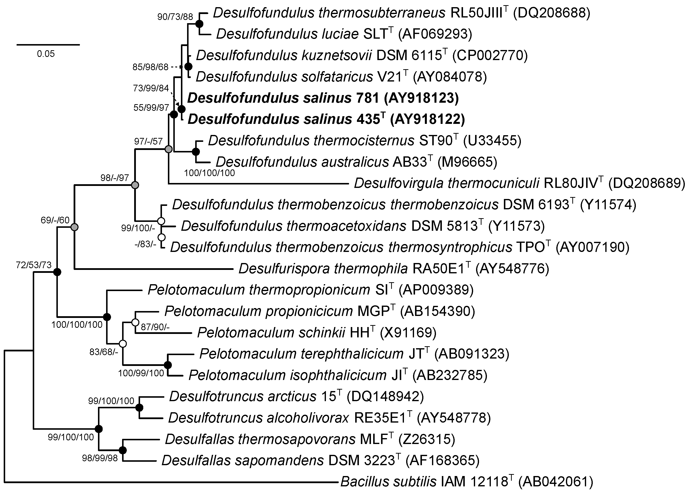
3.1. Phylogenetic Analysis of the 16S rRNA Gene

Phylogenetic analysis of 16S rRNA gene sequences in EzBioCloud and BLAST showed that strain 435T showed a 99.4% similarity with strain 781 and a 98.4%, 98.0%, 97.5%, 95.6%, and 94.7% similarity with the respective genes of the nearest members of the genus Desulfofundulus—D. solfataricus V21T, D. kuznetsovii 17T, D. thermocisternus ST90T, D. thermosubterraneus RL50JIIIT, and D. luciae SLTT. On the phylogenetic tree constructed using the NJ, MP, and ML methods, both strains formed a separate branch within the cluster of phylogenetically closely related Desulfofundulus species, including the type species of this genus D. kuznetsovii 17T, as well as D. solfataricus V21T, D. luciae SLTT, and D. thermosubterraneus RL50JIIIT (Figure 1).
This indicated that the studied strains 435T and 781 belonged to the genus Desulfofundulus. Importantly, when the trees were constructed using the neighbor-joining and maximum likelihood algorithms, the genus Desulfofundulus appears to be polyphyletic. D. thermoacetoxidans and D. thermobenzoicus subsp. thermosyntrophicus formed a separate cluster on the phylogenetic tree, suggesting that further taxonomic revision may be required to fully resolve the evolutionary relationships within this group.

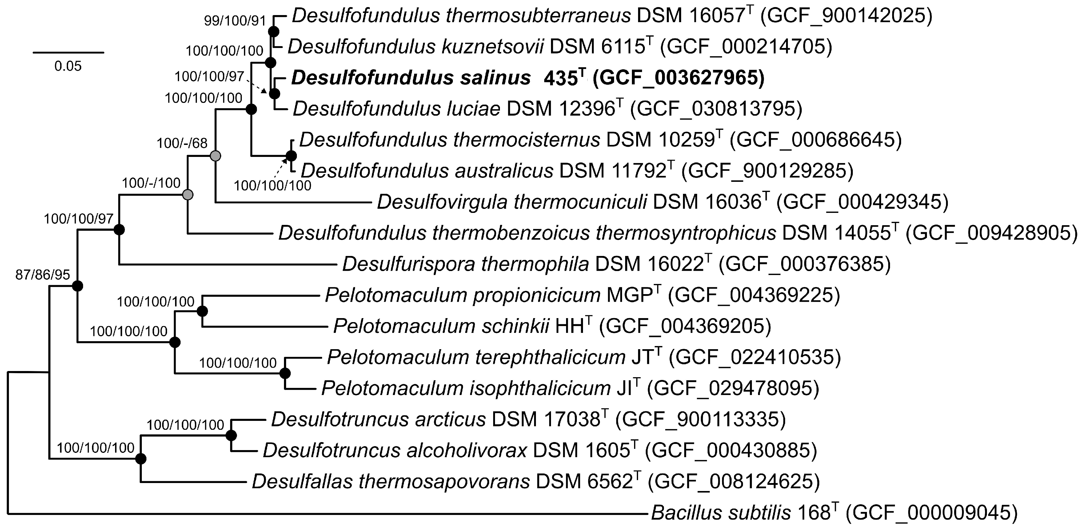
3.2. Genome Features and Phylogeny

The genome of the strain 435T was composed of 10 scaffolds with a total genomic length of 2,886,683 bp, an N50 value of 121.7 kb, a G + C content of 55.1%, and coverage of 137× [9]. The genome comprised 2925 annotated genes, including 2827 protein-coding sequences, 42 pseudogenes, and 56 RNA genes. The ANI and dDDH values of the 435T genome with the genomes of the type strains of the genus Desulfofundulus were in a range 78.7–93.3% and 25.0–52.2%, respectively (Table 1). These values were below the thresholds of 95–96% for ANI and 70% for dDDH, accepted for assigning bacteria to a single species [10,41], which indicates that strain 435T belongs to a new species.
To identify the phylogenetic position of strain 435T, a phylogenetic tree was constructed based on concatenated 120 single-copy proteins. On the phylogenetic tree, strain 435T formed a separate branch in a cluster with the type strains D. kuznetsovii 17T, D. luciae SLTT, and D. thermosubterraneus RL50JIIIT (Figure 2).
As noted, a topology of the ribosomal tree (Figure 1), a low level of similarity of the 16S rRNA genes of D. thermoacetoxidans CAMZT, D. thermobenzoicus subsp. thermobenzoicus TSBT, and D. thermobenzoicus subsp. thermosyntrophicus TPO with those of other members of the genus Desulfofundulus, as well as low values of the genomic relatedness indices dDDH, ANI, and a separate position on the phylogenomic tree of the strain D. thermobenzoicus subsp. thermosyntrophicus TPO, suggests that this phylogenetic group belongs to a separate new genus.
This assumption is also consistent with the GTDB database (release 08-RS214), which assigned D. thermobenzoicus subsp. thermosyntrophicus TPO to the distinct genus Desulfofundulus_A. However, for a reliable description of the new genus, information on the genomes of the type strains D. thermoacetoxidans CAMZT and D. thermobenzoicus subsp. thermobenzoicus TSBT is required.
A comprehensive pangenomic analysis was performed on genomes from seven Desulfofundulus strains and Desulfovirgula thermocuniculi, with the genome of Desulfotomaculum (Dt.) nigrificans as the outgroup. This dataset comprised 28,350 genes organized into 8268 gene clusters (GCs). Within this pangenomic landscape, 951 core clusters were consistently present across all genomes analyzed, of which 766 were single-copy genes (SCGs), as shown in Figure 3. The phylogenetic tree reconstructed from these SCGs, spanning 233,613 amino acid sites, mirrored the topology of the phylogenomic tree derived from concatenated analysis of 120 single-copy proteins. This tree confirmed the placement of strain 435T within the genus Desulfofundulus and also indicated that D. thermobenzoicus subsp. thermosyntrophicus did not cluster with the type strains of Desulfofundulus spp., suggesting a distinct phylogenetic history.
The genome of strain 435T possessed 224 GCs unique to other Desulfofundulus species, 66 of which had a function predicted according to the KEGG database This unique gene set underscores the specialized adaptations of strain 435T, particularly in adhesion, metabolic versatility, nutrient uptake, and defense mechanisms. Among its distinctive features, the genes encoding tight adhesion proteins B and C (K12510, K12511) and pilus assembly proteins (Flp/PilA K02651, CpaB K02279) suggest advanced capabilities for cellular adhesion and potentially for biofilm formation. Metabolic flexibility is indicated by the presence of the genes for 2-oxoglutarate ferredoxin oxidoreductase (K00177) and D-3-phosphoglycerate dehydrogenase (K00058), essential for carbon metabolism and serine biosynthesis.
The genome also harbors unique transport and membrane proteins, including tricarboxylic acid transport (K07793, K07794) and ABC transport system components (e.g., urea transport system ATP-binding protein K11962), coupled with sophisticated defense mechanisms against foreign DNA, as evidenced by the genes encoding components of type I restriction enzymes (K03427, K01154). Notably, the genome of strain 435T contains the genes for urease activity (e.g., urease subunit alpha K01428, urease accessory protein K03188) and unique enzymes such as UDP-2-acetamido-2-deoxy-ribo-hexuluronate aminotransferase (K13017) and lactate racemase (K22373), highlighting its nitrogen utilization efficiency and essential enzymatic functions for cellular maintenance. A significant finding is the gene for long-chain acyl-CoA synthetase (EC: 6.2.1.3) (K01897), which is central to fatty acid metabolism, suggesting that strain 435T has specialized lipid metabolic processes that differ from those of other Desulfofundulus species. This genomic characterization of strain 435T enriches our understanding of its unique physiological capabilities and potential environmental adaptations, providing insights into metabolic diversity and evolutionary strategies within the genus Desulfofundulus.

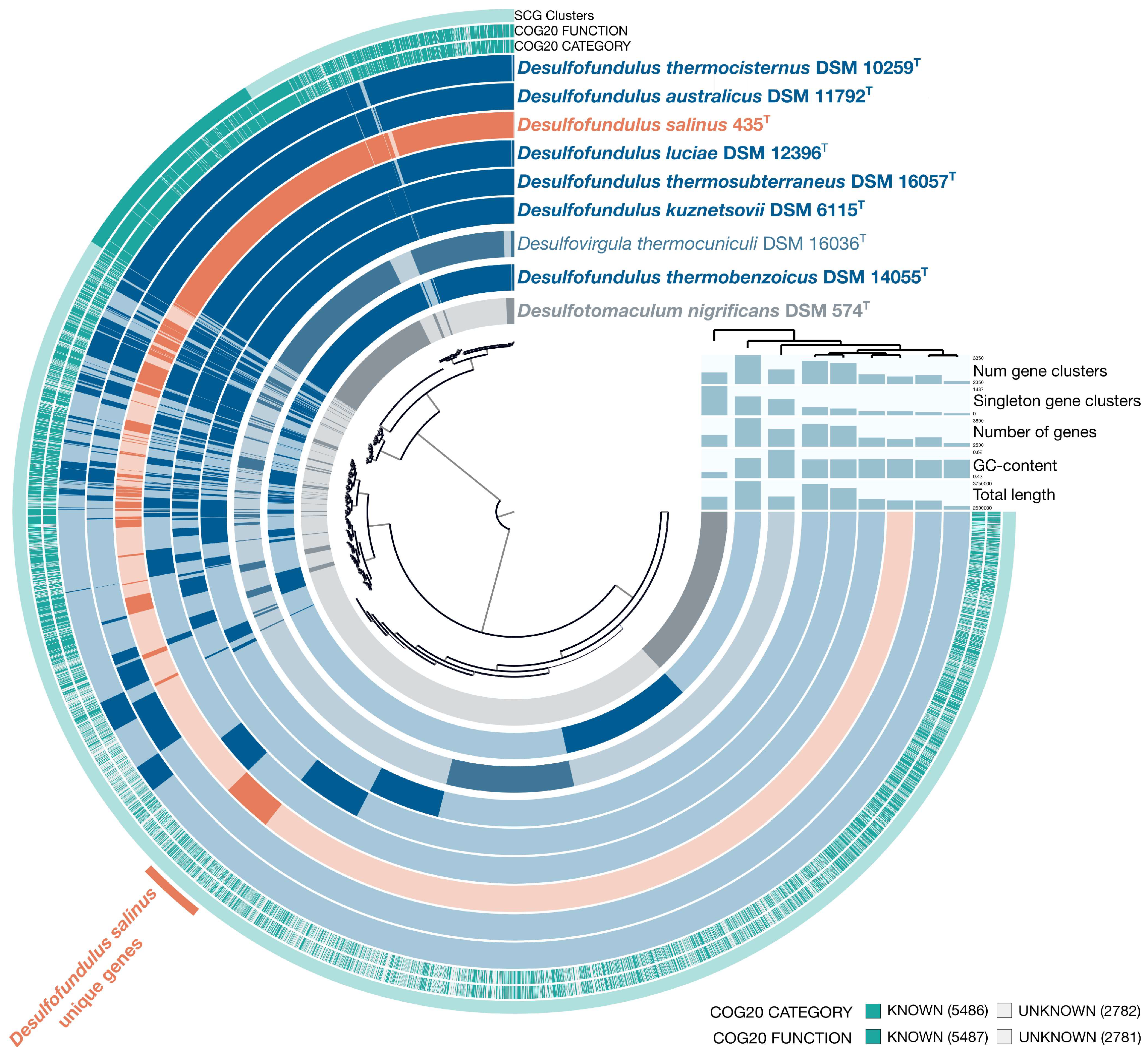
3.3. Genome Insights

The analysis of enzyme composition of the metabolic pathways based on the results of functional protein prediction using the BlastKOALA and RASTtk services provides evidence that the genome of strain 435T contains all the key genes responsible for complete pathways of carbohydrate metabolism: glycolysis (the Embden–Meyerhof pathway) and glucogenesis, pentose phosphate pathway, UDP-N-acetyl-D-glucosamine biosynthesis, and propionate oxidation. Complete sets of the genes comprising the reductive Acetyl-CoA pathway for CO2 fixation (the Wood–Ljundahl pathway) were revealed, as well as those of the pathways for dinitrogen fixation, biosynthesis of the major amino acids, dissimilatory sulfate reduction, biosynthesis and beta-oxidation of fatty acids, dTDP-L-rhamnose biosynthesis, and biosynthesis of vitamins and cofactors as follows: NAD, FAD, biotin (BioW pathway), L-valine, and coenzyme A. No genes of complete pathways for xenobiotic degradation were found. The circular genome map (Figure S1) of strain 435T shows a localization of the genes presumably involved in the metabolic pathways, according to the genome analysis carried out in this work.

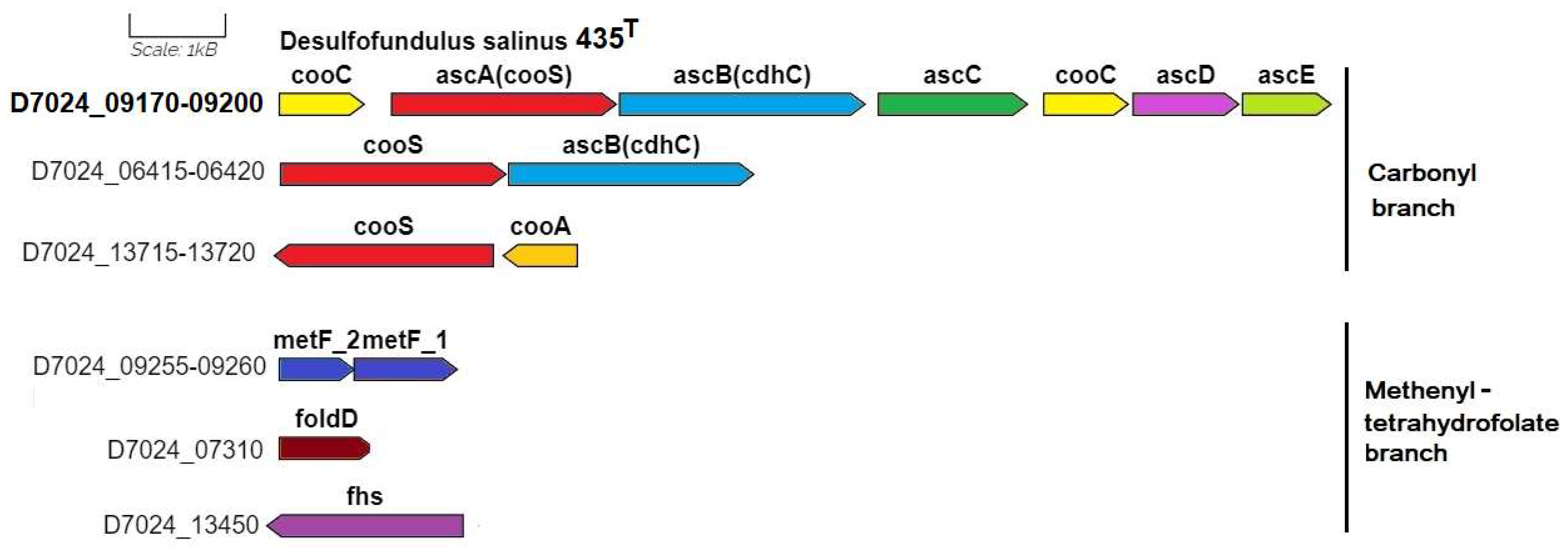
3.3.1. Autotrophic CO2 Fixation

Genomic analysis confirmed the previously shown ability of strain 435T to grow on molecular hydrogen and carbon dioxide (or formate) in the presence of sulfate as an electron acceptor. According to the KEGG map ’Carbon fixation pathways in prokaryotes’ (id. 00720), in the genome of strain 435T, all genes are annotated of the complete reductive acetyl-CoA pathway (the Wood–Ljungdahl pathway), including the genes of key enzymes, anaerobic carbon-monoxide dehydrogenase catalytic subunit (EC: 1.2.7.4) and acetyl-CoA synthase (EC: 2.3.1.169) (Figure S2). This pathway is used by many anaerobic bacteria, but the structure of the gene clusters encoding enzymes of this pathway differs from taxa to taxa. Examples of such different structures are given by Pierce et al. [42] in comparison with the annotated structure in the genome of the type strain Moorella thermoacetica ATCC 39073T.
In the genome of strain 435T, the genes of this pathway are present as the carbonyl branch operon, starting with the cooC gene (D7024_09170) encoding the CO dehydrogenase maturation factor, and the next cluster of genes of the CO complex acsA(cooS)BCDE (D7024_09175-09200) encoding the main and auxiliary genes of dehydrogenase/acetyl-CoA synthase (EC: 1.2.74/2.3.1.169). The structure of this operon is identical for the genomes of the type strains 435T, D. kuznetsovii 17T, and Desulfoscipio (Ds.) gibsoniae DSM 7213T; however, in the genomes of Desulfofarcimen (Df.) acetoxidans DSM 771T and M. thermoacetica ATCC 39073T, this operon differs by the insertion of the ferredoxin gene ahead of the second cooC gene (Figure S3).
The genes encoding the methenyltetrahydrofolate branch in the genome of strain 435T are located separately. They include metF1-2 (D7024_9255-09260), encoding the enzyme methylenetetrahydrofolate reductase (EC: 1.5.1.54), foldD (D7024_07310), encoding a bifunctional enzyme methenyl-tetrahydrofolate cyclohydrolase (EC: 3.5.4.9)/methylenetetrahydrofolate dehydrogenase (NADP+) (EC: 1.5.1.5), and fhs (D7024_13450), encoding formate-tetrahydrofolate ligase (EC: 6.3.4.3). Interestingly, a duplicate of the main carbonyl branch ascA(cooS)B (D7024_6415-06420) genes is additionally annotated in the genome of strain 435T. The cooS gene is annotated in a cluster with the cooA (D7024_13715-13720) gene, which determines the carbon monoxide-responsive transcriptional activator. The localization of all genes encoding enzymes of the reductive acetyl-CoA pathway in the genome of strain 435T is shown in (Figure 4).
Although the growth of strain 435T on CO has not been experimentally tested, the presence of several cooS genes encoding CO hydrogenase in their genome allows us to assume this possibility. The ability to use CO was also observed in a number of other sulfate-reducing bacteria, in particular, in a type strain of Desulfotomaculum nigrificans ATCC 19998T, as well as in another strain of this species, CO-1-SRB, which was previously assigned to a separate species due to its ability to grow on CO [43,44].
The genes fdhAB (d7024_02675_02680) determining the enzyme confurcating selenocysteine-incorporated formate dehydrogenase (NADP+) (EC: 1.17.1.10) are also annotated in the genome of strain 435T. This enzyme catalyzes both the fixation of CO2 via formate and the reverse oxidation reaction of formate to carbon dioxide, which corresponds to the revealed ability of this strain to grow on formate.
The ability to oxidize acetate to CO2 via the reverse acetyl-CoA pathway has been shown for some sulfate reducers, including Df. acetoxidans DSM 771T, based on the isolation of all the relative enzymes and determination of their activity [45,46]. The genome of Df. acetoxidans DSM 771T was subsequently found to contain all the genes required for this process [47]. A similar ability was suggested for D. kuznetsovii type strain 17T, which is related to strain 435T and grows on acetate in the presence of sulfate [14]. The acetyl-CoA pathway gene clusters from genomes of these strains had a similar structure; however, unlike Df. acetoxidans DSM 771T and D. kuznetsovii 17T, the initial study did not report acetate consumption by strain 435T [4]. Belyakova and Rozanova subsequently observed very slow acetate consumption during long-term incubation (56 days) [7]. No acetate consumption was also shown for another sulfate reducer, Ds. gibsoniae DSM 7213T, in the genome of which a similar one was annotated [48]. Four asc genes, coding another pathway of acetate oxidation via acetyl-CoA synthetase (EC: 6.2.1.1), were annotated in the genome of strain 435T: D7024_03470, 06805, 10610, and 10675. These genes are also present in the genomes of D. kuznetsovii 17T and Ds. gibsoniae DSM 7213T, unlike the genome of Df. acetoxidans DSM 771T, in which the genes of other enzymes for acetate oxidation were annotated: ackA encoding acetate kinase (EC: 2.7.2.1) and pta encoding phosphate acetyltransferase (EC: 2.3.1.8). Although it is presently still unclear why sulfate reducers with similar genetic structures for acetate oxidation differ in their ability to grow on this substrate, the detected genes determining acetate consumption enzymes may probably make strain 435T capable of complete oxidation of organic compounds.

3.3.2. Sulfur Metabolism

According to the KEGG map of sulfur metabolism, strain 435T reduces sulfate only via the dissimilatory process (Figure S4). The genes of dissimilatory sulfate reduction to sulfite are represented in the genome of strain 435T by two clusters, one of which comprises the sat gene (D7024_11275) encoding sulfate adenylyltransferase (EC: 2.7.7.4), the genes aprAB (D7024_07245-07250) encoding two subunits of adenylylsulfate reductase (EC: 1.8.99.2), and the qmoABC genes encoding the proteins of adenylsulfate reductase-associated electron transfer complex (Figure 5). Another set of the aprAB genes (11260-11265) is represented by a separate cluster. Further sulfate reduction to sulfide via the intermediary metabolite [DsrC-protein]-trisulfide is carried out by dissimilatory sulfite reductase (EC: 1.8.1.22), two subunits of which are determined by the dsrABD cluster (D7024_00345-00355) and by [DsrC]-trisulfide reductase (EC: 1.8.5.10). The latter is coded by a truncated cluster of genes of the dsrMK (D7024_00375-00380) and by the associated dsrC dissimilatory complex (D7024_00375-00385) encoding sulfur redox associated protein. This localization of the genes of dissimilatory sulfate reduction is identical to the orthological one in the genomes of other Desulfofundulus strains, including D. kuznetsovii 17T.
The KEGG map of sulfur metabolism (Figure S4) also presents the enzyme thiosulfate reductase/polysulfide reductase (EC: 1.8.5.5), which is tentatively involved in thiosulfate disproportionation to sulfate and sulfide. This process has been studied mainly in Gram-negative bacteria, including sulfate-reducing Deltaproteobacteria, although it was hypothesized to be common to all sulfate reducers [49]. While the ability to grow by thiosulfate disproportionation with the formation of stoichiometric amounts of sulfate and sulfide has been shown for the D. thermobenzoicus type strain, this ability was not revealed for the type strains of Dt. nigrificans, Dt. ruminis, and Desulfosporosinus orientis [50]. The genomes of D. thermobenzoicus subsp. thermosyntrophicus strains TPO and 435T (D7024_03870) were found to possess the homologous genes (80% identity of translated amino acid sequences), which were annotated by the BlastKOALA service as the phsA (psrA) genes coding the enzyme thiosulfate reductase/polysulfide reductase (EC: 1.8.5.5), which probably indicated their functional identity. This was in agreement with the previously shown ability of strain 435T to carry out thiosulfate dismutation to sulfate and sulfide [8].
Among the genes encoding the enzymes of assimilatory sulfate reduction, the genome of strain 435T, similar to that of D. kuznetsovii 17T, possessed only cysH (D7024_00280), coding phosphoadenosine phosphosulfate reductase (EC: 1.8.1.8).

3.3.3. Carbohydrate Metabolism and Oxidation of Organic Compounds

Strain 435T, as well as D. kuznetsovii 17T, was unable to grow on glucose or fructose as electron sources in the presence of sulfate [8]. However, the genomes of both strains contained all the genes encoding the enzymes involved in the Embden–Meyerhof–Parnas’s pathway, as well as in the pentose phosphate pathway, a non-oxidative phase. At the same time, the genes of the main enzymes of the Entner–Doudoroff pathway were not detected in the genomes of both strains, as well as most of the genes encoding the enzymes of the Krebs cycle.
Strain 435T oxidizes organic substrates such as lactate, pyruvate, malate, fumarate, and succinate in the presence of sulfates. The genome of strain 435T contains the ldh (D7024_03275) gene encoding L-lactate dehydrogenase (EC: 1.1.1.27), which is involved in the oxidation of lactate to pyruvate. The genome of strain 435T also comprises the genes maeA (D7024_09930), fumAB (D7024_09975-09980), and frdABC (D7024_12330-12340), coding malate dehydrogenase (EC: 1.1.1.38), fumarate hydratase (EC: 4.2.1.2), and succinate dehydrogenase (EC: 1.3.5.1), respectively, which may be involved in the oxidation of malate, fumarate, and succinate to pyruvate. Based on the similarity of the gene clusters (Figure S5) encoding the enzymes of propionate oxidation (Figure S6) with those previously presented for D. kuznetsovii 17T [51], this metabolic process in strain 435T can proceed in a similar way. Strain 435T can use the enzyme pyruvate formate-lyase (EC: 2.3.1.54) encoded by the pflD (D7024_11580) gene to ferment pyruvate to acetyl-CoA.

3.3.4. Methanol Metabolism

Methanol may be consumed via the cobalt-dependent methyl transferase (MT) pathway in combination with the reductive acetyl-CoA pathway, as has been tentatively described for the M. thermoacetica type strain ATCC 39073T [42]. The possibility of the MT pathway for methanol degradation to CO2 was confirmed for D. kuznetsovii 17T by proteomic analysis under conditions of growth in the presence of cobalt and vitamin B12 [52]. Key genes of the MT pathway were annotated in the genome of strain 435T: mtaA (D7024_14080, 14030) [methyl-Co(III) methanol-specific corrinoid protein]:coenzyme M methyltransferase (EC: 2.1.1.246), mtaB (D7024_14075) methanol-corrinoid protein Co-methyltransferase (EC: 2.1.1.90), and mtaC (D7024_14070) methanol methyltransferase corrinoid protein, which, similar to the case of D. kuznetsovii 17T, are components of the complete MT operon. A comparison of the structures of these MT operons (Figure S7) revealed 94% homology of their nucleotide sequences, which indicated the possible existence of the MT pathway for methanol consumption in strain 435T. At the same time, the complete MT operon was not revealed in the genome of another D. kuznetsovii strain, TROSR, and proteomic analysis did not confirm the ability of strain TROSR to carry out methanol degradation via the MT pathway [53].
Methanol-oxidizing activity has also been shown for the partially purified alcohol dehydrogenase (ADH) (EC: 1.1.1.1) with a molecular mass of ~42 kDa from the type strain D. kuznetsovii 17T [54]. The genome of this strain contained several genes determining ADH with a similar mass, which suggested the involvement of these enzymes in methanol assimilation [51]. A proteomic analysis revealed [52] that during growth on methanol in the absence of cobalt in the medium, the activity of the methyl transferase system decreased significantly, while the activity of one of the ADHs increased (Desku_2952), together with that of aldehyde ferredoxin oxidoreductase (Desku_2951), which confirmed the possibility of the existence of another pathway for methanol assimilation via the ADH pathway. Methanol is probably oxidized by ADH to formaldehyde, which is then oxidized to the formate by an aldehyde ferredoxin oxidoreductase. The genome of strain 435T was also found to contain several ADH-determining genes (D7024_02205, 2710, 2725, 11765, 11800, 11860, and 13790). The translated amino acid sequence of one of them, D7024_02725, has a 99% identity with the sequence of the gene of the tentatively active ADH from D. kuznetsovii 17T (Desku_2952). This may indicate the possible functioning of a cobalt-independent, NAD-dependent alcohol dehydrogenase (ADH) pathway of methanol oxidation in strain 435T.
Unlike D. kuznetsovii 17T, in the genome of strain 435T the mdh gene (D7024_11860) was annotated, which encodes methanol dehydrogenase (EC: 1.1.1.244), also oxidizing methanol to formaldehyde. The genes of specific enzymes of formaldehyde assimilation were, however, not annotated (Figure S8). In this strain, methanol dehydrogenase is probably also involved in methanol assimilation via formaldehyde, together with aldehyde ferredoxin oxidoreductase. Further research is required to elucidate the contribution of different metabolic pathways to the processes of methanol degradation by strain 435T.

3.3.5. Methane Metabolism

The formation of small amounts of so-called mini-methane has been previously reported for strains 435T and 781 grown under lithoautotrophic conditions on H2/CO2 [7]. However, the KEGG map did not reveal complete modules of the known types of methanogenesis, including hydrogenotrophic one from H2/CO2, since the genome of strain 435T does not encode the enzyme of the terminal stage of methanogenesis, methyl-coenzyme M reductase (EC: 2.8.4.1), which catalyzes the conversion of methyl-CoM to methane (Figure S8). The previously obtained results were probably caused by an experimental error.

3.3.6. Nitrogen Metabolism

The ability of strain 435T to fix molecular nitrogen, albeit at a very low rate, which has been shown by an acetylene test [6], was analyzed at the genome level. According to the KEGG map of nitrogen metabolism, nitrogenase (EC: 1.18.6.1) is present among its enzymes (Figure S9). The genome of strain 435T contains the nitrogenase reductase nifH gene in the Nif cluster (D7024_03585-D7024_03620), which also comprises the genes of nitrogenase subunits nifD and nifK, the nifB and NifE auxiliary genes, and the genes determining the P-II-family proteins of nitrogen metabolism regulators (Figure S10). The Nif cluster was flanked by the nrgA gene, determining an ammonium transporter protein, and the nifV gene encoding homocitrate synthase. A similar gene cluster was annotated in most genomes of the type strains of Desulfofundulus species, including D. kuznetsovii DSM 6115T (=17T). However, according to the previously obtained physiological data, this strain was completely incapable of nitrogen fixation [14]; for other Desulfofundulus strains, this ability has not been studied.
The reason for the absence or very low level of nitrogen-fixing ability in the studied Desulfofundulus strains, in spite of the presence of the Nif clusters in their genomes, is still unclear. A partially similar nitrogenase gene cluster was annotated in the genomes of Desulfomicrobium baculatum XT [55] and Desulforamulus ruminis DSM 2154T, for which active dinitrogen fixation was shown by the acetylene method [6,56]. However, apart from the Nif gene clusters, similar to those of strain 435T and other Desulfofundulus strains, the genomes of Desulfomicrobium baculatum XT and Desulforamulus ruminis DSM 2154T were also found to contain the nifN genes, which some authors consider essential in the minimal set of auxiliary proteins for nitrogenase maturation [57]. The absence of these genes may be responsible for the very low nitrogenase activity of strain 435T and for the complete absence of this activity in D. kusnetsovii 17T. Another cause may be the complete or partial inactivation of some nitrogenase genes, resulting, for instance, from fragmentation, as has been revealed for the NifD alpha-subunit genes in the genomes of some Desulfofundulus strains. It may be noted that Dt. nigrificans DSM 574T also lacks the ability to fix dinitrogen, in spite of the presence of both auxiliary genes, nifE and nifN, in its Nif cluster [56], probably due to fragmentation of the nifK nitrogenase beta-subunit gene (Figure S10). Further research on nitrogen fixation in Gram-positive sulfate reducers at the physiological and genomic levels is therefore required.
No enzymes for assimilatory and dissimilatory nitrate reduction were annotated in the genome of strain 435T, which was in agreement with the results of physiological tests, showing that strain 435T did not use nitrate as an electron acceptor for growth [8]. According to the results of the KEGG analysis of the nitrogen metabolism pathways (Figure S9), the genes gudB, glnA, and gltB were annotated, which determine the enzymes for assimilation of ammonium, probably produced via nitrogen fixation, namely, NAD-specific glutamate dehydrogenase (EC: 1.4.1.2), glutamine synthetase (EC: 6.3.1.2), and glutamate synthase (EC: 1.4.1.13), respectively.

3.3.7. Hydrogenase Genes

The hydABC genes (D7024_02610-2620) encoding bifurcating trimeric [FeFe] hydrogenase (EC: 1.12.1.4) are present in the genome of strain 435T. This gene cluster is associated with the gene (D7024_02625), which determines the PAS domain-containing protein, tentatively involved in the regulation of the synthesis of this hydrogenase [58]. This hydrogenase was originally found in Thermotoga maritima DSM 3109 [59]. According to the MetaCyc data, it is responsible for the irreversible metabolic pathway hydrogen production I. This heterotrimeric enzyme catalyzes the NADH- and ferredoxin-dependent proton reduction to H2 by a single enzyme complex; it functions only under anoxic conditions. The annotated gene cluster hydABC (D7024_02610-02625), determining trimeric confurcating [FeFe]-hydrogenases, was homologous to one of two clusters of the hydrogenase genes of the D. kuznetsovii 17T genome [51]. In the genome of strain 435T, the genes of the hndABCD operon (D7024_05825-5835, 5815), determining tetraheteromeric NADP-dependent [Fe-Fe] hydrogenase (EC: 1.12.1.3), were annotated. This hydrogenase has been thoroughly studied for the sulfate reducer Solidesulfovibrio fructosivorans DSM 3604 [60]. According to the MetaCyc data, this hydrogenase carries out hydrogen oxidation under anoxic conditions, using reduced sulfur compounds as the terminal electron acceptors. In the absence of these compounds, the reverse process is possible, producing hydrogen during fermentative growth on relevant carbon sources (e.g., pyruvate for strain 435T). The genes hygEFG (D7024_05780-05790), coding the proteins facilitating the maturation of [Fe-Fe] hydrogenase [61], were annotated at the same genome site.

3.3.8. Sporulation Genes

It was previously shown that in the genus Desulfotomaculum, the initial (zero) stage of sporulation is similar to the relevant stage in Bacillus, rather than in Clostridium [62]. It is initiated by the autophosphorylation of a sensory histidine kinase, followed by the sequential transfer of the phosphoryl group to the Spo0F and SP0B proteins, and culminates in the accumulation of Spo0A∼P, the main regulator of sporulation. In the genomes of members of the genus Desulfotomaculum, including D. kuznetsovii 17T, which has previously been assigned to this genus, conserved genetic structures homologous to those of bacilli were identified, including the spo0B gene encoding a sporulation response regulatory protein, as well as rplU, encoding for a ribosomal protein L21, rpmA for a ribosomal protein L27, and obgE for an Obg family GTPase. The genome of strain 435T also has such a structure (D7024_12390-12375), so it can be assumed that the D7024_12385 gene is a homolog of the spo0B gene of D. kuznetsovii 17T. According to the results of the BlastKOALA service, other genes of sporulation stage zero are also annotated in the genome of strain 435T, including spo0F (D7024_00550, 03180) and spo0A (D7024_07375), as well as 48 spo genes of subsequent I–V stages of sporulation and 10 genes of the sensory histidine kinase, which corresponds to the revealed ability of this strain to complete sporulation [5,8].

3.4. Phenotypic Characteristics of Strains 435T and 781

Cells of strains 435T and 781 were rod-shaped or spindle-shaped (lemon-shaped), with sizes of 0.9–1.3 × 2–5 µm and 0.6–1.0 × 2–5 µm, respectively. The cells were motile due to peritrichous flagella, which are spore-forming. Despite the fact that the strains belong to the group of Gram-positive bacteria of the phylum Bacillota, they were Gram-negative stained, which was also noted for other members of this genus [53]. However, electron microscopic examination of the cells showed a cell wall structure typical of Gram-positive bacteria (Figure 6) [8]. The cell wall lacked the external lipoprotein membrane characteristic of Gram-negative bacteria.
Membrane structures that are invaginations of the cytoplasmic membrane, the nucleoid, and polyribosomes were visible in the cytoplasm. It has been shown that cell division is carried out by forming a septum in the central part of the cell, producing two daughter cells of equal sizes (Figure 6a). After division, the cells separated, although chains of two and four cells occurred in the culture. The strains formed spores of oval or spherical shape, located in the center of the cell or subterminal, and slight swelling of the cells. Figure 6b–f show spores at different stages of maturation, spore coat and a cortex, located between the inner and outer membranes of the forespore. Similar to Bacillus species [63,64], during sporulation, the strains 435T and 781 formed a division septum located closer to one pole, producing a smaller forespore and a larger mother cell (Figure 6b–d). Subsequently, the forespore was engulfed by the mother cell, producing an internal endospore that after maturation is released by mother cell lysis (Figure 6e,f).
The main differentiating features of strains 435T and 781 and members of the genus Desulfofundulus are shown in Table 2. Both strains were adapted to the temperature and salinity of the habitats from which they were isolated.
The optimum temperature for the growth of strains 435T and 781 was 55–60 °C, with weak growth observed at 40–45 °C and 65–70 °C, respectively (Figure S11a). Strains 435T and 781 grew in the salinity ranges 0–3 and 0–4.5% NaCl (w/v), respectively, with optima at 1% and 0.5% NaCl (Figure S11b). The strains remained viable after cultivation in a medium with 6% NaCl, probably due to the formation of spores, but no increase in biomass was observed. Both strains were neutrophils, growing in the pH range of 6.0–8.5; the strains accumulated the maximum amount of biomass at pH 7.0, whereas the maximum formation of sulfide in the medium was noted at pH 8.5 [8].
Both strains were obligate anaerobes and had a similar range of electron donors and acceptors used. The strains reduced sulfate to hydrogen sulfide in media with lactate, pyruvate, formate, propionate, butyrate, valerate, palmitate, malate, fumarate, succinate, methanol, ethanol, propanol, and butanol and were capable of autotrophic growth in a mineral medium with a H2/CO2 gas phase. L-alanine, L-serine, L-arginine, asparagine, L-cysteine, glucose, fructose, lactose, benzoate, citrate, tartrate, glycerol, glutamate, threonine, tryptophan, and phenylalanine were not used. The strains carried out an incomplete oxidation of lactate and other organic substrates to acetate. In our experiments (up to 14–30 days), growth on acetate coupled with sulfide production was not observed. However, very slow acetate consumption by both strains after 56 days of cultivation has been noted [7].
The strains 435T and 781 used sulfate, sulfite, and thiosulfate, but not nitrate as electron acceptors and carried out thiosulfate dismutation to sulfate and sulfide. In the absence of sulfate, the bacteria fermented pyruvate with acetate production, and weak growth was also observed on fumarate; lactate was not fermented. The cellular fatty acid profiles of strains 435T, 781, and D. kuznetsovii 17T were dominated by the same fatty acids that were found earlier [8], but in other quantitative ratios. The major fatty acids of strains 435T and 781 were iso-C15:0 (22.1 and 32.7% of the total amount of fatty acids, respectively), iso-C17:0 (9.4 and 18.0%), C16:0 (21.1 and 25.5%), and C18:0 (31.5 and 10.7%) fatty acids (Table S1). In the fatty acids profiles of the type strains of D. luciae [69], D. solfataricus [68], and D. thermosubterraneus [69], iso-C15:0 and iso-C17:0 fatty acids were also predominant. Desulfoviridin was not detected.
The strains 435T and 781 can be distinguished from the closest members of the genus Desulfofundulus by their phenotypic characteristics, including a range of utilized substrates (Table 2). The data presented show that physiological features of strain 435T were supported by the metabolic pathways predicted from its genome sequence. The results of phenotypic studies, phylogenetic position based on the 16S rRNA gene sequencing, and genomic analysis allow us to assign the strains 435T and 781 to a new species of the genus Desulfofundulus with the proposed name Desulfofundulus salinus sp. nov. The description of the species is given in the protologue (Table 3).
The ability to grow autotrophically on H2/CO2, as well as to use lower alcohols and fatty acids, together with the optima of temperature and salinity for strains 435T and 781, indicate the adaptation of the strains to the conditions of the deep subsurface horizons from which they were isolated. A strain distantly related to ‘Desulfotomaculum salinum’ was isolated from the sulfate-reducing culture obtained from a black rust deposit from a borehole on the eastern flank of Juan de Fuca Ridge [73]. A phylotype of ‘Desulfotomaculum salinum’ (99% of 16S rRNA gene sequence similarity) was detected by the DGGE molecular approach in water from a borehole located on the west bank of the river Ob’ (58°50′ N, 81°30′ E) in the Tomsk Region, Western Siberia, Russia [74]. Thus, thermophilic spore-forming sulfate-reducing bacteria of the DesulfotomaculumDesulfofundulus group are commonly found inhabitants of deep subsurface environments [75].

4. Conclusions

In this study, two thermophilic spore-forming sulfate-reducing strains, 435T and 781, isolated 50 years ago from hydrocarbon deposits of Western Siberia (Russia), were taxonomically characterized using physiological, chemotaxonomic, and phylogenetic approaches. Based on phenotypic and 16S rRNA gene-based analyses, the strains were previously assigned to the genus Desulfotomaculum, as ‘Desulfotomaculum salinum’. A genome analysis of the type strain 435T made it possible to bring the taxonomic description of the species in line with the present-day requirements. A phylogenetic analysis based on 16S rRNA gene sequences and 120 conserved proteins from the 435T genome revealed that this strain formed a separate clade within the genus Desulfofundulus. Overall genomic relatedness indices (OGRIs), such as ANI, AAI, and dDDH values, highlighted genomic differences between the new taxon and closely related type strains of Desulfofundulus species. Phenotypic, genomic, and phylogenomic data supported the classification of strains 435T and 781 as members of a novel species with the proposed name Desulfofundulus salinus sp. nov., whose description is reported in Table 3. Comparative genomic analysis of strain 435T unraveled the main aspects of the ecophysiology of bacteria inhabiting deep subsurface environments and increased the taxonomic diversity of the genus Desulfofundulus.

Supplementary Materials

The following supporting information can be downloaded at: https://www.mdpi.com/article/10.3390/microorganisms12061115/s1, Figure S1: Circular genome map of the D. salinus strain 435T. Abbreviations: dsrAB, dissimilatory sulfite reductase subunits A, B; hydABC, trimeric confurcating [FeFe]-hydrogenases; fdhAB, confurcating selenocysteine-incorporated formate dehydrogenase (NADP+); phsA, thiosulfate reductase/polysulfide reductase; hndABCD, tetrageteromeric NADP-dependent [Fe-Fe] hydrogenase; foldD, methenyltetrahydrofolate cyclohydrolase/methylenetetrahydrofolate dehydrogenase (NADP+); metF, 1-2, methylenetetrahydrofolate reductase; acsA (cooS), carbon monoxide dehydrogenase; acsB, acetyl-CoA synthase; ascC, acetyl-CoA synthase corrinoid iron–sulfur protein, large subunit; ascD, acetyl-CoA synthase corrinoid iron–sulfur protein, small subunit; acsE, methyltetrahydrofolate methyltransferase; aprAB, adenylylsulfate reductase subunit A, B; spo0B, sporulation response regulatory protein; fhs, formate-tetrahydrofolate ligase; mtaA, [methyl-Co(III) methanol-specific corrinoid protein]:coenzyme M methyltransferase; mtaB, methanol–corrinoid protein Co-methyltransferase; mtaC, methanol methyltransferase corrinoid protein; Figure S2: KEGG-map of carbon fixation pathways based on the genome analysis of the strain 435T. The enzymes annotated in the genome are highlighted in green. The Wood–Ljungdahl pathway is highlighted in pink; Figure S3: A cluster of genes presumably encoding enzymes of the carbonyl branch of the Wood–Ljungdahl pathway in the genome of the strain D. salinus 435T compared to other bacteria. Abbreviations: acsA (cooS), carbon monoxide dehydrogenase; acsB, acetyl-CoA synthase; ascC, acetyl-CoA synthase corrinoid iron–sulfur protein, large subunit; ascD, acetyl-CoA synthase corrinoid iron-sulfur protein, small subunit; acsE, methyltetrahydrofolate methyltransferase; cooC, carbon monoxide dehydrogenase maturation factor. Scale bar, 1000 bp; Figure S4: KEGG-map of sulfur metabolism pathways based on the genome analysis of strain 435T. The enzymes annotated in the genome are highlighted in green; Figure S5: KEGG-map of propanoate metabolism pathways based on the genome analysis of strain 435T. The enzymes annotated in the genome are highlighted in green; Figure S6: Gene organization of the mmc-cluster encoding propanoate oxidation in D. salinus 435T and D. kuznetsovii 17T. Abbreviations: mmcB, fumarase, N-terminal domain; mmcC, fumarase, C-terminal domain; mmcD1, succinyl-CoA synthetase, beta subunit; mmcD2, succinyl-CoA synthetase, alpha subunit; mmcE, methylmalonyl-CoA mutase, N-terminal domain; mmcG, methylmalonyl-CoA epimerase; mmcH, methylmalonyl-CoA decarboxylase, alpha subunit; mmcI, methylmalonyl-CoA decarboxylase, epsilon subunit; mmcJ, methylmalonyl-CoA decarboxylase, gamma subunit; mmcK, malate dehydrogenase; mmcL, transcarboxylase 5S subunit. Scale bar, 1000 bp; Figure S7: Gene organization of the MT operon encoding a cobalt-dependent methyl transferase pathway in strains 435T and D. kuznetsovii 17T. Abbreviations: 1, cobalamin-binding protein; mtaA, [methyl-Co(III) methanol-specific corrinoid protein]:coenzyme M methyltransferase; mtaB, methanol-corrinoid protein Co-methyltransferase; mtaC, methanol methyltransferase corrinoid protein; 2, cobalamin synthesis protein P47K; 3, ferredoxin iron–sulfur binding domain protein; 4, uroporphyrinogen decarboxylase; 5, methyltransferase cognate corrinoid protein; 6, ferredoxin; 7, tetrahydromethanopterin S-methyltransferase; 8, FMN-binding protein. Scale bar, 1000 bp; Figure S8: KEGG-map of methane metabolism pathways based on the genome analysis of strain 435T. The enzymes annotated in the genome are highlighted in green; Figure S9: KEGG-map of nitrogen metabolism pathways based on the genome analysis of strain 435T. The enzymes annotated in the genome are highlighted in green; Figure S10: Organization of gene clusters presumably encoding nitrogen fixation enzymes in the genome of strain 435T and some other sulfate-reducing bacteria. Abbreviations: nifH, nitrogenase (molybdenum–iron) reductase and maturation protein, (nifDK), nitrogenase (molybdenum–iron) alpha and beta subunits; nifB, nitrogenase FeMo-cofactor synthesis FeS; nitrogen-fixation associated, P-II, nitrogen regulatory proteins; nifN, nitrogenase molybdenum–iron protein; anfO, Fe-only nitrogenase accessory AnfO family protein; nrgA, ammonium transporter; nifV, homocitrate synthase. Scale bar, 1000 bp; Figure S11: H2S production profiles of strains 435T and 781 growing in fumarate–sulfate medium for 14 days at various temperatures (a) and NaCl concentrations (b); Table S1: Cellular fatty acid composition (%) of strains 435T, 781 and D. kuznetsovii 17T.

Author Contributions

Conceptualization, T.N.N.; investigation, T.N.N., S.K.B. and A.B.P.; writing—original draft preparation, T.N.N., T.P.T. and D.S.G.; writing—review and editing, T.N.N. and D.S.G.; project administration, T.N.N.; funding acquisition, T.N.N. All authors have read and agreed to the published version of the manuscript.

Funding

This research was partly funded by the Russian Science Foundation (grant 21-64-00019) (T.P.T., genome analysis).

Data Availability Statement

The whole-genome shotgun project of strain 435T has been deposited at DDBJ/EMBL/GenBank under the accession GCF_003627965.1, and it is the first version described in this paper.

Acknowledgments

We thank Professor Aharon Oren (the Hebrew University of Jerusalem) for nomenclature advice. The bioinformatic analysis was performed using the computer resources of SciBear OU (https://sci-bear.com/, accessed on 5 March 2024). The authors thank D.S. Sokolova for the sequencing of the 16S rRNA genes. The authors also thank the staff of the All-Russian Collection of Microorganisms and the German Collection of Microorganisms and Cell Cultures GmbH for the long-term storage and maintenance of strains.

Conflicts of Interest

The authors declare no conflicts of interest.

References

  1. Campbell, L.L.; Postgate, J.R. Classification of the spore-forming sulfate-reducing bacteria. Bacteriol. Rev. 1965, 29, 359–363. [Google Scholar] [CrossRef] [PubMed]
  2. Stackebrandt, E.; Sproer, C.; Rainey, F.A.; Burghardt, J.; Pauker, O.; Hippe, H. Phylogenetic analysis of the genus Desulfotomaculum: Evidence for the misclassification of Desulfotomaculum guttoideum and description of Desulfotomaculum orientis as Desulfosporosinus orientis gen. nov., comb. nov. Int. J. Syst. Bacteriol. 1997, 47, 1134–1139. [Google Scholar] [CrossRef]
  3. Watanabe, M.; Kojima, H.; Fukui, M. Review of Desulfotomaculum species and proposal of the genera Desulfallas gen. nov., Desulfofundulus gen. nov., Desulfofarcimen gen. nov. and Desulfohalotomaculum gen. nov. Int. J. Syst. Evol. Microbiol. 2018, 68, 2891–2899. [Google Scholar] [CrossRef] [PubMed]
  4. Nazina, T.N.; Rozanova, E.P. Thermophilic sulfate-reducing bacteria from oil strata. Mikrobiologiya 1978, 47, 142–148. (In Russian) [Google Scholar]
  5. Nazina, T.N.; Pivovarova, T.A. Submicroscopic organization and sporulation in Desulfotomaculum nigrificans. Microbiology 1979, 48, 241–246. [Google Scholar]
  6. Nazina, T.N.; Rozanova, E.P.; Kalininskaia, T.A. Fixation of molecular nitrogen by sulfate-reducing bacteria from petroleum strata. Mikrobiologiia 1979, 48, 133–136. (In Russian) [Google Scholar] [PubMed]
  7. Belyakova, E.V.; Rozanova, E.P. Newly discovered properties of spore-forming sulfate-reducing bacteria, Desulfotomaculum strains 435 and 781. Microbiology 2004, 73, 284–286. [Google Scholar] [CrossRef]
  8. Nazina, T.N.; Rozanova, E.P.; Belyakova, E.V.; Lysenko, A.M.; Poltaraus, A.B.; Tourova, T.P.; Osipov, G.A.; Belyaev, S.S. Description of “Desulfotomaculum nigrificans subsp. salinus” as a new species, Desulfotomaculum salinum sp. nov. Microbiology 2005, 74, 567–574. [Google Scholar] [CrossRef]
  9. Grouzdev, D.S.; Bidzhieva, S.K.; Tourova, T.P.; Krutkina, M.S.; Poltaraus, A.B.; Nazina, T.N. Draft genome sequence of a sulfate-reducing bacterium, ‘Desulfofundulus salinum’ 435T, isolated from a high-temperature gas field in Russia. Microbiol. Resour. Announc. 2018, 7, e01408-18. [Google Scholar] [CrossRef]
  10. Chun, J.; Oren, A.; Ventosa, A.; Christensen, H.; Arahal, D.R.; da Costa, M.S.; Rooney, A.P.; Yi, H.; Xu, X.W.; De Meyer, S.; et al. Proposed minimal standards for the use of genome data for the taxonomy of prokaryotes. Int. J. Syst. Evol. Microbiol. 2018, 68, 461–466. [Google Scholar] [CrossRef]
  11. Oren, A.; Garrity, G.M. Notification list. Notification that new names and new combinations have appeared in volume 68, part 9 of the IJSEM. Int. J. Syst. Evol. Microbiol. 2018, 68, 3685–3687. [Google Scholar] [CrossRef] [PubMed]
  12. Stackebrandt, E. The emended family Peptococcaceae and description of the families Desulfitobacteriaceae, Desulfotomaculaceae, and Thermincolaceae. In The Prokaryotes; Rosenberg, E., DeLong, E., Lory, S., Stackebrandt, E., Thompson, F., Eds.; Springer: Berlin/Heidelberg, Germany, 2014; pp. 286–290. [Google Scholar] [CrossRef]
  13. Parte, A.C.; Sardà Carbasse, J.; Meier-Kolthoff, J.P.; Reimer, L.C.; Göker, M. List of Prokaryotic names with Standing in Nomenclature (LPSN) moves to the DSMZ. Int. J. Syst. Evol. Microbiol. 2020, 70, 5607–5612. [Google Scholar] [CrossRef] [PubMed]
  14. Nazina, T.N.; Ivanova, A.E.; Kanchaveli, L.P.; Rozanova, E.P. A new sporeforming thermophilic methylotrophic sulfate-reducing bacterium, Desulfotomaculum kuznetsovii sp. nov. Microbiology 1988, 57, 659–663. [Google Scholar]
  15. Validation of the Publication of New Names and New Combinations Previously Effectively Published Outside the IJSB: List No. 35. Int. J. Syst. Bacteriol. 1990, 40, 470–471. [CrossRef]
  16. Postgate, J.R. Media for sulfur bacteria. Lab. Pract. 1966, 15, 1239–1244. [Google Scholar] [PubMed]
  17. Widdel, F. The genus Desulfotomaculum. In The Prokaryotes, 2nd ed.; Balows, A., Trüper, H.G., Dworkin, M., Harder, W., Schleifer, K.H., Eds.; Springer: New York, NY, USA; Berlin/Heidelberg, Germany, 1992; Volume 1, pp. 1792–1799. [Google Scholar]
  18. Pfennig, N.; Lippert, K.D. Über das Vitamin B12-Bedürfnis phototropher Schwefelbakterien. Arch. Für Mikrobiol. 1966, 55, 245–256. [Google Scholar] [CrossRef]
  19. Cypionka, H.; Pfennig, N. Growth yields of Desulfotomaculum orientis with hydrogen in chemostat culture. Arch. Microbiol. 1986, 143, 366–369. [Google Scholar] [CrossRef]
  20. Trüper, H.G.; Schlegel, H.G. Sulfur metabolism in Thiorhodaceae. I. Quantitative measurements on growing cells of Chromatium okenii. Antonie Van Leeuwenhoek 1964, 30, 321–323. [Google Scholar] [CrossRef]
  21. Lane, D.J. 16S/23S rRNA sequencing. In Nucleic Acid Techniques in Bacterial Systematics; Stackebrandt, E., Goodfellow, M., Eds.; John Wiley & Sons: New York, NY, USA, 1991; pp. 115–175. [Google Scholar]
  22. Jain, C.; Rodriguez-R, L.M.; Phillippy, A.M.; Konstantinidis, K.T.; Aluru, S. High throughput ANI analysis of 90K prokaryotic genomes reveals clear species boundaries. Nat. Commun. 2018, 9, 5114. [Google Scholar] [CrossRef] [PubMed]
  23. Meier-Kolthoff, J.P.; Carbasse, J.S.; Peinado-Olarte, R.L.; Göker, M. TYGS and LPSN: A database tandem for fast and reliable genome-based classification and nomenclature of prokaryotes. Nucleic Acids Res. 2022, 50, D801–D807. [Google Scholar] [CrossRef]
  24. Tatusova, T.; DiCuccio, M.; Badretdin, A.; Chetvernin, V.; Ciufo, S.; Li, W. Prokaryotic genome annotation pipeline. In The NCBI Handbook, 2nd ed.; National Center for Biotechnology Information: Bethesda, MD, USA, 2013. [Google Scholar]
  25. The UniProt Consortium. UniProt: The Universal Protein Knowledgebase in 2023. Nucleic Acids Res. 2023, 51, D523–D531. [Google Scholar] [CrossRef] [PubMed]
  26. Edgar, R.C. MUSCLE: Multiple sequence alignment with high accuracy and high throughput. Nucleic Acids Res. 2004, 32, 1792–1797. [Google Scholar] [CrossRef] [PubMed]
  27. Chaumeil, P.-A.; Mussig, A.J.; Hugenholtz, P.; Parks, D.H. GTDB-Tk: A toolkit to classify genomes with the Genome Taxonomy Database. Bioinformatics 2020, 36, 1925–1927. [Google Scholar] [CrossRef]
  28. Kalyaanamoorthy, S.; Minh, B.Q.; Wong, T.K.F.; von Haeseler, A.; Jermiin, L.S. ModelFinder: Fast model selection for accurate phylogenetic estimates. Nat. Methods 2017, 14, 587–589. [Google Scholar] [CrossRef] [PubMed]
  29. Nguyen, L.T.; Schmidt, H.A.; von Haeseler, A.; Minh, B.Q. IQ-TREE: A fast and effective stochastic algorithm for estimating maximum-likelihood phylogenies. Mol. Biol. Evol. 2015, 32, 268–274. [Google Scholar] [CrossRef] [PubMed]
  30. Hoang, D.T.; Chernomor, O.; von Haeseler, A.; Minh, B.Q.; Vinh, L.S. UFBoot2: Improving the ultrafast bootstrap approximation. Mol. Biol. Evol. 2018, 35, 518–522. [Google Scholar] [CrossRef] [PubMed]
  31. Hoang, D.T.; Vinh, L.S.; Flouri, T.; Stamatakis, A.; von Haeseler, A.; Minh, B.Q. MPBoot: Fast phylogenetic maximum parsimony tree inference and bootstrap approximation. BMC Evol. Biol. 2018, 18, 11. [Google Scholar] [CrossRef] [PubMed]
  32. Tamura, K.; Stecher, G.; Kumar, S. MEGA11: Molecular Evolutionary Genetics Analysis Version 11. Mol. Biol. Evol. 2021, 38, 3022–3027. [Google Scholar] [CrossRef] [PubMed]
  33. Delmont, T.O.; Eren, E.M. Linking pangenomes and metagenomes: The Prochlorococcus metapangenome. PeerJ 2018, 6, e4320. [Google Scholar] [CrossRef]
  34. Eren, A.M.; Esen, O.C.; Quince, C.; Vineis, J.H.; Morrison, H.G.; Sogin, M.L.; Delmont, T.O. Anvi’o: An advanced analysis and visualization platform for ’omics data. PeerJ 2015, 3, e1319. [Google Scholar] [CrossRef]
  35. Wattam, A.R.; Davis, J.J.; Assaf, R.; Boisvert, S.; Brettin, T.; Bun, C.; Conrad, N.; Dietrich, E.M.; Disz, T.; Gabbard, J.L.; et al. Improvements to PATRIC, the all-bacterial Bioinformatics Database and Analysis Resource Center. Nucleic Acids Res. 2017, 45, D535–D542. [Google Scholar] [CrossRef] [PubMed]
  36. Caspi, R.; Billington, R.; Keseler, I.M.; Kothari, A.; Krummenacker, M.; Midford, P.E.; Ong, W.K.; Paley, S.; Subhraveti, P.; Karp, P.D. The MetaCyc database of metabolic pathways and enzymes—A 2019 update. Nucleic Acids Res. 2020, 48, D445–D453. [Google Scholar] [CrossRef] [PubMed]
  37. Aziz, R.K.; Bartels, D.; Best, A.A.; DeJongh, M.; Disz, T.; Edwards, R.A.; Formsma, K.; Gerdes, S.; Glass, E.M.; Kubal, M.; et al. The RAST Server: Rapid annotations using subsystems technology. BMC Genom. 2008, 9, 75. [Google Scholar] [CrossRef] [PubMed]
  38. Kanehisa, M.; Sato, Y.; Morishima, K. BlastKOALA and GhostKOALA: KEGG tools for functional characterization of genome and metagenome sequences. J. Mol. Biol. 2016, 428, 726–731. [Google Scholar] [CrossRef] [PubMed]
  39. Harrison, K.J.; de Crécy-Lagard, V.; Zallot, R. Gene Graphics: A genomic neighborhood data visualization web application. Bioinformatics 2018, 34, 1406–1408. [Google Scholar] [CrossRef] [PubMed]
  40. Grant, J.R.; Enns, E.; Marinier, E.; Mandal, A.; Herman, E.K.; Chen, C.; Graham, M.; Van Domselaar, G.; Stothard, P. Proksee: In-depth characterization and visualization of bacterial genomes. Nucleic Acids Res. 2023, 51, W484–W492. [Google Scholar] [CrossRef] [PubMed]
  41. Meier-Kolthoff, J.P.; Auch, A.F.; Klenk, H.-P.; Göker, M. Genome sequence-based species delimitation with confidence intervals and improved distance functions. BMC Bioinform. 2013, 14, 60. [Google Scholar] [CrossRef] [PubMed]
  42. Pierce, E.; Xie, G.; Barabote, R.D.; Saunders, E.; Han, C.S.; Detter, J.C.; Richardson, P.; Brettin, T.S.; Das, A.; Ljungdahl, L.G.; et al. The complete genome sequence of Moorella thermoacetica (f. Clostridium thermoaceticum). Environ. Microbiol. 2008, 10, 2550–2573. [Google Scholar] [CrossRef] [PubMed]
  43. Parshina, S.N.; Sipma, J.; Nakashimada, Y.; Henstra, A.M.; Smidt, H.; Lysenko, A.M.; Lens, P.N.L.; Lettinga, G.; Stams, A.J.M. Desulfotomaculum carboxydivorans sp. nov.; a novel sulfate-reducing bacterium capable of growth at 100% CO. Int. J. Syst. Evol. Microbiol. 2005, 55, 2159–2165. [Google Scholar] [CrossRef] [PubMed]
  44. Visser, M.; Parshina, S.N.; Alves, J.I.; Sousa, D.Z.; Pereira, I.A.; Muyzer, G.; Kuever, J.; Lebedinsky, A.V.; Koehorst, J.J.; Worm, P.; et al. Genome analyses of the carboxydotrophic sulfate-reducers Desulfotomaculum nigrificans and Desulfotomaculum carboxydivorans and reclassification of Desulfotomaculum caboxydivorans as a later synonym of Desulfotomaculum nigrificans. Stand. Genom. Sci. 2014, 9, 655–675. [Google Scholar] [CrossRef]
  45. Spormann, A.M.; Thauer, R.K. Anaerobic acetate oxidation to CO2 in Desulfotomaculum acetoxidans. Demonstration of enzymes required for the operation of an oxidative acetyl-CoA/carbon monoxide dehydrogenase pathway. Arch. Microbiol. 1988, 150, 374–380. [Google Scholar] [CrossRef]
  46. Spormann, A.M.; Thauer, R.K. Anaerobic acetate oxidation to CO2 by Desulfotomaculum acetoxidans. Arch. Microbiol. 1989, 152, 189–195. [Google Scholar] [CrossRef]
  47. Spring, S.; Lapidus, A.; Schröder, M.; Gleim, D.; Sims, D.; Meincke, L.; Glavina Del Rio, T.; Tice, H.; Copeland, A.; Cheng, J.F.; et al. Complete genome sequence of Desulfotomaculum acetoxidans type strain (5575). Stand. Genom. Sci. 2009, 1, 242–253. [Google Scholar] [CrossRef] [PubMed]
  48. Kuever, J.; Visser, M.; Loeffler, C.; Boll, M.; Worm, P.; Sousa, D.Z.; Plugge, C.M.; Schaap, P.J.; Muyzer, G.; Pereira, I.A.C.; et al. Genome analysis of Desulfotomaculum gibsoniae strain GrollT a highly versatile Gram-positive sulfate-reducing bacterium. Stand. Genom. Sci. 2014, 9, 821–839. [Google Scholar] [CrossRef] [PubMed]
  49. Krämer, M.; Cypionka, H. Sulfate formation via ATP sulfurylase in thiosulfate- and sulfite-disproportionating bacteria. Arch. Microbiol. 1989, 151, 232–237. [Google Scholar] [CrossRef]
  50. Jackson, B.E.; McInerney, M.J. Thiosulfate disproportionation by Desulfotomaculum thermobenzoicum. Appl. Environ. Microbiol. 2000, 66, 3650–3653. [Google Scholar] [CrossRef] [PubMed]
  51. Visser, M.; Worm, P.; Muyzer, G.; Pereira, I.A.C.; Schaap, P.; Plugge, C.M.; Kuever, J.; Parshina, S.N.; Nazina, T.N.; Ivanova, A.E.; et al. Genome analysis of Desulfotomaculum kuznetsovii strain 17T reveals a physiological similarity with Pelotomaculum thermopropionicum strain SIT. Stand. Genom. Sci. 2013, 8, 69–87. [Google Scholar] [CrossRef] [PubMed]
  52. Sousa, D.Z.; Visser, M.; van Gelder, A.H.; Boeren, S.; Pieterse, M.M.; Pinkse, M.W.H.; Verhaert, P.D.E.M.; Vogt, C.; Franke, S.; Kümmel, S.; et al. The deep-subsurface sulfate reducer Desulfotomaculum kuznetsovii employs two methanol-degrading pathways. Nat. Commun. 2018, 9, 239. [Google Scholar] [CrossRef] [PubMed]
  53. Friedeheim, L.; Boeren, S.; Sánchez-Andrea, I.; Stams, A.J.M.; Sousa, D.Z. Alcohol dehydrogenase system acts as the sole pathway for methanol oxidation in Desulfofundulus kuznetsovii strain TPOSR. Antonie Van Leeuwenhoek 2024, 117, 47. [Google Scholar] [CrossRef]
  54. Goorissen, H.P.; Stams, A.J.M.; Hansen, T.A. Methanol dissimilation in Desulfotomaculum kuznetsovii. In Thermophilic Methanol Utilization by Sulfate Reducing Bacteria; Ph.D. Dissertation; University of Groningen: Groningen, The Netherlands, 2002; Chapter 3; pp. 55–61. [Google Scholar]
  55. Copeland, A.; Spring, S.; Goker, M.; Schneider, S.; Lapidus, A.; Del Rio, T.G.; Tice, H.; Cheng, J.F.; Chen, F.; Nolan, M.; et al. Complete genome sequence of Desulfomicrobium baculatum type strain (X). Stand. Genom. Sci. 2009, 1, 29–37. [Google Scholar] [CrossRef]
  56. Postgate, J.R. Nitrogen fixation by sporulating sulphate reducing bacteria including rumen strains. J. Gen. Microbiol. 1970, 63, 137–139. [Google Scholar] [CrossRef] [PubMed]
  57. Dos Santos, P.C.; Fang, Z.; Mason, S.W.; Setubal, J.C.; Dixon, R. Distribution of nitrogen fixation and nitrogenase-like sequences amongst microbial genomes. BMC Genom. 2012, 13, 162. [Google Scholar] [CrossRef]
  58. Taylor, B.L.; Zhulin, I.B. PAS domains: Internal sensors of oxygen, redox potential, and light. Microbiol. Mol. Biol. Rev. 1999, 63, 479–506. [Google Scholar] [CrossRef] [PubMed]
  59. Schut, G.J.; Adams, M.W. The iron-hydrogenase of Thermotoga maritima utilizes ferredoxin and NADH synergistically: A new perspective on anaerobic hydrogen production. J. Bacteriol. 2009, 191, 4451–4457. [Google Scholar] [CrossRef] [PubMed]
  60. Malki, S.; Saimmaime, I.; De Luca, G.; Rousset, M.; Dermoun, Z.; Belaich, J.P. Characterization of an operon encoding an NADP-reducing hydrogenase in Desulfovibrio fructosovorans. J. Bacteriol. 1995, 177, 2628–2636. [Google Scholar] [CrossRef] [PubMed]
  61. Böck, A.; King, P.W.; Blokesch, M.; Posewitz, M.C. Maturation of Hydrogenases. Adv. Microb. Physiol. 2006, 51, 1–225. [Google Scholar] [CrossRef] [PubMed]
  62. Dalla Vecchia, E.; Visser, M.; Stams, A.J.M.; Bernier-Latmani, R. Sporulation in the Desulfotomaculum genus. Environ. Microbiol. Rep. 2014, 6, 756–766. [Google Scholar] [CrossRef]
  63. Khanna, K.; Lopez-Garrido, J.; Pogliano, K. Shaping an endospore: Architectural transformations during Bacillus subtilis sporulation. Annu. Rev. Microbiol. 2020, 74, 361–386. [Google Scholar] [CrossRef]
  64. Khanna, K.; Lopez-Garrido, J.; Sugie, J.; Pogliano, K.; Villa, E. Asymmetric localization of the cell division machinery during Bacillus subtilis sporulation. Elife 2021, 10, e62204. [Google Scholar] [CrossRef]
  65. Love, A.C.; Patel, B.K.; Nichols, P.D.; Stackebrandt, E. Desulfotomaculum australicum, sp. nov., a thermophilic sulfate-reducing bacterium isolated from the Great Artesian Basin in Australia. Syst. Appl. Microbiol. 1993, 16, 244–251. [Google Scholar] [CrossRef]
  66. Nilsen, R.K.; Torsvik, T.; Lien, T. Desulfotomaculum thermocisternum sp. nov., a sulfate reducer isolated from a hot North Sea oil reservoir. Int. J. Syst. Bacteriol. 1996, 46, 397–402. [Google Scholar] [CrossRef]
  67. Liu, V.; Karnauchow, T.M.; Jarrell, K.F.; Balkwill, D.L.; Drake, G.R.; Ringelberg, D.; Clarno, R.; Boone, D.R. Description of two new thermophilic Desulfotomaculum spp., Desulfotomaculum putei sp. nov., from a deep terrestrial subsurface, and Desulfotomaculum luciae sp. nov., from a hot spring. Int. J. Syst. Bacteriol. 1997, 47, 615–621. [Google Scholar] [CrossRef]
  68. Goorissen, H.P.; Boschker, T.S.; Stams, A.J.M.; Hansen, T.A. Isolation of thermophilic Desulfotomaculum strains with methanol and sulfite from solfataric mud pools, and characterization of Desulfotomaculum solfataricum sp. nov. Int. J. Syst. Evol. Microbiol. 2003, 53, 1223–1229. [Google Scholar] [CrossRef] [PubMed]
  69. Kaksonen, A.H.; Spring, S.; Schumann, P.; Kroppenstedt, R.M.; Puhakka, J.A. Desulfotomaculum thermosubterraneum sp. nov., a thermophilic sulfate-reducer isolated from an underground mine located in a geothermally active area. Int. J. Syst. Evol. Microbiol. 2006, 56, 2603–2608. [Google Scholar] [CrossRef]
  70. Min, H.; Zinder, S.H. Isolation and characterization of a thermophilic sulfate-reducing bacterium Desulfotomaculum thermoacetoxidans sp. nov. Arch. Microbiol. 1990, 153, 399–404. [Google Scholar] [CrossRef]
  71. Tasaki, M.; Kamagata, Y.; Nakamura, K.; Mikami, E. Isolation and characterization of a thermophilic benzoate-degrading, sulfate-reducing bacterium, Desulfotomaculum thermobenzoicum sp. nov. Arch. Microbiol. 1991, 155, 348–352. [Google Scholar] [CrossRef]
  72. Plugge, C.M.; Balk, M.; Stams, A.J.M. Desulfotomaculum thermobenzoicum subsp. thermosyntrophicum subsp. nov., a thermophilic, syntrophic, propionate-oxidizing, spore-forming bacterium. Int. J. Syst. Evol. Microbiol. 2002, 52, 391–399. [Google Scholar] [CrossRef]
  73. Nakagawa, S.; Inagaki, F.; Suzuki, Y.; Steinsbu, B.O.; Lever, M.A.; Takai, K.; Engelen, B.; Sako, Y.; Wheat, C.G.; Horikoshi, K. Microbial community in black rust exposed to hot ridge flank crustal fluids. Appl. Environ. Microbiol. 2006, 72, 6789–6799. [Google Scholar] [CrossRef]
  74. Frank, Y.A.; Kadnikov, V.V.; Gavrilov, S.N.; Banks, D.; Gerasimchuk, A.L.; Podosokorskaya, O.A.; Merkel, A.Y.; Chernyh, N.A.; Mardanov, A.V.; Ravin, N.V.; et al. Stable and variable parts of microbial community in Siberian deep subsurface thermal aquifer system revealed in a long-term monitoring study. Front. Microbiol. 2016, 7, 2101. [Google Scholar] [CrossRef]
  75. Aüllo, T.; Ranchou-Peyruse, A.; Ollivier, B.; Magot, M. Desulfotomaculum spp. and related gram-positive sulfate-reducing bacteria in deep subsurface environments. Front. Microbiol. 2013, 4, 362. [Google Scholar] [CrossRef]
Figure 1. The maximum likelihood phylogenetic tree based on the 16S rRNA gene sequences (1369 nucleotide sites) reconstructed with the GTR + F + I + G4 evolutionary model, showing the position of strains 435T and 781 within the genus Desulfofunfulus. Gray circles indicate that the corresponding nodes were recovered in the reconstructed tree based on the maximum parsimony algorithm; white circles indicate that the corresponding nodes were recovered using the neighbor-joining algorithm; black circles indicate that the corresponding nodes were also recovered based on the neighbor-joining and maximum parsimony algorithms. The bootstrap values (>50%) are listed as percentages at the branching points. Bar: 0.05 substitutions per nucleotide position. The tree was rooted using Bacillus subtilis IAM 12118T as the outgroup. The GenBank accession numbers for 16S rRNA genes are indicated in brackets. The names of the strains described in this study are marked in boldface.
Microorganisms 12 01115 g001
Figure 2. The maximum likelihood phylogenetic tree derived from concatenated 120 single-copy proteins showing the position of strain 435T and closely related members within the genus Desulfofundulus. A phylogenetic analysis was performed with the LG + F + I + G4 model using 37,730 amino acid positions. Gray circles indicate that the corresponding nodes were recovered in the reconstructed tree based on the maximum parsimony algorithm; black circles indicate that the corresponding nodes were also recovered based on the neighbor-joining and maximum parsimony algorithms. Bar: 0.05 amino acid substitutions per site. The bootstrap values (>50%) are listed as percentages at the branching points. The tree was rooted using Bacillus subtilis 168T as the outgroup. Accession numbers for the genomic assemblies are indicated in brackets.
Microorganisms 12 01115 g002
Figure 3. A pangenome analysis of type strains of Desulfofunfulus spp., Desulfovirgula thermocuniculi, and Desulfotomaculum nigrificans calculated with Anvi’o v. 7.0. The central dendrogram represents the relationships between 8268 gene clusters (28,350 genes) in the analyzed genomes. Dark circular regions represent the genes found in those areas for each genome. The phylogenetic tree was reconstructed using the single-copy genes.
Microorganisms 12 01115 g003
Figure 4. The genes presumably encoding the enzymes of the Wood–Ljungdahl pathway in the genome of the D. salinus strain 435T. Abbreviations: acsA (cooS), carbon monoxide dehydrogenase; acsB, acetyl-CoA synthase; ascC, acetyl-CoA synthase corrinoid iron-sulfur protein, large subunit; ascD, acetyl-CoA synthase corrinoid iron-sulfur protein, small subunit; acsE, methyltetrahydrofolate methyltransferase; cooC, carbon monoxide dehydrogenase maturation factor; metF_1-2, methylenetetrahydrofolate reductase; foldD, methenyltetrahydrofolate cyclohydrolase/methylenetetrahydrofolate dehydrogenase (NADP+); and fhs, formate-tetrahydrofolate ligase. Scale bar, 1000 bp.
Microorganisms 12 01115 g004
Figure 5. The genes presumably encoding the process of dissimilatory sulfate reduction in the genome of D. salinus strain 435T. Abbreviations: sat, sulfate adenylyltransferase; aprAB, adenylylsulfate reductase; qmoABC, proteins of adenylsulfate reductase-associated electron transfer complex; dsrABD, dissimilatory sulfite reductase; dsrMK, [DsrC]-trisulfide reductase; dsrC, sulfur redox associated protein; and cobB, protein similar to cobyrinic acid a,c-diamide synthetase clustered with dissimilatory sulfite reductase. Scale bar, 1000 bp.
Microorganisms 12 01115 g005
Figure 6. Transmission electron photomicrographs of thin sections of cell division in strain 781 during vegetative growth (a), sporulation (bf), ultrastructure of strain 435T cells (g), and spores (h). Designations: CM, cytoplasmic membrane; CW, cell wall; C, cortex; MC, microcapsule; FS, forespore; and S, spore. Bar (a,d,e,f), 0.5 µm; bar (b,c,g,h), 1.0 µm.
Microorganisms 12 01115 g006
Table 1. The similarity level (%) of the 16S rRNA gene sequences and genome relatedness indices (%) between strain 435T and type strains of the Desulfofundulus species.
Type StrainGenomeGenome Size, MbGenomic
G + C Content, mol.%
Strain 435T
16S rRNAdDDHANI
Strain 435TGCF_003627965.12.88 55.1 100.0100.0100.0
D. australicus AB33TGCF_9001292852.88 54.6 93.627.083.0
D. kuznetsovii 17TGCF_0002147053.60 54.9 98.051.192.7
D. luciae SLT TGCF_0308137952.9555.294.752.293.3
D. thermocisternus ST90TGCF_0006866452.65 54.7 97.527.883.4
D. thermosubterraneus RL50JIIITGCF_9001420253.41 54.9 95.647.592.1
D. solfataricus V21TND *NDND98.4NDND
D. thermoacetoxidans CAMZT NDNDND94.3NDND
D. thermobenzoicus subsp.
thermobenzoicus TSBT
NDNDND92.2NDND
D. thermobenzoicus subsp.
thermosyntrophicus TPO
GCF_009428905 3.7155.895.225.078.7
* ND, no data.
Table 2. A comparison of the characteristics of strains 435T and 781 with those of reference strains of the genus Desulfofundulus.
CharacteristicStrain, Species
1234567891011
Motility++++++++++
Temperature range, °C45–7045–7050–8540–7441–7550–7048–6550–7245–6540–7045–62
Temperature optimum, °C55–6055–6060–65686260–656061–6655–606255
NaCl range/optimum, % (w/v)0–3/
1
0–4.5/
0.5
0–3/
0
>0/
ND
0–4.7/
0.3–1.2
0–3/
<1
6.4–7.9/7.30–1.5/
0–1
0–1.5/
0
0/
0
ND
pH range6.0–8.56.0–8.5ND5.5–8.56.2–8.96.3–8.30–1.56.4–7.86.0–7.56–86–8
pH optimum7.0–8.07.0–8.07.0–7.27–7.46.77.0–7.907.2–7.46.57.27.0
Electron donors with sulfate:
Formate++++++++ND
Acetate+++++
Propionate+++++++++
Butyrate++++++++
Valerate+++ +ND + +ND
Hexadecanoate+++++ND++NDND
Fumarate+++ ND++ND+ND
Malate+++ ++++
Succinate+++ +++
Benzoate+ND++
Methanol++++ND
Ethanol+++++++++
Propanol+++ND+ND++++
Butanol+++ND+++++
Glucose +W
FructoseND+ND
L-AlanineND ND++NDND
Electron acceptors:
Thiosulfate+++ND++++++
Sulfite+++ND++–+++
SulfurND++++ND
NitrateND+
Lactate fermentation++ND+++++
Strains: 1, 435T, this study and [8,9]; 2, 781, this study and [8]; 3, D. kuznetsovii 17T, this study and [14]; 4, D. australicus AB33T [65]; 5, D. thermocisternus ST90T [66]; 6, D. luciae SLTT [67]; 7, D. solfataricus V21T [68]; 8, D. thermosubterraneus RL50JIIIT [69]; 9, D. thermoacetoxidans CAMZT [70]; 10, D. thermobenzoicus subsp. thermobenzoicus TSBT [71]; and 11, D. thermobenzoicus subsp. thermosyntrophicus TPO [72]. All taxa were characterized by rod-shaped cells, spore formation, fermentation of pyruvate, and utilization of lactate, pyruvate, and H2/CO2 as carbon and energy donors for sulfate-reduction. Designations: ‘+’, positive reaction; (W), weakly positive reaction; ‘−’, negative reaction; ND, not determined.
Table 3. Protologue description of Desulfofundulus salinus sp. nov.
Parameter Desulfofundulus salinus sp. nov.
Genus name Desulfofundulus
Species name Desulfofundulus salinus
Species status sp. nov.
Species etymology sa.li’nus. N.L. masc. adj. salinus, salty
Designation of the Type Strain 435T
Strain Collection NumbersVKM B-1492T = DSM 23196T
Genome accession numberGCF_003627965.1
Genome status Incomplete
Genome size 2886 kbp
GC mol% 55.1
16S rRNA gene accession nr. AY918122
Description of the new taxon and diagnostic traits The cells are rod-shaped or spindle-shaped (lemon-shaped), motile due to peritrichous flagella, stained Gram-negative, but have cell wall structure typical of Gram-positive bacteria. Endospores are spherical, located centrally or subterminally and slightly distend the cells. Growth is observed in the presence of 0–4.5% (w/v) NaCl (optimum, 0.5–1% NaCl), at a pH of 6.0–8.5 (optimum, pH 7.0), and at 45–70 °C (optimum, 55–60 °C) in a sulfate-reducing condition. Strictly anaerobic. Reduces sulfate to sulfide in media with H2/CO2, formate, lactate, pyruvate, malate, fumarate, succinate, methanol, ethanol, propanol, butanol, propionate, butyrate, valerate, and palmitate as carbon and energy sources, but does not use L-alanine, L-serine, L-arginine, L-cysteine, glucose, fructose, lactose, benzoate, citrate, tartrate, glycerol, glutamate, threonine, tryptophan, asparagine, glutamate, and phenylalanine. Lactate is oxidized with the production of acetate. No vitamins or other growth factors are required, although the addition of yeast extract stimulates growth. Pyruvate and (weakly) fumarate are used for fermentative growth, but lactate is not fermented. Utilizes sulfate, thiosulfate, and sulfite as electron acceptors in the presence of lactate, but does not use nitrate. The predominant cellular fatty acids are iso-C15:0, iso-C17:0, C16:0, and C18:0. The genome size of the type strain is 2.886 Mb with a genomic G + C content of 55.1 mol%. The type strain, 435T (=VKM B-1492T =DSM 23196T), was isolated from the Igrim gas field, in Khanty-Mansiysk Autonomous Okrug, Yugra, Russian Federation. The GenBank/EMBL/DDBJ accession number for the 16S rRNA gene sequence is AY918122 and the genomic assembly accession number is GCF_003627965.1. The reference strain is 781 (VKM B-1379).
Country and region of origin Russian Federation, Khanty-Mansiysk Autonomous Okrug, Yugra
Date of isolation 1973
Source of isolation A mixture of condensation and reservoir water from the Igrim gas field
Sampling date 1973
Latitude, Longitude 63°06′2.12″ N, 64°19′54.15″ E
Depth (meters below sea level) 1600–1620
Number of strains in study 2
Source of non-type strain Strain 781 was isolated from the Ust-Balyk oil and gas field, Russia
Information related to the Nagoya Protocol Not applicable
Disclaimer/Publisher’s Note: The statements, opinions and data contained in all publications are solely those of the individual author(s) and contributor(s) and not of MDPI and/or the editor(s). MDPI and/or the editor(s) disclaim responsibility for any injury to people or property resulting from any ideas, methods, instructions or products referred to in the content.

